to · increased exposure of rbc membrane outer leaflet phosphatidylserine (ps) and...
TRANSCRIPT

Increased adherence of sickled andphosphatidylserine-enriched humanerythrocytes to cultured human peripheralblood monocytes.
R S Schwartz, … , B Lubin, A J Schroit
J Clin Invest. 1985;75(6):1965-1972. https://doi.org/10.1172/JCI111913.
The precise mechanism by which sickle erythrocytes (RBC) are removed from thecirculation is controversial, although it is possible that enhanced recognition of these cellsby circulating mononuclear phagocytes could contribute to this process. We investigatedthis possibility by interacting sickle cells with cultured human peripheral blood monocytes.Our results show that both irreversibly sickled cells (ISC) and deoxygenated reversiblysickled cells (RSC) had a higher avidity for adherence to monocytes than did oxygenatedsickle and normal RBC. ISC were the most adherent cell type. Adherence of RSC tomonocytes was found to be reversible; reoxygenation of deoxygenated RSC resulted in asignificant decrease in RSC--monocyte adherence. Concomitant with alterations in sickleRBC adherence were alterations in the organization and bilayer distribution of membranephospholipids in these cells. Specifically, enhanced adherence was associated withincreased exposure of RBC membrane outer leaflet phosphatidylserine (PS) andphosphatidylethanolamine, whereas lack of adherence was associated with normal patternsof membrane phospholipid distribution. To investigate the possibility of whether theexposure of PS in the outer membrane leaflet of these cells might be responsible for theirrecognition by monocytes, the membranes of normal RBC were enriched with thefluorescent PS analogue 1-acyl-2[(N-4-nitro-benzo-2-oxa-1,3-diazole)aminocaproyl]-phosphatidy lse rine (NBD-PS) via transfer of the exogenous lipid from a population ofdonor phospholipid vesicles (liposomes). RBC enriched with NBD-PS exhibited enhanced[…]
Research Article
Find the latest version:
http://jci.me/111913-pdf
Increased Adherence of Sickled and Phosphatidylserine-elriched HumanErythrocytes to Cultured Human Peripheral Blood MonocytesRobert S. Schwartz, Yutaka Tanaka, Isaiah J. Fidler, Danny Tsun-Yee Chiu, Bertram Lubin, and Alan J. SchroitBruce Lyon Memorial Research Laboratory, Children's Hospital Medical Center, Oakland, California 94609, and Department ofCell Biology, University of Texas-M. D. Anderson Hospital and Tumor Institute at Houston, Houston, Texas 77030
Abstract
The precise mechanism by which sickle erythrocytes (RBC)are removed from the circulation is controversial, although itis possible that enhanced recognition of these cells by circulatingmononuclear phagocytes could contribute to this process. Weinvestigated this possibility by interacting sickle cells withcultured human peripheral blood monocytes. Our results showthat both irreversibly sickled cells (ISC) and deoxygenatedreversibly sickled cells (RSC) had a higher avidity for adherenceto monocytes than did oxygenated sickle and normal RBC.ISC were the most adherent cell type. Adherence of RSC tomonocytes was found to be reversible; reoxygenation of deox-ygenated RSC resulted in a significant decrease in RSC-monocyte adherence. Concomitant with alterations in sickleRBCadherence were alterations in the organization and bilayerdistribution of membrane phospholipids in these cells. Specif-ically, enhanced adherence was associated with increased ex-posure of RBCmembrane outer leaflet phosphatidylserine (PS)and phosphatidylethanolamine, whereas lack of adherence wasassociated with normal patterns of membrane phospholipiddistribution. To investigate the possibility of whether theexposure of PS in the outer membrane leaflet of these cellsmight be responsible for their recognition by monocytes, themembranes of normal RBCwere enriched with the fluorescentPS analogue 1-acyl-21(N4-nitro-benzo-2-oxa-1,3-diazole)amino-caproyll-phosphatidylserine (NBD-PS) via transfer of the ex-ogenous lipid from a population of donor phospholipid vesicles(liposomes). RBCenriched with NBD-PS exhibited enhancedadherence to monocytes, whereas adherence of RBCenrichedwith similar amounts of NBD-phosphatidylcholine (NBD-PC)was not increased. Furthermore, preincubation of monocyteswith PS liposomes resulted in a -60% inhibition of ISCadherence to monocytes, whereas no inhibition occurred whenmonocytes were preincubated with PC liposomes. These findingsstrongly suggest that erythrocyte surface PS may be a ligandrecognized by receptors on human peripheral blood monocytesand that abnormal exposure of PS in the outer leaflet of theRBCmembrane, as found in sickle RBC, might serve to trigger
A preliminary report of this work was presented at the annual meetingof the American Society of Hematology, San Francisco, CA, 3-6December 1983, and was published as an abstract, 1983. Blood.62(Suppl. 1):6 la.
Address reprint requests to Dr. Schwartz, Bruce Lyon ResearchLaboratory, Children's Hospital Medical Center, Oakland, CA 94609.
Received for publication 2 May 1984 and in revised form 15February 1985.
their recognition by circulating monocytes. Our results furthersuggest that abnormalities in the organization of erythrocytemembrane phospholipids may have significant pathophysiologicimplications, possibly including shortened cell survival.
Introduction
Although the average lifespan of circulating sickle erythrocytes(RBC)' is considerably shorter compared with that of normalRBC(1), the precise mechanism by which this pathologic cellis removed from the circulation is unknown. It has beensuggested that a component underlying decreased sickle cellsurvival is the decreased deformability of these cells (2),resulting in an increased mechanical fragility of the sickleRI3C. However, recent demonstrations that sickle RBC areunusually adherent to cultured endothelial cells (3, 4), artificiallipid vesicles (5), and macrophages (6) suggest that sickle RBCmay possess a membrane abnormality which increases theirpropensity for intermembrane interactions. In this context, itis noteworthy that Tanaka and Schroit (7) have recently shownthat the insertion of exogenously supplied analogs of phospha-tidylserine (PS) into mouse RBCmembranes stimulated theiradherence to and phagocytosis by cultured mouse peritonealmacrophages, whereas similar insertion of an analogue ofphosphatidylcholine (PC) was without effect. It was suggestedby these authors that PS in the outer leaflet of mouse RBCmight serve as a ligand which could be recognized by specificPS receptors on murine macrophages.
It is well known that, in normal RIQC, PS is foundexclusively in the inner leaflet of the RBCplasma membrane(8). However, in irreversibly sickled RBC(ISC) or deoxygenatedreversibly sickled RBC (RSC), PS is found in both the innerand outer membrane leaflets (9). These observations have ledus to investigate the relationship between the abnormal exposureof outer leaftlet PS in sickle RBC and their adherence tocultured human peripheral blood monocytes as a possibleexplanation for the increased hemolytic component (i.e., de-creased cell survival) associated with sickle cell disease.
Methods
MaterialsI -acyl-2[(N-4-nitro-benzo-2-oxa- 1,3-diazole)aminocaproyl]-phosphat-idylcholine (NBD-PC) was purchased from Avanti Polar Lipids,
1. Abbreviations used in this paper: AR, adherence ratio; DQ, de-quenching; ISC, irreversibly sickled cell; NBD-PC, l-acyl-2[(N-4-nitro-benzo-2-oxa-1,3-diazole)aminocaproyl]-phosphatidylcholine; NBD-PS,NBD-phosphatidylserine, PC, phosphatidylcholine; PS, phosphatidyl-serine; RBC, erythrocyte; RSC, reversibly sickled cell; SUV, smallunilamellar vesicle; WBC, leukocyte.
Adherence of Sickle Erythrocytes to HumanMonocytes 1965
J. Clin. Invest.C The American Society for Clinical Investigation, Inc.0021-9738/85/06/1965/08 $ 1.00Volume 75, June 1985, 1965-1972

Inc. (Birmingham, AL). 1-acyl-2[N-4-nitro-benzo-2-oxa-1,3-diazole)-aminocaproyl]-phosphatidylserine) (NBD-PS) was prepared from NBD-PC by phospholipase D-catalyzed base exchange in the presence of L-serine and purified as described previously (7). Analysis of the productby thin-layer chromatography in basic, acidic, and neutral systemsrevealed a single fluorescent phosphate- and ninhydrin-positive spot.Stractan II was purchased from St. Regis Paper Co. (Tacoma, WA)and prepared as described previously (5). Hanks' balanced salt solution(HBSS), Earle's balanced salt solution, Eagle's minimal essential me-dium, and Roswell Park Memorial Institute cell culture medium 1640(RPMI 1640) were obtained from Grand Island Biological Co. (GrandIsland, NY). Percoll and Ficoll were purchased from Sigma ChemicalCo. (St. Louis, MO). All other chemicals were reagent grade fromstandard sources.
ProceduresRBCpreparation. After obtaining informed consent, fresh blood samplesfrom healthy normal individuals (normal hemoglobin, Hb AA) orpatients with sickle cell disease (homozygous sickle hemoglobin, HbSS) were collected in sodium heparin. Cells were separated fromplasma by centrifugation (at 700 g for 5 min at 4VC), leukocytes(WBC) were removed by aspiration of the buffy coat, and the resultingRBCsuspension was washed three times with phosphate-buffered saline(PBS) and resuspended to 20% hematocrit in PBS. Cell counts (RBCand WBC) were obtained using the Coulter model S electronic cellcounter (Coulter Electronics Inc., Hialeah, FL). WBCcontaminationgenerally represented <500 cells/Ml of RBCsuspension and was similarfor both normal and sickle RBC preparations. To quantify RBCuptake by monocytes, 5"Cr-labeled RBC from normal and sickle cellpatients were used. The cells were labeled by the addition of -200gCi of Na25'Cr]04 to 2 X 10⁹ RBC for 1 h at 370C. The RBCwerethen washed and subsequently employed in the experimental proceduresdetailed below. Deoxygenated RBC were prepared by incubating theRBCin a stoppered flask that was continuously flushed with humidifiednitrogen for I h at 370C. Oxygenated RBC were incubated in roomair. Samples of RBCafter oxygenation or deoxygenation were fixed inPBS containing 2% (final concentration) paraformaldehyde in whichoxygenated or deoxygenated conditions were maintained.
Preparation of cohort RBCpopulations. Blood (20-40 ml) fromnormal and sickle cell patients was collected in heparinized tubes,RBCwere separated from WBCand plasma by centrifugation (100 gfor 5 min at 4°C), and the RBC were further washed three times in15 vol of PBS containing 5 mMpotassium and 11 mMglucose. Toprepare cohort RBCpopulations, RBCwere washed and resuspendedto 20% hematocrit in PBS containing 5 mMpotassium and 11 mMglucose and layered onto a discontinuous gradient of Stractan II,prepared as described previously (5). Gradients were centrifuged for30 min at 4°C in a Beckman SW25.1 rotor (Beckman Instruments,Inc., Fullerton, CA) at 20,000 g. RBCfrom the top (21-22% Stractan),middle (23-24% Stractan), and bottom (25-27% Stractan) fractionswere collected, washed three times with PBS, and resuspended to 20%hematocrit in PBS. Top fractions of normal and sickle RBCcontained<8%and >25% reticulocytes, respectively, as determined by methyleneblue-stained smears. Middle and bottom fractions of both normal andsickle RBC contained <2% reticulocytes. Bottom fractions of -sickleRBCcontained -50% ISC.
Isolation and culture of human monocytes from mononuclear bloodleukocytes. Mononuclear blood leukocytes were collected from theperipheral blood of normal donors by separation on lymphocyteseparation medium (Litton Bionetics, Kensington, MD) and washedtwice in HBSS. Peripheral blood monocytes were isolated from theleukocytes by further separation on a preformed continuous Percollgradient (10). Briefly, the leukocytes (4 X 10⁷) were layered ontopreformed Percoll gradients in 1 5-ml polycarbonate tubes and spun inswing-out buckets in a refrigerated centrifuge at 1,000 g for 20 min.Upon centrifugation, cell populations layered on top of the Percollgradient separated on the basis of their relative densities into twodistinct bands. The upper band was enriched in monocytes (80-90%)
as determined by nonspecific esterase staining and morphologic ex-amination. The cells from this band were harvested, washed twice inHBSS, and then resuspended in RPMI 1640 with 5% heat-inactivatedhuman AB serum. After esterase staining, the suspension was adjustedto contain I X 106 monocytes/ml. Monocytes (1 X 10⁵) were addedto each well of a 96-well flat-bottomed Microtest II plate (FalconPlastics, Oxnard, CA) that had been pretreated with fetal bovine serumfor I h at 370C. After incubation, the nonadherent cells were removedby washing with medium and the plates were washed three times withRPMI 1640. The plating efficiency of the monocytes was "90%. Thepurity of monocytes at this point was >99%, as assessed by the abilityof the cells to ingest carbon particles, by examination of the cellmorphology, and by uptake of nonspecific esterase staining by thecells. Moreover, practically all of the adherent cells stained positivelywith the monoclonal antibody 61 D3 directed against human monocytes(Bethesda Research Laboratories, Gaithersburg, MD).
Preparation of vesicles. Small unilamellar vesicles (SUV) wereprepared from NBD-PC (mol wt 757) or NBD-PS (mol wt 741) (10gg lipid/ml PBS) by sonication for I h at 10C in a bath-type sonicatorunder nitrogen. Contaminating multilamellar vesicles were removed(pelleted) from the SUVsuspension by centrifugation at 100,000 g for1 h at 100C.
Vesicle-RBC incubations. Washed 5tCr-labeled RBC(107 RBC/ml)were incubated with the indicated SUV suspensions for 30 min at370C. RBC were collected by centrifugation (1,000 g for 5 min at22°C) and then washed three times with 15 vol of warm PBS. Thewashed RBCwere then resuspended to 107 RBC/ml in PBS.
To quantitate the amount of NBDlipid transferred from the SUVto the RBC, aliquots of the treated RBC (10' cells) in I ml of PBSwere lysed by the addition of 0.1 vol of 10% sodium dodecyl sulfate(SDS) (1% final concentration). The relative fluorescence was thencompared to a standard curve of relative NBDfluorescence at 525- nm(Xex 470 nm) with known amounts of NBD-PS or NBD-PC in 1 mlof PBS containing 107 RBCand 1% SDS. The increase in fluorescencewas linear in the range of interest (0-500 ng of NBD-phospho-lipid/ml).
Fluorescence measurements. Steady-state emission spectra wereobtained by using an Aminco SPF-500 fluorescence spectrophotofluo-rometer (Aminco, American Instrument Co., Silver Spring, MD). Allsamples were excited at 470 nm and emission was recorded at 525 nmusing narrow band-pass slits to minimize scatter effects. Confirmationof lipid transfer from the population of donor vesicles to the RBCwasobtained using an assay based on lipid dequenching (DQ) upon transferand subsequent dilution of fluorescent lipid analogues into the cellmembranes, as previously described (7). The extent of DQin a samplewas calculated from the relative intensities of the NBD-labeled lipidsin washed vesicle-treated RBCin the absence and presence of detergent(Fc) compared with the ratio of fluorescence of the initial vesiclepopulation in the absence and presence of detergent (Fv) by thefollowing relationship:
DQ= (Fc - Fv)/(l - Fv) X 100. (1)Fc and Fv were corrected for sample dilution by the addition of
detergent and the differences in quantum yield of the fluorophore inthe various assay systems, as described previously (7).
Monocyte-RBC adherence assay. Aliquots of 5"Cr-labeled RBCfrom normal and sickle cell patients (0.2 ml, 5 X 10⁷ RBC/ml of PBS)were added to wells containing the cultured human monocytes (RBCto monocyte ratio of 100:1) and incubated for I h at 370C in ahumidified CO2 incubator. The cells were then washed three timeswith PBS to remove unbound RBCand the remaining adherent cellswere lysed by the addition of 0.1 ml of 0.1 N sodium hydroxide. Thelysate was then monitored for 5"Cr radiation using a gammaradiationspectrometer. In those experiments where deoxygenated RBC wereused, the RBC-monocyte incubation and subsequent cell washingswere performed in a sealed chamber constantly flushed with humidifiednitrogen. In some experiments, results are presented as a RBC-monocyte adherence ratio (AR), that is, the ratio of the percentage of
1966 Schwartz, Tanaka, Fidler, Chiu, Lubin, and Schroit
deoxygenated/oxygenated RBCadhering to monocytes or the percentageof sickle/normal RBCadhering to monocytes. It should be noted thatmonocytes are capable of ingesting adherent cells; indeed, phagocytosisnormally proceeds through an adherence - endocytosis process. How-ever, the latter event requires the involvement of monocyte oxidativeglycolysis. Because some of our experiments were conducted in anoxicenvironments, only erythrocyte adherence to monocytes under theseconditions would occur. Thus, to be consistent with terminologythroughout this paper we have referred to monocyte-erythrocyteuptake as representing monocyte-erythrocyte adherence, although thepotential for erythrophagocytosis cannot be ruled out in those experi-ments conducted in oxygen-adequate environments.
Microscopy. RBC-monocyte adherence assays were performed asdescribed above except the monocytes were grown on 12-mm glasscoverslips placed in 15-mm diameter 24-well culture plates. Afterappropriate incubation times, the cells were fixed by the addition of2% paraformaldehyde and observed by Zeiss Nomarski differentialinterference-contrast microscopy (Carl Zeiss, Inc., Thornwood, NY).In some instances, the specimens were further processed for scanningelectron microscopy, as described previously (1 1). Photomicrographswere taken of selected fields to show the morphological characteristicsof the adherence phenomenon.
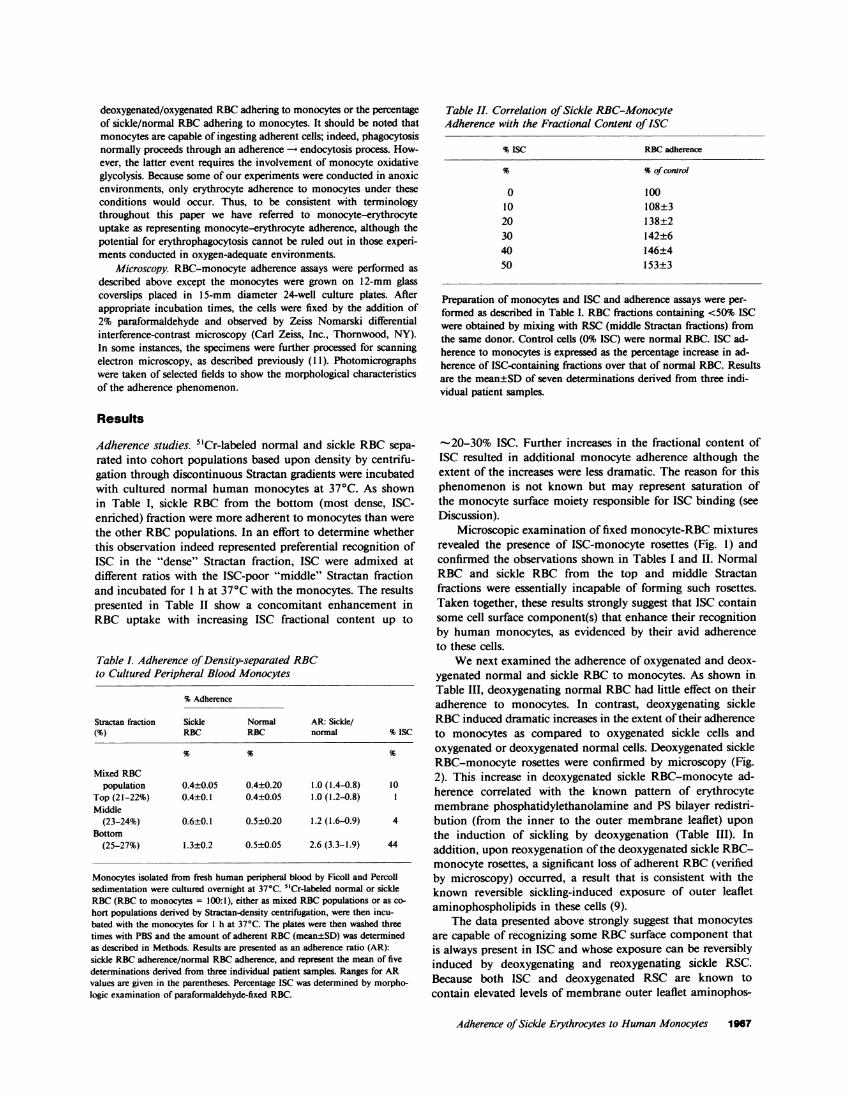
Table II. Correlation of Sickle RBC-MonocyteAdherence with the Fractional Content of ISC
% ISC RBCadherence
% %of control
0 10010 108±320 138±230 142±640 146±450 153±3
Preparation of monocytes and ISC and adherence assays were per-formed as described in Table I. RBCfractions containing <50% ISCwere obtained by mixing with RSC(middle Stractan fractions) fromthe same donor. Control cells (0% ISC) were normal RBC. ISC ad-herence to monocytes is expressed as the percentage increase in ad-herence of ISC-containing fractions over that of normal RBC. Resultsare the mean±SD of seven determinations derived from three indi-vidual patient samples.
Results
Adherence studies. 5"Cr-labeled normal and sickle RBCsepa-rated into cohort populations based upon density by centrifu-gation through discontinuous Stractan gradients were incubatedwith cultured normal human monocytes at 370C. As shownin Table I, sickle RBC from the bottom (most dense, ISC-enriched) fraction were more adherent to monocytes than werethe other RBCpopulations. In an effort to determine whetherthis observation indeed represented preferential recognition ofISC in the "dense" Stractan fraction, ISC were admixed atdifferent ratios with the ISC-poor "middle" Stractan fractionand incubated for 1 h at 370C with the monocytes. The resultspresented in Table II show a concomitant enhancement inRBC uptake with increasing ISC fractional content up to
Table I. Adherence of Density-separated RBCto Cultured Peripheral Blood Monocytes
%Adherence
Stractan fraction Sickle Normal AR: Sickle/(%) RBC RBC normal % ISC
Mixed RBCpopulation 0.4±0.05 0.4±0.20 1.0 (1.4-0.8) 10
Top (21-22%) 0.4±0.1 0.4±0.05 1.0 (1.2-0.8) 1Middle
(23-24%) 0.6±0.1 0.5±0.20 1.2 (1.6-0.9) 4Bottom
(25-27%) 1.3±0.2 0.5±0.05 2.6 (3.3-1.9) 44
Monocytes isolated from fresh human peripheral blood by Ficoll and Percollsedimentation were cultured overnight at 370C. 5"Cr-labeled normal or sickleRBC(RBC to monocytes = 100:1), either as mixed RBCpopulations or as co-hort populations derived by Stractan-density centrifugation, were then incu-bated with the monocytes for I h at 370C. The plates were then washed threetimes with PBS and the amount of adherent RBC(mean±SD) was determinedas described in Methods. Results are presented as an adherence ratio (AR):sickle RBCadherence/normal RBCadherence, and represent the mean of fivedeterminations derived from three individual patient samples. Ranges for ARvalues are given in the parentheses. Percentage ISC was determined by morpho-logic examination of paraformaldehyde-fixed RBC.
-20-30% ISC. Further increases in the fractional content ofISC resulted in additional monocyte adherence although theextent of the increases were less dramatic. The reason for thisphenomenon is not known but may represent saturation ofthe monocyte surface moiety responsible for ISC binding (seeDiscussion).
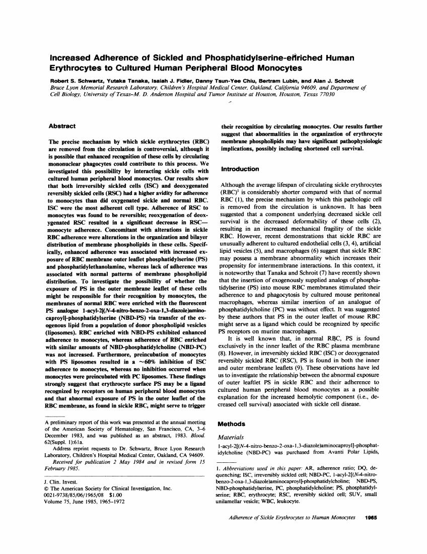
Microscopic examination of fixed monocyte-RBC mixturesrevealed the presence of ISC-monocyte rosettes (Fig. 1) andconfirmed the observations shown in Tables I and II. NormalRBC and sickle RBC from the top and middle Stractanfractions were essentially incapable of forming such rosettes.Taken together, these results strongly suggest that ISC containsome cell surface component(s) that enhance their recognitionby human monocytes, as evidenced by their avid adherenceto these cells.
Wenext examined the adherence of oxygenated and deox-ygenated normal and sickle RBCto monocytes. As shown inTable III, deoxygenating normal RBChad little effect on theiradherence to monocytes. In contrast, deoxygenating sickleRBCinduced dramatic increases in the extent of their adherenceto monocytes as compared to oxygenated sickle cells andoxygenated or deoxygenated normal cells. Deoxygenated sickleRBC-monocyte rosettes were confirmed by microscopy (Fig.2). This increase in deoxygenated sickle RBC-monocyte ad-herence correlated with the known pattern of erythrocytemembrane phosphatidylethanolamine and PS bilayer redistri-bution (from the inner to the outer membrane leaflet) uponthe induction of sickling by deoxygenation (Table III). Inaddition, upon reoxygenation of the deoxygenated sickle RBC-monocyte rosettes, a significant loss of adherent RBC(verifiedby microscopy) occurred, a result that is consistent with theknown reversible sickling-induced exposure of outer leafletaminophospholipids in these cells (9).
The data presented above strongly suggest that monocytesare capable of recognizing some RBCsurface component thatis always present in ISC and whose exposure can be reversiblyinduced by deoxygenating and reoxygenating sickle RSC.Because both ISC and deoxygenated RSC are known tocontain elevated levels of membrane outer leaflet aminophos-
Adherence of Sickle Erythrocytes to Human Monocytes 1967
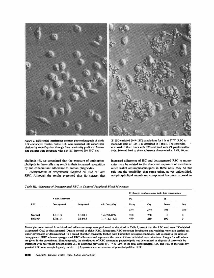
Figure 1. Differential interference-contrast photomicrograph of sickleRBC-monocyte rosettes. Sickle RBCwere separated into cohort pop-ulations by centrifugation through Stractan-density gradients. Mono-cyte cultures were incubated with (A) ISC-depleted [1% ISCI and
pholipids (9), we speculated that the exposure of aminophos-pholipids in these cells may result in their increased recognitionby and concomitant adherence to human phagocytes.
Incorporation of exogenously supplied PS and PC intoRBC. Although the results presented thus far suggest that
(B) ISC-enriched [44% ISCI populations for 1 h at 370C (RBC tomonocyte ratio of 100:1), as described in Table I. The coverslipswere washed three times with PBS and fixed with 2% paraformalde-hyde. Selected field to show adherence characteristics. BAR, 10 ,um.
increased adherence of ISC and deoxygenated RSCto mono-cytes may be related to the abnormal exposure of membraneouter leaflet aminophospholipids in these cells, they do notrule out the possibility that some other, as yet unidentified,nonphospholipid membrane component becomes exposed in
Table III. Adherence of Deoxygenated RBCto Cultured Peripheral Blood Monocytes
Erythrocyte membrane outer leaflet lipid concentration
%RBCadherence PE PS
RBC Deoxygenated Oxygenated AR: Deoxy/Oxy Deoxy Oxy Deoxy Oxy
Normal 1.8±1.3 1.3±0.1 1.4 (2.0-0.9) 260 260 0 0Sickled* 5.7±1.5 0.8±0.5 7.1 (11.7-4.7) 440 260 100 0
Monocytes were isolated from blood and adherence assays were performed as described in Table I, except that the RBCused were 51Cr-labeledoxygenated (Oxy) or deoxygenated (Deoxy) normal or sickle RBC. Subsequent RBC-monocyte incubations and washings were also carried outunder oxygenated or deoxygenated (in a sealed chamber constantly flushed with humidified nitrogen) conditions. ARis equal to the ratio ofdeoxygenated RBCadherence/oxygenated RBCadherence and represents the mean of three individual determinations. Ranges for AR valuesare given in the parentheses. Simultaneously, the distribution of RBCmembrane phospholipids was determined in aliquots of these cells bytreatment with bee venom phospholipase A2, as described previously (9). * 80-90% of the total deoxygenated RBCand 10% of the total oxy-genated RBCwere morphologically sickled. t Approximate concentration of phospholipid/liter RBC.
1968 Schwartz, Tanaka, Fidler, Chiu, Lubin, and Schroit

Figure 2. Scanning electron (A) and light (B) photomicrographs ofdeoxygenated sickle-RBC monocyte rosettes. Monocyte cultures wereincubated with sickle RBC(RBC to monocyte ratio of 100:1) under
sickle RBCas a consequence of sickling, which might conferto these cells an increased propensity for adherence to mono-cytes. Therefore, to define further the physiologic significancethat alterations in RBCmembrane phospholipid organizationmay have on RBC recognition by monocytes, we performedsimilar RBC-monocyte adherence assays using normal RBCwhose membrane phospholipid composition was manipulatedexperimentally. This was accomplished by enriching normalRBCwith exogenously supplied phospholipids via incubationwith artificial lipid vesicles that contained the transferrablefluorescent phospholipid analogues NBD-PS and NBD-PC.
The addition of NBD-PCor NBD-PS vesicles to a suspen-sion of RBCresulted in rapid fluorescence dequenching (DQ)with maximum fluorescence intensity developing within 1-2min (Fig. 3). No enhancement of fluorescence occurred in theabsence of RBCnor was there an increase in fluorescence afterremoval of the RBCby centrifugation, which suggests that theobserved increase in fluorescence intensity was the result ofNBD-phospholipid that had been transferred to and concom-itantly diluted into the RBCmembrane. These results, however,do not rule out the possibility that in addition to phospholipidtransfer, intact vesicles had adhered to the RBC surface.Differentiation between NBD-phospholipid transfer and vesicleadherence was accomplished by comparing fluorescence inten-sity in the absence of (a measure of NBD-phospholipid trans-fered to and diluted into the RBCmembrane) and presenceof (a measure of total RBC-associated NBD-phospholipid)SDS. The measured fluorescence intensities were corrected fordifferences in the probes' quantum yield (whether the lipidwas in RBC, phospholipid vesicles, or detergent micelles), as
Ideoxygenated conditions for 1 h at 370C, as described in Table I.The coverslips were washed three times with degassed PBS and fixedwith 2% paraformaldehyde. Bar, 10 Mm.
56 ng PS
42 ng PS
31 ngPC
00
U- /15 ngPC
0 2Minutes
Figure 3. Dequenching of NBD-fluorescence by insertion (dilution)of lipid into RBCmembranes. NBD-phospholipid vesicles (15-56 ngof lipid) were rapidly mixed in a cuvette with 1 ml of RBCsuspen-sion (107 RBC) and the fluorescence (Xex 470 nm, Xem 525 nm) wascontinuously monitored at 20'C. Light scattering was prevented byusing a high-pass filter (Coming 2A-12, cut-off 510 nm) before theemission monochromator. Lower traces, vesicles and RBCalone;upper traces, vesicle-RBC mixtures.
Adherence of Sickle Erythrocytes to HumanMonocytes 1969
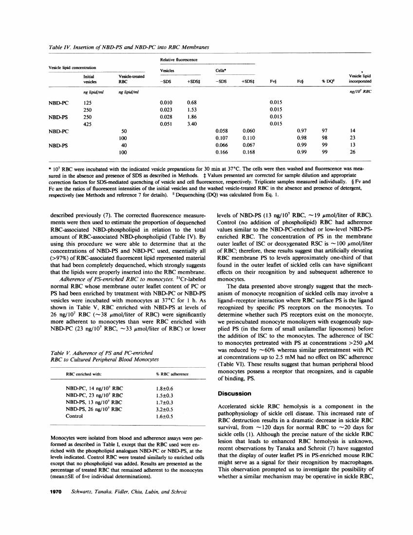
Table IV. Insertion of NBD-PSand NBD-PCinto RBCMembranes
Relative fluorescence
Vesicle lipid concentration Vesicles Cells*
Initial Vesicle-treated Vesicle lipidvesicles RBC -SDS +SDS* -SDS +SDSt Fv§ Fc§ %DQ"1 incorporated
ng lipid/ml ng lipid/ml ng/107 RBC
NBD-PC 125 0.010 0.68 0.015250 0.023 1.53 0.015
NBD-PS 250 0.028 1.86 0.015425 0.051 3.40 0.015
NBD-PC 50 0.058 0.060 0.97 97 14100 0.107 0.110 0.98 98 23
NBD-PS 40 0.066 0.067 0.99 99 13100 0.166 0.168 0.99 99 26
* 107 RBCwere incubated with the indicated vesicle preparations for 30 min at 370C. The cells were then washed and fluorescence was mea-
sured in the absence and presence of SDS as described in Methods. t Values presented are corrected for sample dilution and appropriatecorrection factors for SDS-mediated quenching of vesicle and cell fluorescence, respectively. Triplicate samples measured individually. § Fv andFc are the ratios of fluorescent intensities of the initial vesicles and the washed vesicle-treated RBCin the absence and presence of detergent,respectively (see Methods and reference 7 for details). 1" Dequenching (DQ) was calculated from Eq. 1.
described previously (7). The corrected fluorescence measure-ments were then used to estimate the proportion of dequenchedRBC-associated NBD-phospholipid in relation to the totalamount of RBC-associated NBD-phospholipid (Table IV). Byusing this procedure we were able to determine that at theconcentrations of NBD-PS and NBD-PC used, essentially all(>97%) of RBC-associated fluorescent lipid represented materialthat had been completely dequenched, which strongly suggeststhat the lipids were properly inserted into the RBCmembrane.
Adherence of PS-enriched RBCto monocytes. 5"Cr-labelednormal RBCwhose membrane outer leaflet content of PC orPS had been enriched by treatment with NBD-PCor NBD-PSvesicles were incubated with monocytes at 370C for 1 h. Asshown in Table V, RBCenriched with NBD-PS at levels of26 ng/107 RBC (-38 ismol/liter of RBC) were significantlymore adherent to monocytes than were RBC enriched withNBD-PC (23 ng/107 RBC, -33 ,gmol/liter of RBC) or lower
Table V. Adherence of PS and PC-enrichedRBCto Cultured Peripheral Blood Monocytes
RBCenriched with: %RBCadherence
NBD-PC, 14 ng/107 RBC 1.8±0.6NBD-PC, 23 ng/107 RBC 1.5±0.3NBD-PS, 13 ng/ 107 RBC 1.7±0.3NBD-PS, 26 ng/107 RBC 3.2±0.5Control 1.6±0.5
Monocytes were isolated from blood and adherence assays were per-formed as described in Table I, except that the RBCused were en-riched with the phospholipid analogues NBD-PC or NBD-PS, at thelevels indicated. Control RBCwere treated similarly to enriched cellsexcept that no phospholipid was added. Results are presented as thepercentage of treated RBCthat remained adherent to the monocytes(mean±SE of five individual determinations).
levels of NBD-PS (13 ng/ 107 RBC, - 19 umol/liter of RBC).Control (no addition of phospholipid) RBC had adherencevalues similar to the NBD-PC-enriched or low-level NBD-PS-enriched RBC. The concentration of PS in the membraneouter leaflet of ISC or deoxygenated RSC is -100 ,Amol/literof RBC; therefore, these results suggest that artificially elevatingRBCmembrane PS to levels approximately one-third of thatfound in the outer leaflet of sickled cells can have significanteffects on their recognition by and subsequent adherence tomonocytes.
The data presented above strongly suggest that the mech-anism of monocyte recognition of sickled cells may involve aligand-receptor interaction where RBCsurface PS is the ligandrecognized by specific PS receptors on the monocytes. Todetermine whether such PS receptors exist on the monocyte,we preincubated monocyte monolayers with exogenously sup-plied PS (in the form of small unilamellar liposomes) beforethe addition of ISC to the monocytes. The adherence of ISCto monocytes pretreated with PS at concentrations >250 ,uMwas reduced by -60% whereas similar pretreatment with PCat concentrations up to 2.5 mMhad no effect on ISC adherence(Table VI). These results suggest that human peripheral bloodmonocytes possess a receptor that recognizes, and is capableof binding, PS.
Discussion
Accelerated sickle RBC hemolysis is a component in thepathophysiology of sickle cell disease. This increased rate ofRBCdestruction results in a dramatic decrease in sickle RBCsurvival, from -120 days for normal RBC to '-20 days forsickle cells (1). Although the precise nature of the sickle RBClesion that leads to enhanced RBC hemolysis is unknown,recent observations by Tanaka and Schroit (7) have suggestedthat the display of outer leaflet PS in PS-enriched mouse RBCmight serve as a signal for their recognition by macrophages.This observation prompted us to investigate the possibility ofwhether a similar mechanism may be operative in sickle RBC,
1970 Schwartz, Tanaka, Fidler, Chiu, Lubin, and Schroit

Table VI. PS-mediated Inhibition of ISC Adherenceto Cultured Peripheral Blood Monocytes
Lipid added ISC adherence
mM %of control
Control 100PS (0.25) 40±4PS (0.5) 42±6PS (2.5) 32±3PC (0.25) 90±6PC (0.5) 88±8PC (2.5) 90±4
Preparation of monocytes and ISC (50%, determined morphologi-cally) and adherence assays were performed as described in Table I,except that monocyte monolayers were preincubated with PS or PCsmall unilamellar vesicles, at the indicated concentration, for 30 minat 370C before the addition of ISC. Adherence is expressed as thepercentage change in ISC binding to PS or PC pretreated monocytesvs that to control (PBS-treated) monocytes. Results are the mean±SDof seven individual determinations.
where sickling-induced alterations in membrane phospholipidorganization results in an increased exposure of outer leafletPS (9).
Separation of normal and sickle RBC into cohort popula-tions based upon density revealed that the most-dense fractionsof sickle RBC(enriched in ISC) were 2.5-fold more adherentto human peripheral blood monocytes than were the most-dense populations of normal RBC, a finding similar to therecent observations of Hebbel and Miller (6) who showedenhanced erythrophagocytosis of dense sickle cells to culturedbone marrow macrophages. We found no significant increasein the adherence of mixed (unfractionated) or least-denseStractan populations of sickle cells to monocytes, a finding incontrast to that reported by Hebbel and Miller (6), whoreported a twofold increase in macrophage erythrophagocytosisof least-dense sickle cell populations. Such differences may berelated to different mechanisms involved in the RBCadherenceto monocytes vs. RBCerythrophagocytosis by macrophages.
Microscopic examination of monocytes that had beenincubated with the dense (ISC-rich) sickle RBC populationsrevealed the presence of sickle RBC-monocyte rosettes, wherethe predominant RBCassociated with the monocytes appearedmorphologically to be ISC. In contrast, examination of mono-cytes incubated with oxygenated, mixed sickle cell populations,less-dense (ISC-poor) sickle RBCpopulations, or with any ofthe density-separated fractions of normal RBC, failed to dem-onstrate the presence of any significant monocyte-RBC rosetteformation, corroborating the previous results of Abramson etal. (12), who found that mixed populations of oxygenatedsickle RBC did not form rosettes when added to culturedhuman leukocyte preparations.
Deoxygenation of sickle RBCinduced a dramatic increasein the adherence of these cells to monocytes, whereas similardeoxygenation of normal RBC had essentially no effect ontheir adherence as compared to the adherence of oxygenatedsickle and normal cells, respectively. Exposure of the deoxy-genated sickle RBC-monocyte mixture to room air resultedin a significant loss (>90%) of adherent erythrocytes (micro-scopic observation), which demonstrates the reversibility of
sickle RBC-monocyte adherence. Correlating with these re-versible changes in sickle RBC adherence was the knownreversible alteration in sickle RBC membrane outer leafletaminophospholipid (PE and PS) content (9). Taken together,these results suggest that the membrane content of RBCouterleaflet aminophospholipids may serve as a trigger for recognitionby and adherence to peripheral blood monocytes, a findingstrikingly similar to the many observations that cells of themononuclear phagocyte series avidly recognize synthetic phos-pholipid vesicles containing PS (13, 14).
These results, however, do not rule out the possibility thatother sickle RBCmembrane components may also be partic-ipating in their enhanced adherence to monocytes. Indeed,sickle RBCare known to contain multiple membrane abnor-malities, including elevated levels of surface glycoproteins (15),calcium (16), and hemichrome (17), as well as alterations inthe structures of the cytoskeletal proteins (18), increased sus-ceptibility to lipid peroxidation (19), and reduced cellulardeformability (20). Petz et al. (21) have recently demonstratedthat a subpopulation of sickle RBCcontained elevated levelsof surface-bound IgG. In that monocytes readily recognizeopsonized RBC(12), it is possible that increased adherence ofsickle RBC to monocytes is mediated by cell surface immu-noglobulin. Indeed, Hebbel and Miller (6) have recently shownpartial inhibition of sickle RBCphagocytosis by macrophagesfollowing Fc receptor blockade with IgG. However, the exposureand reversibility of the surface component examined in ourstudies was rapidly inducible by deoxygenation of sickle cellsand was reversible by reoxygenation, suggesting that the moietyresponsible for increased sickle RBCadherence examined inour studies was probably not antibody related.2
To clarify further the role that RBC membrane amino-phospholipids might have in promoting sickled RBC-monocyteadherence, we enriched normal RBC with the fluorescentphospholipid analogues NBD-PS and NBD-PC. These NBD-phospholipids are readily transferred from donor vesicles toacceptor cell membranes (7), and the inherent fluorescenceself-quenching properties of these compounds allow one todistinguish between genuine phospholipid insertion and uptakeof intact phospholipid vesicles. By using this technique, wewere able to incorporate NBD-PCor NBD-PS into intact RBCunder conditions where binding of intact vesicles to RBCdidnot occur. Our results indicate that NBD-PS-enriched RBCwere more adherent to monocytes than were NBD-PC-enrichedor control (PBS-treated) RBC. The amount of incorporatedNBD-PS required to enhanced RBC adherence (-38 ,umol/liter of RBC) was approximately one-third the level of PSfound in the outer leaflet of sickled RBC (-100 ,umol/literRBC, [9]), whereas enrichment of RBCwith NBD-PS belowthis apparent threshold amount had little effect on adherence,strongly suggesting that monocyte recognition of PS-enrichedRBC is sensitive to the actual amount of PS in the RBCmembrane. Although we have not carried out detailed exper-iments defining the relationship between the minimum amountof erythrocyte PS required for enhanced adherence to mono-cytes, one should be aware of the possibility that the actualamount of uniformly dispersed PS in RBC(as obtained in the
2. Whether conditions of low oxygen tension induce an alteration innormal (i.e., oxygenated) monocyte surface characteristics, which mightspecifically enhance their interaction with sickle cells, is not known.
Adherence of Sickle Erythrocytes to HumanMonocytes 1971

NBD-PS treated RBC) may not be as crucial as its distributionin the membrane. Indeed, the precise distribution of erythrocytemembrane PS in sickled cells is unknown; one could speculatethat localization of exposed PS molecules in sickled cells intospecific membrane domains, as has been suggested for eryth-rocyte membrane PC (22) and phosphatidylethanolamine(23), could lead to an even greater adherence of these cellsto monocytes than was measured using the NBD-PS-en-riched RBC.
To determine further whether a binding site capable ofrecognizing PS exists on the monocyte membrane, inhibitionstudies were performed where monocyte monolayers werepreincubated with PS liposomes. Preincubation of the mono-cytes with PS at concentrations >250 ,gM resulted in a -60%inhibition of ISC adherence, whereas similar preincubationwith PC at concentrations up to 2.5 mMhad no effect on ISCadherence. These findings suggest that a surface moiety onhuman monocytes may recognize PS, a finding similar to theone reported by Tanaka and Schroit (7) for murine macro-phages.
Our findings represent but one example of a broadercategory of sickle cell properties regarding the enhanced sus-ceptibility of these cells to undergo intermembrane interactions.Other examples of this phenomenon are an increased bindingto cultured endothelial cells (3, 4), increased uptake of PS-richphospholipid vesicles (5), and an enhanced ability of thesecells to stimulate in vitro blood coagulation (24).
In conclusion, it appears that abnormal exposure of outerleaflet PS in sickle RBCmay significantly affect their propensityfor recognition by circulating human phagocytes and may,furthermore, represent a homeostatic mechanism for the re-moval of pathologic RBC. Although we have not specificallyruled out the involvement of other, as yet unidentified, RBCmembrane components in this process, the reversible natureof both adherence to monocytes and expression of surface PSin sickled RBC strongly suggests that alterations in RBCphospholipid asymmetry may have significant pathophysiologicimplications.
Acknowledaments
The authors are grateful for the competent technical assistance providedby John Madsen, the expertise of Dr. E. S. Kleinerman and Dr. S.Claster in the establishment of the monocyte cultures, and to MarilynFile and Marion Douglass for preparation of this manuscript.
This work was supported by grants HL-20985 and HL-27059 fromthe National Institutes of Health and by developmental fund grant175416 to Dr. Schroit from the University of Texas-M. D. AndersonHospital and Tumor Institute at Houston.
References
1. McCurdy, P. R. 1969. 32DFP and 5"Cr for measurement of redcell life span in abnormal hemoglobin syndromes. Blood. 33:214-224.
2. Mohandas, N., and M. Bessis. 1979. Red cell deformabilitychanges in sickle cell anemia. In Development of Therapeutic Agentsfor Sickle Cell Disease. J. Rosa, Y. Beuzard, and J. Hercules, editors.North Holland Publishing Co., New York. 15-29.
3. Hebbel, R. P., 0. Yamuda, C. F. Moldow, H. S. Jacob, J. G.White, and J. W. Eaton. 1980. Abnormal adherence of sickle eryth-rocytes to cultured vascular endothelium: possible mechanism formicrovascular occlusion in sickle cell disease. J. Clin. Invest. 65:154-160.
4. Hoover, R., R. Rubin, G. Wise, and R. Warren. 1979. Adhesionof normal and sickle erythrocytes to endothelial monolayer cultures.Blood. 54:872-876.
5. Schwartz, R. S., N. Diizgunes, D. T.-Y. Chiu, and B. Lubin.1983. Interaction of phosphatidylserine-phosphatidylcholine liposomeswith sickle erythrocytes: evidence for altered membrane surface prop-erties. J. Clin. Invest. 71:1570-1580.
6. Hebbel, R. P., and W. J. Miller. 1984. Phagocytosis of sickleerythrocytes: immunologic and oxidative determinants of hemolyticanemia. Blood. 64:733-741.
7. Tanaka, Y., and A. J. Schroit. 1983. Insertion of fluorescentphosphatidylserine into the plasma membrane of red blood cells:recognition by autologous macrophages. J. Biol. Chem. 258:11335-11343.
8. Op den Kamp, J. A. F. 1979. Lipid asymmetry in membranes.Annu. Rev. Biochem. 48:47-71.
9. Lubin, B., D. Chiu, J. Bastacky, B. Roelofsen, and L. L. M. vanDeenen. 1981. Abnormalities in membrane phospholipid organizationin sickled erythrocytes. J. Clin. Invest. 67:1643-1649.
10. Kleinerman, E. S., A. J. Schroit, W. E. Fogler, and I. J. Fidler.1983. Tumoricidal activity of human monocytes activated in vitro byfree and liposome-encapsulated human lymphokines. J. Clin. Invest.72:304-315.
11. Bucana, C. D., L. C. Hoyer, A. J. Schroit, E. Kleinerman, andI. J. Fidler. 1983. Ultrastructural studies of the interaction betweenliposome-activated human blood monocytes and allogenic tumor cellsin vitro. Am. J. Pathol. 112:10 -11 1.
12. Abramson, N., A. F. Lo Buglio, J. H. Jandl, and R. S. Cotran.1970. The interaction between human monocytes and red cells:binding characteristics. J. Exp. Med. 132:1191-1206.
13. Raz, A., C. Bucana, W. E. Fogler, G. Poste, and I. J. Fidler.1981. Biochemical, morphological and ultrastructural studies on theuptake of liposomes by murine macrophages. Cancer Res. 41:487-494.
14. Schroit, A. J., and I. J. Fidler. 1982. Effects of liposomestructure and lipid composition on the activation of the tumoricidalproperties of macrophages by liposomes containing muramyl dipeptide.Cancer Res. 42:161-167.
15. Fukuda, M., M. N., Fukuda, S. Hakomori, and T. Papayan-nopoulou. 1981. Anomalous cell surface structure of sickle cell anemiaerythrocytes as demonstrated by cell surface labeling and endo-fl-galactosidase treatment. J. Supramol. Struct. Cell. Biochem. 17:289-297.
16. Palek, J. 1977. Red cell calcium content and transmembranecalcium movements in sickle cell anemia. J. Lab. Clin. Med. 89:1365-1374.
17. Campwala, H. Q., and J. F. Desforges. 1982. Membrane-boundhemichrome in density-separated cohorts pf normal (AA) and sickled(SS) cells. J. Lab. Clin. Med. 99:25-28.
18. Lux, S. E., K. M. John, and J. Karnovsky. 1976. Irreversibledeformation of the spectrin-actin lattice in irreversibly sickled cells. J.Clin. Invest. 58:955-962.
19. Lubin, B., and D. Chiu. 1980. Abnormal susceptibility of sickleerythrocytes to lipid peroxidation. In Red Blood Cell and LensMetabolism. S. Srivastava, editor. Elsevier/North Holland, New York.159-162.
20. Chien, S., S. Usami, and J. F. Bertles. 1970. Abnormal rheologyof oxygenated blood in sickle cell anemia. J. Clin. Invest. 49:623-634.
21. Petz, L. D., P. Yam, L. Wilkinson, G. Garratty, B. Lubin, andW. Mentzer. 1984. Increased IgG molecules bound to the surface ofred blood cells of patients with sickle cell anemia. Blood. 64:301-304.
22. Shukla, S. D., and D. J. Hanahan. 1982. Identification ofdomains of phosphatidylcholine in human erythrocyte plasma mem-branes. J. Biol. Chem. 257:2908-2911.
23. Marinetti, G. V., and K Cattieu. 1982. Asymmetric metabolismof phosphatidyl-ethanolamine in the human red cell membrane. J.Biol. Chem. 257:245-248.
24. Chiu, D., B. Lubin, B. Roelofsen, and L. L. M. van Deenen.1981. Sickled erythrocytes accelerate clotting in vitro: an effect ofabnormal membrane lipid asymmetry. Blood. 58:398-401.
1972 Schwartz, Tanaka, Fidler, Chiu, Lubin, and Schroit