topical anti-inflammatory plant species: bioactivity of
TRANSCRIPT

1
Topical anti-inflammatory plant species: Bioactivity of Bryonia dioica,
Tamus communis and Lonicera peryclimenum fruits
MARCO RAFAEL, LILLIAN BARROS, ANA MARIA CARVALHO, ISABEL C.F.R. FERREIRA*
CIMO/Escola Superior Agrária, Instituto Politécnico de Bragança, Campus de Santa
Apolónia, Apartado 1172, 5301-855 Bragança, Portugal.
* Author to whom correspondence should be addressed (e-mail: [email protected]
telephone +351-273-303219; fax +351-273-325405).

2
ABSTRACT
The practice of rubbing different plant material juices or extracts into the skin to relieve
pain and rheumatic symptoms is deeply rooted in folk medicine and has been used for a
long time. Several common species, usually available in agroecosystems of the Iberian
Peninsula, were/are used for topical medicinal preparations as reported in recent
ethnobotanical surveys. Based on these studies, the fruits of three relevant species
(Bryonia dioica or white-bryony, Lonicera periclymenum or common honeysuckle and
Tamus communis or black-bryony) were gathered and different analyses and assays
were performed in order to characterize their phytochemical composition and to find
biologically active compounds for pharmaceutical application. Black-bryony ripened
fruits revealed the highest antioxidant properties which are in agreement to its highest
concentration in phenolics, flavonoids, ascorbic acid, tocopherols and lycopene. The
studied fruits revealed interesting antioxidant properties and bioactive phytochemicals
that could provide scientific evidence for their folk uses as anti-inflammatory species.
Keywords: Medicinal fruits; Topical anti-inflammatory; Antioxidant properties;
Bioactive compounds; Portuguese ethnobotany.

3
1. Introduction
Inflammatory diseases are accompanied by the chronic release of cytokines and reactive
oxygen (ROS) and nitrogen (RNS) species, which may be involved in increased tissue
injury. Much evidence has shown that the production of reactive species such as
superoxide anion radical, hydrogen peroxide, hydroxyl radical and peroxynitrite occurs
at the site of inflammation and contributes to tissue damage (Conner and Grisham,
1996; Nardi et al., 2007).
Under normal circumstances, ROS and RNS are detoxified by an efficient antioxidant
system that includes enzymes such as superoxide dismutase, catalase and glutathione
peroxidises (Halliwell, 1994; Roome et al., 2008). In acute and chronic inflammation,
high concentrations of those species are produced, which generate an oxidative
imbalance and decrease the capacity of the endogenous antioxidant enzymes to remove
them, contributing to tissue damage (Nardi et al., 2007) and a variety of chronic
inflammatory diseases such as arthritis and atherosclerosis as well as other ailments viz.
cancer, diabetes, hepatitis, neurodegeneration and early aging (Halliwell, 1994; Roome
et al., 2008). Therefore, considering the involvement of oxidative stress in
inflammation, topical antioxidants might bring health benefits (Casagrande et al., 2006).
Particularly, antioxidants from natural products present novel possibilities for the
treatment and prevention of oxidative stress-mediated inflammatory diseases.
Plants contain many constituents with local physical impact on body tissues and the
topical use of herbal remedies is among the most noticeable in the simplest traditions of
healthcare (Marc et al., 2008). Recent ethnobotanical surveys conducted in the Iberian
Peninsula (Neves, 2009; Benitez et al., 2010; Carvalho, 2010; Gonzalez et al., 2010)

4
have recorded interesting uses of very common plant materials applied as topical
homemade remedies.
Three of the most cited and topically used wild species (Carvalho, 2010) are Bryonia
dioica Jacq. (white-bryony; port. norça), Lonicera periclymenum L. (common
honeysuckle; port. madressilva) and Tamus communis L. (black-bryony; port. budanha),
perennial, deciduous climbers often found in woodland, in hedgerows or scrubland and,
sometimes, occurring also in orchards and homegardens. These three species are
generally and popularly considered toxic to humans (Carvalho, 2010). Besides the
medicinal use and the very specific edible use of bryonies sprouts as greens in the
Iberian Peninsula (Martins et al., 2011), every people interviewed were conscious of
their poisonous effects, in particularly of the fruits and of the underground organs
(tuberous rootstalks) (Carvalho, 2010). It seems that the noxious effects are mainly due
to triterpene glucosides and calcium oxalate crystals that are found mainly in the fruit
(Castroviejo et al., 2005)
Traditional practices include mainly different liquid dosage forms such as plant juices,
tinctures and related products (alcoholic or hydroalcoholic solutions prepared from
botanicals). Freshly harvest plant parts (leaves, flowers and mainly fruits) are frequently
applied directly or macerated in water, alcohol or brandy. Their use is claimed to have
local anti-inflammatory effects on minor wounds and lesions and on the management
and relief of inflammations affecting joins, muscles and other subcutaneous tissues.
Although these topical remedies have been used for hundreds of years and liquid herbal
preparations do appear to be quite efficient for the extraction of a wide variety of
compounds found in medicinal plants, a number of studies have highlighted the
importance of the ratio (the correct choice of the alcohol percentage to maximize the

5
quality of the preparation) and little is known about the chemical composition of plant
juices or their possible reactions in aqueous and in alcoholic media.
In the present work, the bioactive phytochemicals of fruits of the three previously
mentioned species were characterized. Furthermore, the evaluation of their antioxidant
properties could support folk uses as topical anti-inflammatory species.
2. Materials and methods
2.1. Samples
Both immature fruits (hard green fruits in early summer) and ripened fruits (fleshy and
soft red fruits in late autumn) of the three selected species were gathered in Bragança,
Trás-os-Montes, North-eastern Portugal. The samples for analysis were collected and
prepared according to the main medicinal topical applications as described by
informants from this Portuguese region (Carvalho, 2010) (Table 1). All the vegetal
material was gathered at random from several plants inside a selected area which has
been visited twice considering the two different stages of fruit maturity. Fruits of
Bryonia dioica and Tamus communis were collected in July (immature fruits) and
September (ripened fruits); the fruits of Lonicera periclymenum were collected in July
and September of 2010.
Local traditional practitioners usually rubbed the juice of fresh immature fruits on body
surfaces to relieve pains and minor wounds; the ripened fruits were used in the form of
alcoholic extracts for arthritis and rheumatic complains; vapors (steam) from the hot
decoction of common honeysuckle’ fruits were recommended for asthma crisis (Neves,
2009; Carvalho, 2010; Gonzalez et al., 2010).

6
Morphological key characters from the Flora Iberica were used for plant identification.
Voucher specimens (ETBO33, ETBO36, ETBO53) are deposited in the herbarium of
the Escola Superior Agrária de Bragança (BRESA). Each sample was lyophilized (Ly-
8-FM-ULE, Snijders, Holland) and stored in the deep-freezer at -20ºC for subsequent
analysis.
2.2. Standards and Reagents
Acetonitrile 99.9%, n-hexane 95%, and ethyl acetate 99.8% were of HPLC grade (Lab-
Scan, Lisbon, Portugal). The fatty acids methyl ester (FAME) reference standard
mixture 37 (standard 47885-U) was purchased from Sigma (St. Louis, MO), as also
other individual fatty acid isomers, ascorbic acid, tocopherols (, , , and -
tocopherols), and sugars (D(-)-fructose, D(+)-glucose anhydrous, D(+)-melezitose
hydrate, D(+)-raffinose pentahydrate, D(+)-sucrose, and D(+)-trehalose) standards, Trolox
(6-hydroxy-2,5,7,8-tetramethylchroman-2-carboxylic acid), gallic acid, and (+)-
catechin. Racemic tocol, 50 mg/mL, was purchased from Matreya (Chalfont, PA). 2,2-
Diphenyl-1-picrylhydrazyl (DPPH) was obtained from Alfa Aesar (Ward Hill, MA). All
other chemicals and solvents were of analytical grade and purchased from common
sources. Water was treated in a Milli-Q water purification system (Pure Water Systems,
Brea, CA).
2.3. In vitro evaluation of antioxidant properties
2.3.1. Preparation of the methanolic extracts
A fine dried powder (20 mesh; ~1 g) was extracted by stirring with 30 mL of methanol
at 25 ºC at 150 rpm for 1 h and filtered through Whatman No. 4 paper. The residue was

7
then extracted with one additional 30 mL portion of methanol (this procedure was
followed according to the extraction conditions optimized by us in a previous report
(Barros et al., 2010a)). The combined methanolic extracts were evaporated at 40ºC
under reduced pressure, re-dissolved in methanol at a concentration of 20 mg/mL, and
stored at 4 ºC for further use. In vitro assays which have already been described by the
authors (Barros et al., 2010b) were applied to evaluate the antioxidant activity of all the
samples. Different concentrations of the extracts were used to find EC50 values.
2.3.2. DPPH radical-scavenging activity
This methodology was performed using an ELX800 Microplate Reader (BioTek
Instruments, Inc., Winooski, VT). The reaction mixture in each of the 96-wells
consisted of one of the different concentrations of the extracts (30 μL) and aqueous
methanolic solution (80:20, v/v, 270 μL) containing DPPH radicals (6 10-5
mol/L).
The mixture was left to stand for 60 min in the dark. The reduction of the DPPH radical
was determined by measuring the absorption at 515 nm. The radical scavenging activity
(RSA) was calculated as a percentage of DPPH discolouration using the equation: %
RSA = [(ADPPH-AS)/ADPPH] 100, where AS is the absorbance of the solution when the
sample extract has been added at a particular level, and ADPPH is the absorbance of the
DPPH solution. The extract concentration providing 50% of radicals scavenging activity
(EC50) was calculated from the graph of RSA percentage against extract concentration.
Trolox was used as standard.
2.3.3. Reducing power

8
This methodology was performed using the Microplate Reader described above. The
different concentrations of the extracts (0.5 mL) were mixed with sodium phosphate
buffer (200 mmol/L, pH 6.6, 0.5 mL) and potassium ferricyanide (1%, w/v, 0.5 mL).
The mixture was incubated at 50 ºC for 20 min, and trichloroacetic acid (10%, w/v, 0.5
mL) was added. The mixture (0.8 mL) was poured in the 48-wells, as also deionised
water (0.8 mL) and ferric chloride (0.1%, w/v, 0.16 mL), and the absorbance was
measured at 690 nm. The extract concentration providing 0.5 of absorbance (EC50) was
calculated from the graph of absorbance at 690 nm against extract concentration. Trolox
was used as standard.
2.3.4. Inhibition of -carotene bleaching
A solution of -carotene was prepared by dissolving -carotene (2 mg) in chloroform
(10 mL). Two millilitres of this solution were pipetted into a round-bottom flask. After
the chloroform was removed at 40ºC under vacuum, linoleic acid (40 mg), Tween 80
emulsifier (400 mg), and distilled water (100 mL) were added to the flask with vigorous
shaking. Aliquots (4.8 mL) of this emulsion were transferred into different test tubes
containing different concentrations of the extracts (0.2 mL). The tubes were shaken and
incubated at 50ºC in a water bath. As soon as the emulsion was added to each tube, the
zero time absorbance was measured at 470 nm using a 200-2004 spectrophotometer
(Analytikjena, Jena, Germany). β-Carotene bleaching inhibition was calculated using
the following equation: (-carotene content after 2h of assay/initial -carotene content)
100. The extract concentration providing 50% antioxidant activity (EC50) was
calculated by interpolation from the graph of β-carotene bleaching inhibition percentage
against extract concentration. Trolox was used as standard.

9
2.3.5. Inhibition of lipid peroxidation using thiobarbituric acid reactive substances
(TBARS)
Brains were obtained from pig (Sus scrofa) of body weight ~150 Kg, dissected and
homogenized with a Polytron in ice-cold Tris–HCl buffer (20 mM, pH 7.4) to produce a
1:2 (w/v) brain tissue homogenate which was centrifuged at 3000g for 10 min. An
aliquot (0.1 mL) of the supernatant was incubated with the different concentrations of
the extracts (0.2 mL) in the presence of FeSO4 (10 M; 0.1 mL) and ascorbic acid (0.1
mM; 0.1 mL) at 37ºC for 1 h. The reaction was stopped by the addition of
trichloroacetic acid (28%, w/v, 0.5 mL), followed by thiobarbituric acid (TBA, 2%, w/v,
0.38 mL), and the mixture was then heated at 80 ºC for 20 min. After centrifugation at
3000g (Centorion K24OR- 2003 refrigerated centrifuge) for 10 min to remove the
precipitated protein, the colour intensity of the malondialdehyde (MDA)-TBA complex
in the supernatant was measured by its absorbance at 532 nm. The inhibition ratio (%)
was calculated using the following formula: Inhibition ratio (%) = [(A – B)/A] x 100%,
where A and B were the absorbance of the control and the compound solution,
respectively. The extract concentration providing 50% lipid peroxidation inhibition
(EC50) was calculated from the graph of TBARS inhibition percentage against extract
concentration. Trolox was used as standard.
2.4. Bioactive compounds
2.4.1. Phenolics
For total phenolics estimation an aliquot of the methanolic extract solution (1 mL) was
mixed with Folin-Ciocalteu reagent (5 mL, previously diluted with water 1:10, v/v) and

10
sodium carbonate (75 g/L, 4 mL). The tubes were vortexed for 15 s and allowed to stand
for 30 min at 40 °C for colour development. Absorbance was then measured at 765 nm
(Wolfe et al., 2003). Gallic acid was used to calculate the standard curve (9.4 10-3
-
0.15 mg/mL), and the results were expressed as mg of gallic acid equivalents (GAE) per
g of extract.
2.4.2. Flavonoids
For total flavonoids content determination, an aliquot (0.5 mL) of the methanolic extract
solution was mixed with distilled water (2 mL) and subsequently with NaNO2 solution
(5%, 0.15 mL). After 6 min, AlCl3 solution (10%, 0.15 mL) was added and allowed to
stand further 6 min, thereafter, NaOH solution (4%, 2 mL) was added to the mixture.
Immediately, distilled water was added to bring the final volume to 5 mL. Then the
mixture was properly mixed and allowed to stand for 15 min. The intensity of pink
colour was measured at 510 nm (Jia et al., 1999). (+)-Catechin was used to calculate the
standard curve (4.5 10-3
-0.29 mg/mL) and the results were expressed as mg of (+)-
catechin equivalents (CE) per g of extract.
2.4.3. Ascorbic acid
A fine dried powder (20 mesh; 150 mg) was extracted with metaphosphoric acid (1%,
10 mL) for 45 min at room temperature and filtered through Whatman Nº 4 filter paper.
The filtrate (1 mL) was mixed with 2,6-dichloroindophenol (9 mL) and the absorbance
was measured within 30 min at 515 nm (Klein and Perry, 1982). Content of ascorbic
acid was calculated on the basis of the calibration curve of authentic L-ascorbic acid
(6.0 10-3
-0.10 mg/mL), and the results were expressed as mg of ascorbic acid per 100 g

11
of dry weight.
2.4.4. Tocopherols
Tocopherols content was determined following a procedure previously described by the
authors (Martins et al., 2011). BHT solution in n-hexane (10 mg/mL; 100 μL) and IS
solution in n-hexane (tocol; 50 μg/mL; 400 μL) were added to the sample prior to the
extraction procedure. The samples (~500 mg) were homogenized with methanol (4 mL)
by vortex mixing (1 min). Subsequently, n-hexane (4 mL) was added and again vortex
mixed for 1 min. After that, saturated NaCl aqueous solution (2 mL) was added, the
mixture was homogenized (1 min), centrifuged (5 min, 4,000g) and the clear upper layer
was carefully transferred to a vial. The sample was re-extracted twice with n-hexane.
The combined extracts were taken to dryness under a nitrogen stream, redissolved in 2
mL of n-hexane, dehydrated with anhydrous sodium sulphate, filtered through 0.2 µm
nylon filters from Whatman and transferred into a dark injection vial. The equipment
consisted of an integrated system with a Smartline 1000 pump (Knauer, Berlin,
Germany), a Smartline manager 5000 degasser, an AS-2057 auto-sampler (Jasco,
Easton, MD) and an FP-2020 fluorescence detector (Jasco, Easton, MD) programmed
for excitation at 290 nm and emission at 330 nm. The column used was a normal-phase
250 mm 4.6 mm i.d., 5 μm, Polyamide II, with a 10 mm 4 mm i.d. guard column of
the same material (YMC Waters, Dinslaken, Germany), operating at 30 ºC. The mobile
phase used was a mixture of n-hexane and ethyl acetate (70:30, v/v) at a flow rate of 1
mL/min. The compounds were identified by chromatographic comparisons with
authentic standards. Quantification was based on the fluorescence signal response, using

12
the internal standard method. Tocopherol contents in the samples are expressed in mg
per 100 g of dry weight.
2.4.5. Pigments
A fine dried powder (150 mg) was vigorously shaken with 10 mL of acetone–hexane
mixture (4:6) for 1 min and filtered through Whatman No. 4 filter paper. The
absorbance of the filtrate was measured at 453, 505, 645 and 663 nm (Nagata and
Yamashita, 1992). Content of -carotene was calculated according to the following
equation: -carotene (mg/100 mL) = 0.216 A663 – 1.220 A645 - 0.304 A505 + 0.452
A453; Lycopene (mg/100 mL) = 0.0458 A663 + 0.204 A645 - 0.304 A505 + 0.452
A453; Chlorophyll a (mg/100 mL) = 0.999 A663 - 0.0989 A645; Chlorophyll b
(mg/100 mL) = - 0.328 A663 + 1.77 A645, and further expressed in mg per 100 g of
dry weight.
2.4.6. Sugars
Free sugars were determined by high performance liquid chromatography coupled to a
refraction index detector (HPLC-RI) as previously described by the authors (Martins et
al., 2011). Dried sample powder (1.0 g) was spiked with the melezitose as internal
standard (IS, 5 mg/ml), and was extracted with 40 mL of 80% aqueous ethanol at 80 ºC
for 30 min. The resulting suspension was centrifuged at 15,000g for 10 min. The
supernatant was concentrated at 60 ºC under reduced pressure (rotary evaporator Büchi
R-210) and defatted three times with 10 mL of ethyl ether, successively. After
concentration at 40 ºC, the solid residues were dissolved in water to a final volume of 5
mL and filtered through 0.2 µm nylon filters from Whatman. The equipment described

13
above was connected to a Smartline 2300 RI detector. Data were analysed using Clarity
DataApex 2.4 Software. The column used was a 250 mm 4.6 mm i.d., 5 μm,
Eurospher 100-5 NH2 with a 5 mm 4mm i.d. guard column of the same material
(Knauer, Berlin, Germany), operating at 30 ºC in a 7971 R Grace oven. The mobile
phase was acetonitrile/deionized water, 7:3 (v/v) at a flow rate of 1 mL/min. Sugar
identification was made by comparing the relative retention times of sample peaks with
standards. Quantification was made by internal normalization of the chromatographic
peak area and the results are expressed in g per 100 g of dry weight.
2.4.7. Fatty Acids
Fat was extracted with petroleum ether in a Soxhlet apparatus. Fatty acids were
determined by gas chromatography with flame ionization detection (GC-FID) as
described previously by the authors (Martins et al., 2011), and after the following
transesterification procedure: fatty acids (obtained after Soxhlet extraction) were
methylated with 5 mL of methanol:sulphuric acid:toluene 2:1:1 (v/v/v), during at least
12 h in a water bath at 50 ºC and 160 rpm; then 3 mL of deionised water were added to
obtain phase separation; the FAME were recovered with 3 mL of diethyl ether by
shaking in vortex, and the upper phase was dehydrated with sodium sulphate anhydrous;
finally the sample was filtered with 0.2 µm nylon filter from Whatman. The equipment
was a DANI model GC 1000 with a split/splitless injector, and a FID. The column used
was a 30 m 0.32 mm i.d., 0.25 µm, 50% cyanopropyl-methyl-50%
phenylmethylpolysiloxane (Macherey-Nagel, Düren, Germany). The FID temperature
was 260 ºC. The oven temperature program was as follows: the initial temperature of
the column was 50 ºC, held for 2 min, then a 30ºC/min ramp to 125 ºC, 5ºC/min ramp to

14
160 ºC, 20ºC/min ramp to 180 ºC, 3ºC/min ramp to 200 ºC, 20ºC/min ramp to 220 ºC
and held for 15 min. The carrier gas (hydrogen) flow-rate was 4.0 mL/min (0.61 bar),
measured at 50 ºC. Split injection (1:40) was carried out at 250 ºC. Fatty acid
identification was made by comparing the relative retention times of FAME peaks from
samples with standards. The results were recorded and processed using CSW DataApex
1.7 software and expressed in relative percentage of each fatty acid.
2.5. Statistical analysis
For each species, three samples were analysed and the assays were carried out in
triplicate. The results are expressed as mean values and standard deviation (SD), and.
were analyzed using one-way analysis of variance (ANOVA) followed by Tukey’s HSD
Test with α = 0.05 (different letters mean significant differences; the letter a is attributed
to the highest value). This treatment was carried out using SPSS v. 16.0 program.
3. Results and discussion
3.1. In vitro evaluation of antioxidant properties
Four different assays were used for the in vitro evaluation of the antioxidant properties
of immature and ripened fruits used in Portuguese and Spanish folk medicine as
described in Table 1. The extracts were prepared using methanol to mimetize the
ethnopharmacological use of the fruits: maceration in alcohol or brandy.
The results of scavenging activity on DPPH radicals, reducing power, inhibition of -
carotene bleaching, and inhibition of lipid peroxidation in brain tissue homogenates are
shown in Figure 1, and antioxidant activity EC50 values as also phenolic and flavonoids
contents are given in Table 2. Black-bryony ripened fruits proved to have the most

15
promissory antioxidant activity (the lowest EC50 values, ranging from 0.29 to 1.26
mg/mL), with the highest phenolic (119.78 mg GAE/g extract) and flavonoids (52.69
mg CE/g extract; Table 2) contents. Otherwise, white-bryony immature fruits revealed
the lowest antioxidant properties (the highest EC50 values, ranging from 1.07 to 18.01
mg/mL) which are compatible to its lower phenolics (33.37 mg GAE/g extract) and
flavonoids (10.32 mg CE/g extract; Table 2) content. Both species and maturity stage of
fruits seemed to have influence in the antioxidant properties. Nevertheless, all the
samples revealed antioxidant activity, which might be related to its traditional use as
topical anti-inflammatory, as previously explained (Nardi et al., 2007; Roome et al.,
2008).
As far as we know this is the first study dealing with fruits of white (Bryonia dioica)
and black (Tamus communis) bryonies, and common honeysuckle (Lonicera
periclymenum). The antioxidant properties of bryonies edible parts (shoots) were
already reported by our research group (Martins et al., 2001). Other authors reported
antioxidant activity of Lonicera japonica under cultivation (the pharmaceutical name in
Chinese Pharmacopoeia is Flos lonicerae and is used for acute infections of respiratory
tract, the mammary glands and appendicitis) (Liu et al., 2008; Tsai et al., 2008). The
latter authors reported 45.2 1.65% of DPPH radical scavenging activity at 1 mg of
methanolic extract/ml, a higher activity than the observed in Lonicera periclymenum
fruits herein studied (Figure 1). Moreover, the samples obtained in Taiwan (Tsai et al.,
2008) and in China (Liu et al., 2008) revealed lower phenolics content, but higher
flavonoids content than fruits of Lonicera periclymenum herein studied.
3.2. Bioactive compounds
16
Vitamins (tocopherols and ascorbic acid) and pigments (carotenoids and chlorophylls)
contents of the immature and ripened fruits are given in Table 3.
Ascorbic acid was the most abundant vitamin in all the studied samples; Black-bryony
ripened fruits gave the highest levels (292.62 mg/100 g dry weight), and revealed higher
contents than the corresponding immature sample.
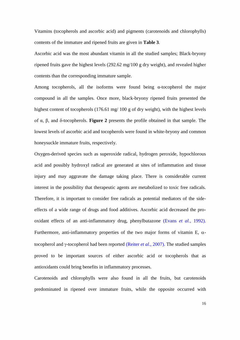
Among tocopherols, all the isoforms were found being α-tocopherol the major
compound in all the samples. Once more, black-bryony ripened fruits presented the
highest content of tocopherols (176.61 mg/ 100 g of dry weight), with the highest levels
of α, β, and δ-tocopherols. Figure 2 presents the profile obtained in that sample. The
lowest levels of ascorbic acid and tocopherols were found in white-bryony and common
honeysuckle immature fruits, respectively.
Oxygen-derived species such as superoxide radical, hydrogen peroxide, hypochlorous
acid and possibly hydroxyl radical are generated at sites of inflammation and tissue
injury and may aggravate the damage taking place. There is considerable current
interest in the possibility that therapeutic agents are metabolized to toxic free radicals.
Therefore, it is important to consider free radicals as potential mediators of the side-
effects of a wide range of drugs and food additives. Ascorbic acid decreased the pro-
oxidant effects of an anti-inflammatory drug, phenylbutazone (Evans et al., 1992).
Furthermore, anti-inflammatory properties of the two major forms of vitamin E, -
tocopherol and -tocopherol had been reported (Reiter et al., 2007). The studied samples
proved to be important sources of either ascorbic acid or tocopherols that as
antioxidants could bring benefits in inflammatory processes.
Carotenoids and chlorophylls were also found in all the fruits, but carotenoids
predominated in ripened over immature fruits, while the opposite occurred with

17
chlorophylls content. Common honeysuckle ripened fruits revealed the highest
concentration of β-carotene (195.68 mg/100 g dry weight). Lycopene and chlorophylls
were found in very low amounts, giving black-bryony ripened fruits the highest
concentration of lycopene (1.85 mg/100 g), and white-bryony immature fruits the
highest content in chlorophylls a (2.43 mg/100 g) and b (1.14 mg/100 g).
In relation to sugar composition (Table 4) common honeysuckle ripened fruits gave the
highest total sugars content (28.15 g/100 g dry weight), with the highest levels of
fructose (10.19 g/100 g) and glucose (11.91 g/100 g). Immature fruits of the same
sample showed the highest levels of trehalose (2.21 g/100 g) and raffinose (1.08 g/100
g). Ripened fruits gave higher total sugars, fructose and glucose content than immature
fruits, as it can be observed in the example of common honeysuckle (Figure 3).
Nevertheless, the amounts of trehalose (disaccharide) and raffinose (trisaccharide)
decreased in the ripened fruits. It should be highlighted that the anti-inflammatory
actions of some sugars such as 2-deoxyglucose, was already reported (Goth, 1968).
The results for fatty acid composition, total saturated fatty acids (SFA),
monounsaturated fatty acids (MUFA), and polyunsaturated fatty acids (PUFA) are
shown in Table 5. Twenty three fatty acids were identified and quantified. The major
fatty acid found in white-bryony and common honeysuckle was linoleic acid (C18:2n6),
while oleic acid (C18:1n9) predominated in black-bryony, contributing to the
prevalence of PUFA in the first two species and MUFA in the last species. Palmitic acid
(C16:0) was also a main fatty acid in all the studied fruits. Overall, white-bryony
immature fruits revealed the highest PUFA levels, including linoleic acid. Some n-3

18
fatty acids were also found in the studied samples. It was reported that n-3, or omega-3
PUFA exhibit anti-inflammatory properties in many inflammatory diseases (Gil, 2002).
Traditional medicinal uses, whose knowledge and practices have been orally transmitted
over the centuries, are important approaches for discovering therapeutic molecules and
compounds. This study provides novel information about phytochemical composition of
wild fruits traditionally used for medicinal purposes, useful to researchers in
phytopharmacology, phytotherapy and phytotoxicology. Black-bryony ripened fruits
revealed the highest antioxidant properties which are in agreement to its highest
concentration in phenolics, flavonoids, ascorbic acid, tocopherols and lycopene.
Otherwise, white-bryony immature fruits gave the lowest antioxidant potential and
hydrophilic antioxidants (phenolics, flavonoids, ascorbic acid and sugars) concentration.
Overall, the studied fruits revealed interesting antioxidant properties and bioactive
phytochemicals such as phenolics, flavonoids, vitamins, carotenoids, sugars, and fatty
acids that could provide scientific evidence for some folk uses as anti-inflammatory
species.
Acknowledgements
The authors are grateful to the Foundation for Science and Technology (FCT, Portugal)
for financial support to the research centre CIMO. L. Barros thanks to FCT, POPH-
QREN and FSE for her grant (SFRH/BPD/4609/2008).

19
References
Barros, L., Heleno, S.A., Carvalho, A.M., Ferreira, I.C.F.R. 2010a. Lamiaceae often
used in Portuguese folk medicine as a source of powerful antioxidants: Vitamins
and phenolics. LWT 43, 544–550.
Barros, L., Oliveira, S., Carvalho, A.M., Ferreira, I.C.F.R. 2010b. In vitro antioxidant
properties and characterization in nutrients and phytochemicals of six medicinal
plants from the Portuguese folk medicine. Ind. Crops Prod. 32, 572–579.
Benitez, G., González-Tejero, M.R., Molero-Mesa, J. 2010. Pharmaceutical ethnobotany
in the western part of Granada province (southern Spain): Ethnopharmacological
synthesis. J. Ethnopharmacol. 129, 87–105.
Carvalho, A.M. 2010. Plantas y sabiduría popular del Parque Natural de Montesinho. Un
estudio etnobotánico en Portugal. Biblioteca de Ciencias nº 35, Consejo Superior
de Investigaciones Científicas, Madrid.
Casagrande, R., Georgetti, S.R., Verri, Jr. W.A., Dorta, D.J., Santos, A.C., Fonseca,
M.J.V. 2006. Protective effect of topical formulations containing quercetin against
UVB-induced oxidative stress in hairless mice. J. Photochem. Photobiol. B: Biol.
84, 21–27.
Castroviejo, S. (coord). 2005. Flora Iberica. Plantas vasculares de la Península Ibérica e
Islas Baleares, Vol. III and Vol. XXI, Real Jardín Botánico, CSIC, Madrid.
Conner, E.M., Grisham, M.B. 1996. Inflammation, free radicals and antioxidants.
Nutrition 12, 274–277.

20
Evans, P.J., Cecchini, R., Halliwell, B. 1992. Oxidative damage to lipids and q-
antiproteinase by phenylbutazone in the presence of haem proteins: protection by
ascorbic acid. Biochem. Pharmacol. 44, 981-984.
Gallego, E.C., Gallego, A.C. 2008. Usos, tradiciones y conocimientos de las plantas por
las gentes de Sayago. ADERISA, Zamora, Spain.
Gil, Á. 2002. Polyunsaturated fatty acids and inflammatory diseases. Biomed.
Pharmacother. 56, 388–396.
González, J.A., García-Barriuso, M., Amich, F. 2010. Ethnobotanical study of medicinal
plants traditionally used in the Arribes del Duero, western Spain. J.
Ethnopharmacol. 131, 343–355.
Goth, A. 1962. Interaction of carbohydrates and anti-inflammatory drugs with mast cells
in the rat. Biochem. Pharmacol. 309-314.
Halliwell, B. 1994. Free radicals, antioxidants and human diseases: curiosity, cause or
consequences. Lancet 334, 721–724.
Jia, Z., Tang, M., Wu, J. 1999. The determination of flavonoid contents in mulberry and
their scavenging effects on superoxide radicals. Food Chem. 64, 555-559.
Klein, B.P., Perry, AK. 1982. Ascorbic acid and vitamin A activity in selected
vegetables from different geographical areas of the United States. J. Food Sci. 47,
941–945.
Liu, H., Qiu, N., Ding, H., Yao, R. 2008. Polyphenols contents and antioxidant capacity
of 68 Chinese herbals suitable for medical or food uses. Food Res. Int. 41, 363–
370.

21
Marc, E.B., Nelly,A., Annick, D.-D., Frederic, D. 2008. Plants used as remedies
antirheumatic and antineuralgic in the traditional medicine of Lebanon. J.
Ethnopharmacol. 120, 315–334.
Martins, D., Barros, L., Carvalho, A.M., Ferreira, I.C.F.R. 2011. Nutritional and in vitro
antioxidant properties of edible wild greens in Iberian Peninsula traditional diet.
Food Chem. 125, 488-494.
Nagata, M., Yamashita, I. 1992. Simple method for simultaneous determination of
chlorophyll and carotenoids in tomato fruit. Nippon Shokuhin Kogyo Gakkaish 39,
925–928.
Nardi, G.M., Junior, J.M.S., Monachec, F.D., Pizzolatti, M.G., Ckless, Ribeiro-do-Valle,
R.M. 2007. Antioxidant and anti-inflammatory effects of products from Croton
celtidifolius Bailon on carrageenan-induced pleurisy in rats. Phytomedicine 14,
115-122.
Neves, J.M., Matosa, C., Moutinho, C., Queiroz, G., Gomes, L.R. 2009.
Ethnopharmacological notes about ancient uses of medicinal plants in Trás-os-
Montes (northern of Portugal). J. Ethnopharmacol. 124, 270–283.
Reiter, E., Jiang, Q., Christen, S. 2007. Anti-inflammatory properties of - and -
tocopherol. Mol. Aspects Med. 28, 668–691.
Roome, T., Dar, A., Ali, S., Naqvi, S., Choudhary, M.I. 2008. A study on antioxidant,
free radical scavenging, anti-inflammatory and hepatoprotective actions of
Aegiceras corniculatum (stem) extracts. J. Ethnopharmacol. 118, 514–521.
Tsai, T.-H., Tsai, T.-H., Chien, Y.-C., Lee, C.-W., Tsai, P.-J. 2008. In vitro antimicrobial
activities against cariogenic streptococci and their antioxidant capacities: A
comparative study of green tea versus different herbs. Food Chem. 110, 859–864.

22
Wolfe, K., Wu, X., Liu, R.H. 2003. Antioxidant activity of apple peels. J. Agric. Food
Chem. 51, 609-614.

23
Table 1. Medicinal uses of the studied fruits reported in ethnobotanical surveys recently carried out in the Iberian Peninsula.
Scientific name
(Botanical family) English name Local name Therapeutic indication Preparation References
Bryonia dioica Jacq.
(Cucurbitaceae) White-bryony Norça, norça-branca, nóscora
Bruises, rheumatic pains,
arthritis, minor wounds
and lesions
Maceration in alcohol or
brandy Gallego and Gallego, 2008
Carvalho, 2010
González et al.., 2010 The juice of crushed fruits
for rubbing into the skin
Tamus communis L.
(Dioscoreaceae) Black -bryony Norça-preta, budanha
Bruises, goat, minor
wounds, rheumatic pains
Maceration in alcohol or
brandy Gallego and Gallego, 2008
Carvalho, 2010
González et al., 2010
The juice of crushed fruits
Crushed fruits mixed with
lard to produce a sort of
paste
Lonicera periclymenum L.
(Caprifoliaceae) Common honeysuckle Madressilva
Asthma and respiratory
affections
Rheumatic pains,
arthritis, minor wounds
Inhalants from the hot
decoction of the fruits
Maceration in alcohol
Neves et al., 2009
Carvalho, 2010
Caution: In spite of their medicinal use, these fruits are considered toxic for humans and domestic animals, therefore their traditional use is only recommended for
external applications. Moreover, in cases of broken skin and individuals with certain contact sensitivities their use should be avoid or extracts must be applied at low
doses.

24
Table 2. Extraction yields, antioxidant activity (EC50 values, mg/mL) and composition in phenolics and flavonoids (mean SD; n=9) of the
studied fruits. In each row different letters mean significant differences (p0.05).
White-bryony
Immature fruits
White-bryony
Ripened fruits
Black-bryony
Immature fruits
Black-bryony
Ripened fruits
Common honeysuckle
Immature fruits
Common honeysuckle
Ripened fruits
(%) 25.32 ± 1.23 24.56 ± 0.87 41.82 ± 2.01 26.74 ± 0.55 79.37 ± 3.25 68.15 ± 1.98
DPPH scavenging activity 18.01 ± 0.64 a 1.21 ± 0.02 dc 1.26 ± 0.12 dc 0.91 ± 0.01 d 1.44 ± 0.06 c 2.97 ± 0.13 b
Reducing power 1.07 ± 0.01 a 0.40 ± 0.00 e 0.57 ± 0.01 d 0.21 ± 0.01 f 0.76 ± 0.01 c 1.05 ± 0.01 b
β-carotene bleaching inhibition 1.87 ± 0.25 a 0.58 ± 0.09 d 0.79 ± 0.04 c 0.21 ± 0.01 e 1.48 ± 0.14 b 1.74 ± 0.09 a
TBARS inhibition 5.18 ± 0.46 a 0.22 ± 0.02 cd 0.29 ± 0.01 cbd 0.15 ± 0.03 d 0.43 ± 0.02 cb 0.49 ± 0.03 b
Phenolics (mg GAE/g extract) 33.37 ± 6.50 e 150.12 ± 1.38 b 119.78 ± 15.70 c 445.96 ± 31.41 a 71.47 ± 1.88 d 55.47 ± 0.37 d
Flavonoids (mg CE/g extract) 10.32 ± 0.49 e 15.77 ± 2.34 d 14.54 ± 1.78 d 52.69 ± 3.51 a 28.15 ± 0.83 b 23.33 ± 0.63 c
EC50 values for the standard trolox: 43 g/ml (DPPH scavenging activity); 30 g/ml (Reducing power); 3 g/ml (β-carotene bleaching inhibition) and 4 g/ml (TBARS
inhibition).

25
Table 3. Composition in vitamins and pigments (mg/100 g dry weight) of the studied fruits (mean SD; n=9). In each row different letters mean
significant differences (p0.05).
White-bryony
Immature fruits
White-bryony
Ripened fruits
Black-bryony
Immature fruits
Black-bryony
Ripened fruits
Common honeysuckle
Immature fruits
Common honeysuckle
Ripened fruits
α-tocopherol 30.11 ± 1.53 d 96.14 ± 9.41 b 59.27 ± 6.97 c 160.21 ± 9.83 a 6.35 ± 0.00 e 9.37 ± 0.30 e
β-tocopherol 0.71 ± 0.02 d 3.55 ± 0.16 b 1.09 ± 0.14 c 5.69 ± 0.04 a 0.25 ± 0.00 e 0.25 ± 0.02 e
γ-tocopherol 21.63 ± 1.05 a 10.44 ± 0.69 b 5.05 ± 0.75 d 7.16 ± 0.14 c 1.42 ± 0.02 f 3.35 ± 0.10 e
δ-tocopherol 1.01 ± 0.16 b 0.87 ± 0.01 cb 0.85 ± 0.17 cb 1.55 ± 0.20 a 0.54 ± 0.00 cd 0.25 ± 0.02 d
Total tocopherols 53.46 ± 2.75 c 111.00 ± 10.26 b 66.26 ± 8.01 c 176.61 ± 10.13 a 8.56 ± 0.02 d 13.22 ± 0.24 d
Ascorbic acid 63.61 ± 6.21 f 217.10 ± 7.36 b 96.08 ± 4.57 e 292.62 ± 1.84 a 141.40 ± 0.03 d 150.94 ± 0.10 c
β-carotene 69.82 ± 0.13 d 155.28 ± 6.50 b 0.24 ± 0.00 e 139.92 ± 7.81 c 6.68 ± 0.65 e 195.68 ± 6.30 a
Lycopene 0.01 ± 0.00 e 1.80 ± 0.01 b 0.02 ± 0.00 e 1.85 ± 0.01 a 0.30 ± 0.02 d 0.41 ± 0.03 c
Chlorophyll a 2.43 ± 0.00 a 0.11 ± 0.00 e 0.55 ± 0.00 c 0.14 ± 0.00 e 0.94 ± 0.03 b 0.38 ± 0.05 d
Chlorophyll b 1.14 ± 0.00 a 0.18 ± 0.00 d 0.20 ± 0.00 d 0.23 ± 0.00 d 0.81 ± 0.02 b 0.43 ± 0.08 c

26
Table 4. Composition in sugars (g/100 g of dry weight) of the studied fruits (mean
SD; n=9). In each row, different letters mean significant differences (p0.05).
nd- not detected
White-bryony
Immature fruits
White-bryony
Ripened fruits
Black-bryony
Immature fruits
Black-bryony
Ripened fruits
Common honeysuckle
Immature fruits
Common honeysuckle
Ripened fruits
Fructose 2.26 ± 0.09 d 3.24 ± 0.50 c 0.89 ± 0.10 e 6.44 ± 0.01 b 2.22 ± 0.05 d 10.19 ± 0.02 a
Glucose 2.48 ± 0.02 d 2.95 ± 0.34 dc 2.86 ± 0.10 dc 6.26 ± 0.01 b 3.40 ± 0.39 c 11.91 ± 0.12 a
Sucrose 0.46 ± 0.02 d 2.19 ± 0.40 c 4.56 ± 0.20 a 2.58 ± 0.11 c 2.39 ± 0.11 c 3.81 ± 0.02 b
Trehalose 0.59 ± 0.02 c 0.35 ± 0.09 d 0.27 ± 0.02 d 0.30 ± 0.03 d 2.21 ± 0.06 a 1.76 ± 0.02 b
Raffinose 0.12 ± 0.02 c nd nd nd 1.08 ± 0.09 a 0.48 ± 0.02 b
Total sugars 5.92 ± 0.13 e 8.72 ± 1.32 d 8.59 ± 0.41 d 15.57 ± 0.10 b 11.30 ± 0.69 c 28.15 ± 0.13 a

27
Table 5. Composition (percentage) in fatty acids of the studied fruits (mean SD; n=3).
In each column different letters mean significant differences (p0.05).
White-bryony
Immature fruits
White-bryony
Ripened fruits
Black-bryony
Immature fruits
Black-bryony
Ripened fruits
Common honeysuckle
Immature fruits
Common honeysuckle
Ripened fruits
C6:0 0.01 ± 0.00 0.02 ± 0.00 0.03 ± 0.01 0.01 ± 0.00 0.05 ± 0.00 0.01 ± 0.00
C8:0 0.01 ± 0.00 0.03 ± 0.00 0.06 ± 0.02 0.02 ± 0.00 0.03 ± 0.01 0.01 ± 0.00
C10:0 0.01 ± 0.00 0.03 ± 0.01 0.04 ± 0.01 0.02 ± 0.00 0.04 ± 0.00 0.01 ± 0.00
C12:0 0.02 ± 0.00 0.10 ± 0.01 0.10 ± 0.02 0.12 ± 0.02 0.28 ± 0.00 0.08 ± 0.00
C14:0 0.06 ± 0.00 0.23 ± 0.00 0.35 ± 0.07 0.29 ± 0.02 0.36 ± 0.01 0.17 ± 0.01
C14:1 nd nd 0.06 ± 0.01 nd 0.08 ± 0.01 0.02 ± 0.00
C15:0 0.05 ± 0.01 0.09 ± 0.00 0.21 ± 0.03 0.11 ± 0.00 0.08 ± 0.00 0.03 ± 0.00
C16:0 7.80 ± 0.04 11.60 ± 0.31 13.15 ± 1.11 13.54 ± 0.58 8.35 ± 0.00 6.11 ± 0.01
C16:1 0.09 ± 0.01 0.11 ± 0.01 0.34 ± 0.07 0.08 ± 0.01 0.18 ± 0.00 0.17 ± 0.00
C17:0 0.10 ± 0.00 0.16 ± 0.01 0.24 ± 0.08 0.18 ± 0.02 0.20 ± 0.00 0.08 ± 0.01
C18:0 3.27 ± 0.05 2.85 ± 0.09 3.33 ± 0.81 2.30 ± 0.16 2.59 ± 0.11 2.40 ± 0.01
C18:1n9 17.07 ± 0.13 28.47 ± 2.62 56.40 ± 0.58 51.40 ± 2.19 17.40 ± 0.28 31.82 ± 0.09
C18:2n6 66.83 ± 0.35 50.49 ± 3.30 21.01 ± 1.51 24.65 ± 0.58 44.96 ± 0.33 54.64 ± 0.37
C18:3n3 3.04 ± 0.09 2.92 ± 0.06 2.14 ± 0.01 3.96 ± 0.83 9.38 ± 0.05 1.43 ± 0.03
C20:0 0.19 ± 0.03 0.39 ± 0.03 0.57 ± 0.04 0.53 ± 0.04 0.36 ± 0.01 0.15 ± 0.00
C20:1 0.11 ± 0.01 0.30 ± 0.05 0.56 ± 0.04 0.47 ± 0.01 0.07 ± 0.00 0.12 ± 0.01
C20:2 0.01 ± 0.00 0.02 ± 0.00 0.03 ± 0.01 0.02 ± 0.00 0.07 ± 0.00 0.02 ± 0.00
C20:3n6 0.29 ± 0.03 0.01 ± 0.00 nd nd nd nd
C20:3n3+C21:0 0.25 ± 0.09 0.21 ± 0.01 0.06 ± 0.00 0.07 ± 0.01 0.12 ± 0.01 nd
C20:5n3 0.39 ± 0.07 0.26 ± 0.03 0.04 ± 0.00 0.01 ± 0.00 nd nd
C22:0 0.13 ± 0.01 0.70 ± 0.05 0.60 ± 0.03 0.87 ± 0.15 1.30 ± 0.08 0.23 ± 0.01
C23:0 0.01 ± 0.00 0.05 ± 0.01 0.05 ± 0.01 0.05 ± 0.00 8.04 ± 0.29 1.40 ± 0.19
C24:0 0.25 ± 0.01 0.97 ± 0.06 0.65 ± 0.03 1.29 ± 0.11 6.07 ± 0.17 1.10 ± 0.09
Total SFA 11.91 ± 0.11 d 17.21 ± 0.54 c 19.36 ± 2.00 b 19.33 ± 0.72 b 27.74 ± 0.63 a 11.78 ± 0.30 d
Total MUFA 17.26 ± 0.15 e 28.88 ± 2.67 d 57.35 ± 0.55 a 51.96 ± 2.17 b 17.73 ± 0.28 e 32.13 ± 0.19 c
Total PUFA 70.82 ± 0.25 a 53.91 ± 3.21 b 23.28 ± 1.44 d 28.71 ± 1.45 c 54.53 ± 0.35 b 56.10 ± 0.09 b
Total fat (g/100 g
dry weight)
1.52 ± 0.04 e 1.90 ± 0.14 e 3.26 ± 0.05 d 11.54 ± 0.49 c 16.49 ± 0.18 b 23.04 ± 0.26 a
nd- not detected.

28
0
10
20
30
40
50
60
70
80
90
100
0 0.2 0.4 0.6 0.8 1 1.2 1.4 1.6 1.8 2
DP
PH
ra
dic
al-
scav
eng
ing a
cti
vit
y (
%)
Extract concentration (mg/ml)
0
0.2
0.4
0.6
0.8
1
1.2
1.4
1.6
1.8
2
0 0.1 0.2 0.3 0.4 0.5 0.6 0.7 0.8 0.9 1
Ab
s 6
90
nm
Exytract concentration (mg/ml)

29
0
10
20
30
40
50
60
70
80
90
0 0.2 0.4 0.6 0.8 1 1.2 1.4 1.6 1.8 2
β-c
aro
ten
e b
lea
ch
ing
in
hib
itio
n (
%)
Extract concentration (mg/ml)
0
10
20
30
40
50
60
70
80
90
0 0.2 0.4 0.6 0.8 1 1.2 1.4 1.6 1.8 2
TB
AR
S in
hib
itio
n (
%)
Extract concentration (mg/ml)
Figure 1. Antioxidant activity of white-bryony immature fruits ( ); white-bryony
ripened fruits ( ); black-bryony immature fruits ( ); black-bryony ripened
fruits ( ); common honeysuckle immature fruits ( ); common honeysuckle
ripened fruits ( ).

30
Time (min.)5 10 15 20 25
Vo
lta
ge
(V)
0.0
0.5
1.0
1.5
2.0
1
12 14 16 18 20
24
3
5
0
Figure 2. Individual tocopherols chromatograms of black-bryony ripened fruits: 1- α-
tocopherol; 2-β-tocopherol; 3- γ-tocopherol; 4- δ-tocopherol; 5-tocol (IS).

31
Time (min)0 2 4 6 8 10 12
Vo
lta
ge
(mV
)
0
50
100
150
200
250
1
2
3
4 5
6
Common honeysuckle Ripened fruits
Common honeysuckle Immature fruits
Figure 3. Individual sugars chromatogram of common honeysuckle fruits: 1- fructose;
2- glucose; 3- sucrose; 4- trehalose; 5- melezitose (IS); 6- raffinose.