topics -defense mechanisms -systems -non...
TRANSCRIPT
1
Chapter 14
Topics- Defense Mechanisms- Systems - Non-specific immunity
2
Defense Mechanisms
• Inate and nonspecific– Firstline of defense– Secondline of defense
• Acquired and specific– Thirdline of defense
3
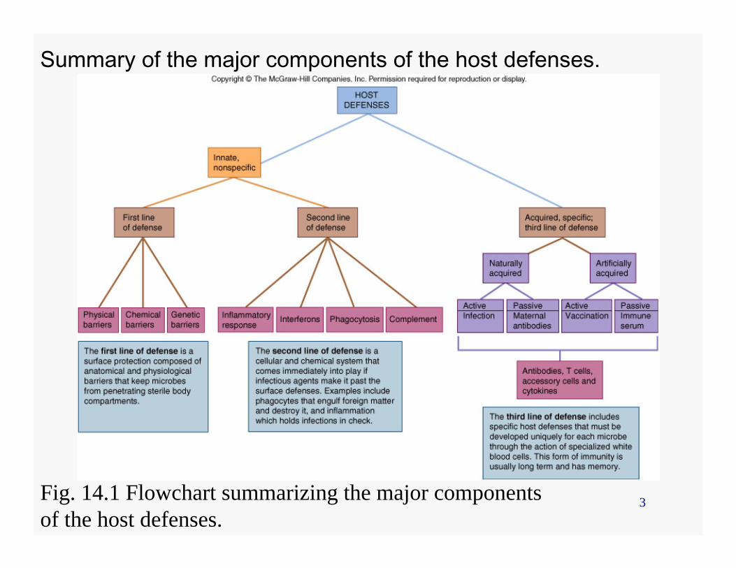
Summary of the major components of the host defenses.
Fig. 14.1 Flowchart summarizing the major componentsof the host defenses.
4
First line of defense
• Barriers– Anatomical– Chemical– Genetic
5
Anatomical barriers• Skin
– Outermost layer– Hair follicles– Skin glands
• Mucous membrane– Digestive– Urinary– Respiratory– Eye

6
The trachea contain cilia that entrap and propel particles out of the respiratory tract.
F
Fig. 14.3 The ciliary defense of the respiratory tree.
7
Chemical barriers• Sebaceous secretions• Eyelid glands – meibomian gland• Tears and saliva – lysozyme• Acidic pH
– Sweat – Stomach– Skin – Vagina
• Semen
8
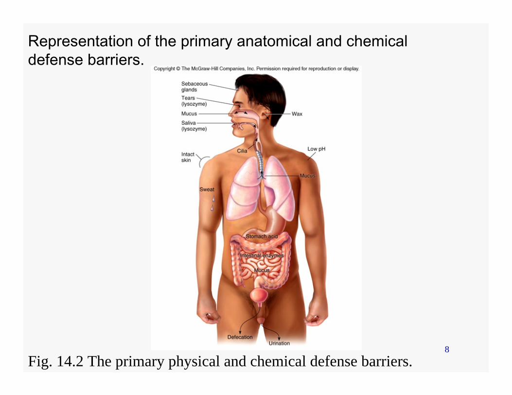
Representation of the primary anatomical and chemical defense barriers.
Fig. 14.2 The primary physical and chemical defense barriers.
9
Genetic barriers
• Different level of sensitivity and resistance to infectious agents– Malaria– Tuberculosis– Leprosy– Fungal infections
10
Second line and Third line of defense
• Defines immunology• Protective cells
11
Immunology• Study of the development of resistance to
infectious agents by the body– Surveillance of the body– Recognition of foreign material– Destruction of foreign material or agent
• Involve nonspecific and specific immune defense systems
• White blood cells (wbc) or leukocytes are involved
12
WBC
• WBC recognize self markers on the host cell – Do not attack or do not respond to host cell
• WBC recognize nonself markers on the invading microbe– Attack or respond to microbe
13
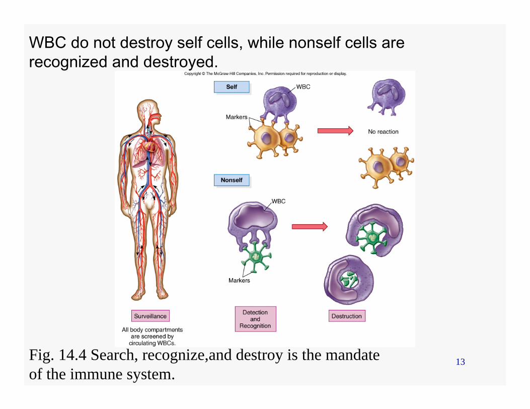
WBC do not destroy self cells, while nonself cells are recognized and destroyed.
Fig. 14.4 Search, recognize,and destroy is the mandateof the immune system.
14
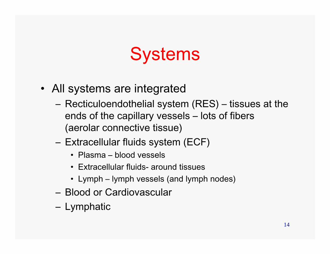
Systems
• All systems are integrated– Recticuloendothelial system (RES) – tissues at the
ends of the capillary vessels – lots of fibers (aerolar connective tissue)
– Extracellular fluids system (ECF)• Plasma – blood vessels• Extracellular fluids- around tissues• Lymph – lymph vessels (and lymph nodes)
– Blood or Cardiovascular– Lymphatic
15
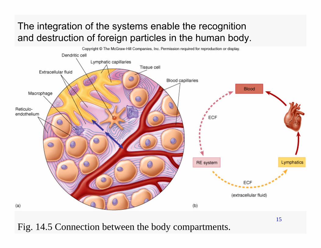
The integration of the systems enable the recognition and destruction of foreign particles in the human body.
Fig. 14.5 Connection between the body compartments.
16
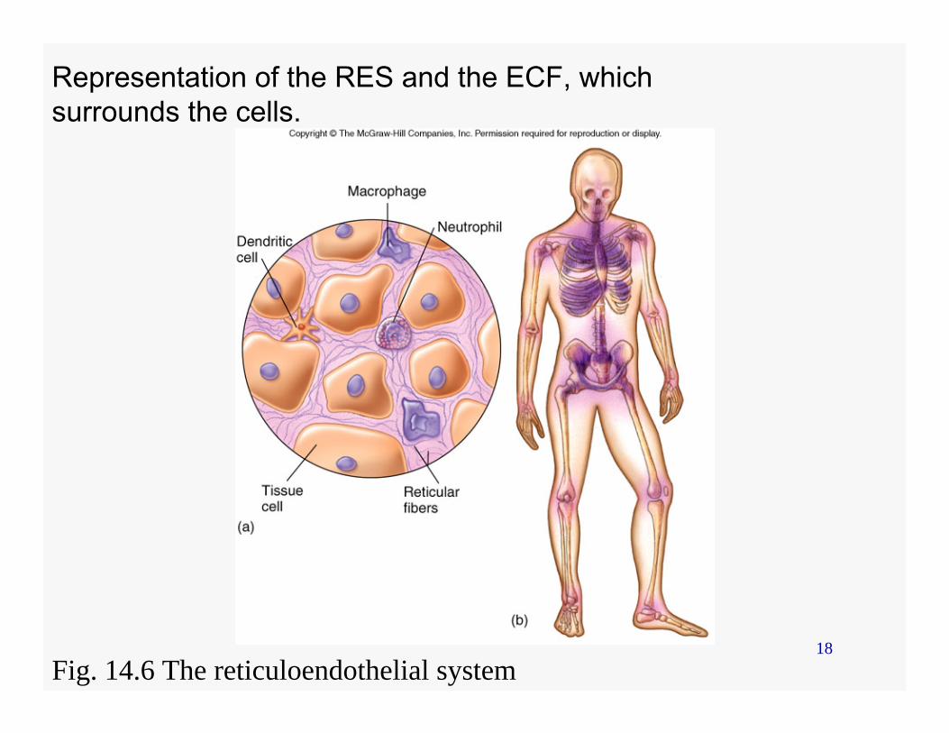
Reticuloendothelial (RES)
• Network of connective tissue fibers (Reticulum)
• Interconnects cells• Allows immune cells to bind and move
outside the blood and lymphatic system
17
Extracellular fluid (ECF)
• The spaces surrounding tissue cells and RES
• Enable immune cells to move
18
Representation of the RES and the ECF, which surrounds the cells.
Fig. 14.6 The reticuloendothelial system
19
Blood
• Stem cells precursors• Hemopoiesis• Components
20
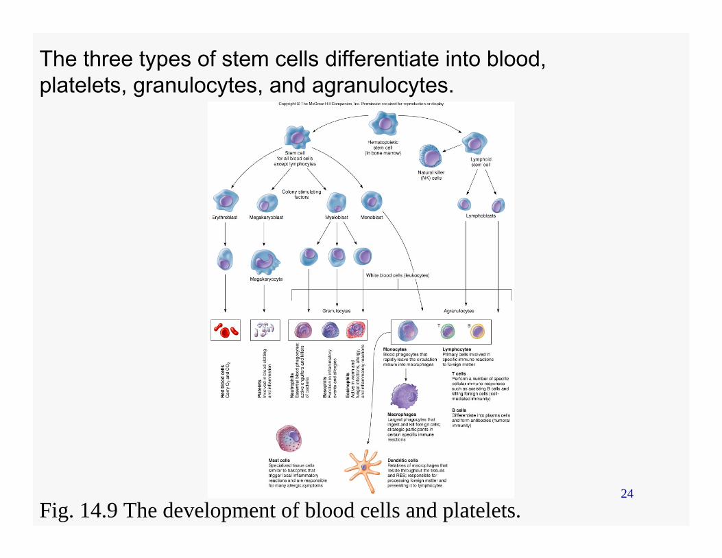
Stem cells
• From blood cells– Rbc– platelets
• Hematopoietic stem cells in bone marrow– Neutraphils, basophils, eosinophils, monocytes
• Lymphoid stem cells– T cells– B cells
21
Hemopoiesis
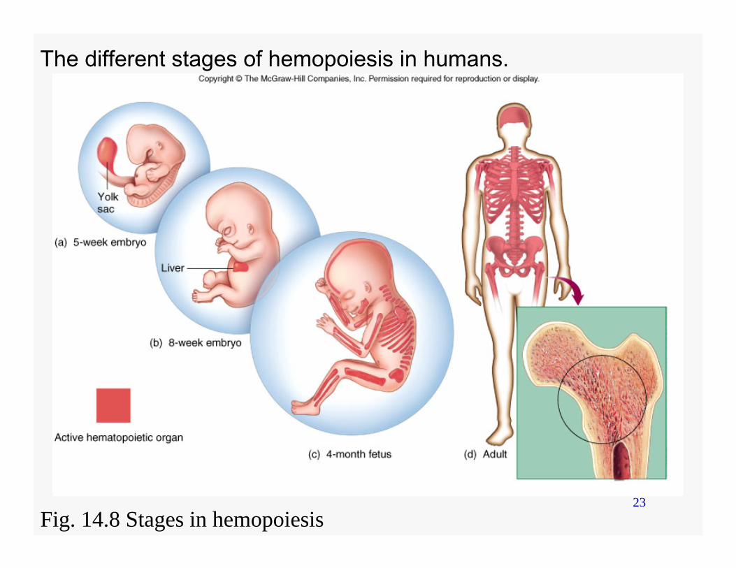
• Production of blood– Starts at the embryonic stage
• Yolk sac and liver– Continues during adult stage
• Bone marrow
22
Components of blood
• White blood cells (WBC) or leukocytes• Red blood cells (RBC)• Platelets
23
The different stages of hemopoiesis in humans.
Fig. 14.8 Stages in hemopoiesis
24
The three types of stem cells differentiate into blood, platelets, granulocytes, and agranulocytes.
Fig. 14.9 The development of blood cells and platelets.
25
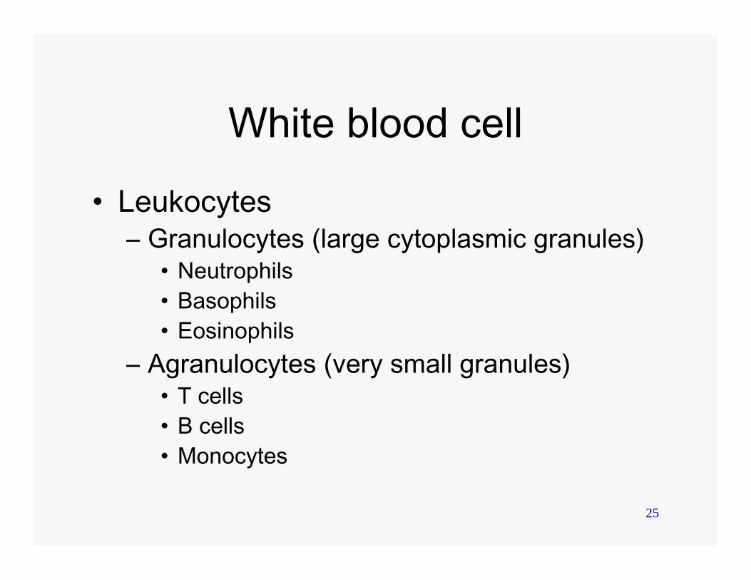
White blood cell
• Leukocytes– Granulocytes (large cytoplasmic granules)
• Neutrophils• Basophils• Eosinophils
– Agranulocytes (very small granules)• T cells• B cells • Monocytes
26
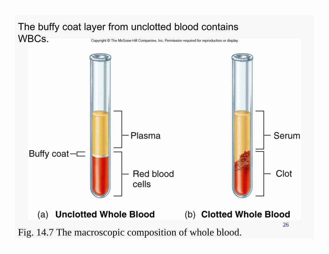
The buffy coat layer from unclotted blood contains WBCs.
Fig. 14.7 The macroscopic composition of whole blood.
27
Neutrophils
• Nuclei - horse shoe or polymorphic nuclei
• Present in high numbers in blood and tissue
• Phagocytizes bacteria – granules are digestive enzymes
• First to arrive during an immune response (inflammation)
28
Eosinophils
• Nuclei – bilobed• Present in the bone marrow and spleen• Attach and destroy eucaryotic
pathogens• Associated with inflammation and
allergies
29
Basophils
• Nuclei – constricted• Present in low in number in the body• Function is similar to eosinophils• Localized basophils are called mast
cells
30
Lymphocytes
• Specific immunity– T cells – B cells
• Present throughout the body
31
Monocytes
• Agranulocyte• Differentiate into macrophages
(circulation and lymphatics) and dendritic cells (tissue associated)
• Phagocytosis
32
Lymphatic system• Network of vessels that extend to most body
areas • Connected to the blood system• Provides an auxiliary route for the return of
extracellular fluid to the circulatory system• “Drain off” system for inflammatory response• Contains lymphocytes, phagocytes and
antibodies
33
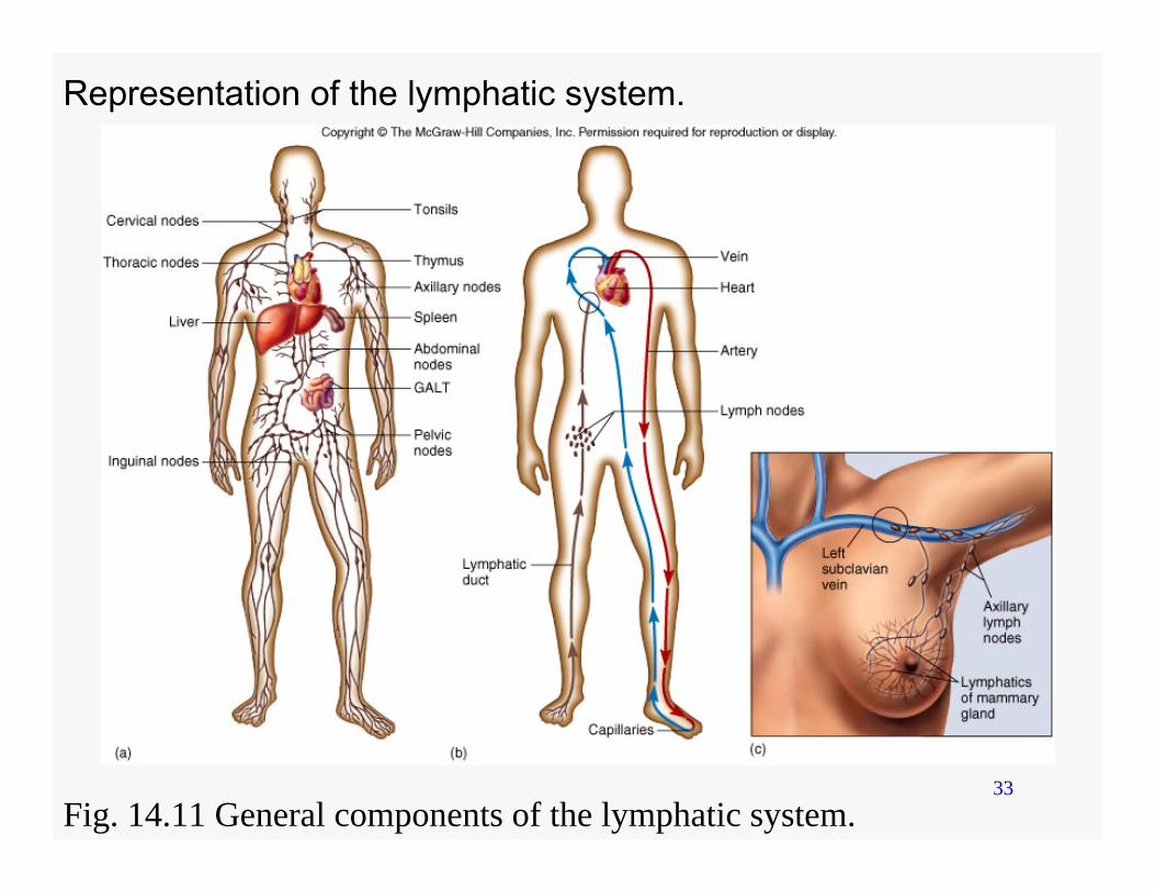
Representation of the lymphatic system.
Fig. 14.11 General components of the lymphatic system.
34
Lymphatic system
• Fluids• Vessels• Nodes• Spleen• Thymus• Miscellaneous
35
Fluids
• Plasma-like fluid (lymph)– Water– Dissolved salts– Proteins (antibodies, albumin)– White blood cells– No red blood cells
• Formed from blood components– Diffuse into the lymphatic capillaries
36
Vessels
• Parallels the blood system• Returns lymph to the blood system• Movement of lymph depends on muscle
contractions• Permeate all parts of the body except
the central nervous system, bone, placenta, and thymus.
37
Lymph nodes
• Exist in clusters• Located
– along the lymphatic channels and blood vessels– in the thoracic and abdominal cavity regions,
armpit, groin and neck• Filter for the lymph• Provide environment for immune reactions
38
Spleen
• Located in the upper left portion of the abdominal cavity
• Filter for blood – traps pathogens and phagocytizes
pathogens• Adults can survive without spleen• Asplenic children are severely
immunocompromised
39
Thymus
• Embryo – two lobes in the pharyngeal region– High activity (releases mature T cells) until
puberty• Adult
– Gradually shrinks– Lymph node and spleen supply mature T
cells
40
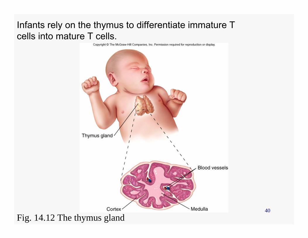
Infants rely on the thymus to differentiate immature T cells into mature T cells.
Fig. 14.12 The thymus gland
41
Gut-associated lymphoid tissue (GALT)
• Recognized incoming microbes from food
• Supply lymphocytes for antibody response
• Ex. Appendix, lacteals, Peyer’s patches
42
Non-specific Immunity
• Inflammation• Phagocytosis• Interferon• Complement
43
Inflammation
• Five major symptoms– Redness– Warmth– Swelling– Pain– Loss of function
44
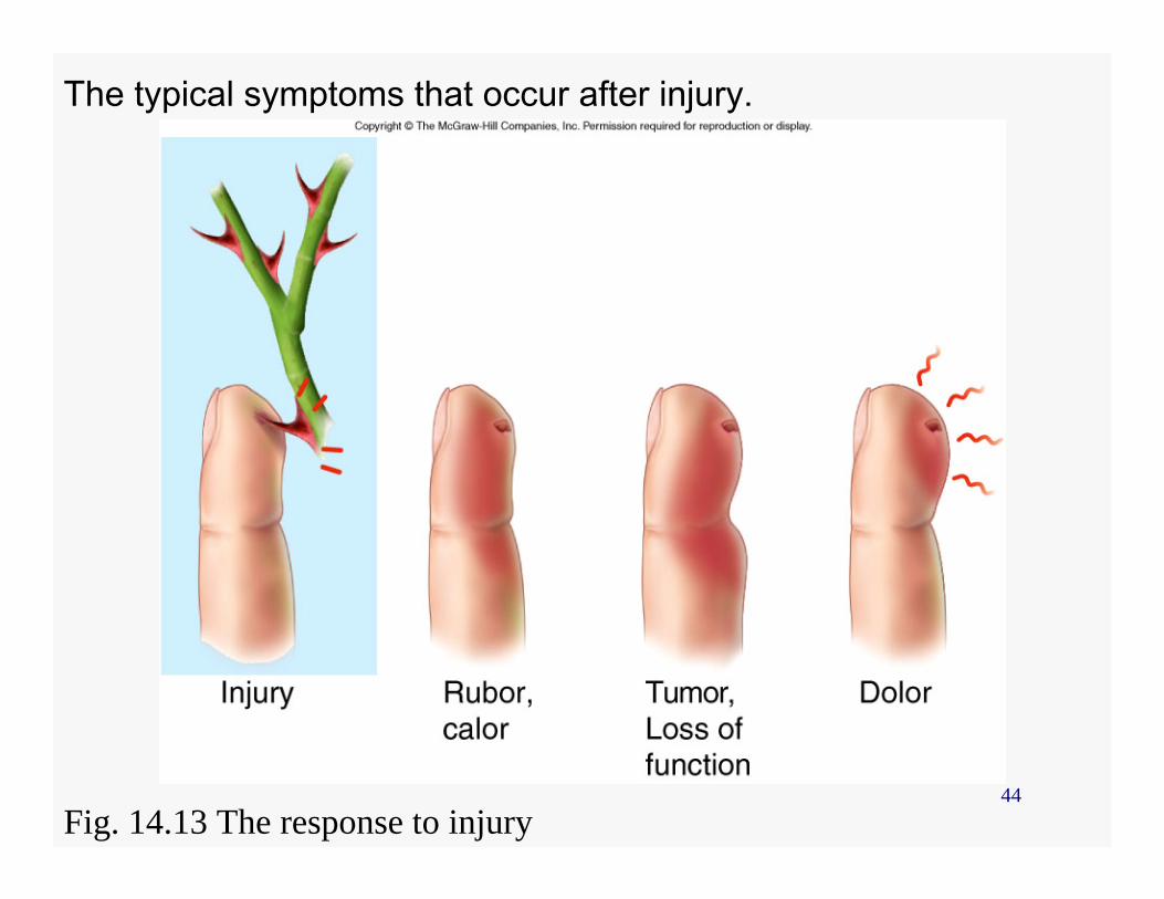
The typical symptoms that occur after injury.
Fig. 14.13 The response to injury
45
Inflammation
• Causes• Function• Stages
46
Causes
• Trauma • Tissue injury due to physical or
chemical agents • Specific immune reactions
47
Function
• Mobilize and attract immune components to the site of injury
• Aid in the repair of tissue damage• Localized and remove harmful
substances• Destroy microbes and block their
invasion
48
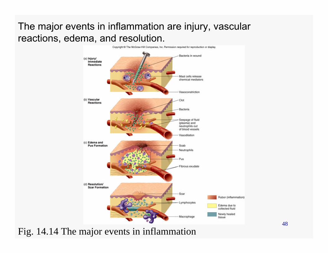
The major events in inflammation are injury, vascular reactions, edema, and resolution.
Fig. 14.14 The major events in inflammation
49
Stages
• Vascular changes• Edema• Fever
50
Vascular changes
• Blood cells, tissue cells, and platelets release chemical mediators and cytokines
• Chemical mediators– Vasoactive
• Affect endothelial cells, smooth muscles of blood vessels
– Chemotactic (chemokines)• Affect WBC
51
Chemical mediators
• Cause fever, stimulate lymphocytes, prevent virus spread, cause allergic reactions– Vasoactive mediators
• Affect endothelial cells, smooth muscles of blood vessels
– Chemotactic (chemokines) mediators• Affect WBC
52
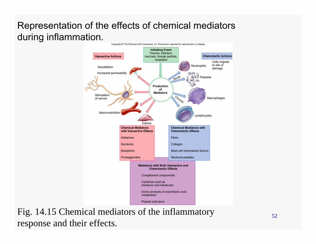
Representation of the effects of chemical mediators during inflammation.
Fig. 14.15 Chemical mediators of the inflammatory response and their effects.
53
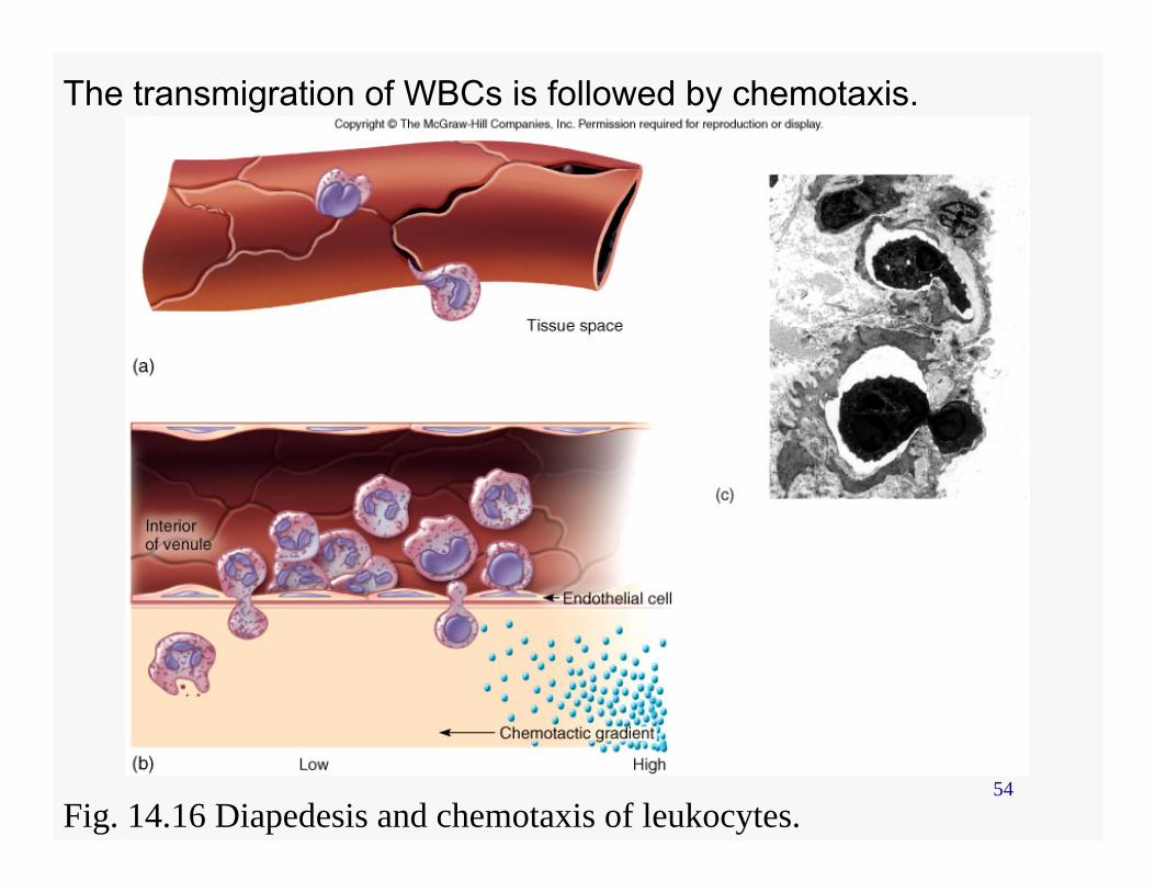
Edema
• Leakage of vascular fluid (exudate) into tissue
• Exudate - plasma proteins, blood cells (wbc), debris, and pus
• Migration of wbc is called diapedesis or transmigration– Chemotaxis
54
The transmigration of WBCs is followed by chemotaxis.
Fig. 14.16 Diapedesis and chemotaxis of leukocytes.
55
Fever• Caused by pyrogens
– reset the hypothalamic thermostat (increase temperature)
– Vasoconstriction• Pyrogens
– Microbes and their products (ex. Lipopolysaccharides)
– Leukocyte products (ex. lnterleukins) • Inhibits microbe and viral multiplication,
reduces nutrient availability, increases immune reactions
56
Phagocytosis
• Neutrophils and eosinophils• Macrophages• Mechanism
57
Neutrophils and eosinophils
• Early responders to inflammation• Neutrophils are primary components of
pus• Eosinophils are primary responders to
parasitic infections
58
Macrophages
• Monocytes transform into macrophages• Scavengers
– Histiocytes – reside in one location (ex. Alveolar, Kupffer, Langerhans)
– Drift throughout the RES• Undergo phagocytosis, • Interact with B and T cells
59
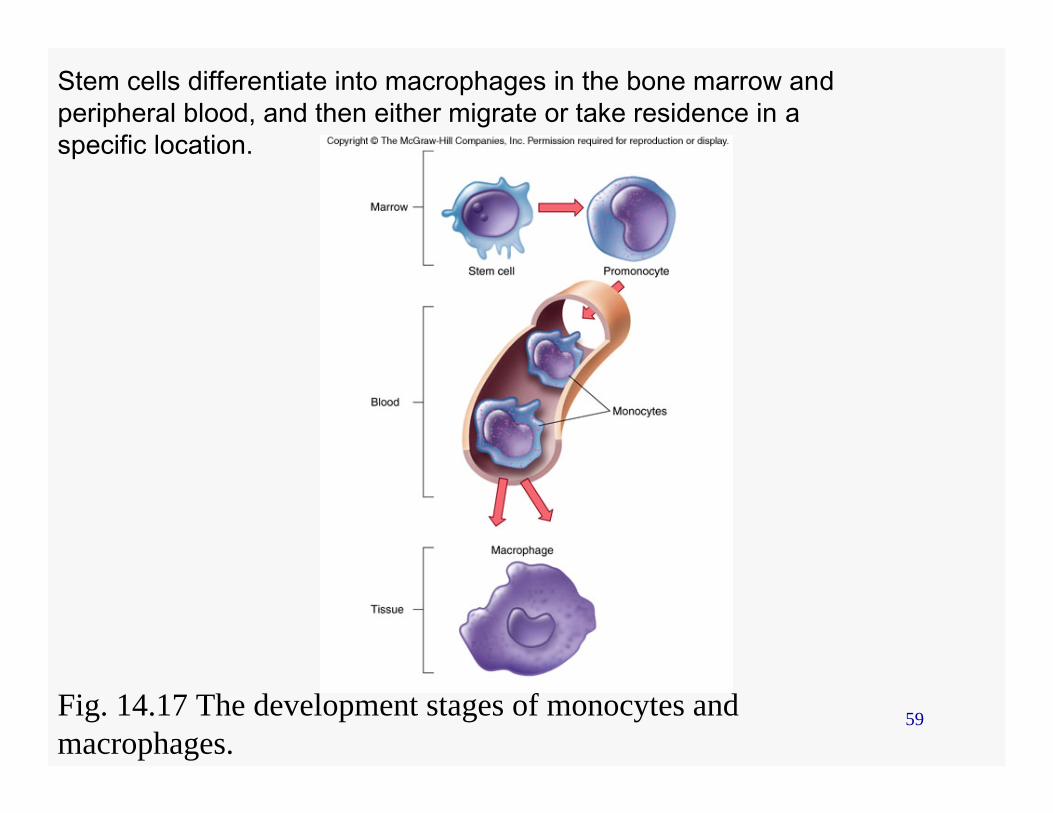
Stem cells differentiate into macrophages in the bone marrow andperipheral blood, and then either migrate or take residence in aspecific location.
Fig. 14.17 The development stages of monocytes and macrophages.
60
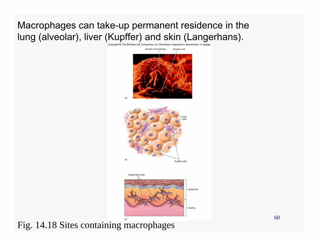
Macrophages can take-up permanent residence in the lung (alveolar), liver (Kupffer) and skin (Langerhans).
Fig. 14.18 Sites containing macrophages
61
Mechanism of Phagocytosis
• Chemotaxis• Ingestion• Phagolysosome• Destruction
62
Chemotaxis
• Directed by – Pathogen-associated molecular patterns
(PAMPs) • Peptidoglycan• LPS
– Foreign debris
63
Ingestion
• Pseudopods enclose the pathogen or foreign material
• Form a phagosome
64
Phagolysosome
• Lysosomes fuse with the phagosome• Other antimicrobials chemicals are
released into the phagolysosome
65
Destruction
• Within the phagolysosome– Oxygen-dependent system
• Oxidative burst (oxidizing agents)– Enzymes – Nitric oxide
• Undigestible debris are released
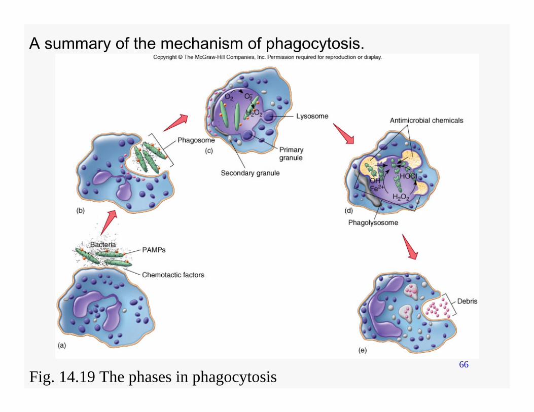
66
A summary of the mechanism of phagocytosis.
Fig. 14.19 The phases in phagocytosis
67
Interferon
• Produced due to viral infections, microbe infections, RNA, immune products, and antigens
68
Interferon Activity
• Ex. Virus - binds to host cell • A signal is sent to the nucleus to synthesized
(transcription and translation) interferon• Interferon is secreted • Binds to other host cells• Host cells produce antiviral proteins
– inhibit viral multiplication or translation• Not virus-specific
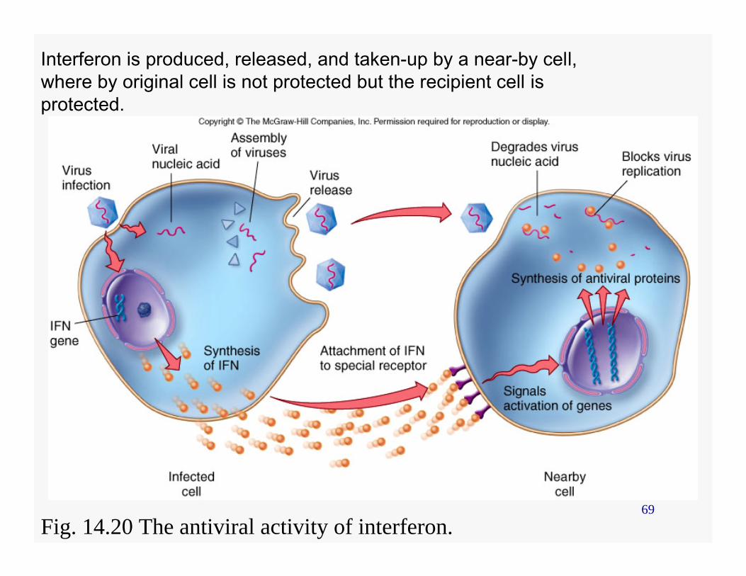
69
Interferon is produced, released, and taken-up by a near-by cell, where by original cell is not protected but the recipient cell is protected.
Fig. 14.20 The antiviral activity of interferon.
70
Other Roles of Interferon
• Activates and instructs T and B cell development
• Inhibits cancer cells• Activates macrophages
71
Complement
• Consist of 26 blood proteins• Produced by liver hepatocytes,
lymphocytes, and monocytes• Pathways • Cascade reaction• Stages
72
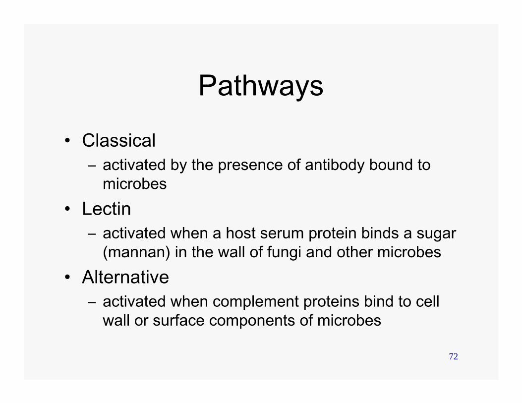
Pathways
• Classical – activated by the presence of antibody bound to
microbes• Lectin
– activated when a host serum protein binds a sugar (mannan) in the wall of fungi and other microbes
• Alternative – activated when complement proteins bind to cell
wall or surface components of microbes
73
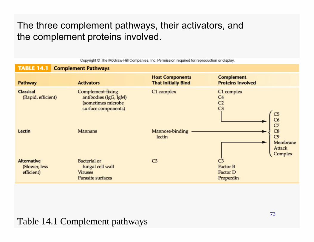
The three complement pathways, their activators, and the complement proteins involved.
Table 14.1 Complement pathways
74
Stages
• Initiation• Amplification and cascade• Polymerization• Membrane attack
75
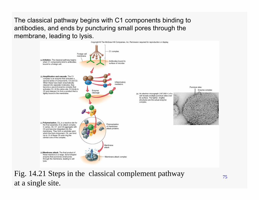
The classical pathway begins with C1 components binding to antibodies, and ends by puncturing small pores through the membrane, leading to lysis.
Fig. 14.21 Steps in the classical complement pathwayat a single site.