topoisomerase i-mediated integration of hepadnavirus
TRANSCRIPT

JOURNAL OF VIROLOGY, May 1991, p. 2381-23920022-538X/91/052381-12$02.00/0Copyright C) 1991, American Society for Microbiology
Topoisomerase I-Mediated Integration of HepadnavirusDNA In Vitro
HAI-PING WANG AND CHARLES E. ROGLER*Marion Bessin Liver Research Center, Department of Medicine, Albert Einstein
College of Medicine, 1300 Morris Park Avenue, Bronx, New York 10461
Received 26 November 1990/Accepted 11 February 1991
Hepadnaviruses integrate in cellular DNA via an illegitimate recombination mechanism, and clonallypropagated integrations are present in most hepatocellular carcinomas which arise in hepadnavirus carriers.Although integration is not specific for any viral or cellular sequence, highly preferred integration sites havebeen identified near the DR1 and DR2 sequences and in the cohesive overlap region of virion DNA. We havemapped a set of preferred topoisomerase I (Topo I) cleavage sites in the region ofDR1 on plus-strand DNA andin the cohesive overlap near DR2 and have tested whether Topo I is capable of mediating illegitimaterecombination of woodchuck hepatitis virus (WHV) DNA with cellular DNA by developing an in vitro assay forTopo I-mediated linking. Four in vitro-generated virus-cell hybrid molecules have been cloned, and sequenceanalysis demonstrated that Topo I can mediate both linkage of WHV DNA to 5'OH acceptor ends ofheterologous DNA fragments and linkage of WHV DNA into internal sites of a linear double-stranded cellularDNA. The in vitro integrations occurred at preferred Topo I cleavage sites in WHV DNA adjacent to the DR1and were nearly identical to a subset of integrations cloned from hepatocellular carcinomas. The end specificityand polarity of viral sequences in the integrations allows us to propose a prototype integration mechanism forboth ends of a linearized hepadnavirus DNA molecule.
Eucaryotic cells contain two classes of DNA topoisom-erases, types I and II (Topo I and Topo II). These enzymes
change the superhelical state of DNA through nicking-closing reactions. Topo II makes a transient, staggered,double-stranded break and passes the unbroken DNA strandthrough the double-strand break (56). Topo I introduces atransient, single-strand break which allows the nicked strandto unwind (11, 56). The enzyme remains covalently bound tothe 3' phosphate at the nick site during swivelling, thusremaining in position to catalyze the closing reaction (12-14). However, if Topo I cleaves a DNA molecule in a
single-stranded region, the nicked strands separate and theclosing reaction is interrupted. Under these circumstances,3' covalently bound Topo I molecules participate in inter-strand linking reactions with heterologous DNA moleculescontaining 5'OH ends (24, 31).Although Topo I is a universal enzyme existing in all living
organisms, its function has only recently begun to be under-stood. In Saccharomyces cerevisiae, mutants deleted forTopo I are still viable, and Topo II compensates for the lossof Topo I activity (51). The phenotype of yeast Topo I plusTopo II double mutants suggests that Topo I is involved inboth DNA replication and transcription (19, 23). Support fora role in DNA replication has been obtained from studiesdemonstrating preferential association of Topo I with repli-cating simian virus 40 (SV40) molecules (15, 36) and other invitro studies (61). Regarding transcription, Topo I has beenfound at sites of actively transcribing genes in diversesystems (5, 22, 47). Its role in DNA replication and tran-scription may be to relieve superhelical tension generated bymovement of the replication or transcription complexesalong the DNA (17, 60, 61).A role for eucaryotic Topo I in illegitimate recombination
is suggested by a growing body of evidence (16). Mapping of
* Corresponding author.
preferred Topo I cleavage sites on SV40 DNA has shownthat Topo I cleaves SV40 DNA in vitro in the same positionsthat viral excision and rearrangement occur in vivo (6, 7).Surveys of nonhomologous recombination sites in mamma-lian DNA have shown that trinucleotides that are preferen-tially cleaved by rat liver Topo I in vitro are present in theimmediate vicinity of 92% of the crossover sites (27). Themost compelling evidence that Topo I can promote illegiti-mate recombination in vivo is the recent study showing thatTopo I from vaccinia virus causes lambda prophage excisionby an illegitimate recombination mechanism (46).
Hepadnaviruses infect primarily the liver and cause per-sistent infections in approximately 5 to 10% of infectedindividuals (21, 30). Long-term persistent infection is asso-
ciated with a high risk of hepatocellular carcinoma (HCC)(2). Early studies of HCCs from hepatitis B virus (HBV)carriers revealed that most of the HCCs contained clonalHBV DNA integrations (3, 4, 10, 28, 35, 43). Initial cloningand sequencing data from integrations revealed that theywere the result of illegitimate recombination between viralDNA and the cellular genome (18, 44) and that virus-celljunctions occurred at many sites in both the viral and cellularDNA (32). In addition, rearrangements of integrated viralDNA, including deletions and direct and inverted duplica-tions, are common (35, 40, 44). Cellular sequences flankingintegrations also contain micro- and macrodeletions (32, 39)and inverted duplications (52, 59) and have served as sitesfor HBV-associated chromosome translocations in severalinstances (26, 52).
Since a large number of integrations have been cloned andsequenced, including integrations from both precancerousliver and tumors at various stages of malignant progression,it has become clear that two classes of integrations existin vivo (32, 58). These include simple linear integrations(genome length or near genome length) and rearrangedintegrations containing either very short or very long (great-er-than-genome-length) segments of viral DNA. Nucleotide
2381
Vol. 65, No. 5

2382 WANG AND ROGLER
2400 800
- \ \
-o / \ \\ 5*'
(+) 3'-TCCGTmTTTTGTCTGTGTCA ACZCAATI AGCGTT AA primer(-)5-AGGCAAAAACATACGTTA TCGCAATTC AGACAAGCAACACG-3'(-)
1941
FIG. 1. Illustration of the circular structure of WHV Dane par-ticle DNA (referred to as WHV DNA in the text), including thenucleotide sequences in the vicinity of the 5' ends of the minus (-)and plus (+) strands. The positions of the envelope (S gene), core (Cgene), terminal protein-reverse transcriptase (P gene), and X genesare noted by arrows around the circular map. Roman numerals I, II,III, and IV with associated arrows refer to the prototype integrationtypes I to IV described by Shih et al. (45). Directly repeatedsequences DR1 and DR2 are denoted as boxed areas of the genome.Topo I cleavage sites mapped by primer extension are illustrated byvertical arrows above the WHV (+) strand. Topo I cleavage motifsin the region adjacent to the DR1 sequence are denoted by solid linesbelow the WHV (+) and (-) strands. The solid double line denotesa break in the WHV sequence.
sequence analysis of many virus-cell junctions, particularlythose from the simple linear integrations, has revealedseveral regions of the viral genome which are highly pre-ferred integration sites (18, 26, 32, 45, 58, 59). The mosthighly preferred sites reside near two 11-bp directly repeatedsequences, DR1 and DR2, which are the initiation points forminus- and plus-strand DNA synthesis, respectively (21).The region of the genome between these two points, desig-nated the cohesive overlap, is also a preferred region forintegration (Fig. 1). The plus and minus strands of hepadna-virus DNA are nicked at their initiation sites as a conse-quence of the viral DNA replication mechanism, whichincludes a reverse transcription step (48).
Preferred integration in the immediate vicinity of the DR1and DR2 sites has led to the suggestion that linear moleculesare the primary substrates for hepadnavirus integration (25,32, 52) and that hepadnaviruses integrate by a strand inva-sion mechanism (45). One earlier report has pointed out theexistence of preferred Topo I cleavage motifs in HBV andwoodchuck hepatitis virus (WHV) DNA near DR1 andsuggested a possible role of Topo I in viral integration (25).Illegitimate recombination of linear DNAs transfected intomammalian cells is also thought to be mediated by Topo I(27), and linearized hepadnavirus DNA resembles trans-fected DNAs in some ways. However, until this report, aspecific enzymatic mechanism for hepadnavirus integrationhas been lacking.
Integration is not a part of the hepadnavirus replicationcycle (29, 48), and hepadnaviruses do not contain an inte-grase gene (37). Therefore, cellular enzymes must mediatethe viral integration pathway. The studies in this reportdemonstrate that Topo I cleaves WHV DNA at a cluster ofbases near the DR1 sequence in plus-strand WHV DNA andin several regions within the cohesive overlap ofWHV DNA
near DR2. Using an in vitro assay for linking, we havedemonstrated that Topo I can also link viral DNA moleculesinto internal sites in a linear double-stranded cellular targetDNA. The linked molecules closely resemble viral DNAintegrations which occur in vivo, and a prototype integrationmechanism involving Topo I has been suggested.
MATERIALS AND METHODS
Plasmids, enzymes, primers, PCR, and DNA sequencing. (i)Plasmids. Plasmid pW8 (20) contains the complete 3,308-bpWHV genome cloned in the unique EcoRI site ofWHV andwas used as the source of WHV DNA for subcloning and32P-hybridization probes and as template for sequencingreactions requiring WHV standards. Plasmids pW805 andpW803 were subgenomic clones of WHV DNA in plasmidpBR322 which contained the WHV BglII-PstI fragment,nucleotides 2534 to 3047, and the WHV BamHI-HindIIIfragment, nucleotides 1529 to 2190, respectively. PlasmidpGEM-3Z (Promega) linearized at the HinclI site was usedto clone rearranged and integrated WHV DNA moleculesfrom polymerase chain reaction (PCR)-amplified Topo Ireaction mixes.
(ii) Enzymes. Calf thymus Topo I was obtained fromBethesda Research Laboratories, and restriction endonu-cleases, T4 DNA ligase, and T4 polynucleotide kinase wereobtained from New England BioLabs.
(iii) Primers. The oligonucleotide primers used were asfollows: primer a, WHV minus-strand DNA comprisingnucleotides 5'-2030 to 2011-3'; primer b, WHV minus-strandDNA comprising nucleotides 5'-2052 to 2033-3'; primer c,WHV plus-strand DNA comprising nucleotides 5'-1730 to1749-3'; primer d, C3 human DNA containing the nucleotidesequence 5'-CATATAACTCAGATTTCCTT-3'; primer e,C3 human DNA containing the nucleotide sequence 5'-ATCTGCTTAGAAATCTTCAG-3'; primer f, this primer wasused for the WHV sequencing ladder in the primer extensionprotocol and consisted of WHV minus-strand DNA fromnucleotides 5'-2020 to 2001-3'. The WHV nucleotide num-bering system was based on that of Galibert et al. (20).
(iv) PCR. A GeneAmp DNA amplification reagent kit withAmpliTaq DNA polymerase was purchased from Perkin-Elmer-Cetus and the PCRs were performed according to theprotocols described for each experiment. The PCR reactionproducts were analyzed by agarose gel electrophoresisthrough a 2.5% Nusieve agarose gel.
(v) DNA sequencing. A Sequenase version 2.0 DNA se-quencing kit was purchased from United States BiochemicalCorporation, and [35S]dATP (>1,000 Ci/mmol) was pur-chased from Amersham. Dideoxy chain termination reac-tions (42) were performed following the manufacturer'sinstructions and were analyzed by electrophoresis throughan 8% polyacrylamide-8 M urea gel followed by drying ofthe gel under vacuum on a BioRad model 483 slab gel dryer.The gels were autoradiographed with Kodak XAR-5 film at-70°C overnight without intensifying screens.
Isolation ofWHV DNA from Dane particles. Dane particleswere isolated from the serum of a WHV carrier woodchuck(CW 624) as previously described (50), except that the viruswas pelleted twice through a 15 to 30% sucrose step gradientand between the first and second sucrose gradients the pelletwas treated with 1 ,ug of DNase I per ml and 20 ,ug of RNaseA per ml in 10 mM MgSO4-10 mM Tris HCl (pH 7.4) at 37°Cfor 30 min. Endogenous labeling of WHV Dane particleDNA was carried out in a 100-[d reaction mix containing 7mM MgSO4, 50 mM NaCl, 1 mM dithiothreitol, 50 mM Tris
J. VIROL.

TOPO I-MEDIATED INTEGRATION OF HEPADNAVIRUS DNA
HCl, 0.1% Triton X-100, 100 FM (each) dATP, dGTP, dTTP,and 100 ,uCi of [32P]dCTP (3,000 Ci/mmol; Amersham Re-search Products) at 37°C for 10 min. This reaction wasfollowed by the addition of unlabeled dCTP to 100 ,uM andfurther incubation for an additional 2 h to allow completionof the plus strand. Endogenously labeled WHV Dane parti-cle DNA was purified by adding EDTA to 10 mM, andsodium dodecyl sulfate (SDS) to 0.1% followed by protein-ase K digestion (100 ,ug/ml) at 37°C for 60 min, phenolextraction, and ethanol precipitation. WHV Dane particleDNA in which the incomplete plus strand was filled in by theendogenous polymerase activity is referred to as WHV DNAin this report. Whenever cloned WHV DNA was used, it isspecifically mentioned in the text.Topo I cleavage of WHV DNA. Purified WHV DNA was
cleaved with calf thymus Topo I (Bethesda Research Labo-ratories) in a Topo I cleavage buffer containing 40 mM TrisHCl (pH 7.5), 120 mM KCl, 10 mM MgCl2, 0.1 mM EDTA,0.1 mM dithiothreitol, and 30 p.g of bovine serum albuminper ml. Various amounts of WHV DNA were incubated with100 U of Topo I (approximately 7 ,ug of protein) at 37°C for10 min. The Topo I reaction was arrested by adding SDS to1% followed by the addition of EDTA and NaCl to 10 mMand 100 mM, respectively. WHV DNA was then digestedwith 100 p,g of proteinase K per ml at 37°C for 30 minfollowed by phenol extraction, addition of 25 ,ug of wheatgerm RNA carrier, and ethanol precipitation. (The reactionsshown in Fig. 2 were treated according to the experimentalprotocol described above). After being pelleted, WHV DNAwas washed with 70% ethanol, air dried, and redissolved inwater. As a control, an equal amount of WHV DNA wascarried through the above procedure without the addition ofTopo I. Topo I-treated and control WHV DNAs were furtherdigested with restriction endonucleases, and the reactionproducts were analyzed by agarose gel electrophoresis andSouthern blotting for the experiments shown in Fig. 3.Primer extension analysis of Topo I-cleaved WHV DNA.
Oligonucleotide primer f was purified through a 20% poly-acrylamide-7 M urea gel and stored frozen in distilled water.Topo I-treated and control WHV DNAs without carrierRNA were denatured in 0.2 N NaOH at room temperaturefor 5 min, neutralized by the addition of 0.4 volumes of 5 Mammonium acetate (pH 7.5), and then precipitated with 4volumes of ethanol (-70°C, 5 min). After being washed with80% ethanol, the pelleted DNA was redissolved in 10 ,ul ofsolution containing 40 mM Tris HCl (pH 7.5), 20 mM MgCl2,50 mM NaCl, and 100 ng of oligonucleotide primer f.Annealing was accomplished by heating the sample at 85°Cfor 2 min and then slowly cooling it to 35°C. A mixturecontaining 1.5 mM (each) dGTP, dCTP, dTTP, 20 mMdithiothreitol plus 1 ,ul of [35S]dATP (1,000 Ci/mmol; Amer-sham Research Products), and 6 U of Sequenase was addedto the primer-annealed WHV DNA solution, and the primerextension reaction was carried out for 5 min at roomtemperature followed by the addition of 10 p.l of chase buffercontaining 80 p.M (each) dGTP, dATP, dCTP, dTTP and 50mM NaCl, followed by further incubation for 5 min at 37°C.The primer extension products were precipitated with etha-nol, redissolved in 3 p.l of sequencing stop buffer (95%formamide, 20 mM EDTA, 0.05% bromophenol blue, and0.05% xylene cyanol FF), and then separated by electropho-resis at 50 W for 1.5 h through an 8% polyacrylamide-8 Murea sequencing gel.A 661-bp WHV subgenomic fragment from plasmid
pW803 (HindIII-BamHI insert) was subcloned into phageM13mpl8, and single-stranded DNA from this phage was
used as template for the sequence ladder in the primerextension experiment.
Protocols for generation and detection of Topo I-mediatedlinking and integration of WHV DNA. The basic reactionmixtures for these experiments contained 200 ng of WHVDNA and 3.5 p.g of Topo I in 20 ,ul of Topo I cleavage buffer,as described above. The reactions were carried out for 1 h at37°C and were stopped by the addition of SDS to 1%,followed by purification of the reaction products as de-scribed above. The final Topo I-treated WHV DNA pelletswere taken up in 10 to 20 p.l of water, and aliquots were usedfor PCR amplification of rare reaction products.
(i) PCR amplification and cloning of WHV DNA linked toC3 human DNA. A 1.6-kb fragment of human C3 DNA (25)was used as a template for PCR reactions to amplify a 139-bpinternal fragment of C3 DNA. The 50-pI mixture contained 2ug of 1.6-kb C3 DNA and 1 p.M C3 DNA primers d and e andwas subjected to 30 cycles of PCR consisting of 1 min ofdenaturation (94°C), 1 min of annealing (50°C), and 1 min ofextension (72°C). The PCR products were purified by phenolextraction and ethanol precipitation followed by agarose gelanalysis for homogeneity of product. The WHV linkingreaction was carried out in a 10-,ul reaction mixture contain-ing 20 ng of WHV DNA and 1.4 pug of Topo I. The reactionwas incubated at 37°C for 10 min followed by the addition of200 ng of previously amplified 139-bp C3 DNA (in 1 p.l) andfurther incubation for 30 min. The same reaction schemeincluding WHV DNA and C3 DNA without added Topo Iwas used as control. The reaction was stopped with 1% SDS,and the DNA was purified as previously described. HybridC3-WHV molecules were selectively amplified by PCR usingC3 primer d and WHV primer c. The amplified DNA wasanalyzed by Nusieve agarose gel electrophoresis, and spe-cific amplified fragments were isolated from the agarose gelwith Geneclean (Bio 101). The isolated fragments werephosphorylated with T4 polynucleotide kinase prior to clon-ing into the HincIl site of pGEM-3Z. Transformed coloniesof Escherichia coli JM109 were identified as white colonieson agar containing isopropyl-3-D-thiogalactopyranoside and5-bromo-4-chloro-3-indolyl-,-D-galactopyranoside and ampi-cillin (100 pug/ml). White colonies containing hybrid C3-WHV pGEM-3Z plasmids were identified as follows. Amicropipet tip was touched to the white colony and thenrinsed in 20 pI of PCR reaction mixture containing the twoprimers (in this case primers c and d) and next in LB plusampicillin (100 ,ug/ml) to establish a stock culture. The 20-,uPCR reaction mixture was heated to 100°C for 10 min todenature the DNA and kill endogenous protease and thenwas cooled on ice. Amplitaq polymerase was added, and thePCR amplification was carried out by using the protocol thatwas used to initially amplify the Topo I-treated WHV DNAfor each separate experiment. Agarose gel analysis of theamplified DNA identified those colonies which containedcloned hybrid C3-WHV DNA by the presence of an ampli-fied band of the predicted size. Minipreparations of plasmidDNA were used for sequence analysis which was carried outfrom both ends of the WHV DNA insert, using both T7 andSp6 primers or WHV primers in some cases. The completesequence of the cloned insert was determined.
(ii) PCR amplification of integrated WHV DNA. The targetDNA for the WHV integration reaction was the 1.6-kb C3human DNA fragment. The Topo I integration reaction wascarried out in a 20-p.l reaction mixture containing 50 ng ofWHV DNA, 200 ng of C3 1.6-kb DNA, and 3.5 p.g of TopoI. The control reaction contained the same componentsminus Topo I. After incubation at 37°C for 1 h, the reaction
VOL. 65, 1991 2383

2384 WANG AND ROGLER
was stopped with SDS and reaction products were purifiedas described above.
Integrated WHV molecules were selectively amplified byPCR, using two combinations of viral and cellular primers.Integrated molecules corresponding to type I integrationswere amplified using WHV primer a and cellular primer e,and type II integrations were amplified using WHV primer cand cellular primer e. Approximately 25% of the purifiedTopo I reaction products were used in each PCR amplifica-tion. A sample of each reaction product was analyzed byagarose gel electrophoresis, and the remaining productswere cloned in the HincII site of pGEM-3Z as describedabove. Recombinant plasmid DNAs containing integratedWHV DNA were identified by PCR amplification using thetwo combinations of primers and the method describedabove. Minipreparations of plasmid DNA were used forDNA sequence analysis, using both T7 and Sp6 promoterprimers to sequence the entire recombinant molecules inboth directions.
RESULTS
Covalent attachment and limited cleavage of WHV DNA byTopo I. Because of the unique structure ofWHV virion DNA(subsequently referred to as WHV DNA), we expected thatTopo I cleavage of the plus and/or minus strands oppositethe DNA replication initiation sites near DR1 and DR2,respectively, would result in linearization of the virionmolecule (Fig. 1). This should allow us to detect accumula-tion of nicked reaction intermediates due to interruption ofthe closing reaction. These sites contain Topo I motifs plusan 8-bp terminally redundant tail (third DNA strand) near theDR1 site and a partially hybridized RNA primer at DR2 (Fig.1). The effect of these structures on the efficiency of Topo Icleavage at those sites was unknown.We first characterized the reaction products produced by
Topo I treatment of WHV DNA. The single-stranded gap invirion DNAs was filled in by using the endogenous polymer-ase reaction during virus purification, and the 5' terminalprotein was removed from purified WHV DNA with protein-ase K. These treatments maintain the nicks in WHV DNAand the unique nucleic acid structures at the DR1 and DR2sites. WHV DNA isolated in this manner was treated withcalf thymus Topo I, and the reactions were stopped by theaddition of SDS (12, 13). Agarose gel electrophoresis (in theabsence of SDS) and Southern blotting of the reactionproducts showed that Topo I-treated WHV DNA formedhigh-molecular-weight complexes (Fig. 2, lane 4). Agarosegel electrophoresis in the presence of SDS resulted indisaggregation of the complexes; however, a significantportion of viral DNA continued to exhibit retarded migrationcompared with that of control WHV DNA (Fig. 2, lanes 5and 6). This suggested that some WHV molecules remaincovalently bound to Topo I after SDS treatment. ProteinaseK treatment caused WHV DNA to migrate as a narrowerband spanning the positions expected for double-strandedopen circular and linear WHV molecules without covalentlybound proteins (Fig. 2, lane 2). Heating of the Topo I plusproteinase K-treated samples caused the WHV DNA tomigrate at the position expected for intact single-strandedviral molecules (Fig. 2, lane 1). These results demonstratedthat individual strands of WHV DNA were largely intactafter Topo I treatment. When proteinase K digestion wasomitted but the sample was heated, a significant fraction (atleast 20%) ofWHV DNA exhibited retarded mobility (Fig. 2,lane 3) because of the presence of covalently bound Topo I.
Lane X 2 3 4 5 6 XTopoI + + + + - +
Proteinase K + + - - - -
Heated + - + - --SDS
2 3 -
9.4 -
6. 7e-Id Id -
+ + +:
- 23
If1
2.q
--6.7
-.L4' -444
-2.3- 2.0
0.5-
* -0.5
FIG. 2. Southern blot analysis of WHV DNA treated with Topol, illustrating limited cleavage of individual strands and covalentattachment of Topo I to WHV DNA. Topo I-treated WHV DNAwas subjected to various combinations of proteinase K and/or heattreatment (100°C, 2 min) as noted above the lanes. The reactionproducts were separated in 0.8% agarose gels in the presence (SDS+) or absence (SDS -) of SDS, and the Southern blot washybridized with a complete 3.3-kb 32P-WHV DNA probe. Theleft-hand lambda standard provides a reference for lanes 1 to 4, andthe right-hand lambda standard provides a reference for lanes 5 and6. Sizes of the HindIll-digested lambda DNA fragments are inkilobases, from bottom to top.
Restriction endonuclease mapping of Topo I cleavage sites inWHV DNA. The presence of genome-length and some near-genome-length (only 250 bp shorter; Fig. 2, lane 3) WHVDNA in the previous experiment indicated that Topo Icleavage probably occurred near the nick sites in viral DNAand in the cohesive overlap region since cleavage of opencircular WHV DNA at other positions would generate smallsubgenomic viral DNA fragments upon denaturation, andsuch molecules were not present. In order to map the TopoI cleavage sites, WHV DNA was digested with single-cutrestriction endonucleases after Topo I treatment and thereaction products were analyzed by Southern blotting. Usinga BglII-PstI subgenomic WHV probe (pW805), we detecteda series of fragments unique to Topo I digestion (Fig. 3A,asterisks) as well as to a set of fragments that were enrichedby Topo I digestion (Fig. 3A, circles). This was particularlyevident for samples treated with HindlIl, PstI, EcoRI, andBamHI. The WHV regions present in each of the unique andenriched fragments are diagrammed as arrows around thecircular WHV map to the right of each Southern blot. TheTopo I-enriched fragments (circles) were present in both theTopo I-treated and control DNAs. The sizes of these frag-ments corresponded to those which would be expected ifWHV molecules became linearized either by melting andfilling in of the cohesive overlap region during the endoge-nous polymerase filling reaction or by Topo I cleavage of theplus strand opposite the nick in the minus strand. From theseresults, we estimated that molecules linearized by meltingand filling in of the cohesive overlap region constitutedapproximately 5% of the native WHV DNA molecules in our
J. VIROL.

TOPO I-MEDIATED INTEGRATION OF HEPADNAVIRUS DNA
A +TOPO I -TOPOIXI Un Pv H Pst E BIIUn Pv H Pst E B I
2. 3- _|9 im a w-2:0- ~ -
Q5~~~~ ~ ~-
0
0.5-
TOPO I - + - +Hind I - - + +
4.4-
2.3- =
0.5-
FIG. 3. Restriction endonuclease mapping of Topo I cleavage sites in WHV DNA by Southern blot hybridization with various 32P-WHVDNA probes. The interpretation of the genomic locations of the various WHV fragments produced by restriction endonuclease cleavage ofTopo I-treated WHV DNA is shown to the right of each blot. (A, left) Southern blot of Topo I-cleaved and control WHV DNA hybridizedwith WHV subgenomic probe pW805. Restriction fragments unique to Topo I cleavage are marked with asterisks and those enriched by TopoI cleavage are marked with circles. Samples were untreated (Un) or treated with PvuI (Pv), HindlIl (H), PstI (Pst), EcoRl (E), or BamHI (B).Lane X, size markers (in kilobases). (A, right) Partial restriction map of circular WHV DNA, with outside arrows denoting the WHV regionspresent in each of the unique and enriched fragments generated by Topo I plus restriction endonuclease. Asterisks and circles adjacent to thearrows correspond to the WHV fragments marked in the Southern blot. Arrows marked with an asterisk represent WHV DNA fragmentswhich are continuous from the origin of the arrow at the appropriate restriction site, and those arrows marked with circles represent fragmentswhich end at the internal arrowhead. (B, left) Southern blot analysis of WHV DNA treated with various combinations of Topo I and/orHindIll and hybridized with WHV subgenomic probe pW803. Treatments are denoted above the lanes, and fragments unique or enriched byTopo I are designated 1, 2, or 3. (B, right) Proposed WHV map positions of fragments 1, 2, and 3, illustrated by arrows around the WHVgenome map. The leftmost lane of the Southern blot contains lambda HindlIl standards (in kilobases). The rightmost lane contains XX174HaeIII fragments (in kilobases).
preparation. The lengths of fragments unique to Topo Itreatment were those expected for WHV molecules whichare cleaved in the minus strand in the vicinity of the DR2sequence (Fig. 3A, asterisks).
Since the BglII-PstI subgenomic probe would not detectWHV fragments generated by Topo I cleavage near both theDR1 and DR2 sites, a separate preparation of WHV DNAwas cleaved by Topo I followed by HindIll digestion and asecond Southern blot was hybridized with a subgenomicprobe spanning the complete cohesive overlap region(pW803). This allowed us to identify fragments produced byTopo I cleavage near the DR1 site (Fig. 3B, bands 1), nearthe DR2 site (Fig. 3B, band 2), or including both the DR1 andDR2 sites (Fig. 3B, bands 3). Again, WHV genomic regionspresent in fragments 1, 2, and 3 are diagrammed by arrowsaround the WHV map to the right of the Southern blot.Fragment 1 is present in WHV DNA treated with HindlIlonly, because of the presence of linear molecules in the
WHV DNA preparation. Fragment 3 was produced bydigestion at both the DR1 and DR2 sites since it was alsopresent in the Topo I only lane.Primer extension analysis of Topo I cleavage sites in WHV
plus-stranded DNA. Since the sequences in the vicinity ofDR1 are the most highly preferred integration sites in vivo,we chose to precisely map the Topo I cleavage sites in thevicinity of DR1 and to focus on this area for our furtherstudies. WHV DNA was digested with Topo I, and theplus-strand cleavage products in the vicinity of DR1 were
analyzed by primer extension, using a minus-strand oligonu-cleotide primer spanning nucleotides 5'-2020 to 2001-3'. Thepositions of the cleavage sites were determined by compar-ison with a WHV sequencing ladder generated with clonedWHV DNA and using the same primer (Fig. 4, lanes 1 to 4,minus-strand sequence shown). This analysis would onlydetect those molecules in which the Topo I closing reactionwas interrupted. According to this analysis, most of the
B
- 1.1
-0.9-0.6
1 1 4'ft- ' -0.3
VOL. 65, 1991 2385

2386 WANG AND ROGLER
A(e)
3' ' 5'
5',,- 3'(d)
PCRS..S'OH5O -
5'0H.X H
(d) \
(e) /I3' 5'OH(d)Hr . 3' "
(d) PCR| (5
1936 -
0..-__
A._
_. _bbf -_as
laA f
_M _
5'A-1914TG
Ic|GAC
GTA
ccA-4
TG
-94---4-
- 1941
assm
C
T
T
GT
T
T-19593'
FIG. 4. Mapping of Topo I cleavage sites in WHV DNA plus
strands by primer extension. WHV minus-strand primer f was usedfor primer extension of Topo I-treated WHV DNA. Lanes 1 to 4,DNA sequence ladder ofWHV minus strands obtained using primerf and cloned WHV DNA. Lane 5, primer extension products ofWHV DNA cleaved with Topo I. Note the unique set of bandsbetween nucleotides 1931 and 1938. Lane 6, primer extensionproducts of control WHV DNA. The nucleotide sequence of plus-strand WHV DNA is illustrated to the right of the sequencing gel,and Topo I cleavage sites are denoted by arrows adjacent to theDNA sequence. The length of the arrows corresponds to the relativeintensity of the Topo I-generated band on the primer extension gel.The uppermost arrow to the right of lane 6 is the fully extendedWHV DNA product.
WHV DNA in both the control and Topo I reactionsremained intact as judged by the common high-molecular-weight primer extension product (Fig. 4, lanes 5 and 6, see
topmost arrow adjacent to lanes). However, Topo I cleavageproducts were identified as a cluster of six Topo I-specific
B
C3 12(e)182
(+)5'
WHV J(d)
(c)
WHV * b C3
e
(-) 5'(-) 3'
AGAATTGCGAACCATGGATT ...... CAAGGACCTTTGGACTCCTTIATCTGCTTAGAAATCTTCAG ...... AAGGAAATCTGAGTTATATG 1 3'TCTTAACGCTTGGTACCTAA... GTTCCTGGAAACCTGAGGAA TAGACGAATCTrTAGAAGTC. TTCCTTTAGACTCAATATACI_---F ,-
1749 1821730d
FIG. 5. (A) Illustration of the experimental protocol and pro-posed mechanism of Topo I-mediated linking of WHV DNA tocellular DNA. (Upper right) WHV DNA. (Upper left) C3 cellularDNA. (Left middle) Internal fragment of C3 DNA amplified by PCRusing cellular primers d and e to generate a 139-bp DNA fragmentwith 5'OH ends. (Middle) Proposed linking mechanism (see text).(Bottom) Selective PCR amplification of virus-cell DNA reactionproducts using cellular primer d and WHV primer c. The narrowarrow inside the circular WHV molecule indicates a Topo I nickingsite in WHV DNA. The Topo I molecule is denoted as a hatchedcircle. (B) Nucleotide sequence of a clone obtained by PCR ampli-fication of a Topo I linking reaction containing WHV DNA and C3DNA, according to the protocol in panel A. The hybrid moleculewas amplified using primers c and d.
bands which corresponded to Topo I cleavage sites on the 3'side of nucleotides 1931, 1934, 1935, 1936, 1937, and 1938(Fig. 4, lane 5). Control WHV DNA was not cleaved at thesesites (Fig. 4, lane 6). These Topo I cleavage sites occurred inWHV plus-strand DNA opposite the nick in the minus strandand were partially within the terminal redundancy in virionminus-strand DNA (vertical arrows in Fig. 1 denote posi-tions of Topo I cleavage sites).Topo I-mediated linking of WHV DNA to 5'OH ends of
cellular DNA. As a first step in studying Topo I-mediatedintegration, we tested whether Topo I could mediate linkingofWHV DNA to free ends of cellular DNA. Previous studieshad demonstrated that DNAs containing 3' covalently boundTopo I molecules could function as donors in linking reac-tions with heterologous DNAs containing 5'OH acceptorends (24). Therefore, we performed an experiment to deter-mine whether WHV molecules treated with Topo I couldfunction as donor molecules in a linking reaction with ahuman cellular DNA containing 5'OH acceptor ends. Wefirst produced a 139-bp cellular DNA fragment containing5'OH ends by PCR (see Materials and Methods) and used itas an acceptor molecule in our linking experiment (Fig. 5). Areaction was carried out in which WHV DNA was firstincubated with Topo I followed by the addition of the 139-bpfragment to the reaction mix. The reaction products wereamplified by PCR using one plus-strand WHV oligonucleo-tide spanning nucleotides 5'-1730 to 1749-3' (primer c) andone human cellular DNA oligonucleotide (primer d) which
J. VIROL.
c
I'

TOPO I-MEDIATED INTEGRATION OF HEPADNAVIRUS DNA
permitted only virus-cell hybrid DNA molecules to be am-plified (Fig. 5A). One of these molecules was cloned intopGEM-3Z, and nucleotide sequence analysis showed that itwas a virus-cell hybrid molecule in which WHV plus-strandDNA at nucleotide 1820 was linked to the 5'OH end ofcellular DNA primer e (Fig. SB). This finding was expectedfrom our experimental design since primers c and d had beenused to amplify hybrid molecules in the PCR reaction. Thisresult demonstrated that a Topo I molecule mediated linkingof WHV plus-strand DNA to the free 5'OH of the cellularDNA fragment. A highly preferred Topo I cleavage motif,5'-CTT-3', was present in WHV DNA between nucleotides1819 and 1821 (27).Topo I-mediated WHV DNA integration into internal posi-
tions of double-stranded cellular DNA. Having establishedTopo I-mediated linking of ends of WHV and cellular DNAfragments, we next focused on testing whether Topo I couldlink WHV DNA at internal positions in double-strandedcellular DNA. For these experiments, our target DNA was a1.6-kb human DNA fragment, C3, which had been previ-ously cloned and partially sequenced. The C3 fragment wasthe normal allele from an HBV-associated HCC in which theother allele had served as an HBV integration site. Knowingthe sequence across the HBV integration site in the normalallele allowed us to design an experiment to determinewhether in vitro linking could occur at the same site asintegration occurred in vivo. Interestingly, the in vivo inte-gration of HBV had caused a microdeletion of C3 DNA atthe integration site, and HBV and C3 cellular DNA shared a5-bp homology at one junction site (25).An in vitro linking reaction was performed by mixing
WHV DNA and C3 DNA in the presence or absence of TopoI. We assayed for rare linked molecules by using a PCRreaction which contained one cellular and one viral primer.In this way, we selectively amplified virus-cell hybrid mol-ecules produced by Topo I-mediated reactions. In separatePCR reactions we used either a WHV primer in the coregene region, primer a, or, in the cohesive overlap region,primer e. In this way, we were able to assay for linked WHVmolecules with different polarities. Using the minus-strandcore gene primer we could amplify DNA molecules contain-ing core gene sequences, and using the plus-strand cohesiveoverlap primer (located immediately adjacent to DR2) wewere able to amplify DNA molecules containing sequencesin the entire cohesive overlap region. The C3 oligonucleotidewe used (primer e) was approximately 70 bp away from theknown HBV integration site in C3 DNA. This would allowus to amplify WHV integrations within 70 bp 3' of the HBVintegration site and, several hundred base pairs upstream, 5'to the integration site.Agarose gel analysis of the products of the PCR reaction
which utilized the WHV core gene primer (primer a) and C3primer e revealed a specific DNA fragment of approximately150 bp unique to the Topo I-treated DNAs (Fig. 6, lanes 4and 5). Similar analysis of the PCR reaction products usingthe WHV cohesive overlap primer c and C3 primer erevealed a major band of approximately 250 bp and twominor bands of approximately 300 and 400 bp which wereunique to the Topo I-treated DNAs (Fig. 6, lanes 2 and 3).Since these fragments were likely to be virus-cell hybridmolecules, we cloned and sequenced the major 150- and250-bp fragments.
Nucleotide sequence analysis of the smaller fragmentrevealed that it was a virus-cell hybrid in which WHV DNAlinked to C3 DNA between WHV nucleotides 1931 and 1935(Fig. 7, lines 1). DNA sequences proceeding toward the
1 2 3 4 5
310-
281 -234 -194 -
118-
FIG. 6. Agarose gel analysis of PCR amplification products fromWHV integration reactions. Lanes: 1, 4X174 RF HaeIII-digestedDNA standards in base pairs (top to bottom); 2 and 3, PCR reactionproducts from control (lane 2) and Topo I-treated (lane 3) reactionsutilizing WHV primer c and cellular primer e to assay for linking inthe cohesive overlap region; 4 and 5, PCR reaction products fromcontrol (lane 4) and Topo I-treated (lane 5) reactions utilizing WHVprimer a and cellular primer e to assay for linkage in the core gene.
WHV core gene were present in the fragment, as expectedfor type I integrations (45). The exact position of the WHVlinkage site could not be determined unambiguously becausethe viral DNA shared a 5-bp homology with C3 DNA at thelinkage site (Fig. 7, lines 3). In addition, WHV DNAintegrated in C3 DNA near the site in which HBV hadintegrated in vivo (9 to 13 bp away) and the nucleotidesbetween the two sites (in vitro versus in vivo) were the sameones which had been deleted when HBV integration oc-curred in this region in vivo (25).
Nucleotide sequence analysis of the larger (-250-bp)fragment from the second PCR reaction revealed that it wasalso a virus-cell hybrid. In this case, WHV linkage occurredbetween nucleotides 1938 and 1941, with WHV sequencesproceeding into the cohesive overlap toward the DR2 se-quence as would be expected for type II integrations (Fig. 7,lines 2). In this case, a 3-bp homology was observed betweenviral and cellular DNA at the linkage site (Fig. 7, lines 3).This linkage reaction occurred 10 to 13 bp (ambiquity due tothe region of homology) downstream from the first in vitrolinkage site (Fig. 7, lines 3) and in the opposite strand of C3DNA. These results require that Topo I cleaved oppositestrands of C3 DNA at staggered positions, 10 to 14 bp apart,in separate reactions. If such cleavage could also occur invivo, it would facilitate breakage of the DNA and generationof two cellular DNA fragments containing cohesive ends.Such a mechanism has been previously proposed (6).
Previous reports have shown that nucleotide sequencescomposed of five or more A's and T's are preferred sitesfor nonhomologous recombination in mammalian cells (27).In agreement with this, the five-nucleotide sequence, 5'ATTTA-3' was found to be present at each of the integrationswhich occurred in the 31-bp region of C3 DNA (sequencedenoted by bars above the C3 sequence in Fig. 7, lines 3).This included the in vivo HBV integration and the two invitro WHV linkage sites. Therefore, the presence of threeA-T stretches in addition to preferred Topo I motifs at eachlinkage site appears to make this region a highly preferredtarget.
Virus-cell hybrid molecules are not produced by PCRamplification of WHV and C3 DNAs treated with Topo Iseparately. Since WHV DNA shared 3 to 5 bp of homologywith C3 DNA at both in vitro linkage sites, the possibilityexisted that WHV DNA cleaved by Topo I could have
VOL. 65, 1991 2387

2388 WANG AND ROGLER
*WHV*I I_C3 e
( (+) 3 GATACAGGTACGGGGTTTCG ...... CGTGTCCACTTTTTCTATGTAATCCATTGTAAATATGTTACAAAGGATAAAGTGACTTCTAAAGATTCGTCTA 5'1 -5 ICTATGTCCATGCCCCAAAGC ...... GCACAGGTGAAAAAGATACATTAGGTAACATTTATACAATGTTTCCTATTTCACTGAAGATTTCTAAGCAGAT 3'
r2030 a 2011 1935 1931
C WHV*| C3
(+) 5I AGAATTGCGAACCATGGATT ...... GCATAAATTCATGCGACTTCTGTAACCATGTATCTTTTAACAATGTTTCCTATTTCACTGAAGATTTCTAAGCAGAT 33')3 TCTTAACGCTTGGTACCTAA ...... CGTATTTACGTACGCTGAAGACATTGGTACATAGAAAATTGTTACAAAGGATAAAGTGACTTCTAAAGATTCGTCTA 5'1730 1749 1938 1941 e
1938 1941l
5' TAAATGCATGCGACTTCTGTAACCATGTATCTTTTTCACCTGTGCCTTGTTTTTGCCTGTGTTCCATGTCCTACT 3' (+) WHV5' TAAAATGCATGCGACTTCTGTAACCATGTATCTTTTATACAATGTTTCCTATTTCACTGAAGATTTCTAAGCAGAT 3' 1938 Integration
(3) 5- CTTAAA TTTAGCAGTATTTACATTAGGTAACATACAATGTTT¢CTATTT¢A¢TGAAGATTTCTAAGCAGAT3 C3 Human cellular sequence5' AGGCACAGGTGAAAAAGA+ACA++AG+GAA'ATTTA+++TACAA+6+++6 A+CAC+GKCA C+++6AA6UA6A+ 3' 1931 Integrations5 AGGCACAGGTGAAAAAG ATGGTTACGGAAGTCGCATGCATTTATGCCTACAGCCTCCTAATACAAATATTG 3' (-) WHV
1935 1931
FIG. 7. (Lines 1) Nucleotide sequence analysis of WHV-C3 DNA hybrid molecule with a virus-cell junction between WHV nucleotides1931 and 1935. The bracketed region with an asterisk denotes a region of 5-bp homology. WHV primer a and C3 primer e were used to amplifythe molecule. (Lines 2) Nucleotide sequence of a WHV C3 DNA hybrid molecule with a virus-cell junction between WHV nucleotides 1938and 1941. The bracketed region with an asterisk denotes a region of 3-bp homology between viral and cellular DNA at the linkage site. WHVprimer c and C3 primer e were used to amplify the molecule. (Lines 3) Comparison of the WHV linkage sites in C3 DNA. The locations ofWHV linkage sites are illustrated in relation to the intact C3 DNA sequence. The WHV DNA sequences of intact plus and minus strandsacross the integration sites are illustrated on the top and bottom lines labeled (+) WHV and (-) WHV, respectively. The 5'-ATTTA-3'triplicate repeats are denoted by a solid bar above the intact C3 DNA sequence. The sequences across the integration sites of the twointegrations are noted in the lines labeled 1931 integration and 1938 integration. The middle line is the intact C3 sequence. HBV integrationoccurred in vivo within the left-hand 5'-ATTTA-3' repeat.
served as a primer for Taq polymerase extension into C3DNA during in the PCR reaction, generating virus-cellhybrid molecules. Although it was unlikely that such shorthomologous regions would be stable at 72°C and able toprime the Taq polymerase extension reaction, we conductedadditional control experiments to test this and other unfor-seen possibilities. In a series of reactions, WHV DNA andC3 DNA were treated separately with Topo I and then theDNAs (purified to remove Topo I) were mixed and used inPCR reactions. We added a new WHV minus-strand primer,primer b, spanning WHV nucleotides 5'-2033 to 2052 3' tothe PCR reactions so that we could distinguish any newhybrid molecule from those which had been previouslyproduced. The same C3 primer, primer e, was used asbefore, so that we could test for additional linkage productsin the same cellular DNA region. If nicked WHV DNA couldfunction as its own primer by hybridizing to C3 DNA,amplified fragments should be produced in the PCR reactionmixtures. PCR reactions with WHV and C3 DNAs alonewere also carried out.Agarose gel analysis of the PCR reactions containing
either WHV DNA, C3 DNA, or the mixture ofWHV plus C3DNA showed that no fragments were amplified (Fig. 8, lanes7, 8, and 9, respectively). Additional control reactions usingWHV, C3, and pGEM-3Z DNAs which were not treatedwith Topo I also did not produce any amplified fragments(Fig. 8, lanes 3, 4, and 5, respectively). PCR reactions withthe same WHV and C3 DNA mixtures, except for differentprimers (WHV primer c in the cohesive overlap plus C3primer e) also did not yield any amplified fragments (Fig. 8,lanes 13, 14, and 15). These control reactions were repeatedseveral times, each time with the same results, leading us toconclude that self priming did not account for the generationof hybrid molecules in our experiments.Topo I-mediated integration occurs preferentially at short
regions of homology. At the same time that we conducted theabove negative controls, we repeated the Topo I integration
experiment we had performed earlier, except that we utilizedthe new WHV minus-strand primer, primer b, 20 bp 5' of thefirst core gene primer. In two separate PCR reactions, wedetected single amplified fragments unique to the TopoI-treated DNAs. These fragments were approximately 20 bplarger than the previous integration we had produced in thecore gene region (Fig. 8, lanes 1 and 10). Control mixturesutilizing the same DNAs and reaction components but notTopo I treated yielded no amplified fragments (Fig. 8, lanes2 and 11). One of the amplified DNA fragments was clonedand sequenced, and the analysis revealed that WHV integra-tion had occurred at the exact same position in both viral andcellular DNA (between WHV nucleotides 1931 and 1935) ashad occurred in the first core gene integration. The core genesequences in the cloned integration extended 22 bp fartherinto the core gene (to nucleotide 2052) than those in the firstintegration, proving that the molecule was produced by aseparate linking reaction (Fig. 9, lower sequence). Theseresults suggest that Topo I-mediated illegitimate recombina-tion is enhanced by short regions of homology between viraland cellular DNA, especially if the regions of homology arecoincident with preferred Topo I cleavage motifs.
DISCUSSION
The observation that Topo I cleavage sites are associatedwith runs of purines (or pyrimidines) and A/T-rich regionshas led to the suggestion that unusual DNA structurespreferentially enhance Topo I-mediated illegitimate recom-bination (8). In support of this, Topo I has been shown to bepreferentially associated with replicating SV40 molecules(15) and camptothecin, which blocks the Topo I closingreaction, induces DNA breakage at replication forks (1).Only 11% of the potential Topo I cleavage sites were cleavedin replicating SV40 molecules, and those that were cleavedwere clustered near the terminus for DNA replication (36).This cleavage also occurs asymmetrically, such that Topo I
J. VIROL.

TOPO I-MEDIATED INTEGRATION OF HEPADNAVIRUS DNA
1 2 3 4 5 6 7 8 9 10 1112 13 14 15 16
FIG. 8. Agarose gel analysis of PCR reaction products fromexperiments in which WHV, C3, and pGEM-3Z DNAs were sub-jected to various combinations of Topo I treatment followed by PCRamplification with WHV and C3 oligonucleotide primers. Lanes: 1and 10, Topo I treatment of WHV plus C3 DNA mixtures followedby PCR using C3 primer e and WHV minus-strand primer b; 2 and11, control reactions for lanes 1 and 10 in which Topo I treatment ofC3 and WHV DNAs was omitted; 3, 4, and 5, PCR amplification ofWHV, C3, and pGEM-3Z DNAs without Topo I treatment, respec-
tively, using the same primers as in lane 1; 7 and 8, PCR amplifica-tion of WHV (lane 7) and C3 (lane 8) DNAs treated separately withTopo I and amplified separately using the same primers as in lane 1;9, PCR amplification of a WHV and C3 DNA mixture in which TopoI-treated DNAs from lanes 7 and 8 were mixed (after separate TopoI treatment and purification) and subjected to PCR amplificationusing the same primers as in lane 1; 13 and 14, PCR amplification of'WHV and C3 DNAs, respectively, treated as in lanes 7 and 8 exceptthat PCR amplification was carried out by using WHV plus-strandprimer c and C3 primer e; 15, PCR amplification of a WHV and C3DNA mixture treated as in lane 9 except that WHV plus-strandprimer c and C3 primer e were used for PCR amplification.
cleavage sites are almost exclusively located on the DNAstrand that is the template for discontinuous DNA synthesis(36, 53).A close analogy can be drawn between Topo I cleavage of
specific regions ofSV40 DNA and Topo I cleavage of WHVDNA in the vicinity of the DRI sequence. As shown in Fig.1, the DR1 region is analogous to a stationary replicationfork in which the WHV plus strand serves as the template forreplication of the minus strand. This, of course, is not thenatural mode of hepadnavirus replication. However, thepresence of a forked structure makes these molecules primetargets for Topo I cleavage. This predisposition is furtherenhanced in WHV DNA by the presence of both preferredTopo I trinucleotide cleavage sites (underlined nucleotides inFig. 1) and an A/T-rich region in plus-strand DNA. Topo Icleavage of WHV DNA in the plus strand opposite the nickin the minus strand would be expected to linearize themolecule and either prevent or slow down the Topo I closingreaction. Linearization was probably responsible for our
ability to detect Topo I cleavage intermediates and to map
the cleavage sites in WHV DNA near DR1.It has previously been proposed that Topo I mediates
illegitimate recombination by cleaving opposite strands of
C3 Priner (-) Viral-Cell Junction5' ATCTGCTTAGAAATCTTCAGTGAAATAGGAAACATTGTATAAATGTTACCTAATGTATCTTTTTC
ACCTGTGCCTTGTTTTTGCCTGTTTTCCATGTCCTACTGTTCAAGCCTCAAGCTGTGCCTTGGATIQIV Pri er(a) pC4-3Z
GGCTTTGGGGCATGGACATAGGACTCTAGAGGATCCCC 3'2030
C3 Prier (e) Viral-Cell Junction5' ATCTGCTTAGAAATCTTCAGTGAAATAGGAAACATTGTATAAATGTTACCTAATGTATCTTTTTC
ACCTGTGCCTTGTTTTTGCCTGTTTTCCATGTCCTACTGTTCAAGCCTCAAGCTGTGCCTTGGATWV Primer 2033-2052 pM-3Z
GGCTTTGGGGCATGGACATAGATCCTTATAAAGAATTTGGTTCGACTCTAGAGGATCCCC 3'2052
FIG. 9. Complete DNA sequence analysis of two in vitro-gener-ated WHV-C3 DNA molecules. WHV linking occurred betweennucleotides 1931 and 1935, as noted by the bracket labeled viral-celljunction. WHV primers a (upper) and b (lower) plus C3 primer ewere used to amplify the virus-cell hybrids.
cellular DNA such that the number of base pairs between thetwo break sites is insufficient to hold the two ends of theDNA molecule together and that these ends then function aseither donor or acceptor ends in intermolecular reactionswith linearized SV40 DNA molecules which are similarlycleaved by Topo I (6). Our in vitro-generated WHV integra-tions occurred in opposite strands of C3 DNA at sites whichwere separated by 10 to 13 bp (Fig. 7, line 3, and Fig. 10,upper right). Therefore, our data provide experimental sup-port for the previously proposed model. In addition, theresults suggest that Topo I cleavage can produce cellularDNA molecules with short cohesive ends. Since thesesingle-strand ends are sensitive to cellular nucleases, theirdeletion or truncation could explain why microdeletionsoccur at many hepadnavirus integration sites.We have constructed molecular models to explain the
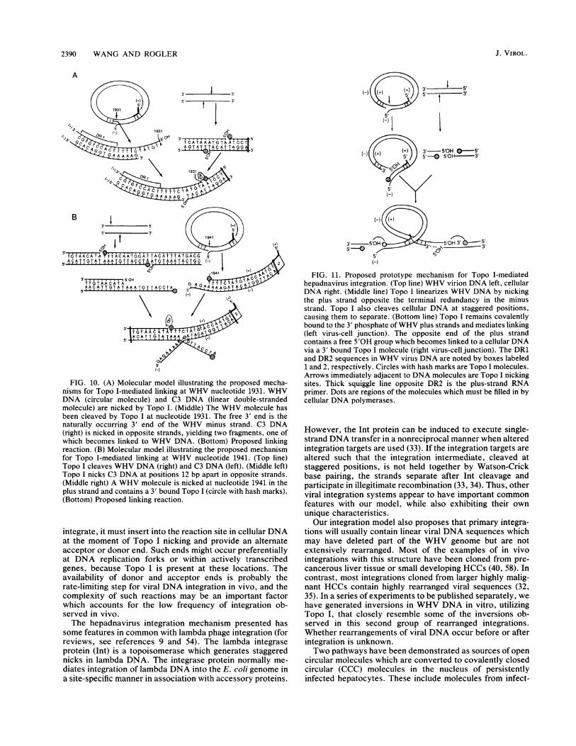
mechanism of the Topo I-mediated linking reactions (Fig.10). The DNA sequence of the first integration is consistentwith a mechanism in which WHV DNA was cleaved byTopo I at nucleotide 1931, resulting in linearization of theWHV molecule (Fig. 10A). The free 5'OH end of the plusstrand was then available to link to cellular DNA whichcontained a 3' bound Topo I. The region of 5-bp homologybetween WHV and cellular DNA suggests that it stabilizedthe formation of the hybrid molecule during the linkingreaction. In the second case (Fig. 10B), we propose thatWHV plus-strand DNA was cleaved at nucleotide 1941 andthat the 3' end of the plus-strand DNA containing a 3' boundTopo I molecule became linked to a 5'OH acceptor end incellular DNA. In this case, a short 3-bp region of homologybetween viral and cellular DNA was evident. According tothese models, WHV DNA served as a 5'OH acceptor end inone case (Fig. 10A) and in the other case (Fig. 10B) itprovided a 3' Topo I-containing donor end. Our experimentsonly tested linking of a single strand of WHV DNA at eachsite. In vivo, it would be necessary to complete the integra-tion of the opposite strand either by a second Topo I linkingreaction or via other cellular DNA polymerases and ligases.The end specificity and polarity of the separate virus-cell
DNA hybrid molecules lead us to propose a prototype modelfor Topo I-mediated integration of both ends of a linearizedWHV molecule (Fig. 11). According to the model, Topo Ilinearizes circular WHV DNA and the Topo I-containing endfunctions as a donor during WHV integration. The oppositeend, containing a 5'OH group, functions as an acceptor sitefor other heterologous cellular DNAs containing 3' boundTopo I molecules. In order for a linearized viral molecule to
VOL. 65, 1991 2389
2390 WANG AND ROGLER
A
3' ~5'0H5'---
5'H-
3' 1 5'S'- t 3'
3'-5'OH -5'5`0 5'01 3'
5'0H 3' 35'
FIG. 10. (A) Molecular model illustrating the proposed mecha-nisms for Topo I-mediated linking at WHV nucleotide 1931. WHVDNA (circular molecule) and C3 DNA (linear double-strandedmolecule) are nicked by Topo I. (Middle) The WHV molecule hasbeen cleaved by Topo I at nucleotide 1931. The free 3' end is thenaturally occurring 3' end of the WHV minus strand. C3 DNA(right) is nicked in opposite strands, yielding two fragments, one ofwhich becomes linked to WHV DNA. (Bottom) Proposed linkingreaction. (B) Molecular model illustrating the proposed mechanismfor Topo I-mediated linking at WHV nucleotide 1941. (Top line)Topo I cleaves WHV DNA (right) and C3 DNA (left). (Middle left)Topo I nicks C3 DNA at positions 12 bp apart in opposite strands.(Middle right) A WHV molecule is nicked at nucleotide 1941 in theplus strand and contains a 3' bound Topo I (circle with hash marks).(Bottom) Proposed linking reaction.
integrate, it must insert into the reaction site in cellular DNAat the moment of Topo I nicking and provide an alternateacceptor or donor end. Such ends might occur preferentiallyat DNA replication forks or within actively transcribedgenes, because Topo I is present at these locations. Theavailability of donor and acceptor ends is probably therate-limiting step for viral DNA integration in vivo, and thecomplexity of such reactions may be an important factorwhich accounts for the low frequency of integration ob-served in vivo.
The hepadnavirus integration mechanism presented hassome features in common with lambda phage integration (forreviews, see references 9 and 54). The lambda integraseprotein (Int) is a topoisomerase which generates staggerednicks in lambda DNA. The integrase protein normally me-
diates integration of lambda DNA into the E. coli genome ina site-specific manner in association with accessory proteins.
FIG. 11. Proposed prototype mechanism for Topo I-mediatedhepadnavirus integration. (Top line) WHV virion DNA left, cellularDNA right. (Middle line) Topo I linearizes WHV DNA by nickingthe plus strand opposite the terminal redundancy in the minusstrand. Topo I also cleaves cellular DNA at staggered positions,causing them to separate. (Bottom line) Topo I remains covalentlybound to the 3' phosphate ofWHV plus strands and mediates linking(left virus-cell junction). The opposite end of the plus strandcontains a free 5'OH group which becomes linked to a cellular DNAvia a 3' bound Topo I molecule (right virus-cell junction). The DR1and DR2 sequences in WHV virus DNA are noted by boxes labeled1 and 2, respectively. Circles with hash marks are Topo I molecules.Arrows immediately adjacent to DNA molecules are Topo I nickingsites. Thick squiggle line opposite DR2 is the plus-strand RNAprimer. Dots are regions of the molecules which must be filled in bycellular DNA polymerases.
However, the Int protein can be induced to execute single-strand DNA transfer in a nonreciprocal manner when alteredintegration targets are used (33). If the integration targets arealtered such that the integration intermediate, cleaved atstaggered positions, is not held together by Watson-Crickbase pairing, the strands separate after Int cleavage andparticipate in illegitimate recombination (33, 34). Thus, otherviral integration systems appear to have important commonfeatures with our model, while also exhibiting their ownunique characteristics.Our integration model also proposes that primary integra-
tions will usually contain linear viral DNA sequences whichmay have deleted part of the WHV genome but are notextensively rearranged. Most of the examples of in vivointegrations with this structure have been cloned from pre-cancerous liver tissue or small developing HCCs (40, 58). Incontrast, most integrations cloned from larger highly malig-nant HCCs contain highly rearranged viral sequences (32,35). In a series of experiments to be published separately, wehave generated inversions in WHV DNA in vitro, utilizingTopo I, that closely resemble some of the inversions ob-served in this second group of rearranged integrations.Whether rearrangements of viral DNA occur before or afterintegration is unknown.Two pathways have been demonstrated as sources of open
circular molecules which are converted to covalently closedcircular (CCC) molecules in the nucleus of persistentlyinfected hepatocytes. These include molecules from infect-
J. VIROL.

TOPO I-MEDIATED INTEGRATION OF HEPADNAVIRUS DNA
ing viral Dane particles and replicative intermediates in coreparticles which are cycled into the nucleus instead of beingexcreted (54, 55, 57). The exact mechanism and site ofconversion of open circular molecules to CCC DNA areunknown. However, it is quite likely that one step in thepathway involves uncoating and release of open circularvirion DNA molecules into the nucleus followed by cleavageof the 5' terminal protein. These open circular moleculeswould be identical in structure to those which we utilized forour in vitro integration experiments and would be expectedto be present in the nucleus and available for cleavage byTopo I.The recent demonstration that recycling of viral DNA into
the nucleus is suppressed to a very low level during produc-tive infection (49) helps to explain why CCC DNA copynumber is stable in the nucleus during persistent infectionand may also explain why integrations are rare under normalconditions. The further demonstration that a blockage inviral envelope production prevents core particle excretionand causes a significant accumulation of open circular andCCC DNAs in the nucleus (49) supports the notion thatconditions can be established during persistent infectionwhich would promote the occurrence of viral integration.This idea is consistent with studies showing that viralintegrations accumulate in the livers of long-term persistentHBV carriers which have developed a blockage in the viralreplication cycle (38).
ACKNOWLEDGMENTS
We thank Okio Hino for providing a plasmid clone of the 1.6-kbC3 human DNA and the nucleotide sequence across the HBVintegration site and Walter Ogston for WHV subgenomic clonespW803 and pW805. The authors also thank David Shafritz, RobertBurk, Raju Kucherlapati, Stewart Shuman, and Peter Bullock forcritical evaluation of the manuscript.
This work was supported by Public Health Service grant CA-37232 from the National Cancer Institute, grant DK-17702 from theDigestive Disease Center grant program, and grant FRA-316 fromthe American Cancer Society.
REFERENCES1. Aveman, K., R. Knippers, T. Koller, and J. M. Sogo. 1988.
Camptothecin, a specific inhibitor of type I DNA topoisomer-ase, induces DNA breakage at replication forks. Mol. Cell. Biol.8:3026-3034.
2. Beasley, R. P., C. C. Kiu, L. Y. Hwang, et al. 1981. Hepatocel-lular carcinoma and hepatitis B virus: a prospective study of22,707 men in Taiwan. Lancet ii:1129-1133.
3. Brechot, C. M., M. Hadchouel, J. Scotto, M. Fonck, F. Potet,G. N. Vyas, and P. Tiollais. 1981. State of hepatitis B virus DNAin hepatocytes of patients with hepatitis B surface antigen-positive and -negative liver diseases. Proc. Natl. Acad. Sci.USA 78:3906-3910.
4. Brechot, C., C. Pourcel, A. Louise, B. Rain, and P. Tiollais.1980. Presence of integrated hepatitis B virus DNA sequences incellular DNA in human hepatocellular carcinoma. Nature (Lon-don) 286:533-535.
5. Brill, S. J., S. DiNardo, K. Voelkel-Meiman, and R. Sternglanz.1987. Need for DNA topoisomerase I activity as a swivel forDNA replication and for transcription of ribosomal RNA.Nature (London) 326:414-416.
6. Bullock, P., J. J. Champoux, and M. Botchan. 1985. Associationof crossover points with topoisomerase I cleavage sites: a modelfor nonhomologous recombination. Science 230:954-958.
7. Bullock, P., W. Forrester, and M. Botchan. 1984. DNA se-quence studies of simian virus 40 chromosomal excision andintegration in rat cells. J. Mol. Biol. 174:55-84.
8. Camilloni, G., E. Di Martino, M. Caserta, and E. di Mauro.1988. Eukaryotic DNA topoisomerase I reaction is topology
dependent. Nucleic Acids Res. 16:7071-7085.9. Campbell, A. 1984. Types of recombination: common problems
and common strategies. Cold Spring Harbor Symp. Quant. Biol.49:839-844.
10. Chakraborty, P. R., N. Ruiz-Opazo, D. Shouval, and D. A.Shafritz. 1980. Identification of integrated hepatitis B virusDNA and expression of viral RNA in an HBsAg-producinghuman hepatocellular carcinoma cell line. Nature (London)286:531-533.
11. Champoux, J. J. 1976. Evidence for an intermediate with asingle-strand break in the reaction catalyzed by the DNAuntwisting enzyme. Proc. Natl. Acad. Sci. USA 73:3488-3491.
12. Champoux, J. J. 1977. Strand breakage by the DNA untwistingenzyme results in the covalent attachment of the enzyme toDNA. Proc. Natl. Acad. Sci. USA 74:3800-3804.
13. Champoux, J. J. 1978. Mechanism of the reaction catalyzed bythe DNA untwisting enzyme: attachment of the enzyme to the3-terminus of the nicked DNA. J. Mol. Biol. 118:441-446.
14. Champoux, J. J. 1981. DNA is linked to the rat liver DNAnicking-closing enzyme by a phosphodiester bond to tyrosine. J.Biol. Chem. 256:4805-4809.
15. Champoux, J. J. 1988. Topoisomerase I is preferentially asso-ciated with isolated replicating simian virus 40 molecules aftertreatment of infected cells with camptothecin. J. Virol. 62:3675-3683.
16. Champoux, J. J., and P. A. Bullock. 1988. A possible role for theeucaryotic type I topoisomerase in illegitimate recombination,p. 655-666. In R. Kucherlapati and G. Smith (ed.), Geneticrecombination. American Society for Microbiology, Washing-ton, D.C.
17. Champoux, J. J., and R. Dulbecco. 1972. An activity frommammalian cells that untwists superhelical DNA: a possibleswivel for DNA replication. Proc. Natl. Acad. Sci. USA 69:143-146.
18. Dejean, A., P. Sonigo, S. Wain-Hobson, and P. Tiollais. 1984.Specific hepatitis B virus integration in hepatocellular carci-noma DNA through a viral 11-base-pair direct repeat. Proc.Natl. Acad. Sci. USA 81:5350-5354.
19. DiNardo, S., K. Voelkel, and R. Sternglanz. 1984. DNA topo-isomerase II mutant of Saccharomyces cerevisiae: topoisomer-ase II is required for segregation of daughter molecules at thetermination of DNA replication. Proc. Natl. Acad. Sci. USA81:2616-2620.
20. Galibert, F., T. N. Chen, and E. Mandart. 1982. Nucleotidesequence of a cloned woodchuck hepatitis virus genome: com-
parison with the hepatitis B virus sequence. J. Virol. 41:51-65.21. Ganem, D., and H. E. Varmus. 1987. The molecular biology of
the hepatitis B virus. Annu. Rev. Biochem. 56:652-693.22. Gilmour, D. S., and S. C. R. Elgin. 1987. Localization of specific
topoisomerase I interactions within the transcribed region ofactive heat shock genes by using the inhibitor camptothecin.Mol. Cell. Biol. 7:141-148.
23. Goto, T., and J. C. Wang. 1985. Cloning of yeast TOP 1, thegene encoding DNA topoisomerase I, and construction ofmutants defective in botl DNA topoisomerase I and DNAtopoisomerase II. Proc. Natl. Acad. Sci. USA 82:7178-7182.
24. Halligan, B. D., J. L. Davis, K. A. Edwards, and L. F. Liu. 1982.Intra- and intermolecular strand transfer by HeLa DNA topo-isomerase I. J. Biol. Chem. 257:3995-4000.
25. Hino, O., K. Ohtake, and C. E. Rogler. 1989. Features of twohepatitis B virus (HBV) DNA integrations suggest mechanismsof HBV integration. J. Virol. 63:2638-2643.
26. Hino, O., T. B. Shows, and C. E. Rogler. 1986. Hepatitis B virusintegration site in hepatocellular carcinoma at chromosome17:18 translocation. Proc. Natl. Acad. Sci. USA 83:8338-8342.
27. Konopka, A. K. 1988. Compilation of DNA strand exchangesites for nonhomologous recombination in somatic cells. Nu-cleic Acids Res. 16:1739-1758.
28. Koshy, R., P. Maupas, R. Muller, and P. H. Hofschneider. 1981.Detection of hepatitis B virus-specific DNA in the genomes ofhuman hepatocellular carcinoma and liver cirrhosis tissues. J.Gen. Virol. 57:95-102.
29. Mason, W. S., M. S. Halpern, J. Newbold, C. E. Rogler, K. L.
VOL. 65, 1991 2391

2392 WANG AND ROGLER
Molnar-Kimber, and J. Summers. 1984. Molecular biology ofthe replication of hepatitis B viruses, p. 23-41. In P. W. J. Rigbyand N. M. Wilkie (ed.), Viruses and cancer. Cambridge Univer-sity Press, Cambridge.
30. Mason, W. S., and J. M. Taylor. 1989. Experimental systems forthe study of hepadnavirus and hepatitis delta virus infections.Hepatology 9:635-645.
31. McCoubrey, W. K., and J. J. Champoux. 1986. The role ofsingle-strand breaks in the catenation reaction catalyzed by therat type I topoisomerase. J. Biol. Chem. 261:5130-5137.
32. Nagaya, T., T. Nakamua, T. Tokino, T. Tsurimoto, M. Imai, T.Mayumi, K. Kamino, K. Yamamura, and K. Matsubara. 1987.The mode of hepatitis B virus DNA integration in chromosomesof human hepatocellular carcinoma. Genes Dev. 1:773-782.
33. Nash, H. A., and C. A. Robertson. 1989. Heteroduplex sub-strates for bacteriophage lambda site-specific recombination:cleavage and strand transfer products. EMBO J. 8:3523-3533.
34. Nunes-Duby, S. E., L. Matsumoto, and A. Landy. 1987. Sitespecific recombination intermediates trapped in suicide sub-strates. Cell 50:779-788.
35. Ogston, W., G. J. Jonak, C. E. Rogler, S. M. Astrin, and J.Summers. 1982. Cloning and structural analysis of integratedwoodchuck hepatitis virus sequences from hepatocellular carci-nomas of woodchucks. Cell 29:385-394.
36. Porter, S. E., and J. J. Champoux. 1988. Mapping in vivotopoisomerase I sites on simian virus 40 DNA: asymmetricdistribution of sites on replicating molecules. Mol. Cell. Biol.9:541-550.
37. Radziwill, G., W. Tucker, and H. Schaller. 1990. Mutationalanalysis of the hepatitis B virus P gene product: domainstructure and RNase H activity. J. Virol. 64:613-620.
38. Raimondo, G., R. D. Burk, H. M. Lieberman, J. Muschel, S. J.Hadziyannis, H. Will, M. C. Kew, J. M. Dusheiko, and D. A.Shafritz. 1988. Interrupted replication of hepatitis B virus inliver tissue of HBsAg carriers with hepatocellular carcinoma.Virology 166:103-112.
39. Rogler, C. E., M. Sherman, C. Y. Su, and D. A. Shafritz. 1985.Deletion in chromosome lip associated with a hepatitis Bintegration site in hepatocellular carcinoma. Science 230:319-322.
40. Rogler, C. E., and J. Summers. 1984. Cloning and structuralanalysis of integrated woodchuck hepatitis virus sequencesfrom a chronically infected liver. J. Virol. 50:832-837.
41. Sadowski, P. 1986. Site-specific recombinases: changing part-ners and doing the twist. J. Bacteriol. 165:341-347.
42. Sanger, F., S. Niklen, and A. R. Coulson. 1977. DNA sequencingwith chain-terminating inhibitors. Proc. Natl. Acad. Sci. USA74:5463-5467.
43. Shafritz, D. A., D. Shouval, H. I. Sherman, S. J. Hadziyannis,and M. C. Kew. 1981. Integration of hepatitis B virus DNA intothe genome of liver cells in chronic liver disease and hepatocel-lular carcinoma. N. Engl. J. Med. 305:1067-1073.
44. Shaul, Y., M. Ziemer, P. D. Garcia, R. Crawford, H. Hsu, P.Valenzuela, and W. J. Rutter. 1984. Cloning and analysis ofintegrated hepatitis virus sequences from a human hepatomacell line. J. Virol. 51:776-787.
45. Shih, C., K. Burke, M.-J. Chow, J. B. Zeldis, C.-S. Yang, C.-S.Lee, K. J. Isselbacher, J. R. Wands, and H. M. Goodman. 1987.Tight clustering of human hepatitis B virus integration sites inhepatomas near a triple-stranded region. J. Virol. 61:3491-3498.
46. Shuman, S. 1989. Vaccinia DNA topoisomerase I promotesillegitimate recombination in Escherichia coli. Proc. Natl. Acad.Sci. USA 86:3489-3493.
47. Stewart, A. F., and G. Schutz. 1987. Camptothecin-induced "invivo" topoisomerase I cleavage in the transcriptionally activetyrosine aminotransferase gene. Cell 50:1109-1117.
48. Summers, J., and W. S. Mason. 1982. Replication of the genomeof a hepatitis B-like virus by reverse transcription of an RNAintermediate. Cell 29:403-415.
49. Summers, J., P. M. Smith, and A. L. Horwich. 1990. Hepadna-virus envelope proteins regulate covalently closed circular DNAamplification. J. Virol. 64:2819-2824.
50. Summers, J., J. Smolec, and R. Snyder. 1978. A virus similar tohuman hepatitis B virus associated with hepatitis and hepatomain woodchucks. Proc. Natl. Acad. Sci. USA 75:4533-4537.
51. Thrash, C., K. Voelkel, S. DiNardo, and R. Sternglanz. 1984.Identification of Saccharomyces cerevisiae mutants deficient inDNA topoisomerase I activity. J. Biol. Chem. 259:1375-1377.
52. Tokino, T., S. Fukushige, T. Nakamura, T. Nagaya, T. Murotsu,K. Shiga, N. Aoki, and K. Matsubara. 1987. Chromosomaltranslocation and inverted duplication associated with inte-grated hepatitis B virus in hepatocellular carcinoma. J. Virol.61:3848-3854.
53. Tsui, S., M. E. Anderson, and P. Tegtmeyer. 1989. Topoisom-erase I sites cluster asymetrically at the ends of the simian virus40 core origin of replication. J. Virol. 63:5175-5183.
54. Tuttleman, J., C. Pourcel, and J. Summers. 1986. Formation ofthe pool of covalently closed circular viral DNA in hepadnavi-rus infected cells. Cell 47:451-460.
55. Tuttleman, J. S., J. C. Pugh, and J. W. Summers. 1986. In vitroexperimental infection of primary duck hepatocyte cultures withduck hepatitis B virus. J. Virol. 58:17-25.
56. Wang, J. C. 1985. DNA topoisomerases. Annu. Rev. Biochem.54:665-697.
57. Wu, T.-T., L. Coates, C. E. Aldvih, J. Summers, and W. S.Mason. 1990. In hepatocytes infected with duck hepatitis Bvirus the template for viral RNA synthesis is amplified by anintracellular pathway. Virology 175:255-261.
58. Yaginuma, K., H. Kobayashi, M. Kobayashi, T. Morishima, K.Matsuyama, and K. Koike. 1987. Multiple integration site ofhepatitis B virus DNA in hepatocellular carcinoma and chronicactive hepatitis tissues from children. J. Virol. 61:1808-1813.
59. Yaginuma, K., M. Kobayashi, E. Yoshida, and K. Koike. 1985.Hepatitis B virus integration hepatocellular carcinoma DNA:duplication of cellular flanking sequences at the integration site.Proc. Natl. Acad. Sci. USA 82:4458-4462.
60. Yang, L., M. S. Wold, J. J. Li, T. J. Kelly, and L. F. Liu. 1987.Roles of DNA topoisomerases I in the transcription of humanribosomal RNA genes. Cell 85:1060-1064.
61. Yang, L., M. S. Wold, J. J. Li, T. J. Kelly, and L. F. Liu. 1987.Roles of DNA topoisomerase in simian virus 40 DNA replica-tion in vitro. Proc. Natl. Acad. Sci. USA 84:950-954.
J. VIROL.