total ginsenosides inhibit the right ventricular ... · tions in right ventricular peak systolic...
TRANSCRIPT

Ginseng, the root of Panax ginseng C. A. MEYER, is a well-known and popular herbal medicine used in China for cen-turies and is now a world wide used natural medicine. Gin-senosides, the pharmacologically active components found inGinseng, are also found in the leaf and stem of Panax gin-seng C. A. MEYER,1) and more than 30 different ginsenosideshave been isolated from Ginseng. Ginsenosides exert variouspharmacological effects on the central nervous, cardiovascu-lar, endocrine, and immune systems.2—4) Notably, it is shownthat Ginsenosides could reduce both Ca2� influx and cate-cholamine secretion in bovine adrenal chromaffin cells,5,6) in-hibit voltage-dependent Ca2� channels in sensory neurons aswell as in chromaffin in cells,7) and decrease the level of in-tracellular free calcium concentration ([Ca2�]i) in dog’s my-ocardium suffered from ischemia-reperfusion injury8); Gin-senosides could also stimulate endogenous product of nitricoxide (NO) in rat kidney9) and in cardiovascular tissues,10)
evoke endothelium-dependent vascular relaxation in rataorta.11) Ca2� elevation has been known to be a critical sig-naling in the development of cardiac hypertrophy induced byvarious hypertrophic stimuli,12) and NO, produced in virtu-ally every cell type in the heart, shows potent antihyper-trophic effects.13) Both effects of Ca2�-decrease and NO-re-lease of ginsenosides indicate that ginsenosides may have apotential inhibitory effect on cardiac hypertrophy.
The mechanism(s) at molecular level of pathological car-diac hypertrophy remains unclear. It has been reported thatCa2� elevation activates the calcineurin (CaN) pathway, andin turn enhances hypertrophy or apoptosis of cardiomy-ocytes.14) In addition, extracellular signal-regulated kinase-1/2 (ERK-1/2, the important members of mitogen-activatedprotein kinase (MAPK) family) likely occupies a central reg-ulatory position in the signaling hierarchy of a cardiac my-
ocyte given its unique ability to respond to virtually everycharacterized hypertrophic agonist and stress stimuli.15)
In the present study, we investigated whether total ginseno-sides (TG) extracted from the leaf and stem of Panax ginsengC. A. MEYER have inhibitory effect on right ventricle hyper-trophy (RVH) induced by injection of monocrotaline (MCT)which lead to pulmonary hypertension by causing injury ofthe lung vasculature, and examine the possible influences ofTG on NO-release, CaN and ERK signaling pathways.
MATERIALS AND METHODS
Materials TG (purity �93%), extracted from the leafand stem of Panax ginseng C. A. MEYER, was presented byprofessor Rui Zhao (Beijing Naturally Occurring Drugs Re-search Institute, China), and was composed of Rb1 (5.26%),Rg1 (5.20%), Re (21.60%), Rd (13.65%), and other ginseno-sides. MCT was purchased from Sigma Chemical Co.(U.S.A.). The antibodies against CnA, MKP-1, actin wereobtained from Stressgen, Santa Cruz Company. Horseradishperoxidase conjugated goat anti-rabbit IgG were from DakoCytomation Company. The reverse transcription kit and theprimers of ANF, CaN, ERK-1 were purchased from TaKaRaBiological Engineering Company.
Animals and RVH Model Male Sprague Dawley rats(n�64, 200�20 g), obtained from Animal Center of theThird Military Medical University (Chongqing, China), werehoused in a standard environment with a 12-h light/12-h darkcycle, where they were free access to food and water. Oneweek after being fed adaptively, the rats were injected (i.p.)with either MCT (60 mg/kg/d, which was dissolved in 0.5 N
HCl and adjusted to pH 7.4 with 0.5 N NaOH, the final con-centration was adjusted to 2% with PBS) or equal volume of
1530 Vol. 31, No. 8
Total Ginsenosides Inhibit the Right Ventricular Hypertrophy Induced byMonocrotaline in Rats
Na QIN,a,b Qi-hai GONG,a Li-wei WEI,c Qin WU,a and Xie-nan HUANG*,a
a Department of Pharmacology, Zunyi Medical College; Zunyi 563000, P. R. of China: b Luoyang Orthopedic Hospital;Luoyang, Henan 471000, P. R. of China: and c 150 Hospital of the People’s Liberation Army; Luoyang, Henan 471000, P.R. of China. Received February 4, 2008; accepted May 21, 2008; published online May 28, 2008
Ginsenosides have been reported to release nitric oxide (NO) and decrease intracellular free Ca2� in cardio-vascular system, which play important roles in antihypertrophic effect. This study investigated the potential in-hibitory effect of total ginsenosides (TG) on right ventricular hypertrophy induced by monocrotaline (MCT,60 mg/kg/d) and examined the possible antihypertrophic mechanism in male Sprague Dawley rats. MCT-intoxi-cated animals were treated with TG (20, 40, 60 mg/kg/d) for 18 d. TG treatment ameliorated MCT-induced eleva-tions in right ventricular peak systolic pressure, right ventricular hypertrophy and the expression of atrial natri-uretic peptide; NG-nitro-L-arginine-methyl ester (L-NAME), an NO synthase (NOS) inhibitor, had no influenceon these inhibitory effects of TG 40 mg/kg/d, and TG at this dose had no any effect on the eNOS mRNA expres-sion, suggesting the limited rule of NO in TG’s effects. To further examine the mechanisms of the protection, theexpression of calcineurin and its catalytic subunit CnA, as well as extracellular signal-regulated kinase-1 (ERK-1) and mitogen-activated protein kinase (MAPK) Phosphatase-1 (MKP-1) was examined. TG treatment signifi-cantly suppressed MCT-induced elevations of these signaling pathways in a dose-dependent manner. In sum-mary, TG is effective in protecting against MCT-induced right ventricle hypertrophy, possibly through loweringpulmonary hypertension. Multiple molecular mechanisms appeared to be involved in this protection, such as thesuppression of MCT-activated calcineurin and ERK signaling pathways.
Key words total ginsenoside; right ventricular hypertrophy; calcineurin; extracellular signal-regulated kinase-1; nitro oxide;monocrotaline
Biol. Pharm. Bull. 31(8) 1530—1535 (2008)
© 2008 Pharmaceutical Society of Japan∗ To whom correspondence should be addressed. e-mail: [email protected]
saline. Then, they were randomly divided into eight groups:(1) vehicle-treated rats (Control group): the rats were giveni.p. with vehicle once a day for 18 d; (2) MCT-treated rats(seven groups): after MCT-treatment, the rats were given i.p.vehicle (MCT group) or TG 20, 40, 60 mg/kg/d or L-arginine(L-arg) 200 mg/kg/d, for analyzing whether TG’s effect wasrelated to NO-release, another two groups were given withNG-nitro-L-arginine-methyl ester (L-NAME) 20 mg/kg/d(p.o.) combined with TG 40 mg/kg/d by i.p. or L-arg 200mg/kg/d by i.p., respectively. The rats in every group wereadministered for 18 d, and n�8 for all groups. All animalstudy procedures are followed by the WHO guideline for hu-mane use of experimental animal.
Measurements of Right Ventricular Peak Systolic Pres-sure and Assessment of RV Hypertrophy In day 19 afterMCT-injection, the rat body weights (BW) in each groupwere weighed, and the right ventricular peak systolic pressure(RVPSP) of the rats were monitored by polygraph systemthrough the cannulation of polyvinyl tube into right ventricle,under the anesthesia with sodium pentobarbital solution(40 mg/kg/d i.p.). the heart was removed and weighedquickly. The heart was separated into the right ventricle (RV)and the left ventricle with septum (LV) and weighed sepa-rately. Finally, RVW/BW (RV weight/BW) and RVH index(RVHI �RV/LV) were calculated. After weighing, the RV tis-sue were quickly frozen and kept at �80 °C for extractingtotal RNA or protein.
Observation of Cardiac Tissue Ultrastructure The RVwall (1 mm3) was fixed by immersion in 2.5% cold glu-taraldehyde solution (pH 7.4), followed by rinsing and post-fixing with 1% osmium tetroxide in 0.1 mol/l PBS for 2 h atroom temperature, then the tissues were dehydrated througha graded series of ethanol to propylene oxide and embeddedin epoxy resin. 600-A sections in thick were made and thenobserved under the transmission electron microscope.
Preparation of Lung Tissue for Morphometric AnalysisFixation was performed by immersion of the lung tissues in a4% paraformaldehyde solution. For paraffin embedding, alllobes from entire lungs were dissected in to tissue blocks,sectioned at 5 mm. H&E and elastica stainings were per-formed according to common histopathological procedures.For examing whether TG can inhibit the pulmonary arteryhypertrophy, the vessel diameter and wall thickness (WT) ofat least 15 pulmonary arteries (50—100 mm in diameter) ineach rat in MCT, control and TG 40 mg/kg/d groups were ob-served by a blinded observer under �40 magnification usinga computerized morphometric system (QWin; Leica). TheWT of each artery was expressed as a percent of external di-ameter (% wall thickness) according to the formula: (2�wallthickness /external diameter)�100.
Cardiac Protein Extract and Western Blotting RV tis-sues were rapidly excised and rinsed in cold phosphate-
buffered saline then homogenized on ice in 1 ml of proteinextract. Homogenates were sonicated on ice for three burstsof 5 s each and centrifuged for 15 min at 14000 g/min at 4 °C.Lysates were kept frozen until used or added with SDS-PAGE loading buffer (125 mM Tris, 4% SDS, 20% glycerol,100 mM dithiothreitol, 0.2% bromphenol blue, pH 6.8) toreach a final concentration of 25%. Lysates were then heatedat 95 °C for 5 min and run in 10% SDS-PAGE, and trans-ferred on polyvinylidene difluoride nylon membranes. Theblots were probed with mouse anti-CnA (1 : 1000), anti-MKP-1 (1 : 400), or anti-actin antibodies (1 : 600) followedby horseradish peroxidase-conjugated goat anti-rabbits IgG(1 : 1000) antibodies. Immunodetection was carried out usinggel image analysis system.
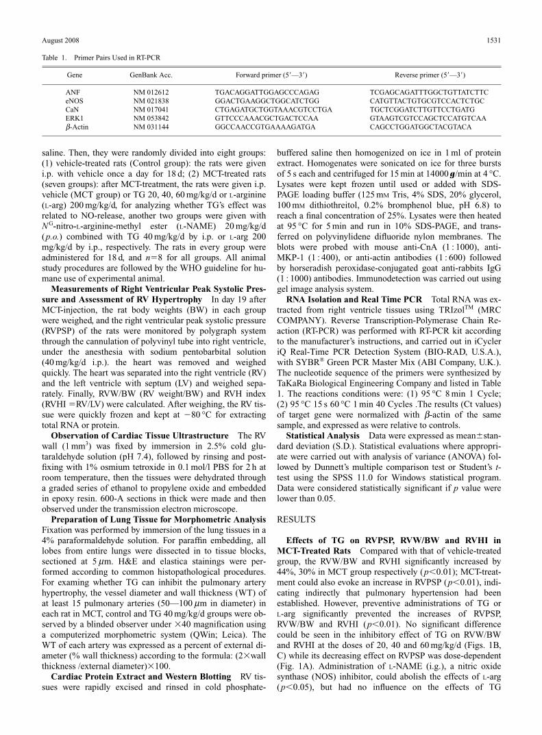
RNA Isolation and Real Time PCR Total RNA was ex-tracted from right ventricle tissues using TRIzolTM (MRCCOMPANY). Reverse Transcription-Polymerase Chain Re-action (RT-PCR) was performed with RT-PCR kit accordingto the manufacturer’s instructions, and carried out in iCycleriQ Real-Time PCR Detection System (BIO-RAD, U.S.A.),with SYBR® Green PCR Master Mix (ABI Company, U.K.).The nucleotide sequence of the primers were synthesized byTaKaRa Biological Engineering Company and listed in Table1. The reactions conditions were: (1) 95 °C 8 min 1 Cycle;(2) 95 °C 15 s 60 °C 1 min 40 Cycles .The results (Ct values)of target gene were normalized with b-actin of the samesample, and expressed as were relative to controls.
Statistical Analysis Data were expressed as mean�stan-dard deviation (S.D.). Statistical evaluations where appropri-ate were carried out with analysis of variance (ANOVA) fol-lowed by Dunnett’s multiple comparison test or Student’s t-test using the SPSS 11.0 for Windows statistical program.Data were considered statistically significant if p value werelower than 0.05.
RESULTS
Effects of TG on RVPSP, RVW/BW and RVHI inMCT-Treated Rats Compared with that of vehicle-treatedgroup, the RVW/BW and RVHI significantly increased by44%, 30% in MCT group respectively (p�0.01); MCT-treat-ment could also evoke an increase in RVPSP (p�0.01), indi-cating indirectly that pulmonary hypertension had been established. However, preventive administrations of TG or L-arg significantly prevented the increases of RVPSP,RVW/BW and RVHI (p�0.01). No significant differencecould be seen in the inhibitory effect of TG on RVW/BWand RVHI at the doses of 20, 40 and 60 mg/kg/d (Figs. 1B,C) while its decreasing effect on RVPSP was dose-dependent(Fig. 1A). Administration of L-NAME (i.g.), a nitric oxidesynthase (NOS) inhibitor, could abolish the effects of L-arg(p�0.05), but had no influence on the effects of TG
August 2008 1531
Table 1. Primer Pairs Used in RT-PCR
Gene GenBank Acc. Forward primer (5�—3�) Reverse primer (5�—3�)
ANF NM 012612 TGACAGGATTGGAGCCCAGAG TCGAGCAGATTTGGCTGTTATCTTCeNOS NM 021838 GGACTGAAGGCTGGCATCTGG CATGTTACTGTGCGTCCACTCTGCCaN NM 017041 CTGAGATGCTGGTAAACGTCCTGA TGCTCGGATCTTGTTCCTGATGERK1 NM 053842 GTTCCCAAACGCTGACTCCAA GTAAGTCGTCCAGCTCCATGTCAAb-Actin NM 031144 GGCCAACCGTGAAAAGATGA CAGCCTGGATGGCTACGTACA

40 mg/kg/d (Figs. 1A, B, C).Myocardial Morphology Figure 2A showed the normal
morphology of the RV tissues in vehicle-treated rats; MCT-induced myocardial injuries in RV were observed, such asdisruption of the intercalated disk, irregular pattern of cross-striation, and aggregation of swollen and misshapen mito-chondria (Fig. 2B). Treatment with TG 40 mg/kg/d signifi-cantly improved the ultrastructure damages to RV, eventhough the swollen mitochondria still existed (Fig. 2C).
Assessment of Lung Morphology The normal lung tis-
sue was shown in Fig. 3A, MCT-treatment induced a markedmedial hypertrophy in pulmonary arteries, widening on alveolar septa, muscularization in the distal arterioles, andmarked periarteritis in vascular artery (Fig. 3B). Administra-tion of TG 40 mg/kg/d significantly ameliorated MCT-in-duced pulmonary vascular remodeling, reduced musculariza-tion of the pulmonary arteries and arterioles, and relieved theperiarteritis (Fig. 3C). For quantitatively analyzing, the pro-tective effect of TG on MCT-induced pulmonary vascular re-modeling, the % wall thickness of pulmonary arteries (50—100 mm in diameter) was calculated. MCT-injection causedan increase in % wall thickness to near 3 times, administra-tion of TG 40 mg/kg/d siginificantly decreased the elevated% wall thickness (p�0.05), (Fig. 3D).
Effects of TG on the Expressions of ANF, eNOS, CaNand ERK-1 mRNA in Hypertrophic RV Tissue Expres-sions of ANF (Fig. 4A), eNOS (Fig. 4B), CaN (Fig. 4C) andERK-1 (Fig. 4D) mRNA from the cardiac RV were shown inFig. 4. It was obvious that the basic expressions of ANF,
1532 Vol. 31, No. 8
Fig. 1. Effects of Preventive Administration with TG on RVPSP (A),RVW/BW (B) and RVHI (C) in Rat Pretreated by MCT (Mean�S.D.)
RVPSP: right ventricular peak systolic pressure; RVW/BW: right ventricularweight/body weight; RVHI: right ventricular hypertrophy index. Group: male SpragueDawley rats were given i.p. a single monocrotaline (MCT, 60 mg/kg/d) (MCT group) orequal volume of saline (control group) and then fed with vehicle for 18 d. In otherMCT-treated groups, total ginsenosides at doses of 20, 40, 60 mg/kg/d, L-arg200 mg/kg/d i.p. and L-NAME 20 mg/kg/d (p.o.) combined with TG 40 mg/kg/d by i.p.or L-arg 200 mg/kg/d by i.p. were given from day 1 to day 18, respectively. MCT groupvs. control group # p�0.01; vs. MCT group ∗ p�0.05; MCT�L-arg 200 mg/kg/d�L-NAME group vs. MCT�L-arg 200 mg/kg/d group � p�0.05.
Fig. 2. Effects of TG 40 mg/kg/d Preventive Administration on the Ultra-structural Changes in RV Myocardial Cell of the Rats Pretreated by MCT
(A) Control group; (B) MCT group, the monocrotaline-treated rats showed some my-ocardial injuries such as disruption of the intercalated disk, irregular pattern of cross-striation, and aggregation of swollen and misshapen mitochondria (arrow); (C)MCT�TG 40 mg/kg/d group, TG 40 mg/kg/d administration significantly amelioratedthe ultrastructure damages to the right ventricular striated patterns and intercalateddisk. Scale bar is 2.2 mm.

eNOS, CaN and ERK-1 mRNA were very low in normalcontrol group. However, their expressions increased by 5.6,1.0, 2.1 and 2.6 times, respectively, in MCT-treated rats(MCT group) (p�0.01). Administration of TG 20, 40,60 mg/kg/d and L-arg 200 mg/kg/d significantly blunted theelevation of ANF mRNA expression, and administration ofL-NAME (i.g.) could abolish the effect of L-arg on this pa-rameter (p�0.05), but not influence on the effect of TG
August 2008 1533
Fig. 3. Effect of Total Ginsenosides 40 mg/kg/d Administration on theHistomorphological Changes of the Small Muscular Arteries (H.E. Staining,40�, A, B, C) and % Wall Thickness (D) in the Rats Pretreated byMonocrotaline
(A) Control group, the normal lung tissue of the vehicle-treated rat; (B) MCT group,the lung tissue of the MCT-treated rat: exhibiting the medial hypertrophy and mascular-ization of the arteriole, and a marked periarteritis; (C) MCT�TG 40 mg/kg/d group,the lung tissue of the rat administered with TG 40 mg/kg/d for 18 d after MCT-treat-ment: exhibiting the relief of the medial hypertrophy and muscularization of arteriole,and the relieving periarteritis. (D) % wall thickness (2�wall thickness/external diame-ter)�100.
Fig. 4. Effects of Total Ginsenosides Administration on the Expressionsof ANF (A), eNOS (B), CaN (C) and ERK1 (D) mRNA in Right Ventricleof Rats Pretreated by Monotrotaline (Mean�S.D.)
Group: male Sprague Dawley rats were given i.p. a single monocrotaline (MCT,60 mg/kg/d) (MCT group) or with vehicle (control group) and then fed for 18 d. Inother MCT-treated groups, total ginsenosides at doses of 20, 40, 60 mg/kg/d and L-arg200 mg/kg/d by i.p., and L-NAME 20 mg/kg/d (p.o.) combined with TG 40 mg/kg/d byi.p. or L-arg 200 mg/kg/d by i.p. were given from day 1 to day 18, respectively. MCTgroup vs. control group # p�0.01; vs. MCT group ∗ p�0.01; MCT�L-arg 200mg/kg/d�L-NAME group vs. MCT�L-arg 200 mg/kg/d group � p�0.01.

40 mg/kg/d (p�0.05). Notably, administrations of TG 20 and40 mg/kg/d had no any effect on eNOS mRNA Expression,but when the dose of TG was increased to 60 mg/kg/d, itcaused an elevation in eNOS mRNA Expression. Similar tothe effect of TG on ANF mRNA expression, the elevations ofCaN and ERK-1mRNA expressions induced MCT were alsoinhibited by the administrations of TG 20, 40, 60 mg/kg/d.
Effects of TG on CnA and MKP-1 Protein Levels in RVThe protein expression levels of CnA were markedly in-creased in MCT-treated rats (p�0.05), but there was a ten-dency for the increase in protein expression level of MKP-1(p�0.05), compared with that of normal control group. TGadministrations could significantly decrease the protein ex-pression level of CnA (p�0.01), and further increase theprotein expression levels of MKP-1 in a dose-dependentmanner (p�0.01), compared with that in MCT-treated group(Fig. 5).
DISCUSSION
It has been well known that MCT causes injury of the vas-culature and leads to pulmonary hypertension and RVHthrough its metabolite MCT-pyrrole.16,17) In the present study,we used MCT-induced RVH in rat to examine the possible in-hibitory effect of TG on cardiac hypertrophy. In MCT-treatedanimals, the increase in RVPSP and the findings in the elec-tron microscopic observation strongly indicated that there ex-isted the formation of pulmonary hypertension. The eleva-tions of RVW/BW, RVHI and ANF mRNA expression (amarker of hypertrophy), as well as the RV tissue morphologi-cal changes, further suggested that the establishment of RVHmodel in our experiment was successful.
Intraperitoneal injection as an administering method wasused widely by many investigators for studies on the pharma-cological effects of ginsenosides in rats.9,18) In this study, wedesigned to administer TG (20, 40, 60 mg/kg/d, i.p.) to MCT-treated rats for 18 d, and found that TG could significantly re-duce the elevated RVW/BW RVHI, ANF mRNA expressioninduced by MCT, suggesting that TG could ameliorate theRVH in this model. However, it was unclear whether TG at-tenuate or delay the genesis of MCT-induced RVH. Our pre-vious study demonstrated that ginsenoside Rb1, an active in-gredient (5.26%) in TG, could reduce the MCT-inducedRVH, when it was administered (i.p.) after 3 weeks of MCT-injection (RVH had been generated) at the dose of40 mg/kg/d for 21 d,19) suggesting that Rb1 could attenuatethe genesis of RVH. Whether the effect of TG is the same asRb1 remains to be studied. To our attention, TG 20 mg/kg/dalmost completely suppressed the increases in RVW/BW andRVHI, suggesting that the optimal dosage for the antihy-pertrophic effect of TG in this model was at 20 mg/kg/d,whereas the suppressing effect of TG on the elevated RVPSPwas dose-dependent at doses of 20, 40 and 60 mg/kg/d, andthe inhibition of TG 40 mg/kg/d on % wall thickness was notcomplete in our experiment. The results indicated that othermechanisms rather than the suppressing effect on pulmonaryhypertension might involve in the antihypertrophic effect ofTG. Furthermore, we also found that TG had an antihyper-trophic effect on cardiomyocyte in vitro which was not re-lated to pulmonary hypertension (data not shown). Thus weconsider that the lowering effect on pulmonary hypertensionis not the only mechanism for the antihypertrophy of my-ocardium.
TG has been reported to stimulate endogenous NO releasein cardiovascular system10) and other tissue,9) and NO waswell-known to be a potent inhibitor of cardiac hypertrophy.13)
However, we observed that L-NAME, an NOS inhibitor,could abolish the inhibitory effects of L-arg on RVH, but had no influence on the antihypertrophic effects of TG40 mg/kg/d, which was consistent with that TG at this dose or20 mg/kg/d had no any effect on eNOS mRNA expression, inspite of it could increase the eNOS expression at the dose of60 mg/kg/d, compared with that in MCT group. The resultssuggested that the antihypertrophic effect of TG might comefrom other mechanism rather than NO release when the dosewas 40 or lower than 40 mg/kg/d, but when the dose of TGwas increased to 60 mg/kg/d, the NO release might be par-tially responsible for its antihypertrophic effect.
In the past several years, a number of studies have impli-
1534 Vol. 31, No. 8
Fig. 5. Effects of Total Ginsenosides Administrations on the Protein Ex-pressions of CnA and MKP-1 in Right Ventricle of the Rats Pretreated byMCT
Group: male Sprague Dawley rats were given i.p. a single monocrotaline (MCT,60 mg/kg/d) (MCT group) or with vehicle (control group) and then fed for 18 d. Inother MCT-treated groups, total ginsenosides at doses of 20, 40, 60 mg/kg/d by i.p.were given from day 1 to day 18, respectively. MCT group vs. control group # p�0.01;vs. MCT group ∗ p�0.01. (A) Lane 1: control group; Lane 2: MCT-treated group; Lane3: MCT�TG 20 mg/kg/d; Lane 4: MCT�TG 40 mg/kg/d; Lane 5: MCT�TG60 mg/kg/d; (B, C) the relative amount of CnA and MKP-1 were quantified by NIHImage program and normalized against the amount of actin.

cated that the Calcineurin signal transduction pathway acti-vated by the increased [Ca2�]i may play an important role inthe cardiomyocyte hypertrophy process,12,20—22) and TG hasbeen reported to reduce the Ca2� influx,5) inhibit the voltage-dependent channels7,23) and decrease the [Ca2�]i
8) in some tis-sues. In the present paper, the fact that the elevated expres-sions of CaN mRNA and CnA (the catalytic subunit of CaN)protein induced by MCT were significantly blunted by TGsuggested that an inhibition on the CaN signaling pathwaymight be involved in the antihypertrophic mechanisms ofTG. Moreover, many studies have indicated that ERK1/2, themembers of the mitogen-activated protein kinase (MAPK)family, are crucial regulators in cardiac hypertrophy15,24,25);while the mitogen-activated protein kinase phosphatase-1(MKP-1) can dephosphorylate and then inactivate ERK,functioning as a negative feedback mechanism in the controlof MAPK activity.26—28) It was interesting to note that TG notonly markedly reduced the elevated ERK-1 mRNA expres-sion induced by MCT, but also increased the MKP-1 proteinexpression in a dose-dependent manner. The results stronglydemonstrated that the molecular mechanism for the antihy-pertrophic effect of TG might be also involved in the inhibi-tion on the ERK signaling pathway. Nevertheless, the MCT-induced RVH was suppressed almost completely by TG atthe dose of 20 mg/lg, but only a part of expressions of CaNmRNA, ERK-1 mRNA and CnA protein was inhibited by TGat this dose. The reason for this discrepancy was unclear, itwas possible that the activities of signaling pathways werealso inhibited by TG through some other mechanisms, ex-cepting the inhibition on the transcription or translation ofthe members of signaling pathways.
In conclusion, our study demonstrates that TG can allevi-ate cardiac hypertrophy induced by MCT in rats, which mayin part be, mediated by lowering pulmonary hypertension;the molecular mechanism for the antihypertrophic effect ofTG may be involved in the inhibitions on the CaN and ERKsignaling pathways.
Acknowledgement This work was supported by theHigher Education Developing Foundation for Natural Sci-ence of Guizhou Province, China (No. 2005110).
REFERENCES
1) Yu C. H., Wei F., He Z. M., Chin. Tradit. Herbal Drugs, 38, 46—50(2007).
2) Attele A. S., Wu J. A., Yuan C. S., Biochem. Pharmacol., 58, 1686—1693 (1999).
3) Gillis C. N., Biochem. Pharmacol., 54, 1—8 (1997).4) Kim J. H., Lee J. H., Jeong S. M., Lee B. H., Yoon I. S., Lee J. H.,
Choi S. H., Nah S. Y., Biol. Pharm. Bull., 28, 2120—2124 (2005).5) Tachikawa E., Kudo K., Kashimoto T., Takahashi E., J. Parmacol. Exp.
Ther., 273, 629—636 (1995).6) Kim H. S., Lee J. H., Goo Y. S., Nah S. Y., Brain Res. Bull., 46, 245—
251 (1998).7) Nah S. Y., Park H. J., McCleskey E. W., Proc. Natl. Acad. Sci. U.S.A.,
92, 8739—8743 (1995).8) Hou M. X., Ao D. C., Chin. J. Thorac. Cardiovasc. Surg., 7, 256—259
(2000).9) Han S. W., Kim H., Int. J. Biochem. Cell Biol., 28, 573—580 (1996).
10) Chen X., Clin. Exp. Pharmacol. Physiol., 23, 728—732 (1996).11) Kim N. D., Kang S. Y., Schini V. B., Gen. Pharmacol., 25, 1071—
1077 (1994).12) Sugden P. H., Ann. Med., 33, 611—622 (2001).13) Kampf T., Wollert K. C., Bioassays, 26, 608—615 (2004).14) Yue T. L., Ohlstein E. H., Ruffolo R. R. Jr., Curr. Opin. Chem. Biol., 3,
474—480 (1999).15) Bueno O. F., Molkentin J. D., Circ. Res., 91, 776—781 (2002).16) Ahn B. H., Park H. K., Cho H. G., Lee H. A., Lee Y. M., Yang E. K.,
Lee W. J., J. Korean Med. Sci., 18, 641—648 (2003).17) Chen L., Gan X. Y., Haist J. V., Feng Q., Lu X., Chakrabarti S., Kar-
mazyn M., J. Pharmacol. Exp. Ther., 298, 469—476 (2001).18) Lee J. H., Kim S. R., Bae C. S., Kim D., Hong H. N., Nah S. Y., Neu-
rosci. Lett., 325, 129—133 (2002).19) Jing Q. S., Huang X. N., Dal Z. K., Yang G. Z., Zhou Q. X., Shi J. S.,
W Q., J. Ethnopharmacol., 111, 567—572 (2007).20) Molkentin J. D., Circ. Res., 87, 731—738 (2000).21) Bush E. W., Hood D. B., Papst P. J., Chapo J. A., Minobe W., Bristow
M. R., Olson E. N., McKinseym T. A., J. Biol. Chem., 281, 33487—33496 (2006).
22) Nakayama H., Wilkin B. J., Bodi I., Molkentin J. D., FASEB J., 20,1660—1670 (2006).
23) Lee J. H., Jeong S. M., Kim J. H., Lee B. H., Yoon I. S., Lee J. H.,Choi S. H., Lee S. M., Park Y. S., Lee J. H., Kim S. S., Kim H. C., LeeB. Y., Nah S. Y., Mol. Cell., 21, 52—62 (2006).
24) Glennon P. E., Kaddoura S., Sale E. M., Sale G. J., Fuller S. J., SugdenP. H., Circ. Res., 78, 954—961 (1996).
25) Bueno O. F., DeWindt L. J., Tymitz K. M., Witt S. A., Kimball T. R.,Klevitsky R., Hewett T. E., Jones S. P., Lefer D. J., Peng C. F., Kitsis R.N., Molkentin J. D., EMBO J., 19, 6341—6350 (2000).
26) Fuller S. J., Davies E. L., Gillespie-Brown J., Sun H., Tonks N. K., J.Biol. Chem., 323 (Pt 2), 313—319 (1997).
27) Sun H., Tonks N. K., Bar-Sagi D., Science, 266, 285—288 (2004).28) Chu Y., Solski P. A., Khosravi-Far R., Der C. J., Kelly K., J. Biol.
Chem., 271, 6497—6501 (1996).
August 2008 1535