towards a phylogenetic clarification of lophiostoma and ... · towards a phylogenetic clarification...
TRANSCRIPT
Fungal Diversity
225
Towards a phylogenetic clarification of Lophiostoma / Massarina and morphologically similar genera in the Pleosporales Zhang, Y.1, Wang, H.K.2, Fournier, J.3, Crous, P.W.4, Jeewon, R.1, Pointing, S.B.1 and Hyde, K.D.5,6*
1Division of Microbiology, School of Biological Sciences, The University of Hong Kong, Pokfulam Road, Hong Kong SAR, P.R. China 2Biotechnology Institute, Zhejiang University, 310029, P.R. China
3Las Muros, Rimont, Ariège, F 09420, France 4Centraalbureau voor Schimmelcultures, Fungal Biodiversity Centre, P.O. Box 85167, 3508 AD, Utrecht, The Netherlands
5School of Science, Mae Fah Luang University, Tasud, Muang, Chiang Rai 57100, Thailand 6International Fungal Research & Development Centre, The Research Institute of Resource Insects, Chinese Academy of Forestry, Kunming, Yunnan, P.R. China 650034 Zhang, Y., Wang, H.K., Fournier, J., Crous, P.W., Jeewon, R., Pointing, S.B. and Hyde, K.D. (2009). Towards a phylogenetic clarification of Lophiostoma / Massarina and morphologically similar genera in the Pleosporales. Fungal Diversity 38: 225-251. Lophiostoma, Lophiotrema and Massarina are similar genera that are difficult to distinguish morphologically. In order to obtain a better understanding of these genera, lectotype material of the generic types, Lophiostoma macrostomum, Lophiotrema nucula and Massarina eburnea were examined and are re-described. The phylogeny of these genera is investigated based on the analysis of 26 Lophiostoma- and Massarina-like taxa and three genes – 18S, 28S rDNA and RPB2. These taxa formed five well-supported sub-clades in Pleosporales. This study confirms that both Lophiostoma and Massarina are polyphyletic. Massarina-like taxa can presently be differentiated into two groups – the Lentithecium group and the Massarina group. Of these, the type species M. eburnea together with the Massarina group represents Massarina sensu stricto. Lophiostoma taxa clustered in two groups – one group, including the type species L. macrostomum, is characterized by fusiform, hyaline one-septate ascospores which are pigmented and 3-septate when senescent, clavate asci, and apical structures which are highly variable, being crest-like in L. macrostomum, an umbilicate pore surrounded by 4-6 radial ridges in L. rugulosum, or papillate in L. glabrotunicatum. The second group comprises Lophiostoma species with heavily pigmented multi-septate ascospores and compressed crests. Lophiotrema species including the type species L. nucula form a monophyletic group. One new genus – Lentithecium with five new species – Lentithecium aquaticum, Lophiostoma glabrotunicatum, L. rugulosum, Lophiotrema brunneosporum and L. lignicola and three new combinations – Lentithecium arundinaceum, L. fluviatile and L. lineare are introduced in this paper. Key words: Lentithecium, Lophiostoma, Lophiotrema, Massarina, phylogeny, type studies
Article Information Received 3 May 2009 Accepted 13 July 2009 Published online 1 October 2009 *Corresponding author: Kevin D. Hyde; e-mail: [email protected] Introduction
There is considerable confusion sur-rounding the genera Lophiostoma, Lophio-trema and Massarina (Scheuer, 1991; Hyde et al., 2002; Liew et al., 2002; Tanaka and Harada, 2003b; Tang et al., 2003). Taxa in these genera mostly have immersed to erum-pent ascomata, cellular pseudoparaphyses, and hyaline (or dark brown in Lophiostoma),
septate ascospores often with sheaths or appendages (Holm and Holm, 1988; Tanaka and Harada, 2003a,b). Delineation has been based on the fact that Lophiostoma has compressed crests, some with raised crests; the unequal thickness of the peridium, which is usually broader near the base; mostly clavate asci; and 1 to several septate, hyaline to dark brown ascospores with terminal appendages or mucilaginous sheaths (Holm and Holm, 1988;

226
Tanaka and Harada, 2003a). Lophiotrema differs in having a peridium (20-30 µm wide) of nearly equal thickness, composed of an outer textura angularis of uniformly pigmented cells, up to 12 µm, and an inner layer of small, hyaline cells, with somewhat thickened walls (Holm and Holm, 1988), and cylindrical or oblong asci (Holm and Holm, 1988; Tanaka and Harada, 2003b). As compared to the above two genera, the distinguishing morphological character of Massarina is its rounded papilla, and its exclusively hyaline ascospores (Holm and Holm, 1988; Aptroot, 1998).
The confusion between these genera was confounded when Hyde (1995) re-examined the generic type specimen of Massarina ebur-nea and found it comprising flattened ascomata immersed in bark of twigs of Fagus sylvatica, which had little resemblance to most species included in Massarina. This resulted in Aptroot (1998) monographing the genus with accep-tance of 43 species. Aptroot (1998) placed much emphasis on the ascospore characters and only provided brief descriptions of hamathe-cium and ascoma structures. Further new species were introduced from aquatic (Hyde and Goh, 1998; Tsui et al., 1999; van Ryckegem and Aptroot, 2001) or terrestrial (Poonyth et al., 1999; Aptroot et al., 2000) environments. Phylogenetic studies indicated the aquatic Massarina species having affinities towards Lophiostoma species (Liew et al., 2002; Eriksson and Hawksworth, 2003). This led to the transfer of several Massarina species to Lophiostoma based on morphology and molecular data (Hyde et al., 2002; Liew et al., 2002).
Lophiostoma possesses typical characters of Lophiostomataceae. These include im-mersed to erumpent ascomata with a laterally compressed- or crest-like apex, mostly clavate asci, and 1- to several-septate, hyaline to dark brown ascospores with terminal appendages or mucilaginous sheaths. Lophiostoma is morpho-logically a well-studied genus (Chesters and Bell, 1970; Holm and Holm, 1988; Barr, 1990; Yuan and Zhao, 1994), and currently it com-prises about 30 species (Tanaka and Harada, 2003a). The crest-like apex is not considered to be a stable character and varies considerably even in the same specimen (Chesters and Bell, 1970; Holm and Holm, 1988).
Lophiotrema is one of Saccardo’s “sporological genera”, introduced based on the oblong to fusiform, one to multi-septate, hyaline ascospores (Saccardo, 1878). Chesters and Bell (1970) regarded Lophiotrema as a synonym of Lophiostoma. Lophiotrema however, can also be distinguished from Lophiostoma by its peridium and ascal shape, i.e. the peridium of Lophiotrema is almost equal in thickness, while it is broader near the base for Lophiostoma (Holm and Holm, 1988; Tanaka and Harada, 2003b), and species of Lophiotrema have cylindrical or oblong asci, while those of Lophiostoma are clavate or oblong (Holm and Holm, 1988). Based on these characters, only the type species – Lophiotrema nucula and another two closely related taxa were included in Lophiotrema by Holm and Holm (1988). This concept has been widely accepted (Barr, 1990; Yuan and Zhao, 1994; Kirk et al., 2008). Tang et al. (2003) introduced a new Lophiotrema species (L. psy-chotriae) with compressed apex and elongated slit-like ostiole, stating that the main difference between Lophiotrema and Lophiostoma were ascomatal dimensions, peridium structure, ascal shape, and ascospore sheaths.
Several Lophiostoma / Massarina-like taxa have been collected from France and Belgium by J. Fournier. We tried to assign these collections to genera (and species) but it was extremely difficult because the signi-ficance of various characters could not be determined. Practically, the taxonomic dif-ferences between these two genera are based on a single morphological character — the shape of the apex (Lophiostoma containing a com-pressed/crest-like apex and Massarina having a rounded papilla). The shape of the apex is however, highly variable, and taxa with a similar apical shape may be heterogeneous (Holm, 1957; Eriksson, 1981; Leuchtmann, 1984; Holm and Holm, 1988).
Because of the difficulties in dis-tinguishing these genera, we examined their generic types, and carried out a phylogenetic analysis in an attempt to better understand the taxonomy of this group. The aims of the present study are to (1) reevaluate the phylo-geny and circumscription of Lophiostoma, Lophiotrema and Massarina; (2) evaluate
Fungal Diversity
227
which morphological character such as asco-spore (shape, colour, number or type of septum, sheath), ascoma or apical shape has phylo-genetic significance.
Materials and methods Sample collection and specimen examination
Eleven fresh specimens were collected from August to October 2006, from France and Belgium by J. Fournier, and returned to the laboratory in plastic bags. In most cases ascomata were collected directly on natural wood without incubation. The samples were processed and examined following the method described in Tsui et al. (2000). Colonies were sub-cultured onto 2% potato-dextrose agar (PDA), synthetic nutrient-poor agar (SNA), 2% malt extract agar (MEA), and oatmeal agar (OA) (Gams et al., 2007), and incubated under continuous near-ultraviolet (320-400 nm, mainly 340 nm) light at 25°C to promote sporulation. Observations and photographs were prepared from material mounted in water, Congo red, Cotton blue, chlorazol black, lactic acid or Indian ink. Other cultures used in this study were obtained from the Centraalbureau voor Schimmelcultures, the Netherlands (CBS). “Verified specimen / strain” used here means the specimen or voucher of particular strain was examined by authors, and compared with the type material or descriptions to make sure it is correctly identified. Fungal isolates and DNA extraction
Isolates were on PDA and MEA, and total genomic DNA extracted from mycelia following the protocols as outlined by Cai et al. (2005, 2006). Forensic Kit (UltraClean™ Forensic Kit, Cambio) was used to extract DNA from specimens directly. DNA amplification and sequencing
DNA amplification was performed by PCR. For partial LSU rDNA amplification, LROR and LR5 primers (Vilgalys and Hester, 1990) were used. Primer pairs NS1 and NS4 were used to amplify a region from the small subunit (18S) of the rDNA (White et al., 1990). The fRPB2-5F and fRPB2-7cR primers were used for the amplification of the partial RPB2 (RNA polymerase subunit 2) gene (Liu et al.,
1999). The amplification reaction for partial LSU, SSU rDNA and partial RPB2 gene was performed in a 50 μl reaction volume as outlined by Jeewon et al. (2004) and Shenoy et al. (2007): 1 × PCR buffer, 0.2 mM d'NTP, 0.3 μM of each primer; 1.5 mM MgCl2, 0.8 units Taq Polymerase and 5-10 ng DNA. The PCR thermal cycle for partial LSU and SSU rDNA amplification was as follows: 95°C for 3 min, followed by 34 cycles of denaturation at 95°C for 1 min, annealing at 52°C for 30 s and elongation at 72°C for 1 min, with a final extension step of 72°C for 10 min (Vilgalys and Hester, 1990). The PCR thermal cycle for partial RPB2 gene amplification consisted of 95°C for 5 min, followed by 35 cycles of denaturation at 95°C for 1 min, annealing at 55°C for 2 min and elongation at 72°C for 1.5 min, with a final extension step of 72°C for 10 min (Liu et al., 1999). The PCR products, spanning approximately 900 bp (partial LSU rDNA) and 1200 bp (partial RPB2 and SSU rDNA), were checked on 1% agarose electrophoresis gels stained with ethidium bromide. The PCR products were then purified using minicolumns, purification resin and buffer according to the manufacturer's protocols (Amersham Biosciences, Bucking hamshire, UK; product code: 27-9602-01). DNA sequencing was performed using the above-mentioned primers in an Applied Biosystem 3730 DNA analyser at the Genome Research Centre, The University of Hong Kong.
Sequence alignment and phylogenetic ana-lyses
Sequences generated from different pri-mers were analyzed with other sequences obtained from the GenBank. A Blast search was performed to find the possible sister groups of the newly sequenced taxa. In addition, fungal members from different families of the Pleosporales and related orders were also included in the analyses. Multiple alignment was done in BioEdit (Hall, 2005) and Clustal X (Thompson et al., 1997) and analyses were performed in PAUP v. 4.0b10 (Swofford, 2002). Prior to phylogenetic ana-lysis, ambiguous sequences at the start and the end were deleted and gaps manually adjusted to optimize alignment. Maximum Parsimony
228
(MP) was conducted using heuristic searches as implemented in PAUP, with the default options method. Analyses were done under different parameters of maximum parsimony criteria as outlined in Jeewon et al. (2004). Clade stability was assessed in a bootstrap analysis with 1000 replicates, random sequence additions with maxtrees set to 1000 and other default parameters as implemented in PAUP. Indepen-dent Bayesian phylogenetic analysis was performed in MrBayes 3.1.2 using a uniform GRT+I+G model, as selected by hLRT in Mrmodeltest 2.2. The Metropolis-coupled Markov Chain Monte Carlo (MCMC) approach were used to calculate posterior probabilities. Chains were analyzed with random starting trees for 1,000,000 generations. Trees collected before the stable likelihood value points were discarded as “burn-in” (Kodsueb et al., 2006). Trees were viewed in Treeview (Page, 1996). The nucleotide sequences reported in this paper have been deposited in GenBank (Table 1). A combined 18S rDNA and 28S rDNA dataset and an individual RPB2 dataset were analysed respectively in this study. The phylogenetic tree are colour coded following Zhang et al. (2008). Results and Discussion DNA sequencing - Combined LSU and SSU rDNA phylogenies
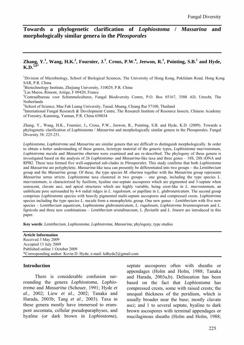
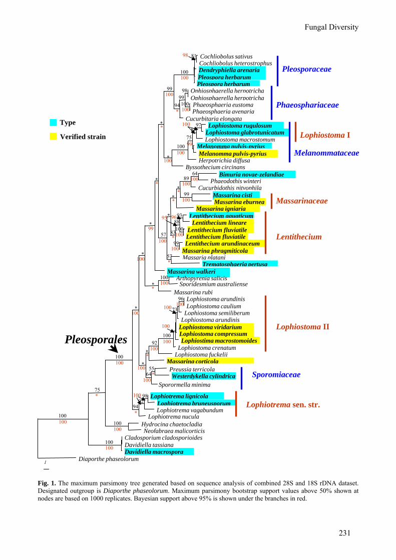
The combined 28S (LSU) and 18S (SSU) rDNA dataset consists of 58 strains. The dataset consists of 1920 characters after alignment, of which 1887 sites are included in the MP analysis. Of the included bases, 350 sites (18.5%) are parsimony-informative. A heuristic search with random addition of taxa (1000 replicates) and treating gaps as missing characters generates six equally parsimonious trees. All trees are similar in topology and not significantly different (figures not shown). A single parsimonious tree (TL = 1528, CI = 0.506, RI = 0.752, RC = 0.380, HI = 0.494) is shown in Fig. 1. Bootstrap support (BS) values (equal to or above 50% based on 1,000 replicates) are shown on the upper branches. Values of the Bayesian posterior probabilities (PP) (equal to or above 95% based on 1,000 replicates) from MCMC analysis are shown under branches in red.
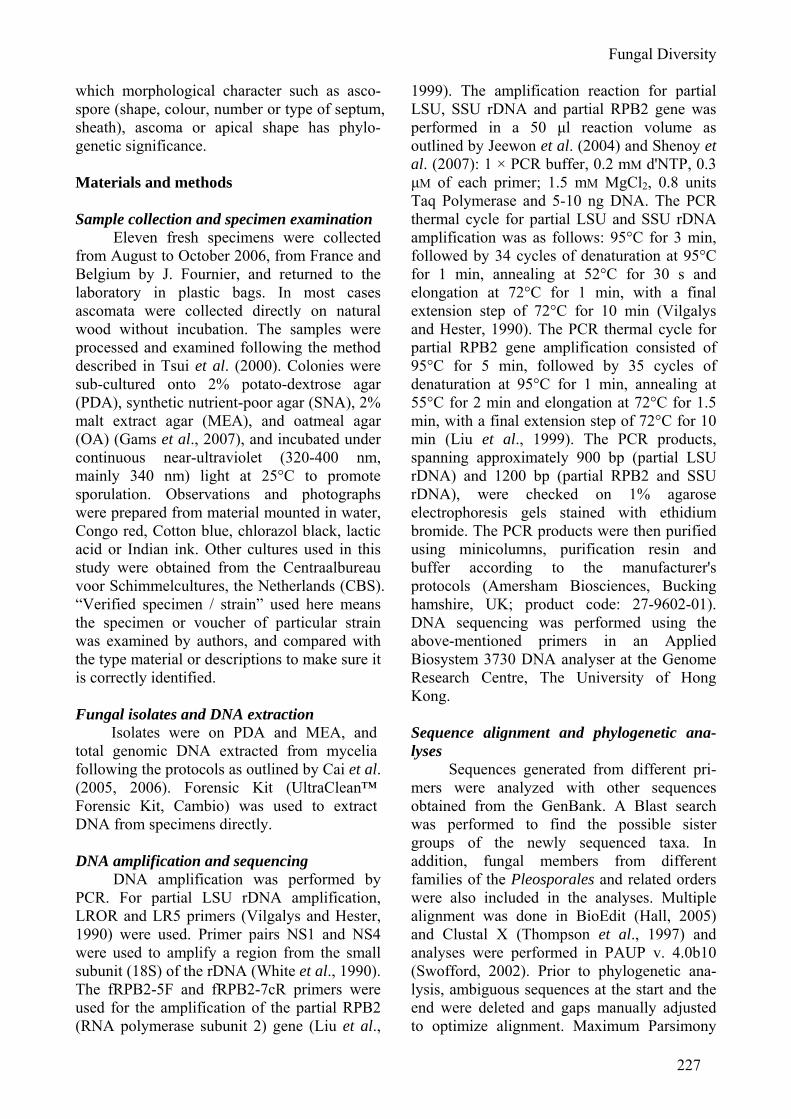
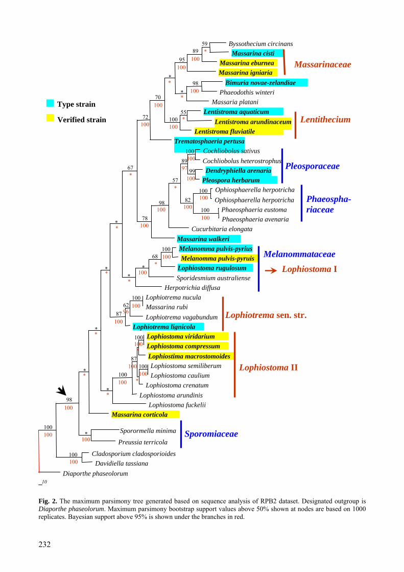
DNA sequencing – RPB2 phylogenies The RPB2 dataset consists of 44 taxa,
with 731 sites after alignment, and all of them are included in the analyses. Of these, 398 sites (54.4%) are parsimony-informative. A heuristic search with random addition of taxa (1000 replicates) and treating gaps as missing charac-ters generates three equally parsimonious trees. All trees are similar in topology and not significantly different (figures not shown). A single parsimonious tree (TL = 3037, CI = 0.285, RI = 0.573, RC = 0.163, HI = 0.715) is shown in Fig. 2. BS values (equal to or above 50% based on 1,000 replicates) are shown on the upper branches. Values of the PP (equal to or above 95% based on 1,000 replicates) from MCMC analysis are shown under branches in red.
Although fewer members are included in the RPB2 dataset, the phylogenetic investiga-tion resulting from both rDNA and RPB2 datasets are almost consistent (Figs 1, 2). The Lophiostoma, Lophiotrema and Massarina taxa clustered in 5 well supported monophyletic clades in the tree based on the combined sequence of the 28S and 18S rDNA or RPB2 dataset (Figs. 1, 2, clades marked in red). In both of the analyses, the Lophiostoma- and Massarina-like taxa clustered throughout the tree and were polyphyletic, and they mainly grouped into four well-supported monophyletic clades. Members of Lophiotrema, including the generic type — L. nucula, form another well supported monophyletic group. All of these five clades are located in Pleosporales. Both Dothideomycetes and Pleosporales form a well supported monophyletic group with high MP and PP bootstrap values (Figs 1, 2).
Clade Lophiostoma I including the type species of Lophiostoma (L. macrostomum), consists of L. glabrotunicatum, Lophiostoma macrostomum and L. rugulosum, and receives high bootstrap support (Fig. 1). These three species are similar in producing 1-septate, hyaline ascospores which become pigmented with age. This group clustered in the Melanom-mataceae with high bootstrap support (Fig. 1). The sequence of L. macrostomum was obtained from GenBank, which was originally from a voucher specimen (Lundqvist 20504 deposited in Swedish Museum of Natural History (S)) from Finland. The identification of this
Fungal Diversity
229
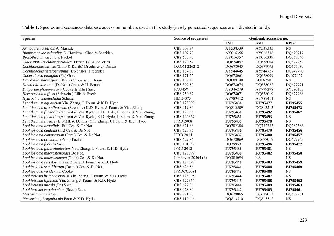
Table 1. Species and sequences database accession numbers used in this study (newly generated sequences are indicated in bold).
GenBank accession no. Species Source of sequences LSU SSU RPB2
Arthopyrenia salicis A. Massal. CBS 368.94 AY538339 AY538333 NS Bimuria novae-zelandiae D. Hawksw., Chea & Sheridan CBS 107.79 AY016356 AY016338 DQ470917 Byssothecium circinans Fuckel CBS 675.92 AY016357 AY016339 DQ767646 Cladosporium cladosporioides (Fresen.) G.A. de Vries CBS 170.54 DQ678057 DQ678004 DQ677952 Cochliobolus sativus (S. Ito & Kurib.) Drechsler ex Dastur DAOM 226212 DQ678045 DQ677995 DQ677939 Cochliobolus heterostrophus (Drechsler) Drechsler CBS 134.39 AY544645 AY544727 DQ247790 Cucurbitaria elongata (Fr.) Grev. CBS 171.55 DQ678061 DQ678009 Dq677657 Davidiella macrospora (Kleb.) Crous & U. Braun CBS 138.40 DQ008148 EU167591 NS Davidiella tassiana (De Not.) Crous & U. Braun CBS 399.80 DQ678074 DQ678022 DQ677971 Diaporthe phaseolorum (Cooke & Ellis) Sacc. FAU458 AY346279 AY779278 AY780175 Herpotrichia diffusa (Schwein.) Ellis & Everh. CBS 250.62 DQ678071 DQ678019 DQ677968 Hydrocina chaetocladia Scheuer HME4375 AY789412 AY789411 NS Lentithecium aquaticum Yin. Zhang, J. Fourn. & K.D. Hyde CBS 123099 FJ795434 FJ795477 FJ795455 Lentithecium arundinaceum (Sowerby) K.D. Hyde, J. Fourn. & Yin. Zhang CBS 619.86 DQ813509 DQ813513 FJ795473 Lentithecium fluviatile (Aptroot & Van Ryck.) K.D. Hyde, J. Fourn. & Yin. Zhang, CBS 123090 FJ795450 FJ795492 FJ795467 Lentithecium fluviatile (Aptroot & Van Ryck.) K.D. Hyde, J. Fourn. & Yin. Zhang, CBS 122367 FJ795451 FJ795493 NS Lentithecium lineare (E. Müll. & Dennis) Yin. Zhang, J. Fourn. & K.D. Hyde IFRD 2008 FJ795435 FJ795478 NS Lophiostoma arundinis (Fr.) Ces. & De Not. CBS 621.86 DQ782384 DQ782383 DQ782386 Lophiostoma caulium (Fr.) Ces. & De Not. CBS 623.86 FJ795436 FJ795479 FJ795456 Lophiostoma compressum (Pers.) Ces. & De Not. IFRD 2014 FJ795437 FJ795480 FJ795457 Lophiostoma crenatum (Pers.) Fuckel CBS 629.86 DQ678069 DQ678017 DQ677965 Lophiostoma fuckelii Sacc. CBS 101952 DQ399531 FJ795496 FJ795472 Lophiostoma glabrotunicatum Yin. Zhang, J. Fourn. & K.D. Hyde IFRD 2012 FJ795438 FJ795481 NS Lophiostoma macrostomoides De Not. CBS 123097 FJ795439 FJ795482 FJ795458 Lophiostoma macrostomum (Tode) Ces. & De Not. Lundqvist 20504 (S) DQ384094 NS NS Lophiostoma rugulosum Yin. Zhang, J. Fourn. & K.D. Hyde CBS 123093 FJ795440 FJ795483 FJ795459 Lophiostoma semiliberum (Desm.) Ces. & De Not. CBS 626.86 FJ795441 FJ795484 FJ795460 Lophiostoma viridarium Cooke IFRDCC2081 FJ795443 FJ795486 NS Lophiotrema brunneosporum Yin. Zhang, J. Fourn. & K.D. Hyde CBS 123095 FJ795444 FJ795487 NS Lophiotrema lignicola Yin. Zhang, J. Fourn. & K.D. Hyde CBS 122364 FJ795445 FJ795488 FJ795462 Lophiotrema nucula (Fr.) Sacc. CBS 627.86 FJ795446 FJ795489 FJ795463 Lophiotrema vagabundum (Sacc.) Sacc. CBS 628.86 FJ795442 FJ795485 FJ795461 Massaria platani Ces. CBS 221.37 DQ678065 DQ678013 DQ677961 Massarina phragmiticola Poon & K.D. Hyde CBS 110446 DQ813510 DQ813512 NS
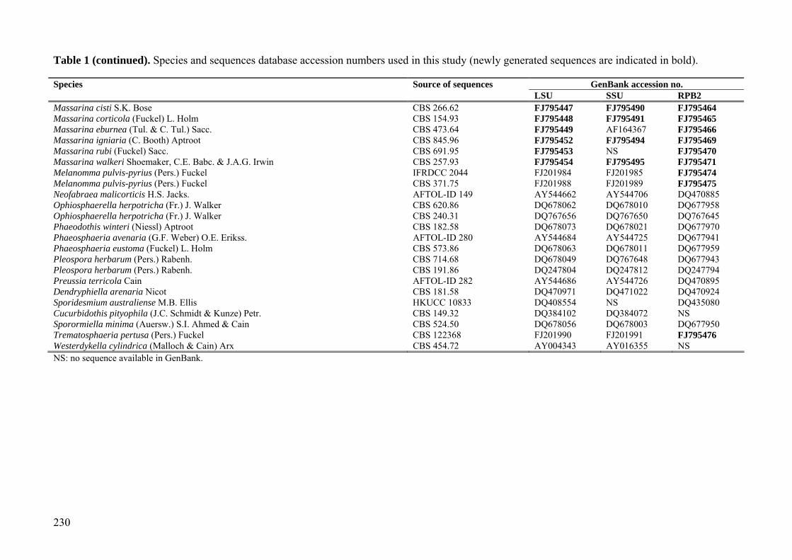
230
Table 1 (continued). Species and sequences database accession numbers used in this study (newly generated sequences are indicated in bold). Species Source of sequences GenBank accession no. LSU SSU RPB2 Massarina cisti S.K. Bose CBS 266.62 FJ795447 FJ795490 FJ795464 Massarina corticola (Fuckel) L. Holm CBS 154.93 FJ795448 FJ795491 FJ795465 Massarina eburnea (Tul. & C. Tul.) Sacc. CBS 473.64 FJ795449 AF164367 FJ795466 Massarina igniaria (C. Booth) Aptroot CBS 845.96 FJ795452 FJ795494 FJ795469 Massarina rubi (Fuckel) Sacc. CBS 691.95 FJ795453 NS FJ795470 Massarina walkeri Shoemaker, C.E. Babc. & J.A.G. Irwin CBS 257.93 FJ795454 FJ795495 FJ795471 Melanomma pulvis-pyrius (Pers.) Fuckel IFRDCC 2044 FJ201984 FJ201985 FJ795474 Melanomma pulvis-pyrius (Pers.) Fuckel CBS 371.75 FJ201988 FJ201989 FJ795475 Neofabraea malicorticis H.S. Jacks. AFTOL-ID 149 AY544662 AY544706 DQ470885 Ophiosphaerella herpotricha (Fr.) J. Walker CBS 620.86 DQ678062 DQ678010 DQ677958 Ophiosphaerella herpotricha (Fr.) J. Walker CBS 240.31 DQ767656 DQ767650 DQ767645 Phaeodothis winteri (Niessl) Aptroot CBS 182.58 DQ678073 DQ678021 DQ677970 Phaeosphaeria avenaria (G.F. Weber) O.E. Erikss. AFTOL-ID 280 AY544684 AY544725 DQ677941 Phaeosphaeria eustoma (Fuckel) L. Holm CBS 573.86 DQ678063 DQ678011 DQ677959 Pleospora herbarum (Pers.) Rabenh. CBS 714.68 DQ678049 DQ767648 DQ677943 Pleospora herbarum (Pers.) Rabenh. CBS 191.86 DQ247804 DQ247812 DQ247794 Preussia terricola Cain AFTOL-ID 282 AY544686 AY544726 DQ470895 Dendryphiella arenaria Nicot CBS 181.58 DQ470971 DQ471022 DQ470924 Sporidesmium australiense M.B. Ellis HKUCC 10833 DQ408554 NS DQ435080 Cucurbidothis pityophila (J.C. Schmidt & Kunze) Petr. CBS 149.32 DQ384102 DQ384072 NS Sporormiella minima (Auersw.) S.I. Ahmed & Cain CBS 524.50 DQ678056 DQ678003 DQ677950 Trematosphaeria pertusa (Pers.) Fuckel CBS 122368 FJ201990 FJ201991 FJ795476 Westerdykella cylindrica (Malloch & Cain) Arx CBS 454.72 AY004343 AY016355 NS NS: no sequence available in GenBank.
Fungal Diversity
231
Fig. 1. The maximum parsimony tree generated based on sequence analysis of combined 28S and 18S rDNA dataset. Designated outgroup is Diaporthe phaseolorum. Maximum parsimony bootstrap support values above 50% shown at nodes are based on 1000 replicates. Bayesian support above 95% is shown under the branches in red.
Melanommataceae
1
Cochliobolus sativus Cochliobolus heterostrophus Dendryphiella arenaria Pleospora herbarum Pleospora herbarum
Ophiosphaerella herpotricha Ophiosphaerella herpotricha Phaeosphaeria eustoma Phaeosphaeria avenaria
Cucurbitaria elongata Lophiostoma rugulosum
Lophiostoma glabrotunicatum Lophiostoma macrostomum
Melanomma pulvis-pyrius Melanomma pulvis-pyrius Herpotrichia diffusa
Byssothecium circinans Bimuria novae-zelandiae
Phaeodothis winteri Cucurbidothis pityophila
Massarina cisti Massarina eburnea
Massarina igniaria Lentithecium aquaticum Lentithecium lineare
Lentithecium fluviatile Lentithecium fluviatile Lentithecium arundinaceum
Massarina phragmiticolaMassaria platani
Trematosphaeria pertusa Massarina walkeri
Arthopyrenia salicis Sporidesmium australiense
Massarina rubi Lophiostoma arundinis Lophiostoma caulium
Lophiostoma semiliberum Lophiostoma arundinis
Lophiostoma viridarium Lophiostoma compressum Lophiostima macrostomoides
Lophiostoma crenatum Lophiostoma fuckelii
Massarina corticola Preussia terricola Westerdykella cylindrica
Sporormella minima
Lophiotrema lignicola Lophiotrema bruneusporum Lophiotrema vagabundum
Lophiotrema nucula Hydrocina chaetocladia Neofabraea malicorticis
Cladosporium cladosporioides Davidiella tassiana Davidiella macrospora
Diaporthe phaseolorum
Type
Verified strain
Pleosporaceae
100
100
9499
9899
81
99
8797
100
100
100
**
*
*
8964
75
83
99
73
8893*
*
*
96
92
57
57
*
*
*
100
100
100
100
*
*
98 92
94
100
64 55
75
Lentithecium
Massarinaceae
Phaeosphariaceae
Sporomiaceae
Lophiotrema sen. str.
95
100
100
100100
10099
96
*
*
100
100100
*
*
100*
*
100
98
*
10099
* 100
100
100
100
100
100
100
100
100
*
*
* 100
100
100
100
100
100
100
Lophiostoma I
Lophiostoma II
Pleosporales
*
232
Fig. 2. The maximum parsimony tree generated based on sequence analysis of RPB2 dataset. Designated outgroup is Diaporthe phaseolorum. Maximum parsimony bootstrap support values above 50% shown at nodes are based on 1000 replicates. Bayesian support above 95% is shown under the branches in red.
10
Byssothecium circinans Massarina cisti
Massarina eburnea Massarina igniaria
Bimuria novae-zelandiae Phaeodothis winteri
Massaria platani Lentistroma aquaticum
Lentistroma arundinaceum Lentistroma fluviatile
Trematosphaeria pertusa Cochliobolus sativus Cochliobolus heterostrophus Dendryphiella arenaria
Pleospora herbarum Ophiosphaerella herpotricha Ophiosphaerella herpotricha
Phaeosphaeria eustoma Phaeosphaeria avenaria
Cucurbitaria elongata Massarina walkeri Melanomma pulvis-pyriusMelanomma pulvis-pyruis
Lophiostoma rugulosum Sporidesmium australiense
Herpotrichia diffusa Lophiotrema nucula Massarina rubi Lophiotrema vagabundum
Lophiotrema lignicola Lophiostoma viridarium Lophiostoma compressum Lophiostima macrostomoides Lophiostoma semiliberum Lophiostoma caulium
Lophiostoma crenatum Lophiostoma arundinis
Lophiostoma fuckelii Massarina corticola
Sporormella minima
Preussia terricola
Cladosporium cladosporioides Davidiella tassiana
Diaporthe phaseolorum
Type strain
Verified strain
100
100
100
100
100
98
89
98
95
89 59
*
*
*
*
*
*
99 67
72
70
55
78
98 82
57
100
* *
*
*
100
68
62
87
87
100
100
100
100
100
97
100
*
*
100 *
96
100
100
100
*
*
100
100
100
100
100
100
100
100
100
100
100
100
*
*
*
*
*
100
100
100 *
100
100
100
100
*
*
*
*
Massarinaceae
Lentithecium
Pleosporaceae
Phaeospha-riaceae
Melanommataceae
Lophiostoma I
Lophiotrema sen. str.
Lophiostoma II
Sporomiaceae
Fungal Diversity
233
specimen is not yet verified. The other two LSU sequences of Lophiostoma macrostomum in GenBank (Accession numbers: AB433273 and AB433274) are from the voucher speci-mens of HHUF:27290 and HHUF:27293 respectively, and they both located in Clade Lophiostoma II (result not shown). According to the hand drawing by Tanaka and Harada (2003a), HHUF:27293 might be wrongly identified as the apex is not crest-like. Thus the identification of these two specimens also needs to be verified. Therefore, it would be premature to separate Lophiostoma sensu stricto from the other Lophiostoma members in Clade Lophiostoma II as the specimens from which the gene sequences were obtained are not verified.
Clade Massarinaceae, from rDNA se-quences, contains only two Massarina species (M. cisti and M. eburnea) and receives high bootstrap support (Fig. 1). Massarina cisti and M. eburnea share several morphological characteristics, including hyaline, broad to narrowly ellipsoidal ascospores and filament-tous, broad, septate and hyaline cellular pseudoparaphyses, that lack or have anasto-moses or branching (Hyde, 1995; Aptroot, 1998; Tanaka and Harada, 2003c). The identification of the two strains used here is verified (Zhang et al., 2008). Liew et al. (2002) pointed out that species of Massarina with broad, ellipsoidal ascospores, together with M. eburnea, should remain in Massarina sensu stricto, and species with narrow, fusiform ascospores must be incorporated in Lophio-stoma. This idea was accepted by other mycologists (Hyde et al., 2002; Eriksson and Hawksworth, 2003). Thus here Clade Massari-naceae includes the generic type of Massarina (M. eburnea) and should represent Massarina sensu stricto. According to RPB2 sequence data, Massarina eburnea and M. cisti clustered together with Byssothecium circinans and M. igniaria (Fig. 2). Byssothecium circinans differs considerably from M. eburnea in mor-phology, such as the black, erumpent ascoma, fusiform ascospores with pigmented central cells and hyaline end cells. The unstable position of B. circinans might be because of the differences of sampling or gene number used in the analysis, and the later has been proved could influence the topology of the tree
(Rokas and Carroll, 2005), thus we are not sure if B. circinans has any relationship with Massarina. The identification of the Massarina igniaria strain (CBS 845.96) used here is verified by checking the voucher specimen (CBS H-006463). Massarina eburnea and M. igniaria share some common morphological characters, such as 3-septate fusiform asco-spores and broad septate pseudoparaphyses. The rDNA sequence dataset however, does not support M. igniaria as belonging in Massarina sensu stricto.
Clade Lentithecium comprises four ligni-colous species of Lentithecium including two transferred from Massarina (M. arundinacea and M. fluviatilis) and one from Keissleriella (K. linearis). This Clade receives moderate bootstrap support from rDNA sequence dataset (Fig. 1), but high bootstrap support from RPB2 sequence dataset (Fig. 2). One well-supported subgroup in this Clade comprises L. arundina-ceum and Massarina phragmiticola (Fig. 1). Morphological characters of these two species are also almost identical (Aptroot, 1998), which suggests these taxa are conspecific. Keissleriella was established by von Höhnel (1919), and its most distinguishing characters are the black setae in and over the small apical papilla and the hyaline one-septate to rarely multi-septate ascospores (Barr, 1990). The familial placement of Keissleriella is debatable, such as in Lophiostomataceae (includes Massarinaceae) by Munk (1957), in Pleospo-raceae by Arx and Müller (1975), in Melano-mmataceae by Barr (1990) and presently in Massarinaceae by Lumbsch and Huhndorf (2007). Keissleriella linearis, as the only taxon having setae in Clade Lentithecium (Fig. 11A), forms a robust Clade with other members, which might indicate that the presence of setae has little phylogenetic significance. Taxa in Clade Lentithecium share similar morpholo-gical characters in having immersed to erumpent lenticular ascomata, clavate asci, anastomosing cellular pseudoparaphyses and hyaline one-septate ascospores. Thus we intro-duce a new genus − Lentithecium to accommo-date this group of fungi. The polyphyletic nature of Massarina has been noticed in previous studies (Hyde and Aptroot, 1998; Hyde et al., 2002; Liew et al., 2002). The lenticular ascoma is a common character

234
shared by all five members in Clade Lentithecium, but it is premature to draw the conclusion that ascomatal shape has any phylogenetic significance at the generic level classification.
Clade Lophiostoma II is a well supported clade containing seven lophiostomataceous species with Lophiostoma fuckelii clustering in a basal position (Fig. 1). The identification of three strains of this group, i.e. L. viridarium, L. compressum and L. macrostomoides, has been verified by carefully checking the fresh specimens we collected and referring to their detailed descriptions by Holm and Holm (1988). Members in this group share a com-pressed apex, mostly brown and multiseptate or even muriform ascospores. Ascospores of L. semiliberum are 1-septate, hyaline, becoming brown with additional septa when senescent. The ascomata with compressed apex are a striking common character possessed by all members of this group. Compared with the crest-like apex possessed by L. macrostomum, the compressed apex is a more stable and evolutionary older character state (Holm and Holm, 1988). This group of fungi seems to represent a natural group at family level but does not include the type of Lophiostoma — L. macrostomum. This conclusion can not be confirmed however, until the verified sequen-ces of L. macrostomum (Clade Melanomma-taceae) are obtained.
The Lophiotrema group includes the type species — L. nucula, and contains four lophiostomataceous species (L. brunneosporum, L. lignicola, L. nucula and L. vagabundum) with consistent high bootstrap (Figs 1, 2). These four species share immersed to erumpent to nearly superficial ascomata, round or elongate ostioles, clavate to cylindrical asci, and 1-septate, hyaline to pigmented, deeply constricted ascospores (Holm and Holm, 1988; Tanaka and Harada, 2003b).
Clade Sporomiaceae contains four species which are moderately supported by parsimony analysis but well supported by MrBayes analyses with M. corticola in the basal position (Figs 1, 2). Sporomiaceae is a coprophilous group of fungi, and previous phylogenetic studies indicated Sporomiaceae to
be a monophyletic group (Kruys et al., 2006; Schoch et al., 2006). The circumscription of some genera in this family has, however, been based on some characters of weak phylogenetic significance (Arenal et al., 2005; Nyberg Kruys, 2006). Taxonomy Lentithecium K.D. Hyde, J. Fourn. & Yin. Zhang, gen. nov. MycoBank: 512790
Etymology: from the Latin “lenti” and “thecium”, in reference to the “lenticular” ascomata.
Ascomata immersa vel partim immersa, lenticular, solitarius vel disseminatus, nigra. Asci clavate vel oblongatus-cylindraceo, pedicellati, bitunicati, fissituni-cati. Ascosporae cymbiforme, 1-septatae, aliquando 3-septatae cum supra-maturus, septum constrictaee, hyaline.
Ascomata immersed to semi-immersed, lenticular. Peridium comprising a few layers of thin-walled cells. Asci bitunicate, fissitunicate, clavate-cylindrical to oblong-cylindrical. Asco-spores hyaline, one-septate.
Type species – Lentithecium fluviatile (Aptroot & Van Ryck.) K.D. Hyde, J. Fourn. & Yin. Zhang, comb. nov. MB 512802
Basionym: Massarina fluviatilis Aptroot & Van Ryck., (2001), Nova Hedwigia 73: 162. (2001) Lentithecium aquaticum Yin. Zhang, J. Fourn. & K.D. Hyde, sp. nov. (Fig. 3) MycoBank: 512791
Etymology: from the Latin “aquaticum”, in reference to the freshwater habit.
Ascomata 130-160 µm alta, 240-320 µm longa, immersa, papillaris, lenticular, disseminatus, nigra. Asci 130-190 × 17-23 µm, 8-spori, clavate, pedicellati, bitunicat. Ascosporae 25-30 × 8-12 µm, fusiformes, hyalinae.
Ascomata 130-160 µm high × 240-320 µm diam., scattered, immersed, lenticular with a flattened base, papillate, the rounded papilla protruding slightly above the wood surface which is stained dark grey; ostioles round (Figs 3A, B). Peridium 13-25 µm thick, thin-walled, pigmented, textura prismatica, thicker and more pigmented in upper part (Figs 3B, C). Hamathecium of dense, long septate pseudopa-raphyses, 2-3 µm broad, mixed in places with much thinner filaments, rarely anastomosing between and above the asci (Fig. 3G). Asci
Fungal Diversity
235
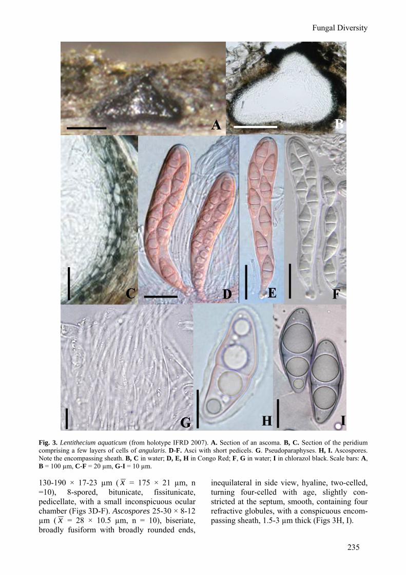
Fig. 3. Lentithecium aquaticum (from holotype IFRD 2007). A. Section of an ascoma. B, C. Section of the peridium comprising a few layers of cells of angularis. D-F. Asci with short pedicels. G. Pseudoparaphyses. H, I. Ascospores. Note the encompassing sheath. B, C in water; D, E, H in Congo Red; F, G in water; I in chlorazol black. Scale bars: A, B = 100 µm, C-F = 20 µm, G-I = 10 µm. 130-190 × 17-23 µm ( x = 175 × 21 µm, n =10), 8-spored, bitunicate, fissitunicate, pedicellate, with a small inconspicuous ocular chamber (Figs 3D-F). Ascospores 25-30 × 8-12 µm ( x = 28 × 10.5 µm, n = 10), biseriate, broadly fusiform with broadly rounded ends,
inequilateral in side view, hyaline, two-celled, turning four-celled with age, slightly con-stricted at the septum, smooth, containing four refractive globules, with a conspicuous encom-passing sheath, 1.5-3 µm thick (Figs 3H, I).

236
Culture characters (ex-holotype: CBS 123099) On OA spreading, up to 15 mm diam.,
smoke-grey with woolly aerial mycelium; hyphae hyaline, smooth, 1.5-3 µm wide, forming hyphal strands with 15-20 hyphae that become dark brown, 4-5 µm wide. On PDA up to 15 mm diam.; central part iron-grey, with a wide cream border; aerial mycelium lacking, slimy or woolly, pale olivaceous-grey; grey-olivaceous in reverse, with cream margin. On MEA spreading, up to 15 mm diam., with moderate, woolly aerial mycelium and smooth, even margins; surface and reverse cream to smoke-grey.
Specimens examined: FRANCE, Ariège, Rimont, Peyrau, on submerged wood of Fraxinus excelsior, 400m, 09 Oct. 2006, leg., det. J. Fournier [holotype: CBS H-20220], ex-holotype living culture deposited in CBS (CBS 123099); 07 Nov. 2006, leg., det. J. Fournier [IFRD2021]; on submerged wood of Alnus glutinosa, 19 Oct. 2006, leg., det. J. Fournier [IFRD2024]; Le Baup brook, along D 18, on submerged wood of Platanus sp., 550m, 10 Nov. 2006, leg., det. J. Fournier, [IFRD 2022].
Notes: Lentithecium aquaticum can be distinguished from other members of Lophio-stoma- and Massarina-like species by its minute, immersed ascomata, which are easily overlooked, and by the narrow conspicuous sheath around ascospores, which is easily visible in water and does not swell in water. Massarina immersa also has tiny immersed ascomata, but the ascomata of M. immersa are globose, and the broadly fusiform ascospores of M. immersa are distinctly smaller than those of Lentithecium aquaticum ((17-)19-22(-24) × 6-8 µm vs. 25-30 × 8-12 µm) (Aptroot, 1998). Ascospore shape is comparable with those of Massarina appendiculata and M. moeszii, but the ascomata of M. moeszii are much larger (500-900 µm), and the ascomata of M. appendiculata are round and larger (to 550 µm) (Aptroot, 1998). It is a commonly encountered species in all regions of France sampled, and is remarkably still present in its ascigenous state in winter, and occurs on various substrates. Lentithecium arundinaceum (Sowerby) K.D. Hyde, J. Fourn. & Yin. Zhang, comb. nov. MycoBank: 512819
Basionym: Sphaeria arundinacea Sowerby, Coloured Figures of English Fungi 3: t.336 (1803).
= Massarina phragmiticola Poon & K.D. Hyde, Botanica Marina 41: 145 (1998).
Specimens examined: DENMARK, Sjaeland, Frederikskilde, Suserup Skove, Tystrup Lake, on
submerged stems of Phragmites sp., 25 May 2007, leg., det. J. Fournier [IFRD2031]; FRANCE, Haute Garonne, Avignonet, Port Lauragais, Rosel artificial lake, on submerged stems of Phragmites sp., 15 Jul. 2007, leg., det. J. Fournier [IFRD2032]; Vendée, Vouvant, on submerged stems of Phragmites sp., Apr. 2005, leg., det. P. Leroy [IFRD2033]. Lentithecium lineare (E. Müll. & Dennis) K.D. Hyde, J. Fourn. & Yin. Zhang, comb. nov. MycoBank: 512820
Basionym: Keissleriella linearis E. Müll. & Dennis (1964), Kew Bulletin 19: 120. (1964)
Specimen examined: FRANCE, Haute Garonne, Avignonet, Port Lauragais, Rosel artificial lake, on dead stems of Phragmites sp., 06 May 2006, leg., det. J. Fournier & P. Leroy [IFRD2008]. Lophiotrema Lophiotrema nucula (Fr.) Sacc., Michelia 1: 338 (1878). (Fig. 4)
≡ Sphaeria nucula Fr., Kongliga Vetenskaps Academiens Handlingar 38: 266 (1817).
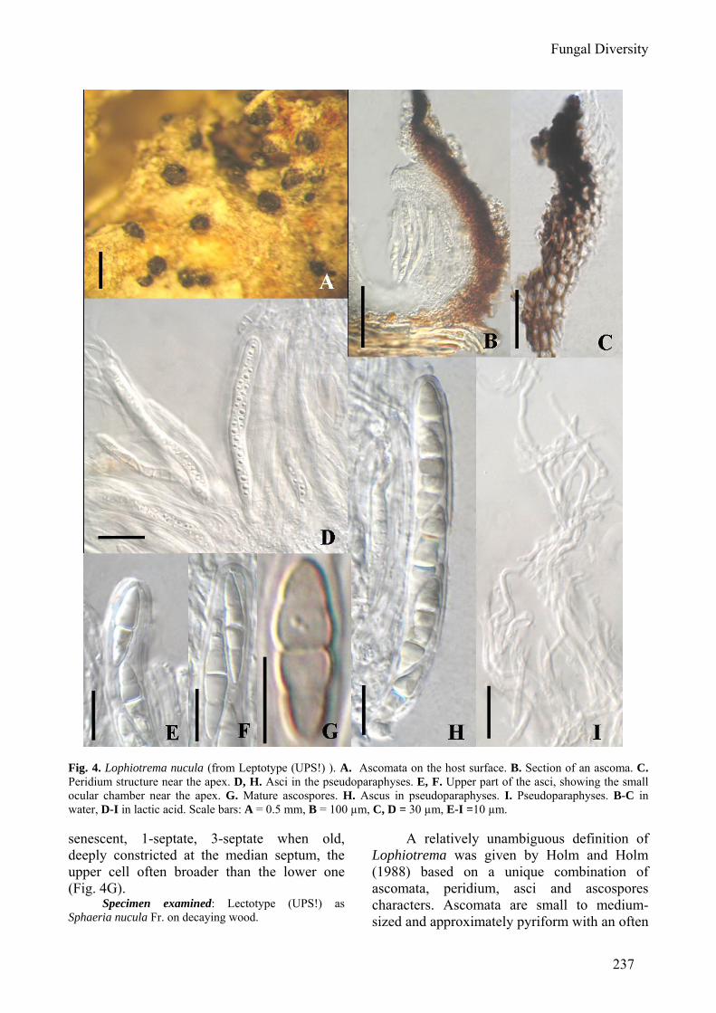
Ascomata 200-240 µm high × 200-280 µm diam., scattered, erumpent to nearly superficial, with basal wall remaining im-mersed in host tissue, globose, subglobose, often laterally flattened, with a flattened base, black, roughened, often bearing remnants of wood fibres; with a cylindrical or laterally compressed papilla, to 120 µm long and 150 µm high (Fig. 4A). Peridium laterally 25-30 µm thick, very thin at the base, composed of heavily pigmented pseudoparenchymatous cells near the apex, cells 2-2 × 6 µm diam., wall 1-3(-4) µm thick, near apex composed of pigmented cells of textura angularis, 3-5 µm diam., wall 0.8-1.5 µm thick, apical wall composed of heavily pigmented and thick walled small cells (Figs 4B, C). Hamathecium of dense, very long septate pseudoparaphyses, 1-2 µm broad, anastomosing and branching between and above asci, embedded in gel matrix (Fig. 4I). Asci 90-115 × 9-11.5 µm (x = 99.5 × 11 µm, n = 10), 8-spored, bitunicate, fissitunicate, cylindrical, with a short, narrowed, furcate pedicel which is up to 10 µm long, with a small ocular chamber (Figs 4D-F, H). Ascospores 17-21(-25) × (4-)5-6.5 µm ( x = 19.5 × 5.5 µm, n = 10), obliquely uniseriate and partially overlapping to biseriate, broad-fusiform, fusiform to narrowly fusiform, with narrowly rounded ends, hyaline and lightly pigmented on very rare occasions when
Fungal Diversity
237
Fig. 4. Lophiotrema nucula (from Leptotype (UPS!) ). A. Ascomata on the host surface. B. Section of an ascoma. C. Peridium structure near the apex. D, H. Asci in the pseudoparaphyses. E, F. Upper part of the asci, showing the small ocular chamber near the apex. G. Mature ascospores. H. Ascus in pseudoparaphyses. I. Pseudoparaphyses. B-C in water, D-I in lactic acid. Scale bars: A = 0.5 mm, B = 100 µm, C, D = 30 µm, E-I =10 µm. senescent, 1-septate, 3-septate when old, deeply constricted at the median septum, the upper cell often broader than the lower one (Fig. 4G).
Specimen examined: Lectotype (UPS!) as Sphaeria nucula Fr. on decaying wood.
A relatively unambiguous definition of Lophiotrema was given by Holm and Holm (1988) based on a unique combination of ascomata, peridium, asci and ascospores characters. Ascomata are small to medium-sized and approximately pyriform with an often
238
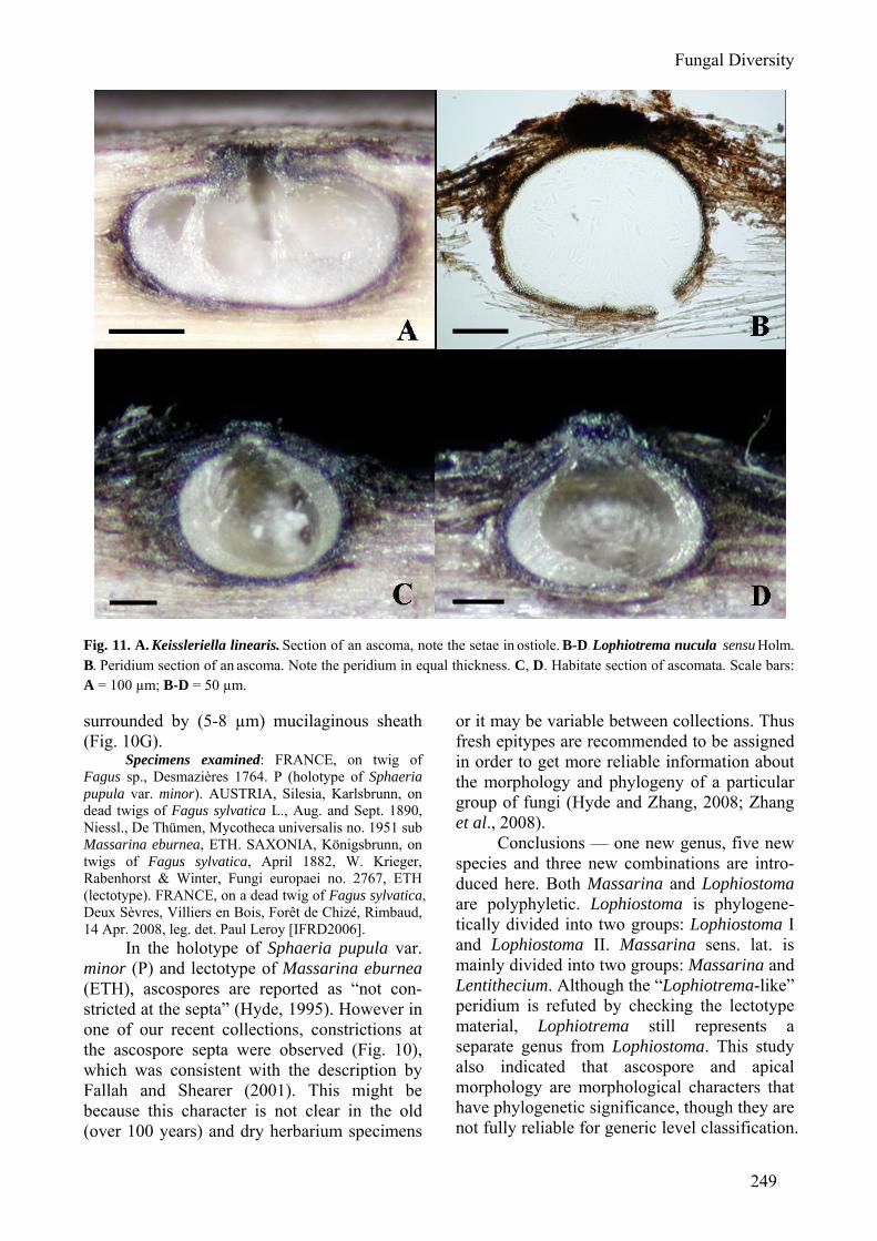
reduced cylindrical neck which may even be absent. The peridium is approximately 20-30 µm of equal thickness, composed of an outer layer of textura angularis of uniformly pigmented cells, up to 12 µm, and an inner layer of very small hyaline cells, with somewhat thickened walls. Asci are cylindrical and spores are hyaline, at first 1-septate, later 3-septate, with distinct guttules, often with a mucilaginous sheath (Holm and Holm, 1988). Much emphasis was placed on ascospore characters by Holm and Holm (1988) when they described and distinguished the three Lophiotrema species: L. boreale, L. nucula and L. vagabundum. This generic concept was widely accepted by later workers (e.g. Barr, 1990; Yuan and Zhao, 1994; Kirk et al., 2008). Emphasis was placed on the peridium and asci by Tanaka and Harada (2003b) to distinguish Lophiotrema from Lophiostoma. This peridium differences however, is not supported in the lectotype specimen we examined here, which has a flattened thin-walled base. While several of our recent collections from Europe fit Holm and Holm’s description well, which differs from the lectotype specimen in: ascomata (immersed to slightly erumpent vs. erumpent to nearly superficial), length of asci (90-115 µm vs. 120-130 µm) and peridium (equal in thickness vs. with thinner base) (Figs 11B-D). Thus here we call these collections as Lophiotrema nucula sensu Holm. The DNA sequences of L. nucula used here are of Holm’s collection. Thus further study is needed to clarify the specific status of L. nucula sensu stricto, and equal-thickness peridium could not serve as a diagnosing character of Lophiotrema. Presently, we temporarily treat L. nucula sensu Holm as L. nucula sensu stricto basing on their mostly comparable morphological characters. Lophiotrema lignicola Yin. Zhang, J. Fourn. & K.D. Hyde, sp. nov. (Fig. 5) MycoBank: 512792
Etymology: from the Latin “lignicola”, in reference to the lignicolous substrate.
Ascomata 260-350 µm alta, 450-530 longa, semi-immersa, papillaris, gregarius. Asci 133-180 × 15-17 µm, 8-spori, latus-cylindrico, pedicellati, bitunicat. Ascosporae 21-25 × 6.5-8 µm, latus- fusiformes, hyalinae.
Ascomata 260-350 µm high × 450-530 µm diam., gregarious, 2/3rds to semi-immersed
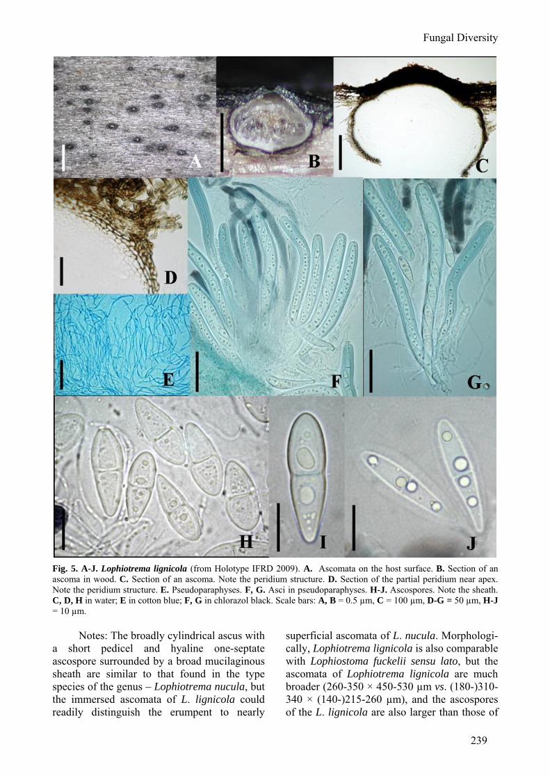
under a blackish pseudostroma, lenticular; papillate, apex often elongate and laterally compressed but not raised or forming a crest, with an elongated slit-like ostiole (Figs 5A, B). Peridium 20-30 µm thick at sides and base, 40-60 µm thick at apex, dark brown, pseudoparen-chymatous, composed of two intergrading layers: outer layer of small cells 4-10 µm long, textura angularis to prismatica, moderately thick-walled at sides and base, very thick-walled at apex, with dark brown hyphal appendages 4-6 µm broad penetrating the surrounding wood and forming a pseudostroma 60-100 µm thick around the papilla; inner layer composed of less pigmented to hyaline cells textura angularis, very thin-walled (Figs 5C, D). Hamathecium of dense, long pseudopara-physes, rarely septate, 1-1.5 µm broad, branching and anastomosing between and above asci, embedded in gelatinous matrix (Fig. 5E). Asci 133-180 µm × 15-17 µm ( x = 160 × 15.5 µm, n = 10), 8-spored, bitunicate, fissitunicate, broad cylindrical, with a short, narrowed, twisted, pedicellate, with an ocular chamber and a minute apical ring visible in immature asci (Figs 5F, G). Ascospores 21-25 × 6.5-8 µm ( x = 23.5 × 7.5 µm, n = 10), uni to biseriate, broadly fusiform with broad to narrow rounded ends, hyaline, yellowish at maturity, 2-celled, cell wall up to 1.8 µm thick, slightly constricted at the median septum, the upper cell often slightly shorter and broader than the lower one, smooth, surrounded by an irregular hyaline gelatinous sheath 2-2.5 µm thick visible in 3% KOH but not seen in water or India ink (Figs 5H-J).
Culture characters (ex-holotype: CBS 122364)
On OA spreading, with sparse aerial mycelium and even, regular margins, reaching 20 mm diam.; surface olivaceous-grey. On PDA spreading with sparse to moderate aerial mycelium and regular margins, reaching 15 mm diam.; surface olivaceous-grey in middle, iron-grey in outer region and in reverse. On MEA erumpent, with moderate aerial myce-lium and smooth, crenate margins, reaching 15 mm diam.; surface pale olivaceous-grey, reverse olivaceous-grey.
Specimen examined: BELGIUM, Hainaut, Orval, ruisseau de Williers, on decorticated trunk of Populus sp., 29 Sept. 2006, leg., det. J. Fournier [CBS H-20221, holotype].
Fungal Diversity
239
Fig. 5. A-J. Lophiotrema lignicola (from Holotype IFRD 2009). A. Ascomata on the host surface. B. Section of an ascoma in wood. C. Section of an ascoma. Note the peridium structure. D. Section of the partial peridium near apex. Note the peridium structure. E. Pseudoparaphyses. F, G. Asci in pseudoparaphyses. H-J. Ascospores. Note the sheath. C, D, H in water; E in cotton blue; F, G in chlorazol black. Scale bars: A, B = 0.5 µm, C = 100 µm, D-G = 50 µm, H-J = 10 µm.
Notes: The broadly cylindrical ascus with a short pedicel and hyaline one-septate ascospore surrounded by a broad mucilaginous sheath are similar to that found in the type species of the genus – Lophiotrema nucula, but the immersed ascomata of L. lignicola could readily distinguish the erumpent to nearly
superficial ascomata of L. nucula. Morphologi-cally, Lophiotrema lignicola is also comparable with Lophiostoma fuckelii sensu lato, but the ascomata of Lophiotrema lignicola are much broader (260-350 × 450-530 µm vs. (180-)310-340 × (140-)215-260 µm), and the ascospores of the L. lignicola are also larger than those of
240
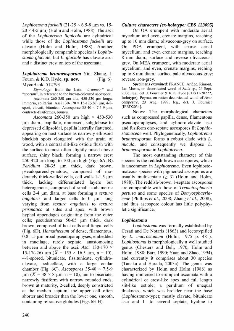
Lophiostoma fuckelii (21-25 × 6.5-8 µm vs. 15-20 × 4-5 µm) (Holm and Holm, 1988). The asci of the Lophiotrema lignicola are cylindrical while those of the Lophiostoma fuckelii are clavate (Holm and Holm, 1988). Another morphologically comparable species is Lophio-stoma glaciale, but L. glaciale has clavate asci and a distinct crest on top of the ascomata. Lophiotrema brunneosporum Yin. Zhang, J. Fourn. & K.D. Hyde, sp. nov. (Fig. 6) MycoBank: 512793
Etymology: from the Latin “brunneo-” and “sporum”, in reference to the brown-coloured ascospore.
Ascomata 260-350 µm alta, 450-530 µm longa, immersa, solitarius. Asci 130-170 × 15-17(-26) µm, 4-8-spori, clavati, bitunicat. Ascosporae 35-40 × 7.5-9 µm, contracte-fusiformes, bruneu.
Ascomata 260-350 µm high × 450-530 µm diam., papillate, immersed, subglobose to depressed ellipsoidal, papilla laterally flattened, appearing on host surface as narrowly ellipsoid blackish spots elongated with the grain of wood, with a central slit-like ostiole flush with the surface to most often slightly raised above surface, shiny black, forming a narrow crest 250-420 µm long, to 100 µm high (Figs 6A, B). Peridium 28-35 µm thick, dark brown, pseudoparenchymatous, composed of mo-derately thick-walled cells, cell walls 1-1.5 µm thick, lacking differentiated layers but heterogenous, composed of small isodiametric cells 2-4 µm diam. at base forming a textura angularis and larger cells 6-10 µm long varying from textura angularis to textura prismatica at sides and apex, with brown hyphal appendages originating from the outer cells; pseudostroma 50-65 µm thick, dark brown, composed of host cells and fungal cells (Fig. 6D). Hamathecium of dense, filamentous, 0.8-1.5 µm broad pseudoparaphyses, embedded in mucilage, rarely septate, anastomosing between and above the asci. Asci 130-170 × 15-17(-26) µm ( x = 155 × 16.5 µm, n = 10), 4-8-spored, bitunicate, fissitunicate, cylindro-clavate, pedicellate, with a large ocular chamber (Fig. 6C). Ascospores 35-40 × 7.5-9 µm ( x = 38 × 8 µm, n = 10), uni to biseriate, narrowly fusiform with narrow rounded ends, brown at maturity, 2-celled, deeply constricted at the median septum, the upper cell often shorter and broader than the lower one, smooth, containing refractive globules (Figs 6E-H).
Culture characters (ex-holotype: CBS 123095) On OA erumpent with moderate aerial
mycelium and even, crenate margins, reaching up to 10 mm diam.; olivaceous-grey on surface. On PDA erumpent, with sparse aerial mycelium, and even crenate margins, reaching 8 mm diam.; surface and reverse olivaceous-grey. On MEA erumpent, with moderate aerial mycelium, and even, crenate margins, reching up to 8 mm diam.; surface pale olivaceous-grey; reverse iron-grey.
Specimens examined: FRANCE, Ariège, Rimont, Las Muros, on decorticated wood of Salix sp., 24 Sept. 2006, leg., det. J. Fournier & K.D. Hyde [CBS H-20222, holotype]; Peyrau, on rotten decorticated wood of Acer campestre, 23 Aug. 1997, leg., det. J. Fournier [IFRD2034].
Notes: The morphological characters such as compressed papilla, dense, filamentous pseudoparaphyses, and cylindro-clavate asci and fusiform one-septate ascospores fit Lophio-stomaceae well. Phylogenetically, Lophiotrema brunneosporum forms a robust clade with L. nucula, and consequently we dispose L. brunneosporum in Lophiotrema.
The most outstanding character of this species is the reddish-brown ascospores, which is uncommon in Lophiotrema. Even lophiosto-mateous species with pigmented ascospores are usually multiseptate (≥ 3) (Holm and Holm, 1988). The reddish-brown 1-septate ascospores are comparable with those of Trematosphaeria pertusa and some species of Botryosphaeria-ceae (Phillips et al., 2008; Zhang et al., 2008), and thus ascospore colour has little polyphy-letic significance. Lophiostoma
Lophiostoma was formally established by Cesati and De Notaris (1863) and lectotypified by L. macrostomum (Holm, 1975 p. 481). Lophiostoma is morphologically a well studied genus (Chesters and Bell, 1970; Holm and Holm, 1988; Barr, 1990; Yuan and Zhao, 1994), and currently it comprises about 30 species (Tanaka and Harada, 2003a). The genus was characterized by Holm and Holm (1988) as having immersed to erumpent ascomata with a cylindrical or crest-like apex and full length slit-like ostiole; a peridium of unequal thickness, which was broader near the base (Lophiostoma-type); mostly clavate, bitunicate asci and 1- to several septate, hyaline to
Fungal Diversity
241
Fig. 6. Lophiotrema brunneusporum (from Holotype). A. Ascomata on the host surface. Note the compressed papilla. B. Section of an ascoma in wood. C. Four-spored mature and some immature asci in pseudoparaphyses. D. Section of the peridium. Note the peridium structure. E-H. Mature ascospores. C in Cotton blue, D-G in water, H in Indian ink. Scale bars: A, B = 0.5 mm, C-H = 20 µm.
242
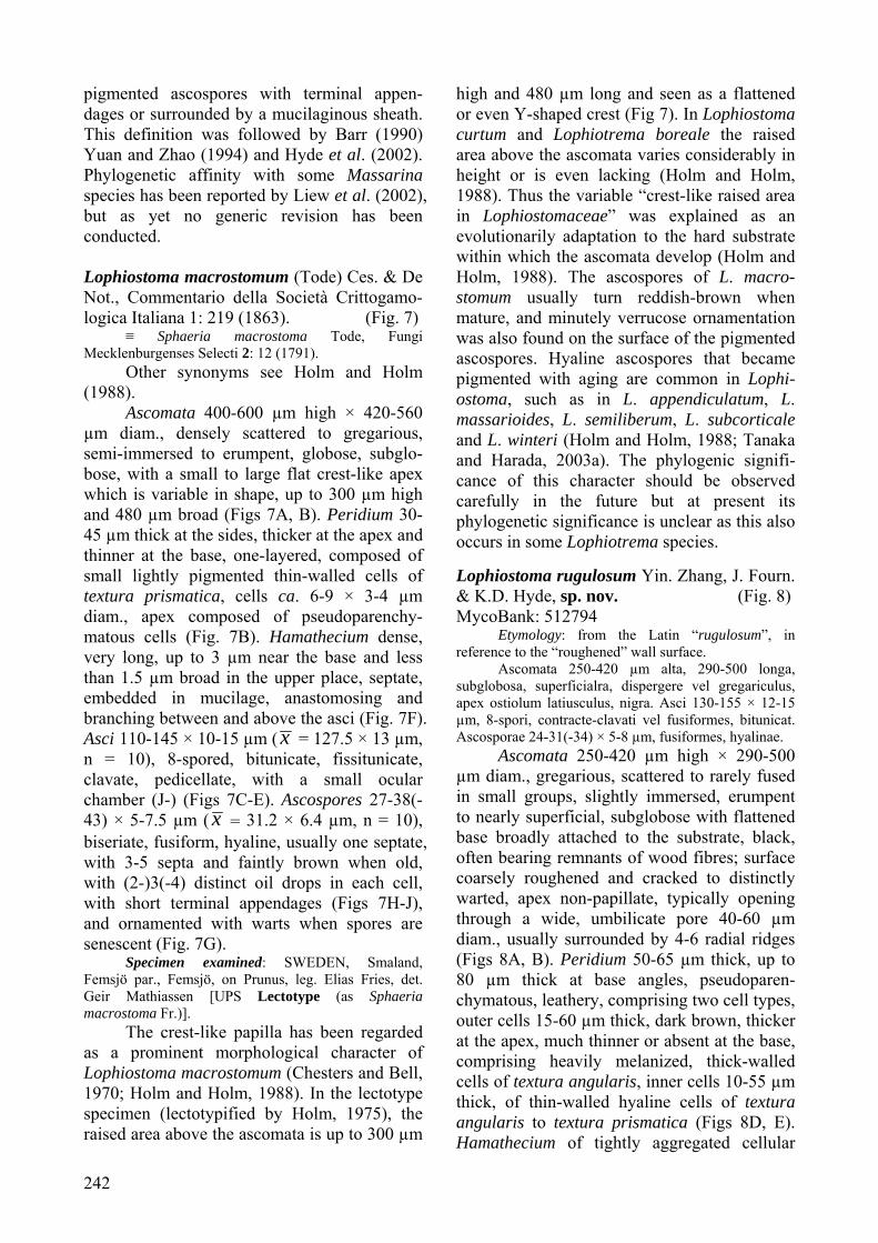
pigmented ascospores with terminal appen-dages or surrounded by a mucilaginous sheath. This definition was followed by Barr (1990) Yuan and Zhao (1994) and Hyde et al. (2002). Phylogenetic affinity with some Massarina species has been reported by Liew et al. (2002), but as yet no generic revision has been conducted. Lophiostoma macrostomum (Tode) Ces. & De Not., Commentario della Società Crittogamo-logica Italiana 1: 219 (1863). (Fig. 7)
≡ Sphaeria macrostoma Tode, Fungi Mecklenburgenses Selecti 2: 12 (1791).
Other synonyms see Holm and Holm (1988).
Ascomata 400-600 µm high × 420-560 µm diam., densely scattered to gregarious, semi-immersed to erumpent, globose, subglo-bose, with a small to large flat crest-like apex which is variable in shape, up to 300 µm high and 480 µm broad (Figs 7A, B). Peridium 30-45 µm thick at the sides, thicker at the apex and thinner at the base, one-layered, composed of small lightly pigmented thin-walled cells of textura prismatica, cells ca. 6-9 × 3-4 µm diam., apex composed of pseudoparenchy-matous cells (Fig. 7B). Hamathecium dense, very long, up to 3 µm near the base and less than 1.5 µm broad in the upper place, septate, embedded in mucilage, anastomosing and branching between and above the asci (Fig. 7F). Asci 110-145 × 10-15 µm ( x = 127.5 × 13 µm, n = 10), 8-spored, bitunicate, fissitunicate, clavate, pedicellate, with a small ocular chamber (J-) (Figs 7C-E). Ascospores 27-38(-43) × 5-7.5 µm ( x = 31.2 × 6.4 µm, n = 10), biseriate, fusiform, hyaline, usually one septate, with 3-5 septa and faintly brown when old, with (2-)3(-4) distinct oil drops in each cell, with short terminal appendages (Figs 7H-J), and ornamented with warts when spores are senescent (Fig. 7G).
Specimen examined: SWEDEN, Smaland, Femsjö par., Femsjö, on Prunus, leg. Elias Fries, det. Geir Mathiassen [UPS Lectotype (as Sphaeria macrostoma Fr.)].
The crest-like papilla has been regarded as a prominent morphological character of Lophiostoma macrostomum (Chesters and Bell, 1970; Holm and Holm, 1988). In the lectotype specimen (lectotypified by Holm, 1975), the raised area above the ascomata is up to 300 µm
high and 480 µm long and seen as a flattened or even Y-shaped crest (Fig 7). In Lophiostoma curtum and Lophiotrema boreale the raised area above the ascomata varies considerably in height or is even lacking (Holm and Holm, 1988). Thus the variable “crest-like raised area in Lophiostomaceae” was explained as an evolutionarily adaptation to the hard substrate within which the ascomata develop (Holm and Holm, 1988). The ascospores of L. macro-stomum usually turn reddish-brown when mature, and minutely verrucose ornamentation was also found on the surface of the pigmented ascospores. Hyaline ascospores that became pigmented with aging are common in Lophi-ostoma, such as in L. appendiculatum, L. massarioides, L. semiliberum, L. subcorticale and L. winteri (Holm and Holm, 1988; Tanaka and Harada, 2003a). The phylogenic signifi-cance of this character should be observed carefully in the future but at present its phylogenetic significance is unclear as this also occurs in some Lophiotrema species. Lophiostoma rugulosum Yin. Zhang, J. Fourn. & K.D. Hyde, sp. nov. (Fig. 8) MycoBank: 512794
Etymology: from the Latin “rugulosum”, in reference to the “roughened” wall surface.
Ascomata 250-420 µm alta, 290-500 longa, subglobosa, superficialra, dispergere vel gregariculus, apex ostiolum latiusculus, nigra. Asci 130-155 × 12-15 µm, 8-spori, contracte-clavati vel fusiformes, bitunicat. Ascosporae 24-31(-34) × 5-8 µm, fusiformes, hyalinae.
Ascomata 250-420 µm high × 290-500 µm diam., gregarious, scattered to rarely fused in small groups, slightly immersed, erumpent to nearly superficial, subglobose with flattened base broadly attached to the substrate, black, often bearing remnants of wood fibres; surface coarsely roughened and cracked to distinctly warted, apex non-papillate, typically opening through a wide, umbilicate pore 40-60 µm diam., usually surrounded by 4-6 radial ridges (Figs 8A, B). Peridium 50-65 µm thick, up to 80 µm thick at base angles, pseudoparen-chymatous, leathery, comprising two cell types, outer cells 15-60 µm thick, dark brown, thicker at the apex, much thinner or absent at the base, comprising heavily melanized, thick-walled cells of textura angularis, inner cells 10-55 µm thick, of thin-walled hyaline cells of textura angularis to textura prismatica (Figs 8D, E). Hamathecium of tightly aggregated cellular
Fungal Diversity
243
Fig. 7. Lophiostoma macrostomum (A-H, J from Leptotype (UPS!), I from IFRD 2005). A. Ascomata on the host surface. B. Section of the peridium. C-E. Ascus. F. Hamathecium. G-J. Ascospores. B-J in water. Scale bars: A = 500 µm, B = 200 µm, C-J = 10 µm. pseudoparaphyses, 1.7-2.5 µm broad, embed-ded in mucilage, ramified and somewhat diverticulate, rarely anastomosing between and above the asci, apically ending into bunches of clavate cells 12-20 µm long × 4-7 µm broad. Asci 130-155 × 12-15 µm (x = 150 × 14 µm, n = 10), 8-spored, bitunicate, fissitunicate, cylindro-clavate, pedicellate, with an faint
ocular chamber (Fig. 8C). Ascospores 24-31(-34) × 5-8 µm ( x = 29 × 7 µm, n = 10), biseriate, fusiform with broad to narrowly rounded to acute ends, with mucilaginous sheath to 3 µm thick, hyaline and one septate when young, turning yellowish-grey and verruculose while in the ascus, dark grey to sooty grey and become 1-3-septate when

244
Fig. 8. Lophiostoma rugulosum (from Holotype IFRD 2011). A. Ascomata on the host surface. B. Section of an ascoma. C. Asci in the pseudoparaphyses. D. Peridium structure. E. Pseudoparaphyses. Note the swelling tips. F, G. Ascospore. C in Congo Red; E in chlorazol black; others in water. Scale bars: A = 500 µm, B, D = 100 µm, C = 20 µm, E-G = 10 µm.
Fungal Diversity
245
senescent, slightly to deeply constricted at the septum, the upper cell often broader than the lower one (Figs 8F, G). Culture characters (ex-type: CBS 123093)
On OA spreading with sparse aerial mycelium and even margins, reaching 10 mm diam.; surface olivaceous-grey with a promi-nent, wide margin of diffuse, bright yellow pigment. On PDA spreading, with sparse aerial mycelium and even crenate margins, reaching 17 mm diam.; surface and reverse iron-grey, smoke-grey at margin, slimy in middle. On OA hyphae smooth, hyaline to pale olivaceous, 3-4 µm wide, becoming swollen below septa, with cells up to 15 µm diam.; in aerial mycelium these cells aggregate at the ends of hyphae, forming densely packed round propagules up to 90 µm diam., consisting of somewhat curved, smooth, olivaceous cells up to 15 µm long and 7 µm wide; presumably these can be dislodged as conidia.
Specimens examined: FRANCE, Ariège, Rimont, Peyrau, 400m, on submerged wood of Alnus glutinosa, 31 Aug. 2006, leg., det. J. Fournier [CBS H-20223, holotype]; 31 Aug. 2000, leg., det. J. Fournier [IFRD2026]; 23 Sept. 2000, leg., det. J. Fournier [IFRD 2027]; on submerged wood of Salix sp., 26 Jul. 2006, leg., det. J. Fournier [IFRD 2028]; on submerged wood of Rhamnus cathartica, 13 Aug. 2006, leg., det. J. Fournier [IFRD 2029]; Lescure, Bois du Pas du Baup, Le Volp, 500m, on partly submerged wood of Quercus, 26 Oct. 2006, leg., det. J. Fournier [IFRD2025].
Notes: The dense, filamentous hamathe-cium, cylindro-clavate asci, and hyaline, one-septate ascospores with mucilaginous sheath fit characters of Lophiostoma well. The peculiar wall structure and thickness, and the broad umbilicate ostioles surrounded by 4-6 radial ridges however, are the most distinctive features of this fungus, which is different from other morphologically comparable species and unusual for this genus. The presence of pseudoparaphyses with apically free ends differentiated into elongated swollen cells is likewise most unusual among Pleosporales and members of Lophiostoma. The ostiolar pore is widely open and empty on dry mature asco-mata, but a whitish plug is present on fresh or rehydrated material. It can be assumed these swollen cells are involved in the formation of the apical plug. They lack pigmented deposits and are not aggregated into an epithecium, as it occurs in Patellariaceae which, moreover are
more cupulate to disc-shaped (Kutorga and Hawksworth, 1997; Zhang and Hyde, 2009). Compressed papilla is a highly variable character that the variation of both form and size can even be seen in the same specimen (Holm and Holm, 1988). Thus, based on phylogenetic result, here we assign Lophio-stoma rugulosum in Lophiostoma despite its broad umbilicate ostioles.
In addition, this taxon appears fairly abundant in the two above mentioned brooks, regardless of any host preference, but was never encountered in other brooks or streams prospected in the same region, which indicates it may have narrow ecological requirements. Lophiostoma glabrotunicatum Yin. Zhang, J. Fourn. & K.D. Hyde, sp. nov. (Fig. 9) MycoBank: 512800
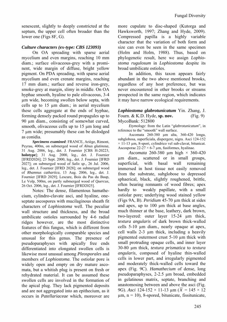
Etymology: from the Latin “glabrotunicatum”, in reference to the “smooth” wall surface.
Ascomata 260-380 µm alta, 360-420 longa, subglobosa, superficiala, dispergere, nigra. Asci 124-152 × 11-13 µm, 8-spori, cylindrico vel sub-clavat, bitunicat. Ascosporae 22-27 × 6-7 µm, fusiformes, hyalinae.
Ascomata 260-380 µm high × 360-420 µm diam., scattered or in small groups, superficial, with basal wall remaining immersed in host tissue not easily removed from the substrate, subglobose to depressed sphaerical, black, slightly roughened, brittle, often bearing remnants of wood fibres; apex hardly to weakly papillate, with a small ostiolar pore; underlying wood stained yellow (Figs 9A, B). Peridium 45-70 µm thick at sides and apex, up to 100 µm thick at base angles, much thinner at the base, leathery, dark brown, two-layered: outer layer 15-24 µm thick, textura angularis of dark brown thick-walled cells 5-10 µm diam., nearly opaque at apex, cell walls 2-3 µm thick, including a heavily pigmented outermost crust 5-10 µm thick with small protruding opaque cells, and inner layer 30-80 µm thick, textura prismatica to textura angularis, composed of hyaline thin-walled cells in lower part, and irregularly pigmented and moderately thick-walled cells toward the apex (Fig. 9C). Hamathecium of dense, long pseudoparaphyses, 2-2.5 µm broad, embedded in gelatinous matrix, septate, branching and anastomosing between and above the asci (Fig. 9G). Asci 124-152 × 11-13 µm ( x = 145 × 12 µm, n = 10), 8-spored, bitunicate, fissitunicate,
246
Fig. 9. Lophiostoma glabrotunicatum (from Holotype IFRD 2012). A. Superficial ascomata on the host surface. B. Section of an ascoma. C. Section of peridium. Note the two layered peridium structure. D-F. Asci with short pedicels in the pseudoparahyses. Note the senescent ascus in F. G. Thick, septate and branching pseudoparaphyses. H, I. Ascospores in sheath. J. Senescent ascospores. C, F, J in water; D, H in chlorazol black; E, G in cotton blue; I in Indian ink. Scale bars: A = 500 µm, B, C = 100 µm, D - F = 20 µm, G - J = 10 µm.
Fungal Diversity
247
clavate-cylindrical, with a short, narrowed, twisted, furcate pedicel which is 6-10 µm long, with a truncate ocular chamber and a minute ring visible on immature asci (Figs 9D-F). Ascospores 22-27 × 6-7 µm ( x = 25 × 6.5 µm, n = 10), obliquely uniseriate and partially overlapping or biseriate in places, fusiform with narrowly rounded ends, hyaline, two-celled, deeply constricted at the median septum, the upper cell often shorter and broader than the lower one, smooth, surrounded by an irregular hyaline gelatinous sheath 2.5-6 µm thick best seen in India ink. Senescent ascospores are brown and 2-3-septate, finely verrucose, a wide gelatinous sheath is present on immature ascospores, disappearing on old ascospores (Figs 9H-J).
Specimens examined: FRANCE, Ariège, Prat Communal, Loumet, on submerged wood of Alnus glutinosa, 1000m, 08 Sept. 2006, leg., det. J. Fournier [CBS H-20225, holotype, CBS H-20224, isotype]; Les Cabannes, Pierrefitte forest, on submerged wood of Fagus sylvatica, 1400 m, 15 Oct. 2007, leg., det. J. Fournier [IFRD2035]; Ustou, Cirque de Cagateille, on submerged wood of Salix sp., 1150 m, 11 Jun. 2007, leg., det. J. Fournier [IFRD2030].
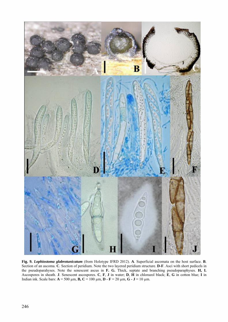
Notes: All of the morphological characters such as dense, long pseudopara-physes, clavate-cylindrical asci, hyaline, one-septate, fusiform ascospores with a broad sheath suggest that this taxon belongs to the Lophiostomataceae, and that L. glabrotunica-tum is phylogenetically closely related with Lophiostoma (Fig. 1). The papilla of this species does not, however, agree with the concept of Lophiostoma. But the widely variable character and young evolutionary status of the crest-like apex of L. macrostomum might help to explain the variable apex morphology of this group (Fig. 1, Clade Melanommataceae) (Holm and Holm, 1988). Ascospores of L. glabrotunicatum are com-parable with those of L. aquaticum and Massarina submediana, but the superficial, subglobose ascomata and the yellow staining of the underlying wood are not in agreement (Aptroot, 1998). Lophiostoma glabrotunicatum has been repeatedly recovered from the mountainous area of France exclusively between 1000-1400 m elevations. This might indicate its ecological preference for cold water in the high altitude mountainous area.
Massarina Massarina was introduced by Saccardo
(1883) for species of pyrenocarpous ascomy-cetes that had previously been placed in Massaria De Not., but typically had hyaline ascospores (Bose, 1961). The family Massarinaceae was described by Munk (1956) to accommodate Massarina. This family was not commonly used and Massarina was later placed within the Lophiostomataceae in the Pleosporales (Bose, 1961; Eriksson and Yue, 1986; Barr, 1987, 1990). Of the 160 epithets listed in his monograph, Aptroot accepted only 43 species (Aptroot, 1998). The concept of Massarina was widely accepted as having single or aggregated, immersed to erumpent, spherical to hemispherical, pseudothecioid ascomata; cellular pseudoparaphyses; bituni-cate, cylindrical to clavate or obpyriform asci; and hyaline, 1-3(-7)-septate, fusiform to long ellipsoid ascospores that mostly have a mucilaginous sheath or appendages (Aptroot, 1998; Hyde and Aptroot, 1998; Tanaka and Harada, 2003c). Recent morphological, mole-cular and anamorphic results indicate, however, that Massarina is polyphyletic (Hyde, 1995; Kirk et al., 2008; Liew et al., 2002). We believe that Massarina sensu stricto should be confined to the generic type (M. eburnea) and very similar species (e.g. M. cisti). Massarina eburnea (Tul. & C. Tul.) Sacc., Syll. Fung. 2: 153 (1883). (Fig. 10)
≡ Massaria eburnea Tul. & C. Tul., Selecta Fungorum Carpologia 2: 239 (1863), nom. nov. [non sensu Tulasne & Tulasne]
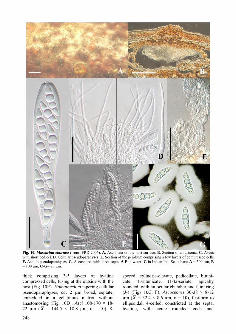
Other synonyms see Hyde (1995). Ascomata to 250 µm high × 500-700 µm
diam., solitary or in small clusters, forming under raised dome-shaped areas, with blackened centres, with a central ostiole, immersed within the cortex of thin dead branches, ellipsoidal, rounded from above, clypeate, neck central, short and barely noticeable on host surface (Fig. 10A). Clypeus ca. 250 µm diam., 60 µm thick, brown, comprising compact brown-walled cells of textura angularis to globulosa beneath host epidermal cells (Fig. 10B). Peridium ca. 20 µm
248
Fig. 10. Massarina eburnea (from IFRD 2006). A. Ascomata on the host surface. B. Section of an ascoma. C. Ascus with short pedicel. D. Cellular pseudoparahyses. E. Section of the peridium comprising a few layers of compressed cells. F. Asci in pseudoparahyses. G. Ascospores with three septa. A-F in water; G in Indian Ink. Scale bars: A = 500 µm, B = 100 µm, C-G= 20 µm. thick comprising 3-5 layers of hyaline compressed cells, fusing at the outside with the host (Fig. 10E). Hamathecium tapering cellular pseudoparaphyses, ca. 2 µm broad, septate, embedded in a gelatinous matrix, without anastomosing (Fig. 10D). Asci 108-170 × 18-22 µm ( x = 144.5 × 18.8 µm, n = 10), 8-
spored, cylindric-clavate, pedicellate, bituni-cate, fissitunicate, (1-)2-seriate, apically rounded, with an ocular chamber and faint ring (J-) (Figs 10C, F). Ascospores 30-38 × 8-12 µm ( x = 32.4 × 8.6 µm, n = 10), fusiform to ellipsoidal, 4-celled, constricted at the septa, hyaline, with acute rounded ends and
Fungal Diversity
249
Fig. 11. A. Keissleriella linearis. Section of an ascoma, note the setae in ostiole. B-D. Lophiotrema nucula sensu Holm. B. Peridium section of an ascoma. Note the peridium in equal thickness. C, D. Habitate section of ascomata. Scale bars: A = 100 µm; B-D = 50 µm. surrounded by (5-8 µm) mucilaginous sheath (Fig. 10G).
Specimens examined: FRANCE, on twig of Fagus sp., Desmazières 1764. P (holotype of Sphaeria pupula var. minor). AUSTRIA, Silesia, Karlsbrunn, on dead twigs of Fagus sylvatica L., Aug. and Sept. 1890, Niessl., De Thümen, Mycotheca universalis no. 1951 sub Massarina eburnea, ETH. SAXONIA, Königsbrunn, on twigs of Fagus sylvatica, April 1882, W. Krieger, Rabenhorst & Winter, Fungi europaei no. 2767, ETH (lectotype). FRANCE, on a dead twig of Fagus sylvatica, Deux Sèvres, Villiers en Bois, Forêt de Chizé, Rimbaud, 14 Apr. 2008, leg. det. Paul Leroy [IFRD2006].
In the holotype of Sphaeria pupula var. minor (P) and lectotype of Massarina eburnea (ETH), ascospores are reported as “not con-stricted at the septa” (Hyde, 1995). However in one of our recent collections, constrictions at the ascospore septa were observed (Fig. 10), which was consistent with the description by Fallah and Shearer (2001). This might be because this character is not clear in the old (over 100 years) and dry herbarium specimens
or it may be variable between collections. Thus fresh epitypes are recommended to be assigned in order to get more reliable information about the morphology and phylogeny of a particular group of fungi (Hyde and Zhang, 2008; Zhang et al., 2008).
Conclusions — one new genus, five new species and three new combinations are intro-duced here. Both Massarina and Lophiostoma are polyphyletic. Lophiostoma is phylogene-tically divided into two groups: Lophiostoma I and Lophiostoma II. Massarina sens. lat. is mainly divided into two groups: Massarina and Lentithecium. Although the “Lophiotrema-like” peridium is refuted by checking the lectotype material, Lophiotrema still represents a separate genus from Lophiostoma. This study also indicated that ascospore and apical morphology are morphological characters that have phylogenetic significance, though they are not fully reliable for generic level classification.
250
Furthermore, a compressed apex has more phylogenetic significance than a crest-like apex. Several new species are described from France, a part of the world where these taxa have been relatively well studied. Massarina and Lophio-stoma species are common in the tropics on submerged as well as terrestrial wood (see Pinruan et al., 2007; Kodsueb et al., 2008), however the correct identity of these taxa need confirmation using molecular techniques. Acknowledgements
Joost Stalpers (Centraalbureau voor Schimmel-cultures, CBS) is thanked for his valuable suggestions on nomenclature. Herbarium Uppsala University (UPS) and the National Herbarium Ethiopia, Addis Abeba University (ETH) and Herbier National de Paris, Muséum National d'Histoire Naturelle, France (PC) are also thanked for their kind cooperation in allowing us to examine and loan specimens. References Aptroot, A. (1998). A world revision of Massarina
(Ascomycota). Nova Hedwigia 66: 89-162. Aptroot, A., Frohlich, J. and Hyde, K.D. (2000). Fungi
from palms. XLIV. Two new Massarina species with pigmented ostioles. Nova Hedwigia 70: 227-232.
Arenal, F., Platas, G. and Pelaez, F. (2005). Two new Preussia species defined based on morphological and molecular evidence. Fungal Diversity 20: 1-15.
Arx, J.A. von and Müller, E. (1975). A re-evaluation of the bitunicate Ascomycetes with keys to families and genera. Studies in Mycology 9: 1-159.
Barr, M.E. (1987). Prodromus to Class Loculoascomy-cetes. Amherst, Massachusetts; University of Massachusetts: 1-168.
Barr, M.E. (1990). North American flora, Melanomma-tales (Loculoascomycetes). Series II, part 13: 1-129.
Bose, S.K. (1961). Studies on Massarina Sacc. and related genera. Phytopathologische Zeitschrift 41: 151-213.
Cai, L., Jeewon, R. and Hyde, K.D. (2005). Phylogenetic evaluation and taxonomic revision of Schizothe-cium based on ribosomal DNA and protein coding genes. Fungal Diversity 19: 1-21.
Cai, L., Jeewon, R. and Hyde, K.D. (2006). Phylogenetic investigations of Sordariaceae based on multiple gene sequences and morphology. Mycological Research 110: 137-150.
Chesters, C.G.C. and Bell, A. (1970). Studies in the Lophiostomataceae. Mycological Paper 120: 1-51.
Eriksson, O. And Yue, J.Z. (1986). Bertiella (Sacc.) Sacc. & Sydow, a synonym of Massarina Sacc. Mycotaxon 27: 247-253.
Eriksson, O.E. (1981). The families of bitunicate ascomycetes. Opera Botanica 60: 1-220.
Eriksson, O.E. and Hawksworth, D.L. (2003). Sacchari-cola, a new genus for two Leptosphaeria species on sugar cane. Mycologia 95: 426-433.
Fallah, P.M. and Shearer, C.A. (2001). Freshwater asco-mycetes: new or noteworthy species from north temperate lakes in Wisconsin. Mycologia 93: 566-602.
Gams, W., Verkleij, G.J.M. and Crous, P.W. (2007). CBS Course of Mycology. 5th edn. Centraalbu-reau voor Schimmelcultures, Utrecht, Netherlands.
Hall, T. (2005). Bioedit version 7.0.4. Department of Microbiology, North Carolina State University.
Höhnel, F. von (1919). Fragmente zur Mykologie. XXIII Mitteilung, Nr. 1154 bis 1188. Sitzungsberichte der Kaiserlichen Akademie der Wissenschaften, Math.-naturw. Klasse, Abt. I. 128: 535-625.
Holm, L. (1957). Etudes taxonomiques sur les Pléosporacées. Symbolae Botanicae Upsalienses 14: 1-188.
Holm, L. and Holm, K. (1988). Studies in the Lophiostmataceae with emphasis on the Swedish species. Symbolae Botanicae Upsalienses 28: 1-50.
Holm, L.M. (1975). Nomenclatural notes on pyrenomy-cetes. Taxon 24: 475-488.
Hyde, K.D. (1995). The genus Massarina, with a description of M. eburnea and an annotated list of Massarina names. Mycological Research 99: 291-296.
Hyde, K.D. and Aptroot, A. (1998). Tropical freshwater species of the genera Massarina and Lophiostoma (ascomycetes). Nova Hedwigia 66: 489-502.
Hyde, K.D. and Goh, T.K. (1998). Fungi on submerged wood in Lake Barrine north Queensland, Australia. Mycological Research 102: 739-749.
Hyde, K.D. and Zhang, Y. (2008). Epitypification: should we epitypify? Journal of Zhejiang University Science 9: 842-846.
Hyde, K.D., Wong, W.S.W. and Aptroot, A. (2002). Marine and estuarine species of Lophiostoma and Massarina. In: Fungi in Marine Environments (ed. K.D. Hyde). Fungal Diversity Research Series 7: 93-109.
Jeewon, R., Liew, E.C.Y. and Hyde, K.D. (2004). Phylogenetic evaluation of species nomenclature of Pestalatiopsis in relation to host association. Fungal Diversity 17: 39-55.
Kirk, P.M., Cannon, P.F., Minter, D.W. and Stalpers, J.A. (2008). Ainsworth & Bisby’s Dictionary of The Fungi. 10th edn. Oxon: CAB INTERNATIONAL.
Kodsueb, R., Jeewon, R., Vijaykrishna, D., McKenzie, E.H.C., Lumyong, P., Lumyong, S. and Hyde, K.D. (2006). Systematic revision of Tubeufiaceae based on morphological and molecular data. Fungal Diversity 21: 105-130.
Kodsueb, R., McKenzie, E.H.C., Lumyong, S. and Hyde, K.D. (2008). Diversity of saprobic fungi on Magnoliaceae. Fungal Diversity 30: 37-53.
Kruys, Å., Eriksson O.E. and Wedin M. (2006). Phylogenetic relationships of coprophilous Pleo-sporales (Dothideomycetes, Ascomycota), and the
Fungal Diversity
251
classification of some bitunicate taxa of unknown position. Mycological Research 110: 527-536.
Kutorga E. and Hawksworth D.L. (1997). Areassessment of the genera referred to the family Patellaria-ceae (Ascomycota). Systema Ascomycetum 15: 1-110.
Leuchtmann, A. (1984). Über Phaeosphaeria Miyake und andere bitunicate Ascomyceten mit mehrfach querseptierten Ascosporen. Sydowia 37: 75-194.
Liew, E.C.Y., Aptroot, A. and Hyde, K.D. (2002). An evaluation of the monophyly of Massarina based on ribosomal DNA sequences. Mycologia 94: 803-813.
Liu, Y.J., Whelen, S. and Hall, B.D. (1999). Phylogenetic relationships among ascomycetes: evidence from an RNA Polymerase II subunit. Molecular Biology and Evolution 16: 1799-1808.
Lumbsch, H.T. and Huhndorf, S.M. (2007). Outline of Ascomycota – 2007. Myconet 13: 1-58.
Munk, A. (1956). On Metasphaeria coccodes (Karst.) Sacc. and other fungi probably related to Massarina Sacc. Massarinaceae n. fam. Friesia 5: 303-308.
Munk, A. (1957). Danish Pyrenomycetes. Dansk Bot. Ark. 17: 1-491.
Nyberg Kruys, Å. (2005). Phylogenetic relationships and species richness of coprophilous ascomycetes. Doctoral thesis, Umeå University.
Phillips, A.J.L., Alves, A., Pennycook, S.R., Johnston, P.R., Ramaley, A., Akulov, A. and Crous, P.W. (2008). Resolving the phylogenetic and taxono-mic status of dark-spored teleomorph genera in the Botryosphaeriaceae. Persoonia 21: 29-55.
Pinruan, U., Hyde, K.D., Lumyong, S., McKenzie, E.H.C. and Jones, E.B.G. (2007). Occurrence of fungi on tissues of the peat swamp palm Licuala longicalycata. Fungal Diversity 25: 157-173.
Poonyth, A.D., Hyde, K.D., Aptroot, A. and Peerally, A. (1999). Three new species of Massarina associated with terrestrial, non-marine parts of mangroves. Fungal Diversity 3: 139-146.
Rokas, A. and Carroll, S.B. (2005). More genes or more taxa? The relative contribution of gene number and taxon number to phylogenetic accuracy. Molecular Biology and Evolution 22: 1337-1344.
Ryckegem, G. van. and Aptroot, A. (2001). A new Massarina and a new Wettsteinina (Ascomycota) from freshwater and tidal reeds. Nova Hedwigia 73: 161-166.
Saccardo, P.A. (1878). Fungi Italici autographice delineati a Prof. P.A. Saccardo. Patavii - Fascicoli V.-VIII. sistentes tab: 161-320.
Saccardo, P.A. (1883). Sylloge Pyrenomycetum, Vol. II. Sylloge Fungorum 2: 153.
Scheuer, Ch. (1991). Massarina tetraploa sp. nov., the teleomorph of Tetraploa aristata. Mycological Research 95: 126-128.
Schoch, C.L., Shoemaker, R.A., Seifert, K.A., Hambleton, S., Spatafora, J.W. and Crous, P.W.
(2006). A multigene phylogeny of the Dothideo-mycetes using four nuclear loci. Mycologia 98: 1041-1052.
Shenoy, B.D., Jeewon, R., Lam, W.H., Bhat, D.J., Than, P.P., Taylor, P.W.J. and Hyde, K.D. (2007). Morpho-molecular characterisation and epity-fication of Collectotrichum capsici (Glomerella-ceae, Sordariomycetes), the causative agent of anthracnose in chilli. Fungal Diversity 27: 197-211.
Swofford, D.L. (2002). PAUP*: Phylogenetic Analysis Using Parsimony (and other methods). Version 4b10. Sinauer Associates, Sunderland, USA.
Tanaka, K. and Harada, Y. (2003a). Pleosporales in Japan (1): the genus Lophiostoma. Mycoscience 44: 85-96.
Tanaka, K. and Harada, Y. (2003b). Pleosporales in Japan (2): the genus Lophiotrema. Mycoscience 44: 115-121.
Tanaka, K. and Harada, Y. (2003c). Pleosporales in Japan (3): the genus Massarina. Mycoscience 44: 173-185.
Tang, A.M.C., Hyde, K.D., Tsui, C.K.M. and Corlett, R.T. (2003). A new species of Lophiotrema from wild fruit in Hong Kong. Persoonia 18: 265-269.
Thompson, J.D., Gibson, T.J., Plewniak, F., Jeanmougin, F. and Higgins, D.G. (1997). The Clustal X windows interface: flexible strategies for multiple sequence alignment aided by quality analysis tools. Nucleic Acids Research 24: 4876-4882.
Tsui, C.K.M., Hyde, K.D. and Hodgkiss, I.J. (2000). Biodiversity of fungi on submerged wood in Hong Kong streams. Aquatic Microbial Ecology 21: 289-298.
Tsui, K.M., Hyde, K.D. and Hodgkiss, I.J. (1999). Massarina proprietunicata sp. nov., from submerged wood in streams in Hong Kong. Mycological Research 103: 1575-1578.
Vilgalys, R. and Hester, M. (1990). Rapid genetic identification and mapping enzymatically ampli-fied ribosomal DNA from several Cryptococcus species. Journal of Bacteriology 172: 4238-4246.
White, T.J., Bruns, T., Lee, S. and Taylor, J. (1990). Amplification and direct sequencing of fungal ribosomal RNA genes for phylogenetics. In: PCR Protocol: a guide to methods and applications.(eds. M.A. Innis, D.H. Gelfand, J.S. Sninsky and T.J. White). San Diego: Academic Press: 315-322.
Yuan, Z.Q. and Zhao, Z.Y. (1994). Studies on lophiostomataceous fungi from Xinjiang, China. Sydowia 46: 162-184.
Zhang, Y. and Hyde, K.D. (2009). Transfer of Pseudoparodia pseudopeziza to Patellariaceae (Patellariales). Nova Hedwigia 88: 211-215.
Zhang, Y., Fournier, J., Pointing, S.B. and Hyde, K.D. (2008). Are Melanomma pulvis-pyrius and Trematosphaeria pertusa congeneric? Fungal Diversity 33: 47-60.