towards an ontology for sharing medical images and regions of interest in neuroimaging
TRANSCRIPT
Journal of Biomedical Informatics 41 (2008) 766–778
Contents lists available at ScienceDirect
Journal of Biomedical Informatics
journal homepage: www.elsevier .com/locate /y jb in
Towards an ontology for sharing medical images and regions of interestin neuroimaging
Lynda Temal a,b,c, Michel Dojat d,e, Gilles Kassel f, Bernard Gibaud a,b,c,*
a INRIA, VisAGes Project-Team, F-35042 Rennes, Franceb INSERM, U746, F-35042 Rennes, Francec University of Rennes 1, CNRS, UMR 6074, IRISA, F-35042 Rennes, Franced INSERM, U836, Grenoble, F-38043, Francee Joseph Fourier University, Institute of Neurosciences, Grenoble, F-38043, Francef LaRIA, CNRS (FRE 2733) and Jules Verne University of Picardie, Amiens, F-80039, France
a r t i c l e i n f o
Article history:Received 6 August 2007Available online 17 March 2008
Keywords:Medical imagingData integrationMediation systemNeuroscienceBiomedical ontologiesSemantic annotationData sharing
1532-0464/$ - see front matter � 2008 Elsevier Inc. Adoi:10.1016/j.jbi.2008.03.002
* Corresponding author. Address: Unité/Projet VisCNRS/U. de Rennes I, IRISA, Faculte de medecine, 2 A35043 Rennes, Cedex, France. Fax: +33 2 99 84 71 71
E-mail addresses: [email protected] (L. Temal),(M. Dojat), [email protected] (G. Kassel),Gibaud).
a b s t r a c t
The goal of the NeuroBase project is to facilitate collaborative research in neuroimaging through a feder-ated system based on semantic web technologies. The cornerstone and focus of this paper is the design ofa common semantic model providing a unified view on all data and tools to be shared. For this purpose,we built a multi-layered and multi-components formal ontology. This paper presents two major contri-butions. The first is related to the general methodology we propose for building an application ontologybased on consistent conceptualization choices provided by the DOLCE foundational ontology and coreontologies of domains that we reuse; the second concerns the domain ontology we designed for neuro-imaging, which encompasses both the objective nature of image data and the subjective nature of imagecontent, through annotations based on regions of interest made by agents (humans or computer pro-grams). We report on realistic domain use-case queries referring to our application ontology.
� 2008 Elsevier Inc. All rights reserved.
1. Introduction Today, such developments in data sharing are considered
Neuroimaging includes a variety of techniques to explore brainstructure and function, such as Magnetic Resonance Imaging (MRI),Computed Tomography (CT), Positron Emission Tomography (PET),Single Photon Emission Computed Tomography (SPECT), and Magne-toencephalography (MEG). It has become a major tool for scientistsand physicians, in their quest for a better understanding of the mech-anisms involved in brain development, brain functions and brain dis-orders. Moreover, neuroimaging is emerging as a prominent tool toassess the efficacy of new drugs against brain pathologies, such ascancer, neurodegenerative diseases or psychiatric disorders.
In addition to standard image interpretation based on humanvisual screening, computer-generated imaging biomarkers providequantitative information useful in medical decision making. Theintroduction of such computerized markers, precisely definedand automatically extracted from the processed images, supportsthe use of well-defined protocols or guidelines to optimize imagingprocessing chains and to standardize image acquisition sequencesand scanner calibration procedures.
ll rights reserved.
AGeS U746, INSERM/INRIA/venue du Pr Leon Bernard, [email protected]@irisa.fr (B.
necessary to improve the relevance and efficacy of large scalemulti-center clinical trials and are therefore strongly encouragedby stakeholders, such as the NIH (National Institute of Health)and FDA (Food and Drug Administration) in the US, and the frame-work programs for research in the EU. Moreover, these develop-ments will benefit basic brain research that highlights therelationships between morphology and function in the central ner-vous system, especially research based on molecular imaging andMR imaging (anatomical and functional MRI, and diffusion-weighted MRI for brain connectivity assessment). This creates astrong need for formal definition of imaging-related information,consisting of acquired data, namely raw data and acquisition con-ditions, as well as processed and interpreted data, namely dataresulting from a specific procedure performed by an agent (humanor machine). Thus, data sharing is a challenging topic in biomedicaldomain and several ongoing efforts are being performed. For in-stance, the Cancer Biomedical Informatics Grid (CaBIG)1 [1] is aNCI (National Cancer Institute) initiative to gather in a common cyb-erarchitecture, a network of cancer centers and research laborato-ries. To reach this goal, CaBIG is developing standards, policies,guidelines, common applications, open source tools and a middle-ware infrastructure. Similar goals are being pursued by the Biomed-
1 https://cabig.nci.nih.gov/.
L. Temal et al. / Journal of Biomedical Informatics 41 (2008) 766–778 767
ical Informatics Research Network (BIRN)2, another initiative sup-ported by the NIH, in the field of neurosciences, in the continuity ofthe Human Brain Project [2]. Related activities concern both the devel-opment of a mediation infrastructure, based on ontologies, and test-bed applications called Morphology BIRN, Function BIRN and MouseBIRN, addressing various kinds of needs in neurosciences [3,4]. Similarefforts exist in Europe, e.g. in the context of the Virtual PhysiologicalHuman initiative, supported by the European Union [5].
The NeuroBase project, launched in France in 2002, pursues thesame general objective: to share images and processing tools in thecontext of distributed and heterogeneous systems [6]. The goal ofthe NeuroBase project is twofold: to manage and share the largequantity of data produced (1Gb/subject), and to provide a feder-ated platform for the interoperability of processing tools. Presently,data and tools are disseminated in three French centers, all part-ners of the NeuroBase project. The objectives can be summarizedin three main points: (i) carrying out large scale experiments bysharing heterogeneous distributed data, (ii) combining existing im-age processing tools to define new data processing pipelines, and(iii) evaluating these heterogeneous pipelines on large datasetsproduced by the imaging centers.
The cornerstone of this project, and the major focus of this pa-per, is the design of a common semantic model, according to anontological approach, which provides a unified view of all dataand tools to be shared via the federated system. Our ultimateaim is: (1) to define an easily maintainable and extensible refer-ence ontology for a broad community of neuroscientists. Currentlytargeted applications concern cognitive science (visual cortexexploration) and neurological pathologies (e.g. neurodegenerativediseases); (2) to integrate conceptualizations from different fields,e.g. neuroanatomy, neurophysiology or neuropathology, into aconsistent whole; and (3) to define an ontology that can bemapped with other ontologies, in order to ensure interoperabilitywith external systems.
To define such an ontology, called OntoNeuroBase, we adopteda multi-layer approach and based our ontological commitments onwell-known ontologies existing at different levels of abstraction,e.g. top-level ontologies or core domain ontologies [7]. We main-tain simultaneously two manifestations of the ontology. The firstmanifestation is specified in the semi-informal language of theOntoSpec methodology [8]. It is semantically rich as it makesuse, in particular, of temporally-indexed relations and meta-prop-erties considered by the OntoClean methodology [9]. As such, thismanifestation is intended to facilitate the mapping of OntoNeuro-Base with other ontologies. The second manifestation is specifiedin the formal Web Ontology Language OWL. This manifestation issemantically poorer but enables to use OntoNeuroBase to performinferences.
Beside its primary role within a federated system to provide acommon unified schema for the mapping of the local databaseschemas, the basic added value of an ontology is that it enablesreasoning about shared information. Such reasoning may concernquerying by introducing new capabilities based on formal seman-tics of the concepts and relations expressed in the ontology. Be-yond querying, reasoning may also be applied to imageannotations with a view to enhance image interpretation, e.g.relating measurements on images having different modalities tothe characteristics of the real-world entities being imaged. Thismay involve representations of space and topological propertiesof these real-world entities. Composing image processing toolsconceived in different contexts to define innovative processingchains requires representing sufficient knowledge about suchtools, as well as the data processed, in order to achieve interoper-ability between the tools.
2 http://www.nbirn.net/index.shtm.
Hence, this paper mainly focuses on three crucial aspects: (1)how neuroimaging data can be organized in a consistent set of cat-egories to facilitate sharing, (2) what the images actually represent,e.g. an MR signal intensity, a 3D volume, or a time sequence of 3Dvolumes, (3) how ‘‘Regions of Interest” (often abbreviated ROIs)can be represented in the images, and what they mean for agents(humans or programs) involved in their creation, querying anduse. This third issue is fundamental for using imaging biomarkersconsistently and relating observations of the same reality throughseveral imaging modalities, and for exploring various aspects ofbrain structure and brain metabolism or function. The literaturecontains many relevant contributions [10–12], but is stillincomplete.
Our work makes two major contributions. The first is amethodology to build a multi-layered application ontology.The second consists of a novel conceptualization of neuroimag-ing data, encompassing both the objective nature of image dataand the subjective nature of annotations made by intelligentagents. This conceptualization is based primarily on the defini-tion of ROIs.
The paper is structured as follows. Section 2 presents our meth-odology to build a multi-layered application ontology. Section 3presents the Datasets Ontology, the principal kernel of the Onto-NeuroBase ontology which specializes I& DA (Information and Dis-course Acts). We focus first on the semantic axes, which allowcategorizing the images; we then describe a way of modelingobjective image content by mathematical functions; lastly, weintroduce ROIs and related annotations, as a means to express sub-jective information. Section 4 provides an illustrative example,based on our current implementation of the ontology. Section 5discusses some of our choices regarding both the general designmethodology and our novel conceptualization of neuroimagingdata. Finally, development perspectives for our project are evokedin Section 6.
2. The ontological reference framework
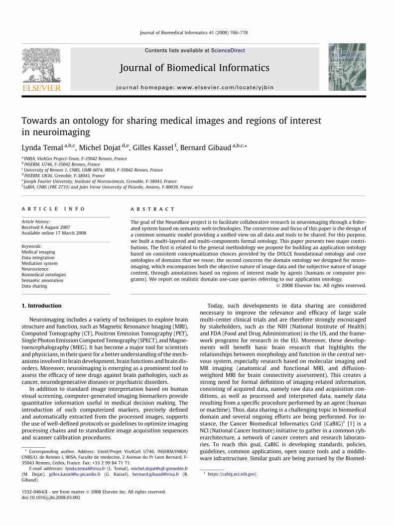
To fulfill our objectives, we chose a design framework thatstructures the ontology at different levels of abstraction (Fig. 1)while respecting common conceptualization choices. In this sec-tion, we focus on the modular approach [13] adopted in the designof the global structure of the OntoNeuroBase conceptualization,disregarding the specification languages.
At the highest level is a top-level ontology that includes abstractconcepts and relationships valid across domains. Foundationalontologies are such top-level ontologies whose concepts and rela-tions share a common philosophical foundation. We adoptedDOLCE (Descriptive Ontology for Linguistic and Cognitive Engineer-ing), which serves as a foundational ontology [14].
We then added Core ontologies, which provide generic, basic andminimal concepts and relations in a specific domain [7]. By mini-mal we mean that core ontologies should include only the mostreusable and widely applicable categories. These kinds of ontolo-gies are essential for sharing intended meaning between differentdomains. We adopted I& DA (Information and Discourse Acts), acore ontology initially built for classifying documents as a functionof their content [15]. We use it to model medical images, which weconsider as types of documents. Participant Roles [16] is the coreontology we use to describe the modes of image participation indata processing. I& DA and Participant Roles are built accordingto DOLCE ontological commitments.
On the basis of these two layers, we constructed our Domainontology dedicated to conceptualizing a specific domain, in thiscase neuroimaging. Obviously, large domains such as neuroimag-ing can be divided into sub-domains for the sake ofmodularization.
(Particulars)
Participant roles(Documents)
(Knowledge roles)
(Medical images)
Foundationalontology
Coreontologies
Domainontologies
DOLCEDOLCE
I&DAI&DA
OntoKADSOntoKADS
Regions of Interest
ROI Annotations
Neuroimaging Math Functions
specializes
references
Reusedonto-logies
Contri-bution
Mathematical Functions
Datasets OntologyDatasets Ontology
Fig. 1. An overview of the application ontology OntoNeuroBase framework: A solid line going down from sub-ontology O1 to O2 means that the entities of O2 (concepts andrelations) specialize the entities of O1; a dashed line between two sub-ontologies, from O1 to O2, means that O2 concepts reference O1 concepts without specialization.
Particular
Endurant Perdurant Quality Abstract
PhysicalObject
AgentivePhysicalObject
Non-PhysicalObject
MentalObject
AgentiveSocialObject
Event Stative
Achievement
Accomplishment
State Process
ActionCollection
Fig. 2. An excerpt from DOLCE’s top-level taxonomy. A solid line between twoconcepts represents a direct specialization relation. A dashed line reflects the exi-stence of intermediate concepts.
768 L. Temal et al. / Journal of Biomedical Informatics 41 (2008) 766–778
This complete framework (Fig. 1) constitutes OntoNeuroBase,our Application ontology. Various domain ontologies can be addedto extend OntoNeuroBase, but should respect the ontological com-mitments driving our conceptualization. In the following pages, wedetail these main conceptualization choices underlying the foun-dational and core ontologies we have adopted.
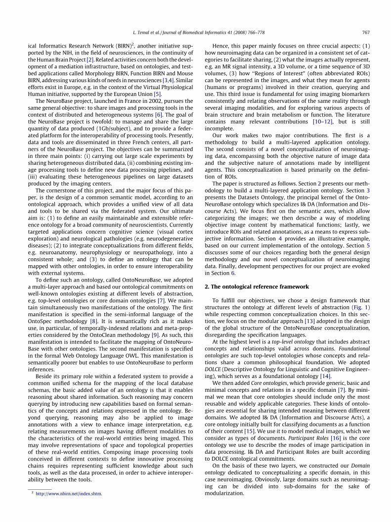
2.1. DOLCE (Particulars)
DOLCE3 [14] (Descriptive Ontology for Linguistic and CognitiveEngineering) is a foundational ontology of Particulars which are clas-sified into four separate categories, depending on their modes ofexistence (Fig. 2).
� Endurants are entities that ‘‘are wholly present in time” (e.g. youand your parts). Among Endurants, and according to whether theentity has direct spatial qualities, Physical Objects (e.g. yourbrain) are distinguished from Non-Physical Objects (e.g. yourknowledge about neuroimaging), which cover social and cogni-tive entities. The notion of Collection was added recently [17] asa specialization of Non-physical Object, in order to represent plu-ral entities (e.g. fiber collection).
� Perdurants are entities that ‘‘occur in time” (e.g. cerebral bloodcirculation) in which Endurants (e.g. cerebral blood) participate.Among Perdurants, Statives are distinguished from Eventsaccording to whether the Perdurants are cumulative4 or not.Events are divided into Achievements and Accomplishments accord-ing to whether they are atomic or not. Actions are Accomplishmentswhich are intentionally controlled by Agents.
� Endurants and Perdurants are characterized by inherent Qualities,which are seen as the basic properties we can perceive or mea-sure (e.g. the density of tissues in a specific anatomical struc-ture). Qualities are divided into Temporal Qualities, PhysicalQualities, and Abstract Qualities, which are, respectively, inherentto Perdurants, Physical Endurants, and Non-Physical Endurants.
� Qualities take ‘‘values”, called Quales (e.g. a particular grey levelencoded by the number 225), within quality region spaces.Quales are Abstract entities.
3 http://www.loa-cnr.it/DOLCE.html.4 Perdurants are stative or eventive according to whether they are the mereological
sum of two of their instances [6].
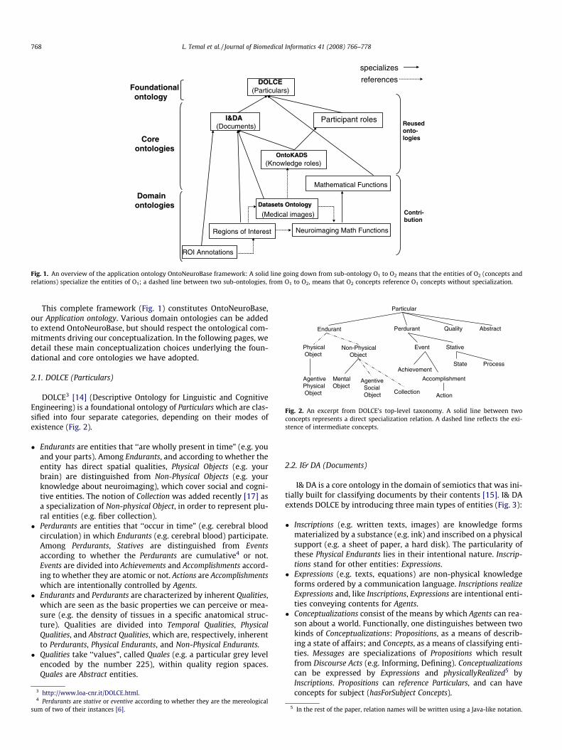
2.2. I& DA (Documents)
I& DA is a core ontology in the domain of semiotics that was ini-tially built for classifying documents by their contents [15]. I& DAextends DOLCE by introducing three main types of entities (Fig. 3):
� Inscriptions (e.g. written texts, images) are knowledge formsmaterialized by a substance (e.g. ink) and inscribed on a physicalsupport (e.g. a sheet of paper, a hard disk). The particularity ofthese Physical Endurants lies in their intentional nature. Inscrip-tions stand for other entities: Expressions.
� Expressions (e.g. texts, equations) are non-physical knowledgeforms ordered by a communication language. Inscriptions realizeExpressions and, like Inscriptions, Expressions are intentional enti-ties conveying contents for Agents.
� Conceptualizations consist of the means by which Agents can rea-son about a world. Functionally, one distinguishes between twokinds of Conceptualizations: Propositions, as a means of describ-ing a state of affairs; and Concepts, as a means of classifying enti-ties. Messages are specializations of Propositions which resultfrom Discourse Acts (e.g. Informing, Defining). Conceptualizationscan be expressed by Expressions and physicallyRealized5 byInscriptions. Propositions can reference Particulars, and can haveconcepts for subject (hasForSubject Concepts).
5
In the rest of the paper, relation names will be written using a Java-like notation.
Endurant
Physical Object Non-Physical Object
Inscription
Written text Image
Expression
LinguisticExpression
FormalExpression
Conceptualization
ConceptProposition
Message
Demand Description
Authorization
Information Definition
Comment
Fig. 3. I& DA’s top-level taxonomy.
6 DICOM (Digital Imaging and Communications in Medicine) is a standard for theexchange of medical images and related data developed by the DICOM StandardsCommittee. The Analyze format is an image format developed by the Mayo Clinic andused in their image processing Package Analyze. The NIFTI image format wasproposed in the context of the Neuroimaging Informatics Technology Initiative. VTK isa file format developed by Kitware for its Visualization Toolkit Package. GIS is animage file format introduced in France in the 90s in the context of the Grouped’Intérêt Scientifique ‘‘Sciences de la Cognition”.
L. Temal et al. / Journal of Biomedical Informatics 41 (2008) 766–778 769
Note that I& DA made the choice to consider three distinct enti-ties, rather than three different points of view on one entity. Wewill see that this modeling choice has important consequencesfor our modeling of medical images and their content.
2.3. Participant roles and knowledge roles
In this section, we introduce the notion of participant roles [16]and their relation with Perdurants. This notion is important for rep-resenting how image content is processed.
The participant roles inform us about the manner in which anEndurant participates in a Perdurant. The particularity of roles isthey are anti-rigid, in the sense that they are non-essential for alltheir instances [18].
The distinction is made between Determinants and Patientsaccording to whether they control the Perdurant in which they par-ticipate, or whether they are affected by it. Among Patients, Dataand Results are introduced to model the participation modes ofNon-Physical Objects in a particular Action. These roles (Data, Re-sults) are knowledge roles [19], which can be played by non-phys-ical objects (Conceptualizations, Expressions). This is particularlyuseful for the modeling of our domain, where we essentially dealwith image content, which is the result of particular Actions, e.g.image processing.
3. Neuroimaging domain (OntoNeurobase)
OntoNeuroBase is an application ontology which covers twoessential domains: medical images and medical image processingtools [20]. In this paper, we focus on the medical images domain.
3.1. Rationale for image modeling
Querying and processing neuroimaging data in a heterogeneousand distributed environment is based on the assumption that thecommon characteristics of the data are properly identified andmanaged. However, this is currently hampered by several factors.One of them is inherent to the ambiguities of the term ‘‘image”in user discourse; it sometimes refers to the physical instances ofthe images, but may also be used to designate the visual renderingof the images or their actual content. Even in terms of image con-tent, an ambiguity may exist between the objective content of animage (e.g. measurement of some physical quantity using imagingequipment) and what users may describe subjectively (e.g. refer-ring to real-world entities such as the subject’s anatomy).
3.1.1. The ‘‘Tower of Babel” of image formatsCreating an ontology of neuroimaging data implies removing
such ambiguities by proposing and organizing meaningful catego-ries of image data in a taxonomy. The basic information to buildsuch categories sometimes exists, and can be found in existing im-
age formats, mainly DICOM, Analyze, NIFTI, VTK, GIS6, etc. A prob-lem arises from the multiplicity of these formats, and from the factthat they partly overlap, and that they do not represent the datastructure and semantics in an explicit and consistent way. Such for-mats usually separate metadata (data describing the structure andsemantics of image data), from the image data itself, sometimesusing different physical files. The DICOM format is primarily usedas a native format to represent images created by acquisition equip-ment. It is not yet well suited to representing processed images, orprocesses of image processing. It is rather complete in terms of con-textual and technical metadata, but it has no ontological foundation,which would facilitate automatic reasoning. DICOM format specifi-cations are primarily organized according to imaging modalities(MRI, PET, CT, etc). In contrast, formats like Analyze, NIFTI, etc., areneutral in terms of modalities and more often used for representingprocessed data, such as segmentation results or statistical maps.However, their specifications are not rigorous; for example, theydo not explicitly distinguish between mandatory and optional infor-mation. Generally speaking, they are not conceived to support inter-operability of independent applications; they simply support imagerepresentation and storage in small communities that share com-mon (non-explicit) ways to represent data.
The extraction of a common, sharable conceptualization of im-age categories is not facilitated by image formats. For instance, T1weighted MR images are acquired for exploring brain anatomy.Based on image format alone, the information can only be retrievedindirectly: via tree file organization, the relevant files being locatedin a special directory called ‘‘anatomical”; or via DICOM, by com-bining various data elements that may or may not be present, sincetheir presence is not required by the standard. Several pieces ofinformation are also not explicitly described and thus not easily re-trieved, such as the physical nature of the sampling variables, thepixel or voxel values, and the relation between them. Such infor-mation is necessary as soon as one tries to represent semanticallythe constraints that govern the applicability of a given processingtool for particular sets of images, e.g. to express that a rigid regis-tration assumes that both the source and target images are sam-pled according to similar X Y Z space variables.
3.1.2. Towards representing imaging biomarkersConcerning image content, what is at stake is the ability to
share consistent representations of imaging biomarkers derivedfrom image processing, as well as the image regions from whichthey have been specifically derived. This is essential to conductingwide-scale studies with thousands of subjects, on pathologies likeAlzheimer’s disease or other dementia, in order to quantify brainchanges over time, both morphologically (volume of specific corti-cal or sub-cortical regions) and functionally (using fMRI). This kindof need is addressed in the DICOM standard with the structuredreporting paradigm (DICOM SR), based on tree representation ofthe various observations and facets of each observation (e.g. nu-meric value of a measurement, code, etc). This paradigm has beensuccessfully used in CAD (Computer Assisted Detection) applica-tions, e.g. for chest or mammography CAD, but so far it has re-ceived little attention in neuroimaging. The need to depictregions of interest has been taken into account in DICOM SR, usingthe notion of SCOORD tree node (spatial coordinates), but in a waythat is not fully relevant for neuroimaging applications. Moreover,
GISFile
AnalyzeFile
3D Image
2D Image
ColorImage
Black&WhiteImage
Expression
GIS DatasetExpression
DICOM DatasetExpression
Analyze DatasetExpression
DICOMFile
Dataset
Proposition
Set of Values
DatasetExpression
Inscription
ImageFileMeta Data
... ... ... … …
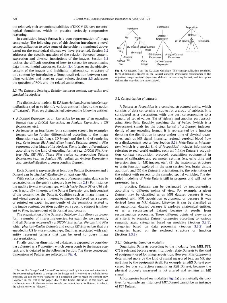
Fig. 4. An excerpt from the Datasets Ontology. This conceptualization considersthree dimensions present in the Dataset concept: Proposition corresponds to theobjective image content, Expression defines the encoding format, and Inscriptiondefines the way data are materialized.
770 L. Temal et al. / Journal of Biomedical Informatics 41 (2008) 766–778
the relatively rich semantic capabilities of DICOM SR have no onto-logical foundation, which in practice seriously compromisesreasoning.
In conclusion, image format is a poor representation of imagecomplexity. The following part of this Section introduces a novelconceptualization to solve some of the problems mentioned above,based on the ontological choices we have presented. Section 3.2addresses the specific question of the relation between content,expression and physical inscriptions of the images. Section 3.3tackles the difficult question of how to categorize neuroimagingdata in meaningful categories. Section 3.4 focuses on the objectivecontent of the images and highlights mathematical structure ofthis content by introducing a (functional) relation between sam-pling variables and pixel or voxel values. Section 3.5 addressesthe question of ROIs and the related annotations.
3.2. The Datasets Ontology: Relation between content, expression andphysical inscriptions
The distinctions made in I& DA (Inscriptions/Expressions/Concep-tualizations) led us to identify various entities linked to the notionof ‘‘dataset”.7 First, we distinguished between the following entities:
� A Dataset Expression as an Expression by means of an encodingformat (e.g. a DICOM Expression, an Analyze Expression, a GISExpression, etc).
� An Image as an Inscription (on a computer screen, for example).Images can be further differentiated according to the imagedimension (e.g. 2D Image, 3D Image) and the kind of rendering(e.g. Color Image, Black and White Image). Datasets stored in Filesrepresent other kinds of Inscriptions. File is further differentiatedaccording to the kind of encoding format (e.g. DICOM File, Ana-lyze File, GIS File). These Files realize corresponding DatasetExpressions (e.g. an Analyze File realizes an Analyze Expression),and physicallyRealizes a corresponding Dataset.
Each Dataset is expressedBy at least one Dataset Expression and aDataset can be physicallyRealizedBy at least one File.
With such a model, various aspects of neuroimaging data can beseparated using the quality category (see Section 2.1). For instance,the quality format encoding type, which hasForQuale U8 or U16 val-ues, is naturally inherent to the Dataset Expression and independentof the content, i.e. the Dataset. Qualities such as image intensityand visual aspects are inherent to Images displayed on a screen,or printed on paper, independently of the semantics related tothe image content. Location quality on a specific support is inher-ent to Files, independent of its format and content.
The organization of the Datasets Ontology thus allows us to per-form a number of interesting queries. For example, we can easilyfind all Datasets expressedBy a DICOM Expression. We can find Fileswhich physicallyRealize Datasets and realize GIS Expressions that areencoded in U8 format encoding type. Qualities associated with eachentity represent criteria that can be used to query imagerepresentations.
Finally, another dimension of a dataset is captured by consider-ing a Dataset as a Proposition, which corresponds to the image con-tent, and is detailed in the following Section. The three conceptualdimensions of Dataset are reflected in Fig. 4.
7 Terms like ‘‘image” and ‘‘dataset” are widely used by clinicians and scientists inthe neuroimaging domain to designate the image and its content as a whole. In ourontology, we use the word ‘‘Dataset” in a dedicated way to mean the content only.However, in the rest of the paper, due to the broad utilization of this word, wecontinue to use it in the two senses: to refer to content, we write Dataset; to refer tothe whole, we write ‘‘dataset”.
3.3. Categorization of datasets
A Dataset as Proposition is a complex, structured entity, whichconsists of data concerning a subject or a group of subjects. It isconsidered as a description, with one part corresponding to astructured set of values (Set of Values), and another part associ-ating Meta-Data. Roughly speaking, Set of Values (which is aProposition), stands for the actual kernel of a Dataset, indepen-dently of any encoding format. It is represented by a functiondenoting the distribution in space and/or time of physical quan-tities, such as MR signal intensity, regional cerebral blood flowor a displacement vector (see Section 5.3). Meta-Data as Informa-tion (which is a special kind of Proposition) includes informationreferring to real-world entities, such as: (1) the Dataset’s acquisi-tion context (acquisition protocol, acquisition equipment) interms of calibration and parameter settings (e.g. echo time andinversion time for MR images, etc.), (2) the anatomical structureor brain function explored in the scan session (e.g. brain, vision,audition), and (3) the Dataset’s orientation, i.e. the orientation ofthe subject with respect to the sampled spatial variables. The de-tailed modeling of Meta-Data is still in progress and will not bepresented here.
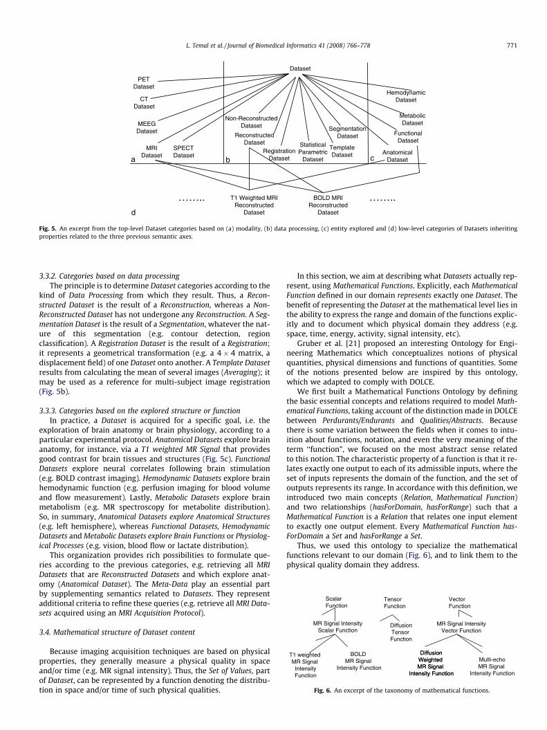
In practice, Datasets can be designated by neuroscientistsaccording to different points of view. For example, a givenDataset may be classified as an MRI dataset because it wasacquired with MRI acquisition equipment, or because it wasderived from an MRI dataset. Likewise, it can be classified asan anatomical dataset because it explores anatomical entities,or as a reconstructed dataset because it results fromreconstruction processing. These different points of view serveas criteria to organize Dataset categories according to varioussemantic axes: categories based on modality (Section 3.3.1),categories based on data processing (Section 3.3.2) andcategories based on the explored structure or function(Section 3.3.3).
3.3.1. Categories based on modalityOrganizing Datasets according to the modality (e.g. MRI, PET,
CT) is relevant because users intuitively relate Datasets to the kindof equipment used for image acquisition. However, this category isdetermined more by the kind of signal measured (e.g. an MR sig-nal) than by the equipment itself. For example, an MRI Dataset pro-cessed for bias correction remains an MRI Dataset, because thephysical property measured is not altered and remains an MRsignal.
All categories based on modality (Fig. 5a) are mutually disjunc-tive: for example, an instance of MRI Dataset cannot be an instanceof PET Dataset.
PETDataset
AnatomicalDataset
FunctionalDataset
HemodynamicDataset
MetabolicDataset
Non-ReconstructedDataset
ReconstructedDataset
Dataset
SegmentationDataset
RegistrationDataset
TemplateDataset
StatisticalParametric
Dataseta
MRIDataset
CTDataset
SPECTDataset
MEEGDataset
b c
T1 Weighted MRI Reconstructed
Dataset
BOLD MRI Reconstructed
Datasetd
…….. ……..
Fig. 5. An excerpt from the top-level Dataset categories based on (a) modality, (b) data processing, (c) entity explored and (d) low-level categories of Datasets inheritingproperties related to the three previous semantic axes.
Multi-echoMR Signal
Intensity Function
ScalarFunction
VectorFunction
MR Signal IntensityVector Function
MR Signal IntensityScalar Function
T1 weighted MR Signal
IntensityFunction
BOLD MR Signal
Intensity Function
DiffusionWeighted MR Signal
Intensity Function
TensorFunction
DiffusionWeighted MR Signal
Intensity Function
DiffusionTensor Function
Fig. 6. An excerpt of the taxonomy of mathematical functions.
L. Temal et al. / Journal of Biomedical Informatics 41 (2008) 766–778 771
3.3.2. Categories based on data processingThe principle is to determine Dataset categories according to the
kind of Data Processing from which they result. Thus, a Recon-structed Dataset is the result of a Reconstruction, whereas a Non-Reconstructed Dataset has not undergone any Reconstruction. A Seg-mentation Dataset is the result of a Segmentation, whatever the nat-ure of this segmentation (e.g. contour detection, regionclassification). A Registration Dataset is the result of a Registration;it represents a geometrical transformation (e.g. a 4 � 4 matrix, adisplacement field) of one Dataset onto another. A Template Datasetresults from calculating the mean of several images (Averaging); itmay be used as a reference for multi-subject image registration(Fig. 5b).
3.3.3. Categories based on the explored structure or functionIn practice, a Dataset is acquired for a specific goal, i.e. the
exploration of brain anatomy or brain physiology, according to aparticular experimental protocol. Anatomical Datasets explore brainanatomy, for instance, via a T1 weighted MR Signal that providesgood contrast for brain tissues and structures (Fig. 5c). FunctionalDatasets explore neural correlates following brain stimulation(e.g. BOLD contrast imaging). Hemodynamic Datasets explore brainhemodynamic function (e.g. perfusion imaging for blood volumeand flow measurement). Lastly, Metabolic Datasets explore brainmetabolism (e.g. MR spectroscopy for metabolite distribution).So, in summary, Anatomical Datasets explore Anatomical Structures(e.g. left hemisphere), whereas Functional Datasets, HemodynamicDatasets and Metabolic Datasets explore Brain Functions or Physiolog-ical Processes (e.g. vision, blood flow or lactate distribution).
This organization provides rich possibilities to formulate que-ries according to the previous categories, e.g. retrieving all MRIDatasets that are Reconstructed Datasets and which explore anat-omy (Anatomical Dataset). The Meta-Data play an essential partby supplementing semantics related to Datasets. They representadditional criteria to refine these queries (e.g. retrieve all MRI Data-sets acquired using an MRI Acquisition Protocol).
3.4. Mathematical structure of Dataset content
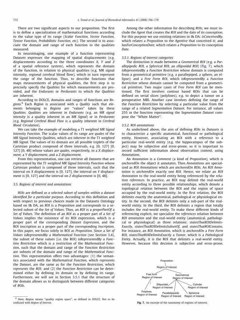
Because imaging acquisition techniques are based on physicalproperties, they generally measure a physical quality in spaceand/or time (e.g. MR signal intensity). Thus, the Set of Values, partof Dataset, can be represented by a function denoting the distribu-tion in space and/or time of such physical qualities.
In this section, we aim at describing what Datasets actually rep-resent, using Mathematical Functions. Explicitly, each MathematicalFunction defined in our domain represents exactly one Dataset. Thebenefit of representing the Dataset at the mathematical level lies inthe ability to express the range and domain of the functions explic-itly and to document which physical domain they address (e.g.space, time, energy, activity, signal intensity, etc).
Gruber et al. [21] proposed an interesting Ontology for Engi-neering Mathematics which conceptualizes notions of physicalquantities, physical dimensions and functions of quantities. Someof the notions presented below are inspired by this ontology,which we adapted to comply with DOLCE.
We first built a Mathematical Functions Ontology by definingthe basic essential concepts and relations required to model Math-ematical Functions, taking account of the distinction made in DOLCEbetween Perdurants/Endurants and Qualities/Abstracts. Becausethere is some variation between the fields when it comes to intu-ition about functions, notation, and even the very meaning of theterm ‘‘function”, we focused on the most abstract sense relatedto this notion. The characteristic property of a function is that it re-lates exactly one output to each of its admissible inputs, where theset of inputs represents the domain of the function, and the set ofoutputs represents its range. In accordance with this definition, weintroduced two main concepts (Relation, Mathematical Function)and two relationships (hasForDomain, hasForRange) such that aMathematical Function is a Relation that relates one input elementto exactly one output element. Every Mathematical Function has-ForDomain a Set and hasForRange a Set.
Thus, we used this ontology to specialize the mathematicalfunctions relevant to our domain (Fig. 6), and to link them to thephysical quality domain they address.
Region of Interest
Proposition
Free formRegion of Interest
GeometricalRegion of Interest
CylinderRegion of Interest
EllipsoidalRegion of Interest
772 L. Temal et al. / Journal of Biomedical Informatics 41 (2008) 766–778
There are two significant aspects to our proposition. The firstis to define a specialization of mathematical functions accordingto the value type of its range (Scalar Function, Vector Function,Tensor Function, Probabilistic Function, etc). The second is to asso-ciate the domain and range of each function to the qualitiesmeasured.
In neuroimaging, one example of a function representingDatasets expresses the mapping of spatial displacements (e.g.displacements according to the three coordinates X, Y and Zof a spatial reference system), which represents the domainof the function, in relation to physical qualities (e.g. MR signalintensity, regional cerebral blood flow), which in turn representthe range of the function. Thus, to describe functions thatmaps measurements of physical qualities, the first step is toprecisely specify the Qualities for which measurements are pro-vided, and the Endurants or Perdurants to which the Qualitiesare inherent.
According to DOLCE, domains and ranges of functions are Re-gions.8 Each Region is associated with a Quality such that ele-ments belonging to Regions are ‘‘values” taken by theseQualities. Qualities are inherent in Endurants (e.g. an MR signalintensity is a quality inherent in an MR Signal) or in Perdurants(e.g. Regional Cerebral Blood Flow is a quality inherent in CerebralBlood Circulation).
We can take the example of modeling a T1 weighted MR SignalIntensity Function. The scalar values of its range are quales of theMR Signal Intensity Qualities, which are inherent in the T1 weightedMR Signal. The values of its domain are all possible triplets of theCartesian product composed of three intervals, e.g. [0, 127] [0,127] [0, 48] whose values are quales, respectively, to a X displace-ment, a Y displacement, and a Z displacement.
From this representation, one can retrieve all Datasets that arerepresented by the T1 weighted MR Signal Intensity Function whoseCartesian product is composed of three intervals, such that theinterval on X displacement is [0, 127], the interval on Y displace-ment is [0, 127] and the interval on Z displacement is [0, 48].
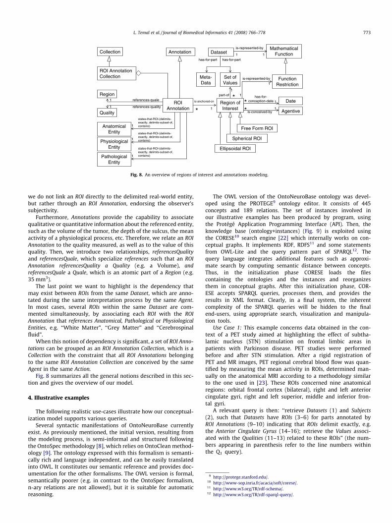
3.5. Regions of interest and annotations
ROIs are defined as a selected subset of samples within a datasetidentified for a particular purpose. According to this definition andwith respect to previous choices made in the Datasets Ontologybased on I& DA, an ROI is a Proposition and corresponds to a se-lected subset of the Set of Values. Thus, an ROI is a properPartOf aSet of Values. The definition of an ROI as a proper part of a Set ofValues implies the existence of its ROI expression, which is aproper part of the corresponding Dataset Expression, and theROI inscription as a proper part of the corresponding Inscription.In this paper, we focus solely in ROI as Proposition. Since a Set ofValues isRepresentedBy a Mathematical Function (see Section 3.4),the subset of these values (i.e. the ROI) isRepresentedBy a Func-tion Restriction which is a restriction of the Mathematical Func-tion, such that the domain and range of the Function Restrictionare subsets of the domain and range of the Mathematical Func-tion. This representation offers two advantages: (1) the seman-tics associated with the Mathematical Function, which representsthe Dataset, are the same as for the Function Restriction, whichrepresents the ROI; and (2) the Function Restriction can be deter-mined either by defining its domain or by defining its range.Furthermore, we will see in Section 3.5.1 that the structure ofthe domain allows us to distinguish between different categoriesof ROIs.
8 Here, Region means ‘‘quality region space”, as defined in DOLCE. Not to beconfused with Region of Interest.
Among the other information for describing ROIs, we must in-clude the Agent that creates the ROI and the date of its conception.For this purpose we use existing relations in I& DA: isConceivedBy,which relates a Proposition to the Agentive that conceived it; andhasForConceptionDate, which relates a Proposition to its conceptionDate.
3.5.1. Regions of interest categoriesThe distinction is made between a Geometrical ROI (e.g. a Par-
allepipedic ROI, a Spherical ROI, an ellipsoidal ROI) (Fig. 7), whichisRepresentedBy a Function Restriction whose domain is computedfrom a geometrical primitive (e.g. a parallepiped, a sphere, an el-lipse); and a Free Form ROI, which isRepresentedBy a FunctionRestriction whose domain cannot be computed from a geometri-cal primitive. Two major cases of Free Form ROI can be men-tioned. The first involves contour based ROIs that can bedefined on serial slices (polylines), e.g. to depict a tumor on apreoperative MRI. Another case involves defining the range ofthe Function Restriction by selecting a particular value from therange of a related Segmentation Dataset. For instance, all values255 in the function representing the Segmentation Dataset com-pose the ‘‘White Matter”.
3.5.2. ROI annotationsAs underlined above, the aim of defining ROIs in Datasets is
to characterize a specific anatomical, functional or pathologicalentity. However, associating an ROI with a reference to aparticular real-world entity (e.g. the hippocampus of the sub-ject) may be subjective and error-prone, so it is important torecord sufficient information about observation context, usingAnnotations.
An Annotation is a Comment (a kind of Proposition), which isanchoredOn the object it annotates. Then Annotations are special-ized as ROI Annotations which are anchoredOn ROIs. An ROI Anno-tation is anchoredOn exactly one ROI. Hence, we relate an ROIAnnotation to the real-world entity being referenced by the rela-tion references. In practice, an ROI may delimit the real-worldentity according to three possible relationships, which denote atopological relation between the ROI and the region of spaceoccupied by the real-world entity. In the first relation, the ROIdelimits exactly the anatomical, pathological or physiological en-tity. In the second, the ROI delimits only a sub-part of the real-world entity. In the third, the ROI delimits a region that totallyincludes the real-world entity. To make these different kinds ofreferencing explicit, we specialize the references relation betweenROI annotation and the real-world entity (anatomical, pathologi-cal or physiological) as three relations: statesThatROIDelimits-Exactly, statesThatROIDelimitsSubsetOf, and statesThatROIContains.For instance, an ROI Annotation, which is anchoredOn a Free FormROI, statesThatROIDelimitsExactly a Tumor, which is a PathologicalEntity. Actually, it is the ROI that delimits a real-world entity.However, because this decision is subjective and error-prone,
ParallelipipedicRegion of Interest
SphericalRegion of Interest
PolylinesRegion of Interest
Fig. 7. An excerpt of the taxonomy of regions of interest.
Collection Annotation DatasetMathematical
Function
is-represented-by
1 1
FunctionRestriction
Meta-Data
Set ofValues
is-represented-by
1
1
ROI Annotation Collection
ROI Annotation
Region
Quality
references-quale
references-quality
AnatomicalEntity
PhysiologicalEntity
PathologicalEntity
Region of Interest
Date
Agentive
has-for-conception-date
is-conceived-by
1
1
Free Form ROI
Spherical ROI
Ellipsoidal ROI
1
1
has-for-part
part-of
states-that-ROI-(delimits-exactly, delimits-subset-of, contains)
states-that-ROI-(delimits-exactly, delimits-subset-of, contains)
states-that-ROI-(delimits-exactly, delimits-subset-of, contains)
has-for-part
is-anchored-on
* 1*
*1
*
1
1
1
Fig. 8. An overview of regions of interest and annotations modeling.
9 http://protege.stanford.edu/.10 http://www-sop.inria.fr/acacia/soft/corese/.11 http://www.w3.org/TR/rdf-schema/.12 http://www.w3.org/TR/rdf-sparql-query/.
L. Temal et al. / Journal of Biomedical Informatics 41 (2008) 766–778 773
we do not link an ROI directly to the delimited real-world entity,but rather through an ROI Annotation, endorsing the observer’ssubjectivity.
Furthermore, Annotations provide the capability to associatequalitative or quantitative information about the referenced entity,such as the volume of the tumor, the depth of the sulcus, the meanactivity of a physiological process, etc. Therefore, we relate an ROIAnnotation to the quality measured, as well as to the value of thisquality. Then, we introduce two relationships, referencesQualityand referencesQuale, which specialize references such that an ROIAnnotation referencesQuality a Quality (e.g. a Volume), andreferencesQuale a Quale, which is an atomic part of a Region (e.g.35 mm3).
The last point we want to highlight is the dependency thatmay exist between ROIs from the same Dataset, which are anno-tated during the same interpretation process by the same Agent.In most cases, several ROIs within the same Dataset are com-mented simultaneously, by associating each ROI with the ROIAnnotation that references Anatomical, Pathological or PhysiologicalEntities, e.g. ‘‘White Matter”, ‘‘Grey Matter” and ‘‘Cerebrospinalfluid”.
When this notion of dependency is significant, a set of ROI Anno-tations can be grouped as an ROI Annotation Collection, which is aCollection with the constraint that all ROI Annotations belongingto the same ROI Annotation Collection are conceived by the sameAgent in the same Action.
Fig. 8 summarizes all the general notions described in this sec-tion and gives the overview of our model.
4. Illustrative examples
The following realistic use-cases illustrate how our conceptual-ization model supports various queries.
Several syntactic manifestations of OntoNeuroBase currentlyexist. As previously mentioned, the initial version, resulting fromthe modeling process, is semi-informal and structured followingthe OntoSpec methodology [8], which relies on OntoClean method-ology [9]. The ontology expressed with this formalism is semanti-cally rich and language independent, and can be easily translatedinto OWL. It constitutes our semantic reference and provides doc-umentation for the other formalisms. The OWL version is formal,semantically poorer (e.g. in contrast to the OntoSpec formalism,n-ary relations are not allowed), but it is suitable for automaticreasoning.
The OWL version of the OntoNeuroBase ontology was devel-oped using the PROTEGE9 ontology editor. It consists of 445concepts and 189 relations. The set of instances involved inour illustrative examples has been produced by program, usingthe Protégé Application Programming Interface (API). Then, theknowledge base (ontology+instances) (Fig. 9) is exploited usingthe CORESE10 search engine [22] which internally works on con-ceptual graphs. It implements RDF, RDFS11 and some statementsfrom OWL-Lite and the query pattern part of SPARQL12. Thequery language integrates additional features such as approxi-mate search by computing semantic distance between concepts.Thus, in the initialization phase CORESE loads the filescontaining the ontologies and the instances and reorganizesthem in conceptual graphs. After this initialization phase, COR-ESE accepts SPARQL queries, processes them, and provides theresults in XML format. Clearly, in a final system, the inherentcomplexity of the SPARQL queries will be hidden to the finalend-users, using appropriate search, visualization and manipula-tion tools.
Use Case 1: This example concerns data obtained in the con-text of a PET study aimed at highlighting the effect of subtha-lamic nucleus (STN) stimulation on frontal limbic areas inpatients with Parkinson disease. PET studies were performedbefore and after STN stimulation. After a rigid registration ofPET and MR images, PET regional cerebral blood flow was quan-tified by measuring the mean activity in ROIs, determined man-ually on the anatomical MRI according to a methodology similarto the one used in [23]. These ROIs concerned nine anatomicalregions: orbital frontal cortex (bilateral), right and left anteriorcingulate gyri, right and left superior, middle and inferior fron-tal gyri.
A relevant query is then: ‘‘retrieve Datasets (1) and Subjects(2), such that Datasets have ROIs (3–6) for parts annotated byROI Annotations (9–10) indicating that ROIs delimit exactly, e.g.the Anterior Cingulate Gyrus (14–16); retrieve the Values associ-ated with the Qualities (11–13) related to these ROIs” (the num-bers appearing in parenthesis refer to the line numbers withinthe Q1 query).
774 L. Temal et al. / Journal of Biomedical Informatics 41 (2008) 766–778
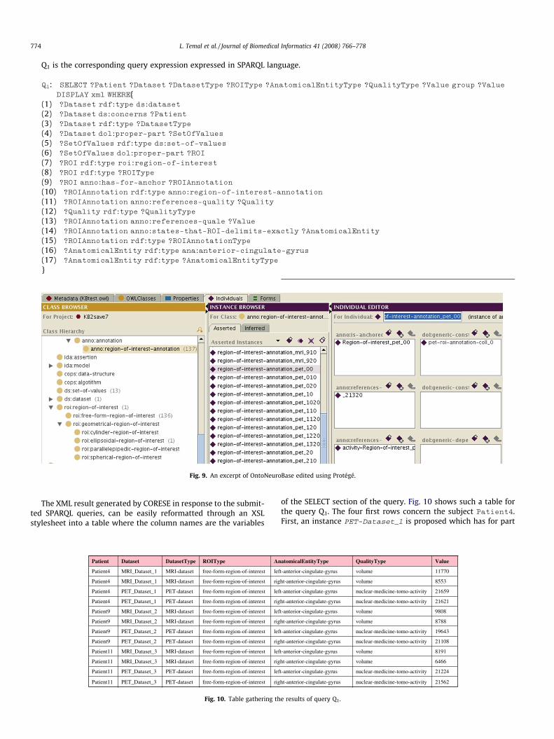
Q1 is the corresponding query expression expressed in SPARQL language.
Q1: SELECT ?Patient ?Dataset ?DatasetType ?ROIType ?AnatomicalEntityType ?QualityType ?Value group ?Value
DISPLAY xml WHERE{(1) ?Dataset rdf:type ds:dataset
(2) ?Dataset ds:concerns ?Patient
(3) ?Dataset rdf:type ?DatasetType
(4) ?Dataset dol:proper-part ?SetOfValues
(5) ?SetOfValues rdf:type ds:set-of-values
(6) ?SetOfValues dol:proper-part ?ROI
(7) ?ROI rdf:type roi:region-of-interest
(8) ?ROI rdf:type ?ROIType
(9) ?ROI anno:has-for-anchor ?ROIAnnotation
(10) ?ROIAnnotation rdf:type anno:region-of-interest-annotation
(11) ?ROIAnnotation anno:references-quality ?Quality
(12) ?Quality rdf:type ?QualityType
(13) ?ROIAnnotation anno:references-quale ?Value
(14) ?ROIAnnotation anno:states-that-ROI-delimits-exactly ?AnatomicalEntity
(15) ?ROIAnnotation rdf:type ?ROIAnnotationType
(16) ?AnatomicalEntity rdf:type ana:anterior-cingulate-gyrus
(17) ?AnatomicalEntity rdf:type ?AnatomicalEntityType
}
Fig. 9. An excerpt of OntoNeuroBase edited using Protégé.
The XML result generated by CORESE in response to the submit-ted SPARQL queries, can be easily reformatted through an XSLstylesheet into a table where the column names are the variables
rigfree-form-region-of-interestPET-datasetPET_Dataset_3Patient11
leffree-form-region-of-interestPET-datasetPET_Dataset_3Patient11
rigfree-form-region-of-interestMRI-datasetMRI_Dataset_3Patient11
leffree-form-region-of-interestMRI-datasetMRI_Dataset_2Patient9
leffree-form-region-of-interestMRI-datasetMRI_Dataset_1Patient4
rigfree-form-region-of-interestMRI-datasetMRI_Dataset_1Patient4
leffree-form-region-of-interestPET-datasetPET_Dataset_1Patient4
rigfree-form-region-of-interestPET-datasetPET_Dataset_1Patient4
leffree-form-region-of-interestMRI-datasetMRI_Dataset_3Patient11
rigfree-form-region-of-interestPET-datasetPET_Dataset_2Patient9
leffree-form-region-of-interestPET-datasetPET_Dataset_2Patient9
rigfree-form-region-of-interestMRI-datasetMRI_Dataset_2Patient9
AnROITypeDatasetTypeDatasetPatient
Fig. 10. Table gathering th
of the SELECT section of the query. Fig. 10 shows such a table forthe query Q1. The four first rows concern the subject Patient4.First, an instance PET-Dataset_1 is proposed which has for part
21562nuclear-medicine-tomo-activityht-anterior-cingulate-gyrus
21224nuclear-medicine-tomo-activityt-anterior-cingulate-gyrus
6466volumeht-anterior-cingulate-gyrus
9808volumet-anterior-cingulate-gyrus
11770volumet-anterior-cingulate-gyrus
8553volumeht-anterior-cingulate-gyrus
21659nuclear-medicine-tomo-activityt-anterior-cingulate-gyrus
21621nuclear-medicine-tomo-activityht-anterior-cingulate-gyrus
8191volumet-anterior-cingulate-gyrus
21108nuclear-medicine-tomo-activityht-anterior-cingulate-gyrus
19643nuclear-medicine-tomo-activityt-anterior-cingulate-gyrus
8788volumeht-anterior-cingulate-gyrus
ValueQualityTypeatomicalEntityType
e results of query Q1.
Z-locationY-locationX-locationT1-weighted-MR-signal-intensity-functionT1-weighted-MR-signal-intensity-function_0MRI_Dataset_1Patient4
Z-locationY-locationX-locationnuclear-medicine-tomo-activity-functionnuclear-medicine-tomo-activity-function_0PET_Dataset_1Patient4
Z-locationY-locationX-locationnuclear-medicine-tomo-activity-functionnuclear-medicine-tomo-activity-function_20PET_Dataset_3Patient11
Z-locationY-locationX-locationT1-weighted-MR-signal-intensity-functionT1-weighted-MR-signal-intensity-function_20MRI_Dataset_3Patient11
Z-locationY-locationX-locationnuclear-medicine-tomo-activity-functionnuclear-medicine-tomo-activity-function_10PET_Dataset_2Patient9
Z-locationY-locationX-locationT1-weighted-MR-signal-intensity-functionT1-weighted-MR-signal-intensity-function_10MRI_Dataset_2Patient9
Var3TypeVar2TypeVar1TypeFunctionTypeFunctionDatasetPatient
Fig. 11. Table gathering the results of query Q2.
L. Temal et al. / Journal of Biomedical Informatics 41 (2008) 766–778 775
an instance of free-form-region-of-interest on which is anchored aninstance of region-of-interest-annotation. It states that the instanceof region-of-interest exactly delimits the left-anterior-cingulate-gyrus whose volume is equal to PET-Dataset_1. Second, for thesame instance PET-Dataset_1, the instance of region-of-interestthat exactly delimits the right-anterior-cingulate-gyrus, whose vol-ume is equal to PET-Dataset_1, is proposed. The same subjectis concerned by PET-Dataset_1 which has for part two instancesof free-form-region-of-interest. On the first ROI is anchored an in-stance of region-of-interest-annotation, that states that the instanceof region-of-interest exactly delimits the left-anterior-cingulate-gyrus whose activity is equal to PET-Dataset_1. Similarly, onthe second ROI is anchored an instance of region-of-interest-annota-tion that states that the instance of region-of-interest exactly delim-its the right-anterior-cingulate-gyrus whose activity is equal to PET-Dataset_1.
Note that, in Q1 only the anatomical entity anterior-cingu-
late-gyrus was initially specified (line 16). However, because inthe ontology left and right anterior cingulate gyrus classes are sub-sumed by anterior-cingulate-gyrus, the system can infer that the leftand the right specializations are also anterior cingulate gyrus and
so retrieves them. The instruction at line 17 of the same query re-trieves the direct type of this anatomical entity and informs theuser that the anatomical entities are the left and the right special-izations of the anterior-cingulate-gyrus.
Similarly, the general concepts of dataset (1) and region-of-inter-est (7) were initially indicated and the system retrieves for in-stance, PET-Dataset_1, an instance of a PET-dataset, and asubclass of region-of-interest, namely free-form-region-of-interest.
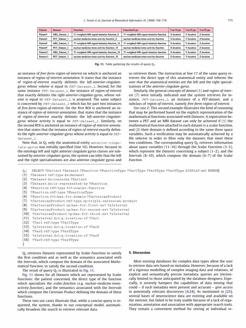
Use case 2: This second example illustrates the kind of reasoningthat may be performed based on the explicit representation of themathematical functions associated with Datasets. A registration be-tween a PET and an MRI dataset can only be achieved if (1) themathematical function attached to each dataset is a scalar function,and (2) their domain is defined according to the same three spacevariables. Such a verification may be automatically achieved by afile selector in order to filter only the datasets that meet thesetwo conditions. The corresponding query Q2 retrieves informationabout space variables (11–16) through the Scalar Functions (3–5),which represent the Datasets concerning a subject (1–2), and theIntervals (8–10), which compose the domain (6–7) of the ScalarFunction.
Q2: SELECT ?Patient ?Dataset ?Function ?FunctionType ?Var1Type ?Var2Type ?Var3Type DISPLAY xml WHERE{(1) ?Dataset rdf:type ds:dataset
(2) ?Dataset ds:concerns ?Patient
(3) ?Dataset ds:is-represented-by ?Function
(4) ?Function rdf:type fct:scalar-function
(5) ?Function rdf:type ?FunctionType
(6) ?Function fct:has-for-domain ?CartesianProduct
(7) ?CartesianProduct rdf:type cp:triple-cartesian-product
(8) ?CartesianProduct cp:has-for-first-set ?xInterval
(9) ?CartesianProduct cp:has-for-second-set ?yInterval
(10) ?CartesianProduct cp:has-for-third-set ?zInterval
(11) ?xInterval dol:q-location-of ?Var1
(12) ?Var1 rdf:type ?Var1Type
(13) ?yInterval dol:q-location-of ?Var2
(14) ?Var2 rdf:type ?Var2Type
(15) ?zInterval dol:q-location-of ?Var3
(16) ?Var3 rdf:type ?Var3Type
}
Q2 retrieves Datasets represented by Scalar Functions to satisfythe first condition and as well as the semantics associated withthe Intervals, which compose the domain of the associated Mathe-matical Function, to satisfy the second condition.
The result of query Q2 is illustrated in Fig. 11.Fig. 11 shows for all Datasets which are represented by Scalar
Functions: the patient concerned, the direct type of the functionwhich specializes the scalar-function (e.g. nuclear-medicine-tomo-activity-function), and the semantics associated with the Intervalswhich compose the Cartesian Product defining the domain of thesefunctions.
These two use cases illustrate that, while a concise query is re-quested, the system, thanks to our conceptual model, automati-cally broadens the search to retrieve relevant data.
5. Discussion
Most existing databases for complex data types allow the userto retrieve data sets based on metadata. However, because of a lackof a rigorous modelling of complex imaging data and relations, ofexplicit and semantically precise metadata, queries are intrinsi-cally limited to those anticipated by the database designers. Espe-cially, it severely hampers the capabilities of data mining thatcould – if such metadata were present and accurate – give accessto potentially relevant discoveries [4,24]. As mentioned in [25],several bases of neuroscience data are existing and available onthe internet, but failed to be truly usable because of a lack of orga-nization, annotation and association with appropriate search tools.They remain a convenient method for storing at individual re-
776 L. Temal et al. / Journal of Biomedical Informatics 41 (2008) 766–778
searcher level or sharing data for well defined multi-centre studies[26], but their integration into federated systems remains verychallenging because of their intrinsic heterogeneity. In contrast,ontology allows making inferences based on the semantics of con-cepts and relations. This capability, mastered by the ontologydesigners, enlarges the set of possible queries compared to a stan-dard data base. A first basic example consists in searching for MRIDatasets expressed in DICOM format. Most classical databaseimplementation would simply use the DICOM format and imagemodality as search criteria. Our model makes it possible to querysuch Datasets at multiple levels of the Dataset hierarchy, e.g. eitherusing a general class MRI Dataset, or using a more specific one likeT1-weighted MRI Reconstructed Dataset or FLAIR-weighted MRIReconstructed Dataset. Similarly, our model makes it possible tospecify, either that any kind of DICOM expression is searched for,i.e. any kind of DICOM Service Object Pair (SOP) Class is allowed,or to specify a particular kind of DICOM expression, i.e. using theregular DICOM MR Image Storage SOP Class or using the DICOM En-hanced MR Image Storage SOP Class. A second example concerns asituation in which a user is searching for cases of patients whoseMR Image Storage images show a Brain Tumor locatedIn the FrontalLobe. Based on the ontology’s knowledge, the system would be ableto retrieve the case of a patient with a Glioblastoma locatedIn theInferior Frontal Gyrus, since a Glioblastoma is a Brain Tumor andthe Inferior Frontal Gyrus isPartOf the Frontal Lobe, and the relation-ship locatedIn satisfies the property that any entity1 which is locat-edIn an entity2, is also locatedIn any entity that hasForPart entity2.Clearly, this kind of result could not be obtained with a standarddata base. Another way of exploiting the semantics embedded inthe ontology amounts to calculate ‘‘semantic distances” betweenconcepts (these distances rely on topological distances calculatedin the graph corresponding to the structure of the ontology). Suchsemantic distances enable query engines to give approximate an-swers. Such a facility is for instance provided by the semantic re-search engine CORESE that we use in our project.
Thus, the use of ontologies helps overcoming the difficultiesencountered within conventional databases, by providing precisedefinition of each concept and relation, and defining the commonunified schema for the mapping of the local database schemas.Our work with OntoNeurobase brings two major contributions.The first is related to the general methodology we propose to builda multi-layered and multi-components application ontology; thesecond concerns the domain ontology we have designed for neuro-imaging. These contributions are then put in perspective with sim-ilar works being carried out in the context of projects such as caBIGand BIRN.
5.1. Multi-layered and multi-components application ontology
Our approach aims at mastering two complexities. The first is aconceptual complexity, arising from the intrinsic nature of the enti-ties belonging to our universe of discourse (i.e. medical images andtheir annotations). The second is a design complexity related to theneed to articulate our model with other models, either existing orin development, addressing the needs of connected domains suchas biology, clinical medicine, anatomy, physiology, etc. so thatour contribution can be managed independently, while fitting intoa consistent whole.
Our basic methodology is to model entities at different levels ofabstraction, by re-using a set of core ontologies, based on a com-mon foundational ontology.
Foundational ontologies provide rigorous logical axiomatisationexpliciting ontological commitments, making possible to reasonabout entities and to map ontologies in the future. DOLCE andthe Basic Formal Ontology (BFO) [27], an ontology developed byIFOMIS and widely used in the biological sciences, are the favorite
candidates which propose rigorous foundational principles tomodel our domain. These ontologies have been elaborated in thecontext of the WonderWeb project [14], whose ultimate aim wasto build a library of foundational ontologies, precisely to establishthe foundations enabling the ‘‘negotiation of meaning” betweenagents. This work leads to the possibility of mapping ontologiesconceived according to different philosophical approaches, as wellas to a better understanding of the difficulties related to suchmappings.
Our choice of DOLCE, considered as a reference by manyauthors, e.g. [28–30], was motivated by three major factors. Thefirst is related to its rich and well-documented axiomatization asto location in space and time, dependence and parthood, and tothe fact that it relies on explicit structuration principles [14]. More-over, it is based on the OntoClean [9] methodology, thus providinga precious guide to structuring application ontologies, especiallyregarding taxonomic relationships. The second argument in favorof DOLCE is the availability of numerous extensions, such asDOLCE-Lite-Plus and many core ontologies, related to participationroles, semiotics, collections, artifacts, and manufactured objects,addressing difficult-to-model domains in which it would havebeen unrealistic to attempt significant work by ourselves. Finally,the third factor lies in the basic principles retained in DOLCE whichwe considered particularly relevant in our context. The deliberatechoice of a ‘‘cognitive bias” (i.e. depending strongly on human per-ception and social conventions) proved relevant for modeling hu-man artifacts such as dataset expressions, or mathematicalconcepts. Similarly, DOLCE’s multiplicative approach (i.e. authoriz-ing several entities to be co-localized in the same space-time)seemed appropriate for modeling spatio-temporally co-localizedentities, such as an anatomical structure and the generator of afunctional activity, although it is quite clear that both are inherentto the same biological reality.
BFO adopts a realist approach and then is reluctant to speakabout categories which are language dependent. Our feeling is thatannotating images and referring to brain pathology would proba-bly require modelling entities such as language acts and cognitivestates, which may be more difficult to introduce based on the BFOphilosophical approach.
However our choice of DOLCE should not hide what is – in ourvision – the most important aspect: the design of an ontology,should be based on a foundational ontology whatever it is. Weclaim that this is more important than the choice of a particularfoundational ontology, since foundational ontologies provide therigorous logical axiomatisation, making possible to reason aboutentities and to map ontologies in the future. In this regard, whatis of paramount importance is to capture sufficient semantics in or-der to enable subsequent mappings between partly overlappingontologies (since such overlapping seems inevitable). The use ofa methodology such as OntoSpec, based on OntoClean, authorizinga semi-informal representation of semantics appears important inthis respect.
It is certainly too early to judge of the added value of DOLCE andthe reused core ontologies in facilitating the integration of multi-domain information (e.g. anatomy, physiology, pathology, imageprocessing, biology), which is expected from any upper layer orfoundational ontology. Clearly, only confrontation with experi-ments would support our claim, and the NeuroLog project (see Sec-tion 6) plans to carry out such work.
5.2. Domain ontology for neuroimaging
5.2.1. Image dataOur modeling helps clarify the various connotations attached to
images. We distinguish what relates to physical entities, such asfile materialization, or rendering on computer screens, from non
L. Temal et al. / Journal of Biomedical Informatics 41 (2008) 766–778 777
physical entities such as expressions according to various formats.Much remains to be done concerning this last point. In the presentwork, we have focused on what images refer to rather than render-ing issues, such as windowing, 3D rendering or ‘‘blending” of mul-timodal values. Our current objectives lie in the sharing and reuseof data and image processing tools rather than in display applica-tions. Data formats are insufficiently explicit, especially regardingthe mathematical aspect, to allow reasoning about data and imageprocessing and the composition of innovative image processingpipelines. Our categorization of datasets is a step towards achiev-ing this goal.
Our accomplishments remain modest, especially when com-pared to a standard like DICOM in which the descriptions of imagestructure, semantics and metadata represent approximately 1000pages of specifications, addressing the details and specificity ofeach imaging modality. However, the orientations we have devel-oped have sufficient generality to enable revisiting the standardbased on ontological principles. This is certainly a huge job, whichshould be conducted progressively to provide significant added va-lue even at the early stages of its completion. Needs in this areahave already emerged, for instance in the context of DICOM Work-ing Group 23, ‘‘Application Hosting”, which addresses the issue ofdefining a standard API for image processing tools (such as plug-ins or Web services). This obviously requires that the semanticsof the image data being processed be properly modeled and shared.
5.2.2. ROI annotationsFor ROIs and annotations, the proposed models constitute a first
step. The objective was to meet the most common requirements,such as referring to real-world entities, with relatively precisesemantics. This allows distinguishing the case where an ROI ex-actly delimits an entity, e.g. an anatomical structure, from caseswhere it contains only a part of it, or conversely, where it belongsto a region of space that contains more than this structure. Theserelationships are intended to be used for spatial reasoning [31] inconjunction with formal ontologies of anatomy that support mere-ological properties, such as the Foundational Model of Anatomy(FMA) [32]. This point raises the issue of aligning FMA, or abrain-related subset of FMA, with foundational ontologies suchas DOLCE or BFO, since they include their own Theory of Parts,whose compatibility with FMA should be assessed with caution.
One limitation of our ROI annotation model is that it requiresrepresentation of a separate instance of ROI Annotation for everyQuality concerning an ROI. Thus, if we wish to represent both themean and standard deviation of a signal intensity over an ROI,we must define two separate ROI Annotations associated with thesame ROI. An alternative would have been to adopt a complexstructured model of ROI Annotations, such as the one used in DI-COM SR. This direction is currently being explored in the contextof the ‘‘Annotation and Image Markup” project, a sub-project ofthe CaBIG initiative. Our feeling is that it may lead to over-compleximplementation, compromising efficient querying in the most fre-quent cases.
5.2.3. ROI annotations and subjectivityWe consider important to establish relationships between the
results of image processing (imaging biomarkers) applied to spe-cific image regions, and real-world entities, while at the same timeunderlining the subjectivity of such relationships. This subjectivityconcerns the whole observation context including the observer.Hence, although it may result from an automatic tool, the resultof any processing is dependent on the specific tools used. Forexample, the numeric value obtained by the hippocampus volumecomputation from a structural image depends on the segmentationalgorithm and if any, the pre-processing steps used, such as biascorrection. We address this issue by using the ‘‘Participant Roles”
core ontology, very helpful in modeling the genealogy of the data.Subjectivity also concerns the categories of real-world entities thatare referred to, which may depend on the observer – i.e. twoobservers may choose to refer to two different entities – but thismay also evolve over time, since relevant new categories may ap-pear based on the progress of knowledge [33]. This may lead to thecreation of new annotations for the data, referring to these newcategories. This possibility should not be underestimated for thefuture, since new categories, especially in the domain of pathology,are likely to emerge, e.g. based on genomic and proteomic data.
5.3. Relations with on going projects
Regarding other projects such as caBIG, interesting work isbeing performed in the context of the ‘‘In vivo Imaging workspace”,especially concerning annotation and image markup. Our work isclearly in line with this effort as well as complementary to thedevelopment of RADLex, a terminological resource for radiologydeveloped by the Radiological Society of North America, althoughthe ontological choices made in either projects are not explicit.
In the fields of neuroscience and neuroimaging, the BiomedicalInformatics Research Network (BIRN) appears to date as the mostadvanced large-scale data integration effort. We share most ofthe general objectives described in the BIRN seminal paper [4],especially regarding the need to adopt a federated approach, andthe need to found mediation on domain ontologies. A lot of effortswere deployed by the BIRN Ontology Task Force to reuse as muchas possible existing terminologies such as UMLS (Unified MedicalLanguage System), NeuroNames, SNOMED (Systematized Nomen-clature of Medicine), GO (Gene Ontology), LOINC (Logical Observa-tions Identifiers Names and Codes) etc., with a mapping betweenthe different resources made via UMLS, or using the BONFIRE tool.With BIRNLex, their most recent work is much more in line withour own approach, i.e. suggesting that such domain terminologiesshould be based on foundational ontologies [34]. However, we con-sider that UMLS has not a sufficient clear ontological foundation tosupport reasoning techniques as required for integrating heteroge-neous data. Several difficulties have been reported using UMLS. Forinstance, Kumar and Smith in [35], based on a concrete exampleconcerning the regulation of blood pressure, illustrate how well-formalized ontologies, contrary to a lightweight ontology such asUMLS, can detect and avoid conflicts. It appears that in UMLS thenotion of cardiac output embraces both continuant and occurrententities, due to a basic confusion between biomedical phenomenaand their measurement in the context of a procedure.
Actually, their most recent achievements rely on work madeunder the auspices of the National Centre of Biomedical Ontol-ogy/Open Biomedical Ontologies foundry (Ontology for BiomedicalInvestigations, Common Anatomy Reference Ontology, BFO, Rela-tion Ontology, GO, etc) and use BFO and RO as a foundationalontology. This alignment will certainly facilitate the inter-operabil-ity, by providing the semantic content which is needed to map theontologies, eventually at different levels of abstraction. However,re-engineering existing terminologies to make them compliant tofoundational ontologies such as BFO or DOLCE will take time. Asfar as we are concerned, our efforts go in the same direction, andwe try to focus our contributions to those fields in which we arethe most competent, i.e. imaging and image processing.
5.4. Extension and Interoperability
Extension and interoperability are key issues in knowledgeengineering and ontology development in particular. As alreadymentioned, our ultimate aim is to define an ontology which is eas-ily extensible, which allows integration of conceptualizations com-ing from different fields, and which ensures interoperability with
778 L. Temal et al. / Journal of Biomedical Informatics 41 (2008) 766–778
other ontologies. Currently, integration of conceptualization of do-mains such as anatomy or physiology is not realized yet. However,we plan in the next step to integrate FMA or some ontologies fromOBO following a vertical strategy as proposed in [36]. According tothis strategy, the most abstract concepts and relations defined inthese ontologies (such as Anatomical structure, Pathological struc-ture, Function, or Physiological state) are mapped to abstract con-cepts present in DOLCE (e.g. Physical object, Feature, State, andProcess, respectively). Furthermore, the interoperability with ontol-ogies conceived in the BFO framework can be facilitated by makinga horizontal mapping between abstract concepts of DOLCE andBFO. On that purpose, we can indicate some correspondences be-tween concepts which are either extensionally equivalent (e.g.bfo:Object = dolce:Physical Object, bfo: Quality = dolce:PhysicalQual-ity) or which hold subsumption relation (e.g. bfo:Boundary of Object< dolce: Feature).
6. Conclusion and perspectives
The next steps of this project will be carried out in the contextof the NeuroLOG Project13, which received a grant from the FrenchNational Research Agency for 2007–2009. A specific workpackageconcerns the consolidation and extension of the OntoNeuroBaseontology, especially with the aim of modeling image processing aswell as processing tools, with the general perspective of sharingand reusing existing processing tools and creating new image pro-cessing pipelines. This extension will take into account three clinicalapplication contexts, namely multiple sclerosis, stroke and brain tu-mors. A federated system will be deployed, associating research cen-ters in Grenoble, Rennes, Sophia-Antipolis and Paris to share theimages and processing tools ‘‘published” by these researchcenters. OntoNeuroBase will be used as a common semanticreference to align and query the potentially heterogeneous dataavailable in the various repositories using OWL-Lite instances. Thisproject gives us the opportunity to assess the validity of our choicesand the relevance of OntoNeuroBase for large scale neuroimagingapplications.
Acknowledgements
L. Temal is supported by a doctoral grant from the region ofBrittany.
This work received financial support from the French Ministryof Research and Technology (‘‘Action Concertée Incitative”).
References
[1] Saltz J, Oster S, Hastings S, Langella S, Kurc T, Sanchez W, et al. caGRID: designand implementation of the core architecture of the cancer biomedicalinformatics grid. Bioinformatics 2006;22(15):1910–6.
[2] Koslow SH, Hirsch MD. Celebrating a decade of neuroscience databases:looking to the future of high-throughput data analysis, data integration, anddiscovery neuroscience. Neuroinformatics 2004;2:267–70.
[3] Gupta A, Ludäscher B, Grethe JS, Martone ME. Towards a formalization ofdisease-specific ontologies in neuroinformatics. Neural Netw2003;16(9):1277–92.
[4] Martone ME, Gupta A, Ellisman MH. e-Neuroscience: challenges and triumphsin integrating distributed data from molecule to brains. Nat Neurosci2004;7(5):467–72.
[5] Seeding the Europhysiome: a roadmap to the virtual physiological human. Co-ordination Action #027642, 2007. Available from: http://www.europhysiome.org/.
[6] Barillot C, Benali H, Dojat M, Gaignard A, Gibaud B, Kinkingnehun S, et al.Federating distributed and heterogeneous information sources inneuroimaging: the NeuroBase project. Stud Health Technol Inform2006;120:3–13.
[7] Gangemi A, Borgo S, editors. Proceedings of the EKAW*04 workshop on coreontologies in ontology engineering. Northamptonshire (UK), 2004. Available
13 http://neurolog.polytech.unice.fr/doku.php.
from: http://sunsite.informatik.rwth-aachen.de/Publications/CEUR-WS/Vol-118/.
[8] Kassel G. Integration of the DOLCE top-level ontology into the OntoSpecmethodology. LaRIA Research Report 2005–2008. Available from: http://hal.ccsd.cnrs.fr/ccsd-00012203. 2005.
[9] Guarino N, Welty C. An overview of OntoClean. In: Staab S, Studer R, editors.Handbook on ontologies. Springer Verlag; 2004. p. 151–71.
[10] Bell DS, Pattison-Gordon E, Greenes RA. Experiments in concept modeling forradiographic image reports. JAMIA 1994;1(3):249–62.
[11] Fielding JM, Marwede D. The image as spatial region: location and adjacencywithin the radiological image. Frontiers in artificial intelligence andapplications. Formal ontology in information systems. In: Bennett B,Fellbaum C, editors. Proceedings of the fourth international conference (FOIS2006), vol. 150, 2006. p. 89–100.
[12] Gemo M, Gouze A, Debande B, Grivegnée A, Macq B. A versatile knowledge-based clinical imaging annotation system for breast cancer screening. SPIEmedical imaging: computer-aided diagnosis. San Diego, California, USA: TheInternational Society for Optical Engineering; 2007.
[13] Rector A, Modularisation of domain ontologies implemented in descriptionlogics and related formalisms including OWL. In: Genari J, editor. Proceedingsof the second international conference on knowledge capture (K-CAP 2003),ACM; 2003. p. 121–8
[14] Masolo C, Borgo S, Gangemi A, Guarino N, Oltramari A, Schneider L. TheWonderWeb Library of Foundational Ontologies and the DOLCE ontology.WonderWeb Deliverable D18, Final Report (v. 1.0, 31-12-2003); 2003.
[15] Fortier JY, Kassel G. Managing knowledge at the information level: anontological approach. In: Proceedings of the ECAI’2004 workshop onknowledge management and organizational memories, Valencia, Spain;2004. p. 39–45.
[16] Sowa JF. Knowledge representation: logical, philosophical, and computationalfoundations. Pacific Grove: Brooks Cole; 2000.
[17] Bottazi E, Catenacci C, Gangemi A, Lehman J. From collective intentionality tointentional collectives: an ontological perspective. Cognitive Sys Res2006;7(2–3):192-208 [Special Issue on Cognition, Joint Action and CollectiveIntentionality].
[18] Guarino N, Welty C. A formal ontology of properties. In: Dieng R, Corby O,editors. Proceedings of the 12th international conference on knowledgeengineering and knowledge management: EKAW2000. Lecture notes oncomputer science. Springer Verlag; 2000. p. 97–112.
[19] Bruaux S, Kassel G, Morel G. A Clarification of the ontological status ofKnowledge Roles. In: Proceedings of the workshop on ‘‘Advances in ConceptualKnowledge Engineering”; 18th international conference on database andexpert systems applications, DEXA 07, Regensburg, Germany; 2007. p. 529–33.
[20] Temal L, Lando P, Gibaud B, Dojat M, Kassel G, Lapujade A. OntoNeuroBase: multi-layered application ontology in neuroimaging. In: Proceedings of the FOMI2006workshop on formal ontology meet industry, Trento, Italy; 2006. p. 3–15.
[21] Gruber T, Olsen G. An ontology for engineering mathematics. In: Doyle J,Torasso P, Sandewall E, editors. Fourth international conference on principlesof knowledge representation and reasoning, Bonn, Germany: GustavStresemann Institut; 1994.
[22] Corby O, Dieng-Kuntz R, Faron-Zucker C. Querying the semantic web with Coresesearch engine. In: Proceedings of the 15th ECAI/PAIS, Valencia, Spain; 2004.
[23] Haegelen C, Verin M, Aubert Broche B, Prigent F, Jannin P, Gibaud B, et al. Doessubthalamic nucleus stimulation affect the frontal limbic areas ? A singlephoton emission computed tomography study using a manual anatomicalsegmentation method. Surg Radiol Anat 2005;27:389–94.
[24] Kotter R. Neuroscience databases: tools for exploring brain structure–functionrelationships. Philos Trans R Soc Lond B Biol Sci 2001;356:1111–20.
[25] Ascoli GA. Mobilizing the base of neuroscience data: the case of neuronalmorphologies. Nat Rev Neurosci 2006;7:318–24.
[26] Hasson U, Skipper JI, Wilde MJ, Nusbaum HC, Small SL. Improving the analysis,storage and sharing of neuroimaging data using relational databases anddistributed computing. NeuroImage 2008;39:693–706.
[27] Grenon P. BFO in a nutshell: a bi-categorical axiomatization of BFO andcomparison with DOLCE, IFOMIS Report, Universität Leipzig, ISSN 1611-4019;2003.
[28] Yu AC. Methods in biomedical ontology. J Biomed Inform 2006;39:252–66.[29] Cimino JJ, Zhu X. The practical impact of ontologies on biomedical informatics.
Methods Inf Med 2006;45(Suppl 1):124–35.[30] Héja G, Varga P, Pallinger P, Surjan G. Restructuring the foundational model of
anatomy. Stud Health Technol Inform 2006;124:755–60.[31] Cohn AG, Hazarika SM. Qualitative spatial representation and reasoning: an
overview. Fundamenta Informaticae 2001;46:1–29.[32] Rosse C, Mejino JLV. A reference ontology for biomedical informatics: the
foundational model of anatomy. J Biomed Inform 2003;36:478–500.[33] Smith B. From concepts to clinical reality: an essay on the benchmarking of
biomedical terminologies. J Biomed Inform 2006;39(3):288–98.[34] Smith B, Ashburner M, Rosse C, Bard J, Bug W, Ceusters W, et al. The OBI
foundry: coordinated evolution of ontologies to support biomedical dataintegration. Nat Biotechnol 2007;25(11):1251–5.
[35] Kumar A, Smith B. The unified medical language system and the geneontology: some critical reflections. In: Kruse R, Günter A, Neumann B, editors.KI 2003: advances in artificial intelligence. Springer Verlag; 2003. p. 135–48.
[36] Rosse C, Kumar A, Mejino JLV, Cook DL, Detwilern LT, Smith B. A Strategy forimproving and integrating biomedical ontologies. In: Proceedings of AMIAsymposium 2005, Washington, DC, 2005. p. 639–43.