toxicity equivalence factors for marine biotoxins ... · food and agriculture organization of the...
TRANSCRIPT

Toxicity Equivalency Factors for Marine Biotoxins Associated with Bivalve Molluscs
TE
CH
NI
CA
L P
AP
ER
JOINT FAO/WHO

Cover photograph: © FAOemergencies

FOOD AND AGRICULTURE ORGANIZATION OF THE UNITED NATIONS
WORLD HEALTH ORGANIZATION
ROME, 2016
Toxicity equivalence factors for marine biotoxins associated with bivalve molluscs
TE
CH
NI
CA
L P
AP
ER
JOINT FAO/WHO

The designations employed and the presentation of material in this publication do not imply the
expression of any opinion whatsoever on the part of the Food and Agriculture Organization of the
United Nations (FAO) or of the World Health Organization (WHO) concerning the legal status of any
country, territory, city or area or of its authorities, or concerning the delimitation of its frontiers or
boundaries. Dotted lines on maps represent approximate border lines for which there may not yet
be full agreement. The mention of specific companies or products of manufacturers, whether or not
these have been patented, does not imply that these are or have been endorsed or recommended
by FAO or WHO in preference to others of a similar nature that are not mentioned. Errors and
omissions excepted, the names of proprietary products are distinguished by initial capital letters.
All reasonable precautions have been taken by FAO and WHO to verify the information contained
in this publication. However, the published material is being distributed without warranty of any
kind, either expressed or implied. The responsibility for the interpretation and use of the material
lies with the reader. In no event shall FAO and WHO be liable for damages arising from its use.
The views expressed herein are those of the authors and do not necessarily represent those of FAO or
WHO.
ISBN 978-92-5-109345-0 (FAO)
ISBN 978-92-4-151148-3 (WHO)
© FAO and WHO, 2016
FAO and WHO encourage the use, reproduction and dissemination of material in this information
product. Except where otherwise indicated, material may be copied, downloaded and printed for
private study, research and teaching purposes, provided that appropriate acknowledgement of FAO
and WHO as the source and copyright holder is given and that FAO and WHO’s endorsement of users’
views, products or services is not implied in any way.
All requests for translation and adaptation rights, and for resale and other commercial use rights should
be made via www.fao.org/contact-us/licence-request or addressed to [email protected].
FAO information products are available on the FAO website (www.fao.org/publications) and can be
purchased through [email protected]
Recommended citation:FAO/WHO. 2016.Technical paper on Toxicity Equivalency Factors for Marine Biotoxins Associated with Bivalve Molluscs. Rome. 108 pp.
Contents
1
2
ACKNOWLEDGEMENTS v
ABBREVIATIONS AND ACRONYMS vi
PREPARATION AND PURPOSE OF THIS DOCUMENT ix
DEFINITIONS OF TERMS USED xi
EXECUTIVE SUMMARY xv
Criteria used for determining TEFs xviBivalve-associated toxins xviMethods for detection of bivalve associated biotoxins xviiToxicity Equivalency Factors (TEFs) for specifi c biotoxins xixData gaps and further considerations for research xxiRecommendations for risk managers xxi
THE NEED FOR AND CONCEPT OF TOXICITY EQUIVALENCY
FACTORS 1
1.1 The need for Toxicity Equivalency Factors 1
1.2 TEF: use of the concept 2
1.3. Deriving TEFs 4
1.3.1 Criteria for assigning TEFs 7
CHEMISTRY AND DETECTION METHODS FOR BIOTOXINS 9
2.1 Chemical nature and molecular structure of analogues 9
2.1.1 Saxitoxin and analogues 102.1.2 Okadaic acid and analogues 132.1.3 Domoic acid and analogues 162.1.4 Azaspiracids and analogues 182.1.5 Tetrodotoxin and analogues 222.1.6 Brevetoxin and analogues 24
2.2 Detection methods 25
2.2.1 Saxitoxins 272.2.2 Okadaic acid and Azaspiracid and their analogues 272.2.3 Domoic acid and analogues 282.2.4 Brevetoxin and analogues 29
TOXICITY EQUIVALENCY FACTORS (TEFS) FOR SPECIFIC
BIOTOXIN GROUPS 30
Considerations for the establishment of TEFs 30
3.1 Saxitoxin and analogues 30
3.1.1 Toxicity of epimers and infl uence of hydrolysis 353.1.2 Conclusion 35
3

FAO/WHO TECHNICAL PAPER ON TOXICITY EQUIVALENCY FACTORS FOR MARINE BIOTOXINS ASSOCIATED WITH BIVALVE MOLLUSCS
iv
4
5
6
3.2 Okadaic acid and analogues 38
3.2.1 Acute toxicity 393.2.2 Toxicity of OA and analogues to cells in vitro 413.2.3 Diarrhetic eff ect 423.2.4 Intestinal mucosa eff ect 443.2.5 Conclusion 45
3.3 Domoic acid and analogues 48
3.3.1 Toxicity 483.3.2 Conclusion 50
3.4 Azaspiracid and analogues 50
3.4.1 Toxicity 503.4.2 In vitro relative potency 533.4.3 Conclusion 54
3.5 Brevetoxins 55
3.5.1 Conclusions 55
DATA GAPS AND RECOMMENDATIONS FOR RESEARCH 57
4.1 Data Gaps for each group for which TEFs were considered 57
4.1.1 STX 574.1.2 OA 574.1.3 Domoic Acid 584.1.4 AZA 58
4.2 Considerations for other biotoxins 58
4.2.1 The special case of tetrodotoxin (TTX) 584.2.2 Yessotoxin and analogues 604.2.3 Pectenotoxin 634.2.4 Palytoxin and analogues 644.2.5 Cyclic imines 65
RECOMMENDATIONS FOR RISK MANAGERS 66
5.1 TEFs recommended for each biotoxin group by the Expert Group 68
Saxitoxins 68OA 69Domoic Acid 69AZAs 70
REFERENCES 71
Annex 1 Oshima relative toxicity 108

ACKNOWLEDGEMENTS v
Acknowledgements
Th e Food and Agriculture Organization of the United Nations (FAO) and the World Health Organization (WHO) would like to express their appreciation to all those who contributed to the preparation of this document through the provision of their time, expertise and other relevant information before, during and aft er the Joint FAO/WHO Expert Meeting on Toxicity Equivalency Factors for Marine Biotoxins, in particular Professor Luis Botana (University of Compostela, Spain), Dr Philipp Hess (IFREMER, France), Dr Rex Munday (AgResearch Ltd, New Zealand), Dr Nathalie Arnich (ANSES, France), Dr Stacey Degrasse (US FDA), Dr Mark Feeley (Health Canada), Dr Toshiyuki Suzuki (National Research Institute of Fisheries Science, Japan) and Professor Martin Van den Berg (Utrecht University, Netherlands). Appreciation is also extended to Gloria Loriente (FAO Fisheries and Aquaculture Department) for her professional work in the layout design and publishing of this report.
Th is work was coordinated by the FAO and WHO Secretariat: Iddya Karunasagar and Esther Garrido Gamarro (Products, Trade and Marketing Branch, Fisheries and Aquaculture Department, FAO); Vittorio Fattori (Food Safety and Quality Unit, Agriculture and Consumer Protection Department, FAO); and Angelika Tritscher and Rei Nakagawa (Department of Food Safety and Zoonoses, WHO).
DECLARATIONS OF INTERESTAll participants in the Joint FAO/WHO Expert Meeting on Toxicity Equivalency Factors for Marine Biotoxins completed a Declaration of Interest form in advance of the meeting. None were considered to present any potential confl ict of interest.

FAO/WHO TECHNICAL PAPER ON TOXICITY EQUIVALENCY FACTORS FOR MARINE BIOTOXINS ASSOCIATED WITH BIVALVE MOLLUSCS
vi
Abbreviations and acronymsAhR aryl hydrocarbon receptorAMPA alphaamino-5-methyl-3-hydroxyisoxazolone-4-propionateAOAC Association of Offi cial Analytical Chemists APHA American Public Health AssociationARfD acute reference doseASP amnesic shellfi sh poisoningAZA azaspiracidAZP azaspiracid poisoningBTX brevetoxinb.w. body weightCAC Codex Alimentarius CommissionCCFFP Codex Committee on Fish and Fishery Products CCMAS Codex Committee for Methods of Analysis and Sampling CEN Comité Européen de Normalisation (European Committee
for Standardization)CID Collusion induced dissociationCONTAM Panel Panel on Contaminants in the Food chain (EFSA)DA domoic aciddcGTX1–4 Decarbamoyl gonyautoxin 1–4dc-NeoSTX Decarbamoyl neosaxitoxindcSTX Decarbamoyl saxitoxin

ABBREVIATIONS AND ACRONYMS vii
DSP diarrhetic shellfi sh poisoningDTX dinophysistoxinEFSA European Food Safety Authority ELISA Enzyme-linked immunosorbent assayEU European UnionFAO Food and Agriculture Organization of the United NationsFL fl uorescence methodGTX gonyautoxinHCl hydrochloric acidHPLC high-performance liquid chromatographyHPLC-FLD High-performance liquid chromatography-fl uorescence
detectionIC50 Inhibitory concentration - the concentration of a substance
that reduces the eff ect by 50%IOC Intergovernmental Oceanographic Commission of
UNESCOISO International Organization for StandardizationIUPAC International Union of Pure and Applied Chemistryi.p. intraperitoneal i.v. intravenousLC liquid chromatographyLC-MS liquid chromatography with mass spectrometry detectionLC-MS/MS liquid chromatography-tandem mass spectrometry LC-UV liquid chromatography with ultraviolet detection LD50 median lethal doseLOAEL lowest observable adverse eff ect levelMBA mouse bioassayMLD minimum lethal doseMS mass spectrometryMU Mouse UnitNeoSTX neosaxitoxinNMDA N-methyl-D-aspartateNMR nuclear magnetic resonanceNOAEL no observable adverse eff ect levelNSP neurotoxic shellfi sh poisoning

FAO/WHO TECHNICAL PAPER ON TOXICITY EQUIVALENCY FACTORS FOR MARINE BIOTOXINS ASSOCIATED WITH BIVALVE MOLLUSCS
viii
NSSP National Shellfi sh Sanitation Program OA okadaic acidPBDD polybrominated dibenzo-p-dioxinPBDF polybrominated dibenzofuranPCB polychlorinated biphenylePCDF polychlorinated dibenzofuranPSP paralytic shellfi sh poisoningRBA receptor-binding assayRfD reference doseREP relative eff ect potencySTX saxitoxinTDI tolerable daily intakeTEF toxicity equivalence factorTFA Trifuoroacetic acidUS-EPA United States Environmental Protection Agency UV ultravioletWHO World Health Organization

PREPARATION AND PURPOSE OF THIS DOCUMENT ix
Preparation and purpose of this document
BACKGROUNDOn the basis of the Joint FAO/IOC/WHO ad hoc Expert Consultation on Biotoxins in Bivalve Molluscs (FAO/IOC/WHO, 2004), the Codex Committee on Fish and Fishery Products (CCFFP) has developed the Standard for Live and Raw Bivalve Molluscs (CODEXSTAN 292–2008, rev.2015). Th e Standard includes the following provisions for biotoxins:
Name of biotoxin group Maximum level per kg of mollusc fl esh
Saxitoxin (STX) group ≤0.8 mg (2HCL) of saxitoxin equivalent
Okadaic acid (OA) group < 0.16 mg of okadaic acid equivalent
Domoic acid (DA) group < 20 mg domoic acid
Brevetoxin (BTX) group < 200 mouse units or equivalent
Azaspiracid (AZA) group < 0.16 mg
Each of these biotoxin groups includes several analogues and the limit in the Codex standard is expressed as the level of a reference toxin.
Th e 34th Session of Codex Committee on Methods of Analysis and Sampling (CCMAS) encouraged CCFFP to elaborate Toxicity Equivalency Factors (TEFs) for all biotoxins listed in the standard and to establish appropriate sampling plans. Th e 33rd Session of CCFFP developed performance criteria for reference and confi rmatory methods for marine biotoxins included in the standard. However, the CCFFP agreed that it was premature to include TEFs as part of the standard and requested FAO/WHO to provide scientifi c advice in this area.

FAO/WHO TECHNICAL PAPER ON TOXICITY EQUIVALENCY FACTORS FOR MARINE BIOTOXINS ASSOCIATED WITH BIVALVE MOLLUSCS
x
FAO/WHO EXPERT MEETING ON TEFS FOR MARINE BIOTOXINSFollowing the request from CCFFP, FAO/WHO agreed to develop a technical document presenting the status of science of marine biotoxins associated with bivalve molluscs, toxin analogues and their biological activity. Professor Luis Botana (University of Compostela, Spain) and Dr Philipp Hess (IFREMER, France) prepared the fi rst draft consisting of two major sections (a) chemistry of biotoxins and methods of analysis, and (b) TEFs for biotoxins. Th e draft was circulated and reviewed by a group of experts including Dr Rex Munday (AgResearch Ltd, New Zealand), Dr Nathalie Arnich (ANSES, France), Dr Stacey Degrasse (United States Food and Drug Administration), Dr Mark Feeley (Health Canada), Dr Toshiyuki Suzuki (National Research Institute of Fisheries Science, Japan) and Professor Martin Van den Berg (Utrecht University, Netherlands). Th e technical paper was discussed and fi nalized by th above Expert Group during the Joint FAO/WHO Expert Meeting on Toxicity Equivalency Factors for Marine Biotoxins held in Rome on 22–24 February 2016. Th e meeting was chaired by Professor Martin Van den Berg and Luis Botana was the rapporteur.

DEFINITIONS OF TERMS USED xi
Defi nitions of terms used
Acute reference dose (ARfD) — Th e estimate of the amount of a substance in food and/or drinking water, expressed on a body weight basis, that can be ingested in a period of 24 hours or less without appreciable health risk to the consumer. It is derived on the basis of all known facts at the time of evaluation.
Analogues — In chemistry, a structural analogue, also known as a chemical analogue or simply an analogue, is a compound having a structure similar to that of another one, but diff ering from it in respect of a certain component. It can diff er in one or more atoms, functional groups or substructures, which are replaced with other atoms, groups or substructures. A structural analogue can be imagined to be formed, at least theoretically, from the other compound.
Apoptosis — Active process of programmed cell death requiring metabolic energy, oft en characterized by fragmentation of DNA, and without associated infl ammation.
Bioavailability — For food additives, contaminants and pesticide residues, a term referring to the proportion of a substance that reaches the systemic circulation unchanged aft er a particular route of administration.
Cardiotoxic — Causing damage to the heart.Certifi ed Reference Material/Certifi ed Calibrant — A certifi ed reference
material or a certifi ed calibrant is a matrix or a calibrant with a documentation issued by an ISO 34 (soon to be ISO 17034) accredited unit, authorized by an accreditation body, that provides one or more specifi ed property values with associated uncertainties and traceability, using valid procedures. Generally, amount and stability are the commonly certifi ed parameters.
Congener — One of two or more substances related to each other by origin, structure or function.

FAO/WHO TECHNICAL PAPER ON TOXICITY EQUIVALENCY FACTORS FOR MARINE BIOTOXINS ASSOCIATED WITH BIVALVE MOLLUSCS
xii
Contaminant — Any substance not intentionally added to food or feed for food producing animals, which is present in such food or feed as a result of the production (including operations carried out in crop husbandry, animal husbandry and veterinary medicine), manufacture, processing, preparation, treatment, packing, packaging, transport or holding of such food or feed, or as a result of environmental contamination. Th e term does not include insect fragments, rodent hairs and other extraneous matter .
Cytotoxic — Causing damage to cell structure or function.Dose — Total amount of an agent administered to, taken up by or absorbed by an
organism, system or (sub)population.Dose-response — Relationship between the amount of an agent administered to,
take up by or absorbed by an organism, system or (sub)population and the change developed in that organism, system or (sub)population in reaction to the agent.
Epimerization — Interconversion of epimers.Epimers — Diastereoisomers that have the opposite confi guration at only one of
two or more tetrahedral stereogenic centres present in the respective molecular entities.
Exposure — Concentration or amount of a particular agent that reaches a target organism, system or (sub)population in a specifi c frequency for a defi ned duration.
Extract — Th e separated phase (oft en – but not necessarily – organic) that contains the material extracted from the other phase.
Gavage — Administration by intragastric intubation.Hazard — A biological, chemical or physical agent in, or condition of, food with
the potential to cause an adverse health eff ect Hepatotoxic — Causing damage to liver cells.Hydrophilic — Water loving. Th e capacity of a molecular entity or of a substituent
to interact with polar solvents, in particular with water, or with other polar groups.
Intraperitoneal (i.p.) — Into the peritoneum (body cavity).Intravenous (i.v.) — Into or inside the vein.in vitro — In glass, referring to a study in the laboratory usually involving isolated
organ, tissue, cell or biochemical systems.in vivo — In the living body, referring to a study performed on a living system.Isomer — One of several species (or molecular entities) that have the same atomic
composition (molecular formula) but diff erent line formulae or diff erent stereochemical formulae and hence diff erent physical and/or chemical properties.
Lethality — May refer to the minimum lethal dose or a dose that killed an unspecifi ed number of animals.

DEFINITIONS OF TERMS USED xiii
Lipophilic — Fat-loving. Applied to molecular entities (or parts of molecular entities) having a tendency to dissolve in fat-like (e.g. hydrocarbon) solvents.
Lowest observed adverse eff ect level (LOAEL) — Lowest concentration or amount of a substance, found by experiment or observation, that causes an adverse alteration of morphology, functional capacity, growth, development or lifespan of the target organism distinguishable from normal (control) organisms of the same species and strain under the same defi ned conditions of exposure.
Mechanism of action — Th e specifi c biochemical interaction through which a substance produces an eff ect on a living organism or in a biochemical system.
Median — Th e midpoint value obtained by ranking all values from highest to lowest and choosing the value in the middle. Th e median divides a population into two equal halves.
Median lethal dose (LD50) — Th e dose that kills half the population of animals tested.
Metabolism — Th e entire physical and chemical processes involved in the maintenance and reproduction of life in which nutrients are broken down to generate energy and to give simpler molecules which by themselves may be used to form more complex molecules.
Metabolite — Any intermediate or product resulting from metabolism.Minimum lethal dose (MLD) — Lowest amount of a substance that, when
introduced into the body, may cause death to individual species of test animals under a defi ned set of conditions.
Mode of action — A biologically plausible sequence of key events leading to an observed eff ect supported by robust experimental observations and mechanistic data. A mode of action describes key cytological and biochemical events – that is, those that are both measurable and necessary to the observed eff ect – in a logical framework.
No observed adverse eff ect level (NOAEL) — Greatest concentration or amount of a substance, found by experiment or observation, that causes no adverse alteration of morphology, functional capacity, growth, development or lifespan of the target organism distinguishable from those observed in normal (control) organisms of the same species and strain under the same defi ned conditions of exposure.
Pharmacokinetics — Description of the fate of drugs in the body, including a mathematical account of their absorption, distribution, metabolism and excretion.
Potency — Pharmacological parameter that defi nes the amount of a compound required for a certain eff ect.
Relative eff ect potency — Value of potency compared to an amount of a reference compound that shares the same mechanism of action (defi ned as the biochemical and signalling interaction between a drug and its target).

FAO/WHO TECHNICAL PAPER ON TOXICITY EQUIVALENCY FACTORS FOR MARINE BIOTOXINS ASSOCIATED WITH BIVALVE MOLLUSCS
xiv
Relative toxicity — Value of toxicity compared to an amount of a reference compound that shares the same mechanism of toxicity (consequence of the same mechanism and mode of action).
Reference dose — An estimate of the daily exposure dose that is likely to be without deleterious eff ect even if continued exposure occurs over a lifetime.
Reference material — Material, suffi ciently homogeneous and stable with respect to one or more specifi ed properties, which has been established to be fi t for its intended use in a measurement process or in examination of nominal properties.
Risk Assessment — A scientifi cally based process consisting of the following steps: (i) hazard identifi cation, (ii) hazard characterization, (iii) exposure assessment, and (iv) risk characterization.
Risk Characterization — Th e qualitative and/or quantitative estimation, including attendant uncertainties, of the probability of occurrence and severity of known or potential adverse health eff ects in a given population based on hazard identifi cation, hazard characterization and exposure assessment.
Tolerable daily intake (TDI) — An estimate of the amount of a substance in food and/or drinking water, normally expressed on a body weight basis, that can be ingested daily over a lifetime without appreciable health risk to the consumer. It is derived on the basis of all the known facts at the time of the evaluation. Analogous to acceptable daily intake, the term tolerable is used for agents such as contaminants that are not deliberately added to foods.
Toxicity — Th e potential of a substance to cause injury (adverse reaction) to a living organism.
Toxicity equivalency factor (TEF) — Th e toxicity ratio of a compound from a chemical group that shares the same mode of action of a reference compound in the same group. Th e toxicity of the congener is expressed as a fraction of the toxicity of the reference compound in terms of potency, which is a phar-macological parameter that defi nes the amount of compound required for a certain eff ect.
Validated Method — An analytical method which has been subjected to a mul-ti-laboratory study for accuracy, precision, reproducibility performance and ruggedness. Concise written procedures for sample selection, preparation, and quantitative analysis are provided for inter-laboratory quality assurance and consistency of results, on which an appropriate regulatory method of analysis can be established.
Validation — Process by which the reliability and relevance of a particular approach, method, process or assessment is established for a defi ned purpose. Diff erent parties defi ne “reliability” as establishing the reproducibility of the outcome of the approach, method, process or assessment over time. “Relevance” is defi ned as establishing the meaningfulness and usefulness of the approach, method, process or assessment for the defi ned purpose.

EXECUTIVE SUMMARY xvxv
Executive summary
Currently, the Codex Standard for Live and Raw Bivalve Molluscs (CODEX STAN 292–2008, rev.2015) includes the following provisions for biotoxins:
Name of biotoxin group Maximum level per kg of mollusc fl esh
Saxitoxin (STX) group ≤ 0.8 mg (2HCL) of saxitoxin equivalent
Okadaic acid (OA) group < 0.16 mg of okadaic acid equivalent
Domoic acid (DA) group < 20 mg domoic acid
Brevetoxin (BTX) group < 200 mouse units or equivalent
Azaspiracid (AZA) group < 0.16 mg
Each of these biotoxin groups includes several analogues, and the limit in the Codex standard is expressed as level of a reference toxin for each group. Th ere is the need to know the relative toxicity of each of the analogues, so that the total toxicity of the material in the shellfi sh extract can be estimated. Th is requires the determination of Toxicity Equivalency Factors (TEFs), defi ned as the toxicity ratio of a compound from a chemical group that shares the same mode of action of a reference compound in the same group. Th e toxicity of the analogue is expressed as a fraction of the toxicity of the reference compound.
Th e 34th Session of the Codex Committee on Fish and Fishery Products (CCFFP) requested FAO/WHO to provide scientifi c advice on TEFs for biotoxins included in the standard. FAO/WHO set up an Expert Group to develop a technical paper providing scientifi c explanations and recommendations on TEFs. Th is technical paper was discussed and fi nalized by the Expert Group during the Joint FAO/WHO Expert Meeting on Toxicity Equivalency Factors for Marine Biotoxins, held in Rome on 22–24 February 2016.
FAO/WHO TECHNICAL PAPER ON TOXICITY EQUIVALENCY FACTORS FOR MARINE BIOTOXINS ASSOCIATED WITH BIVALVE MOLLUSCS
xvi
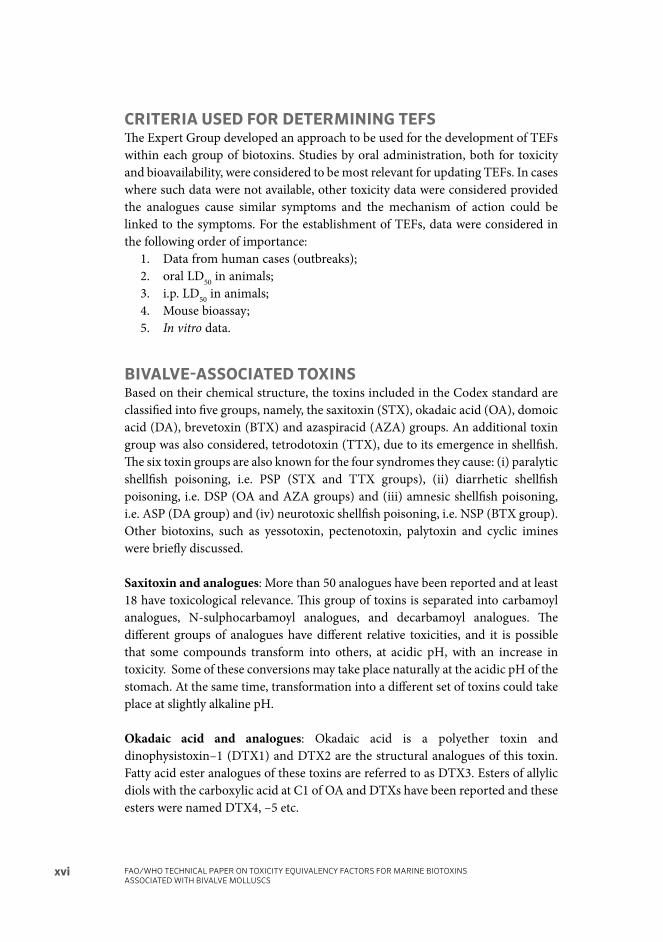
CRITERIA USED FOR DETERMINING TEFSTh e Expert Group developed an approach to be used for the development of TEFs within each group of biotoxins. Studies by oral administration, both for toxicity and bioavailability, were considered to be most relevant for updating TEFs. In cases where such data were not available, other toxicity data were considered provided the analogues cause similar symptoms and the mechanism of action could be linked to the symptoms. For the establishment of TEFs, data were considered in the following order of importance:
1. Data from human cases (outbreaks); 2. oral LD50 in animals; 3. i.p. LD50 in animals; 4. Mouse bioassay; 5. In vitro data.
BIVALVE-ASSOCIATED TOXINS Based on their chemical structure, the toxins included in the Codex standard are classifi ed into fi ve groups, namely, the saxitoxin (STX), okadaic acid (OA), domoic acid (DA), brevetoxin (BTX) and azaspiracid (AZA) groups. An additional toxin group was also considered, tetrodotoxin (TTX), due to its emergence in shellfi sh. Th e six toxin groups are also known for the four syndromes they cause: (i) paralytic shellfi sh poisoning, i.e. PSP (STX and TTX groups), (ii) diarrhetic shellfi sh poisoning, i.e. DSP (OA and AZA groups) and (iii) amnesic shellfi sh poisoning, i.e. ASP (DA group) and (iv) neurotoxic shellfi sh poisoning, i.e. NSP (BTX group). Other biotoxins, such as yessotoxin, pectenotoxin, palytoxin and cyclic imines were briefl y discussed.
Saxitoxin and analogues: More than 50 analogues have been reported and at least 18 have toxicological relevance. Th is group of toxins is separated into carbamoyl analogues, N-sulphocarbamoyl analogues, and decarbamoyl analogues. Th e diff erent groups of analogues have diff erent relative toxicities, and it is possible that some compounds transform into others, at acidic pH, with an increase in toxicity. Some of these conversions may take place naturally at the acidic pH of the stomach. At the same time, transformation into a diff erent set of toxins could take place at slightly alkaline pH.
Okadaic acid and analogues: Okadaic acid is a polyether toxin and dinophysistoxin–1 (DTX1) and DTX2 are the structural analogues of this toxin. Fatty acid ester analogues of these toxins are referred to as DTX3. Esters of allylic diols with the carboxylic acid at C1 of OA and DTXs have been reported and these esters were named DTX4, –5 etc.

EXECUTIVE SUMMARY xvii
Domoic acid and analogues: Domoic acid is a cyclic amino acid and although several isomers of DA, epi-domoic acid and isodomoic acids, have been reported, so far only DA and epi-DA have been shown to be of toxicological relevance. DA transforms into epi-DA through long-term storage and epimerisation is accelerated with warming.
Azaspiracid and analogues: Azaspiracids are polyether toxins with a unique spiral ring assembly, a cyclic amine, or aza group. Over 30 AZA analogues have been identifi ed in phytoplankton and shellfi sh. AZAs have been named by numbering in the chronological order of their detection or postulation. AZA1–10, 26, 33–34, 36–37 have been isolated and fully characterized. Shellfi sh are known to transform AZAs by two diff erent types of reactions: hydroxylation and carboxylation.
Brevetoxin group: Brevetoxins are cyclic polyether toxins grouped based on their backbone structure into group A type and group B type with the former having ten transfused ether rings and the latter eleven. Side chains account for a number of analogues. Brevetoxins are extensively metabolized in shellfi sh by oxidation, reduction, hydrolysis and conjugation, which may lead to formation of further analogues.
Tetrodotoxin and analogues: Tetrodotoxins (TTXs) are traditionally known as the main toxins found in puff er fi sh and a range of other animals. TTXs are also frequently found in other marine animals, including some species of gobies, octopuses, sea stars, crabs, bivalves and gastropods. Bacteria such as Vibrio spp., e.g. Vibrio alginolyticus, also produce TTXs, although the source of these toxins in marine animals has not yet been conclusively identifi ed. About 30 known analogues of TTX have been described. Profi les in shellfi sh included TTX, 4-epi-TTX, 4,9-anhydro-TTX and 5,6,11-tri-deoxy-TTX.
METHODS FOR DETECTION OF BIVALVE ASSOCIATED BIOTOXINSSaxitoxin and analogues: Th e mouse bioassay (MBA) for detection of paralytic shellfi sh toxins has been formally validated in an interlaboratory trial and a standardized AOAC method is available. Th e test does not provide analogue-specifi c data but gives a result in equivalents of STX in European Union and United States of America protocols or in equivalents of dc-STX in Japan.

FAO/WHO TECHNICAL PAPER ON TOXICITY EQUIVALENCY FACTORS FOR MARINE BIOTOXINS ASSOCIATED WITH BIVALVE MOLLUSCS
xviii
Th e receptor-binding assay (RBA) using tritiated STX is also an assay that gives a sum value for STX-equivalents. Th is test also has undergone formal interlaboratory validation and has reached a good level of acceptance in some countries. Analogue-specifi c methods for analysis of STXs are based on separation by liquid chromatography and fl uorescence or mass spectrometric detection, and a number of protocols have been validated. However, these methods require several analytical runs per sample in the case of complex natural toxin profi les.
Okadaic acid, azaspiracid and their analogues: OA and AZA and their analogues may all be detected by liquid chromatography coupled to tandem mass spectrometry (LC-MS/MS). Th is analogue-specifi c methodology has now replaced the lipophilic MBA in some countries, although for practical reasons the MBA continues to be used in many countries. It should be noted, however, that the MBA has never been formally validated for lipophilic marine biotoxins. For the OA-group of toxins, there is an enzyme-based assay, based on the inhibition of phosphoprotein-phosphatase 2a (PP2a). Th is assay has also been recently validated and is accepted in some countries. Th is assay will provide a sum of OA-equivalent toxicity present in a sample.
Domoic acid: Initial eff orts in method validation focused on using the extraction protocol for the PSP MBA, but this method has been superseded by the knowledge that DA is not very stable in strongly acidic conditions. Hence, methods using an extraction protocol based on aqueous methanol with HPLC-UV detection are now generally preferred and such methods have undergone collaborative trials for validation. As DA is a relatively simple compound with one major analogue and a single epimer, an ELISA has also been developed and a collaborative trial permitted interlaboratory validation and formal standardization as an AOAC method.
Brevetoxins: Th e MBA is widely used for detection of BTX and the Codex standard also specifi es the limit in mouse units. Chemical methods like LC-MS have been studied, but the challenge is that purifi ed standards are not available for many of the analogues. Other methods that are in various stages of validation include radioimmunoassay, ELISA, neuroblastoma assay and receptor binding assay. None of these detect individual analogues but indicate overall toxicity.

EXECUTIVE SUMMARY xix
TOXICITY EQUIVALENCY FACTORS (TEFS) FOR SPECIFIC BIOTOXINSSaxitoxin, okadaic acid, azaspiracid and analogues
Th e TEFs agreed by the Expert Group and the rationale for the decisions are indicated in the following table.
CompoundRecommended
TEFRationale
Saxotoxin and analogues
Saxitoxin 1.0
NeoSTX 2.0 Both oral studies support higher toxicity than STX. A
value of 2.0 is recommended and supported by some
Na channel in vitro results (range of relative potency
0.7–3.7).
GTX1 1.0 No new data1
GTX2 0.4 No new data
GTX3 0.6 No new data
GTX4 0.7 No new data
GTX5 0.1 Relative potency values from oral LD50
studies
suggest a lower TEF than from LD50
ip. 0.1 TEF also in
agreement with original Oshima TEF. As with NeoSTX,
a number of in vitro Na channel assays also support a
TEF of 0.1.
GTX6 0.05 New oral data show lower than 0.1.
C1 0.01 No new data (rounded up)
C2 0.1 No new data
C3 0.01 No new data
C4 0.1 No new data
dcSTX 0.5 From recent oral data (more weight on oral data, also
supported by ip toxicity data)
dcNeoSTX 0.2 From recent oral data (more weight on oral data, also
supported by in vitro data)
dcGTX2 0.2 No new data
dcGTX3 0.4 No new data
Okadaic acid and analogues
OA 1.0

FAO/WHO TECHNICAL PAPER ON TOXICITY EQUIVALENCY FACTORS FOR MARINE BIOTOXINS ASSOCIATED WITH BIVALVE MOLLUSCS
xx
CompoundRecommended
TEFRationale
DTX1 1.0 In the case of the OA group of toxins, several case
reports of human intoxication are available, and these
reports are analogue specific. As outlined in the recent
risk assessment by EFSA, a human poisoning event in
Japan with DTX1 as main analogue suggested a LOAEL
of 48 ug DTX1 per person. This dose is smilar to that
reported to induce toxic effects in poisoning events
in Sweden, Norway, UK and Portugal. Thus, it is not
surprising that the currently used TEF of 1.0 in some
countries appears protective for public health. Still, it
should be noted that multiple in vitro studies suggest
that the intrinsic potency of DTX1 could be higher than
that of OA. However, large uncertainty is associated
with these studies (a factor of approximately a 5-fold
difference between results depending on the cell line
used). Therefore, the recommended TEF of 1.0 for
DTX1 should be verified in future studies to corroborate
the observations in humans, and also through more
controlled oral toxicity studies in vivo (in animals).
DTX2 0.5 Consistent among the different assays; based on
acute oral, i.p. and in vitro toxicity in mice, DTX2 is
on average half as toxic as DTX1. This value is also
supported by the various in vitro data.
DTX3 No TEF is recommended since DTX3 esters per se
are non-toxic. They may however be hydrolysed after
ingestion to release the parent compound(s) (OA,
DTX1, DTX2) in the gastrointestinal tract and more
information on this possibility is required
Azaspiracid and analogues
AZA 1 1.0
AZA 2 0.7 Based on recent oral data (also consistent with recent
i.p. data)
AZA 3 0.5 Based on recent oral data (also consistent with recent
i.p. data)
AZA 4 Cannot determine TEF due to lack of data.
AZA 5 Cannot determine TEF due to lack of data.
AZA 6 0.7 No oral data; based on recent i.p. data.
1 In the case of saxitoxin analogues, for which no oral toxicity data were available, TEFs recommended are based on intra-
peritoneal toxicity data of Oshima (Appendix I).

EXECUTIVE SUMMARY xxi
Domoic acid
Domoic acid’s analogues, isodomoic acids D, E and F, are found in shellfi sh, but they have no toxicological signifi cance. DA transforms to epi-DA in storage or by ultraviolet light, and in general DA and epi-DA are considered as one toxin and expressed as DA. Hence, no TEFs are necessary for this group.
DATA GAPS AND FURTHER CONSIDERATIONS FOR RESEARCHTh e Expert Group identifi ed several data gaps and areas for further research, including:
• Lack of information on pharmacokinetics for all groups of marine biotoxins. • Limited information on oral toxicity of analogues for all groups of marine
biotoxins. • Need to establish TEFs for TTX analogues found in bivalves. • Need for comparative studies on the oral toxicity of TTX congeners.• Lack of information on chronic toxicity of DA, OA and AZA groups.
RECOMMENDATIONS FOR RISK MANAGERS• Th e Expert Group recommended that the proposed TEF values be reviewed
and re-evaluated in a 5 to 10-year time-frame, refl ecting new data that might be available. Th is process could be facilitated through the establishment of a database on relative potency studies and related data at FAO/WHO.
• In view of evolving geographical distribution of biotoxins, the Expert Group recommended that national toxin monitoring programmes be updated and expanded to include new and emerging toxins. Also, monitoring should occur during shellfi sh production.
• Th e Expert Group recommended that the analytical methods and monitoring strategies be strengthened and expanded to include the diff erent range of biotoxin analogues.
• Th e Expert Group recommended that if a method cannot separate some of the analogues, the highest TEF should be used to provide maximum consumer protection.
1CHAPTER 1 - THE NEED FOR AND CONCEPT OF TOXICITY EQUIVALENCY FACTORS
1The need for and concept of Toxicity Equivalency Factors
1.1 THE NEED FOR TOXICITY EQUIVALENCY FACTORSShellfi sh can be contaminated with toxic substances derived from marine microalgae. In order to protect consumers from poisoning by these toxins, regulatory authorities have defi ned limits as to the maximum amount of such toxins in shellfi sh intended for human consumption, and analyses must be conducted in order to ensure that such limits are not exceeded.
Several groups of shellfi sh toxins have been described, such as the paralytic, diarrhetic and amnesic toxins. Within each group, several structurally-related toxins may be present that contribute to the overall toxic potential.
In the past, testing has largely relied on the mouse bioassay (MBA). Th is involves injecting an extract of a sample of shellfi sh into the peritoneum of a group of mice, and recording the time that the mice take to die. Th e test gives an estimate of the total toxicity of the extract, i.e. the sum of the toxicities of the various congeners present in the sample. Th e MBA, however, has raised ethical concerns. It is widely accepted that the use of animals should be reduced, refi ned or replaced as far as is practicable. Furthermore, this route of administration is not relevant to the human situation, and the MBA is an assay, not a toxicological parameter. When comparisons have been made, it has been shown that there is no correlation between acute toxicity and the results obtained in the MBA.

FAO/WHO TECHNICAL PAPER ON TOXICITY EQUIVALENCY FACTORS FOR MARINE BIOTOXINS ASSOCIATED WITH BIVALVE MOLLUSCS
2
Over the last few years, it has proved possible to isolate pure samples of shellfi sh toxins for use as analytical standards. Th e development of new analytical techniques, particularly LC-MS, allows individual concentrations of toxin congeners in a shellfi sh extract to be determined with accuracy and precision.
For risk assessment and management, however, knowledge of the amount of toxin congeners in the shellfi sh is not suffi cient. Th ere is also the need to know the relative toxicity of each of the congeners, so that the total toxicity of the material in the extract can be estimated. Th is requires the determination of Toxicity Equivalency Factors (TEFs), defi ned as the toxicity ratio of a compound from a chemical group that shares the same mode of action of a reference compound in the same group (Van den Berg et al., 2006; Botana et al., 2010). Th e toxicity of the congener is expressed as a fraction of the toxicity of the reference compound in terms of potency, which is a pharmacological parameter that defi nes the amount of compound required for a certain eff ect.
1.2 TEF: USE OF THE CONCEPT In general, risk assessment of complex mixtures is done on the basis of individual compounds. However, there are many situations where humans are exposed to a combination of compounds that exhibit a similar mechanism of action, e.g. dioxin-like compounds and organophosphate pesticides. Based on this common mechanism of action, the toxicity equivalency concept has been developed during the last decades to support risk assessment of specifi c complex mixtures.
From a methodological point of view the total toxicity equivalent (TEQ) is defi ned by the sum of the concentrations of each compound multiplied by individual TEF values, which provides an estimate of the total toxicological or biological activity relative to a reference compound. Independent of the nature of the compounds used with a TEF concept, the major prerequisite for using it is based on additivity and similar mechanisms or modes of action.
At present, the TEF concept has been most thoroughly developed for the dioxin-like compounds. Since the early 1990s, the World Health Organization (WHO) regularly organized expert meetings to develop and harmonize TEFs for halogenated dioxins, polychlorinated dibenzodioxins (PCDDs), polybrominated dibenzo-p-dioxin (PBDDs) and dioxin-like compounds – polychlorinated dibenzofurans (PCDFs), polybrominated dibenzofurans (PBDFs) and polychlorinated biphenyls (PCBs) – at an international level, thereby giving recommendations to national regulatory authorities (Van den Berg et al., 1998, 2005, 2014).

CHAPTER 1 - THE NEED FOR AND CONCEPT OF TOXICITY EQUIVALENCY FACTORS 3
In the case of dioxin-like compounds, the TEFs have undergone changes over the years, based on emerging toxicological data and knowledge about the mechanism of action. Nevertheless, the criteria to include a compound in the TEF concept of dioxin-like compounds have remained the same and these specifi cally include:
• show a structural relationship to the PCDDs and PCDFs;• bind to the aryl hydrocarbon receptor (AhR);• elicit AhR-mediated biochemical and toxic responses; and• be persistent and accumulate in the food chain.
In 2010, the United States Environmental Protection Agency (US-EPA) added that “the Ah receptor mediates most if not all of the biologic and toxic eff ects of tetrachlorodibenzodioxin (TCDD) and the DLCs” (dioxin-like compounds) (Agency, 2010). In the European regulation for dioxin-like compounds (Commission Regulation (EC) No 1881/2006), TEF values range from 1 to 0.00003 (a factor of 33 333 between the lowest and the highest values). It should be noted that the TEF concept has not been applied to polycyclic aromatic hydrocarbons (PAHs) by the European Food Safety Authority (EFSA) for the sum of 4 congeners (EFSA, 2008d).
“Th e CONTAM Panel explored whether a toxicity equivalency factor (TEF) approach in the risk characterization of the PAH mixtures in food could be applied and concluded that the TEF approach is not scientifi cally valid because of the lack of data from oral carcinogenicity studies on individual PAHs, their diff erent modes of action and the evidence of poor predictivity of the carcinogenic potency of PAH mixtures based on the currently proposed TEF values”.
Th e principles for risk characterization of chemicals in food have been reviewed by FAO and WHO in Environmental Health Criteria, where chapter 7 specifi cally addresses the use of TEFs (FAO/WHO, 2009).
In addition, the assignment of WHO TEF values for dioxin-like compounds was done in either increments of 0.01, 0.05, 0.1, etc. (van den Berg et al., 1998) or a half order-of-magnitude increments on a logarithmic scale of 0.03, 0.1, 0.3, etc. (van den Berg et al., 2005). Although the most recent WHO re-evaluations of TEF values using the range of relative eff ect potencies (REPs) derived from individual studies initially, the fi nal decision on a TEF value for an individual isomer or congener was based on expert judgment, for which the type of study most relevant for human risk assessment was given more weight. Because, in time, new emerging toxicological data of dioxin-like compounds became available, regular changes in a TEF value were made. In view of this dynamic character of TEF values for dioxin-like compounds, it was decided to defi ne these as “interim” values for risk assessment purposes.

FAO/WHO TECHNICAL PAPER ON TOXICITY EQUIVALENCY FACTORS FOR MARINE BIOTOXINS ASSOCIATED WITH BIVALVE MOLLUSCS
4
1.3. DERIVING TEFSTh e most common potency parameter used is the median lethal dose (LD50), which is the dose that kills half the population of animals tested, and this is clearly the most appropriate parameter for some toxins, such as the paralytic shellfi sh toxins, that are known to cause death in humans. With other toxins, however, the LD50 may not be relevant. For example, no deaths have been reported aft er consumption of azaspiracids or the diarrhetic shellfi sh toxins. Th e eff ects seen in humans are gastrointestinal, involving nausea, vomiting, diarrhoea and stomach cramps. Although rodents are unable to vomit, they do suff er diarrhoea when dosed orally with these substances, and TEFs based on relative diarrhoeagenic activity could be considered.
In the past, it was diffi cult to obtain enough pure toxin congeners in order to conduct acute toxicity studies in animals. Furthermore, some of the early studies were carried out with toxins for which the concentration and stability was not properly assessed. Many of the toxicological results reported only the “minimum lethal dose (MLD)” or “lethality” of a compound, rather than the defi ned parameter of the LD50. Without information on the doses used to identify the MLD, such results cannot be compared with LD50 values. “Lethality” is an ambiguous term that could indicate an MLD or a dose that killed an unspecifi ed number of animals in a group, and is therefore useless for making comparisons.
Several approaches toward the determination of TEFs have been taken. However, because of the shortage of test material, early studies focussed on toxicity by intraperitoneal (i.p.) injection, which is inappropriate for the defi nition of TEFs, since the natural route of exposure is oral ingestion (Botana, 2012). At the same time, for biotoxins with similar bioavailability, i.p. administration can still provide useful toxicity comparison information. Materials dosed by i.p. injection are rapidly and extensively absorbed from the peritoneal cavity, whereas the gastrointestinal tract is designed to minimize absorption of many harmful substances. I.p. injection will therefore usually give an unrealistically high estimate of the toxicity of compounds that require absorption for expression of their harmful eff ects. At the same time, if a toxin were to be metabolized to a more toxic analogue in the gastrointestinal tract, then the i.p. route may give a low estimate (as may happen with neosaxitoxin, which is more toxic than saxitoxin). It should be noted that no correlation exists between i.p. LD50 values and those following oral administration (Table 1).

CHAPTER 1 - THE NEED FOR AND CONCEPT OF TOXICITY EQUIVALENCY FACTORS 5
TABLE 1. Comparison of LD50
values of toxins by i.p. injection versus gavage
Compound Ratio of toxicity by gavage to toxicity by i.p. injection
Palytoxin 708
GTX–1&4 110
Neosaxitoxin 79
Pinnatoxin E 49
Saxitoxin 43
Spirolide A 15
13-Desmethyl spirolide C 23
Gymnodimine 8
Pinnatoxin F 2
Sources: Munday, 2006; Munday et al., 2004; 2012a, 2012b, 2013; Selwood et al., 2010.
TEFs have also been estimated by comparing toxicity to cultured cells in vitro. Considerable eff ort has been made toward developing in vitro tests to replace in vivo acute toxicity evaluations, as undertaken by Th e Interagency Coordinating Committee on the Validation of Alternative Methods in the United States of America and the European Centre for the Validation of Alternative Methods. It has been concluded, however, that at the present time, no in vitro test method is suffi ciently accurate to replace animals for regulatory hazard classifi cation purposes (ICCVAM, 2006a, b). Th ere may, however, be an exception with marine toxins as the mechanism by which the toxins cause death is known, as discussed below for the paralytic shellfi sh poisons. Cell-based assays may, in the future, also be appropriate for other toxins when the target site and mode of action have been unambiguously identifi ed.
Since toxins are ingested by humans, toxicity by oral administration is the most relevant parameter for determining TEFs, but even here, care must be taken. Because of the semi-solid content of the rodent stomach, material administered by gavage can fl ow around the material and rapidly enter the absorptive area of the duodenum, rather than mixing with the food in the stomach as occurs with the liquid stomach contents of humans. Such mixing does occur when materials are dosed acutely to rodents by voluntary consumption. While training of animals is required to ensure that they eat small quantities of food containing the toxin within seconds, this technique is the most relevant to the human situation (Munday, 2014). It is only recently that suffi cient amounts of certifi ed material have become available for such studies to be conducted.

FAO/WHO TECHNICAL PAPER ON TOXICITY EQUIVALENCY FACTORS FOR MARINE BIOTOXINS ASSOCIATED WITH BIVALVE MOLLUSCS
6
At present, TEFs for shellfi sh toxins are based on acute eff ects. Very little information on the chronic toxicity of these substances is available. Acute toxicity studies provide information on the “Acute Reference Dose” (ARfD) of a compound for humans, defi ned as:
“An estimate of the amount of a substance in food and/or drinking water, normally expressed on a milligram per kilogram basis, that can be ingested in a period of 24 hours or less without appreciable health risk to the consumer.”
Repeated doses at low levels may also be toxic, and for risk assessment of certain marine toxins, particularly those that are slowly eliminated from the body and may therefore accumulate aft er repeated exposure, an estimate of the “Tolerable Daily Intake” (TDI), defi ned as “An estimate of the amount of a substance in food and/or drinking water, normally expressed on a milligram per kilogram body weight basis, that can be ingested daily over a lifetime without appreciable health risk to the consumer” would be valuable. Estimates of the TDI require studies in mice continuously fed the toxin over a period of weeks.
Although the TEF concept is rather simple, for marine toxins it is very complex to implement, for several reasons:
• It has been diffi cult to obtain the required quantities of the compound to do acute oral toxicity studies in animals. In the past, many of the studies were carried out with toxins or extracts for which the concentration and stability were not properly assessed, and this is a major source of variation in reported toxicological studies.
• Th e death of an animal does not necessarily show the real risk in humans of a compound that may be ingested in low doses or chronically. Many of the toxicological results only provide the concentration that causes the death of mice, and this value is oft en not as the LD50 but as a lethal dose (see above), so that variation in the way the experiments are conducted and in the way that the results are expressed makes it very diffi cult to compare data.
• Th e death of an animal is the end point of a mechanism of action over activated, and with lower doses the toxic eff ects are, in many cases, quite diff erent.
• Th e mechanism of action of a toxic compound may involve several targets, and those leading to the death of the animal may not be the targets responsible for the toxic symptoms observed in humans.
• If the compounds target several receptors, the sensitivity of each receptor can be diff erent, and this would cause a progressive appearance of symptoms as each of the diff erent targets are being activated.

CHAPTER 1 - THE NEED FOR AND CONCEPT OF TOXICITY EQUIVALENCY FACTORS 7
• In many cases, not all targets are known. For this reason, it is imperative to know the mechanism of action of the compound and the implications, at a systemic level, of the modifi cation of the target.
• Th e target of the compound may show very diff erent eff ects in diff erent systems or organs and, on many occasions, the physiology of the specifi c target in the organ is not well understood.
Given the complexity of the subject, the most appropriate TEF is the one that identifi es the potency of each congener of the same toxic group (analogues sharing the same mode of action) in the specifi c organ or system where the toxicity is reported, relative to a defi ned compound. In general, this is diffi cult to obtain. Finally, it is important to bear in mind that TEFs are needed only for those scenarios in which humans are exposed to a mixture of related compounds, i.e. with the same toxicological profi le but diff erent toxic potency, that can be detected together, in order to assess the total toxicity of the mixture.
Given the complexity of the derivation of a TEF for each desired analogue in a toxin group, and due to the scarcity of solid information at the time, the Working Group on Marine Biotoxins, which was part of the Contaminants Panel of the European Food Safety Authority (EFSA), defi ned TEFs for most of the marine toxin groups based on the i.p. acute toxicity of each analogue (EFSA, 2008c). Th ese TEFs are biased in some cases because they take into account neither the oral bioavailability nor the local eff ect of some toxin groups, but at least they provided a value to be used as a reference. However, TEF values should not be used unless there is data to prove that the compounds share the same mode of action and that this mode of action explains the symptoms observed in humans. Clearly, as more mechanistic information is obtained with each of the toxin groups, an update on the use of TEFs becomes appropriate.
It is now time to engage in such a refl ection at an international level to develop a common view. Th e question is: Is it possible at present to apply the TEF concept to marine biotoxins included in the Codex standard (CODEX STAN 292–2008) In the following sections, the criteria used for assigning TEFs, the values agreed during the meeting and the rationale have been explained.
1.3.1 Criteria for assigning TEFs
Th e Expert Group discussed the criteria for using diff erent data for assigning TEFs. Th ey conclude:
• Results of studies in which certifi ed reference materials were not used or where no quality control of the toxins was taken must be considered with caution. Some commercial supplies may provide quantifi cation errors that invalidate calculations of the lethal doses (Crespo et al., 2015).

FAO/WHO TECHNICAL PAPER ON TOXICITY EQUIVALENCY FACTORS FOR MARINE BIOTOXINS ASSOCIATED WITH BIVALVE MOLLUSCS
8
• Studies by oral administration are most relevant. • In order to permit evaluation by in vitro studies, it is essential to defi ne the
mechanism of action responsible for the toxic eff ect, or the diff erent targets responsible for each toxic eff ect.
• With some toxins, sub-chronic toxicity studies, involving dietary administration of the material to mice for 28 or 90 days, following Organisation for Economic Co-operation and Development (OECD) Guidelines (OECD 1998a, 1998b) are required in order to assess possible harmful eff ects aft er repeated exposure.
Based on the above criteria, a scheme was used to decide whether TEFs could be assigned during the review by the Expert Group (Figure 1).
FIGURE 1. Schema to decide whether TEF assignment was applicable in the
frame of the Expert Group

9CHAPTER 2 - CHEMISTRY AND DETECTION METHODS FOR BIOTOXINS
2Chemistry and detection methods for biotoxins
2.1 CHEMICAL NATURE AND MOLECULAR STRUCTURE OF ANALOGUES Shellfi sh toxins, a subgroup of marine toxins, are mainly produced by micro-organisms (bacteria, cyanobacteria and micro-algae). Based on their chemical structure, the toxins included in Codex Standard 292–2008 can be classifi ed into fi ve groups, namely, the saxitoxin (STX), okadaic acid (OA), domoic acid (DA), brevetoxin (BTX), and azaspiracid (AZA) groups. An additional toxin group will also be considered in Section 4.2.1, tetrodotoxin (TTX), due to its emergence in shellfi sh. Th e fi ve toxin groups are also known for the four syndromes they cause: (i) paralytic shellfi sh poisoning (PSP) (STX and TTX groups); (ii) diarrhetic shellfi sh poisoning (DSP) (AZA and OA groups); and (iii) amnesic shellfi sh poisoning (ASP) (DA group) and neurotoxic shellfi sh poisoning ie NSP (BTX). While TTXs have not been specifi cally mentioned in the Codex standard, they have the same mode of action as STXs (for which analogue-specifi c methods are under discussion) and belong to the group of paralytic shellfi sh toxins. Th ey also have been recently detected in bivalves (Turner et al., 2015b; Vlamis et al., 2015). Th is section briefl y describes the chemical nature of the molecules involved and their structures.
Th e toxins of these fi ve groups are molecules with a range from around 200 to 900 Da, and the structures can be classifi ed according to various chemical classes (Table 2).

FAO/WHO TECHNICAL PAPER ON TOXICITY EQUIVALENCY FACTORS FOR MARINE BIOTOXINS ASSOCIATED WITH BIVALVE MOLLUSCS
10
TABLE 2. Overview of characteristics of key toxins, including fi ve groups in Codex Standard 292–2008
Toxin Syndrome Chemical class Formula
Molar
weight
g/mol
UV [nm] Lipophilicity
Saxitoxin(1) PSP tetrahydro-purine
alkaloid
C10
H17
N7O
4299 n/a(2) hydrophilic
Okadaic acid(1) DSP polyether, spiro-keto
ring assembly
C44
H68
O13
804 n/a lipophilic
Domoic acid(1) ASP cyclic amino acid, 3
carboxy groups
C15
H21
NO6
311 242 hydrophilic
Brevetoxin(1) NSP Polyether with
contiguously fused
rings
C49
H70
O13
867 n/a lipophilic
Azaspiracid(1) DSP(4) polyether, second
amine, 3-spiro-ring
assembly
C47
H71
NO12
841 n/a lipophilic
Tetrodotoxin PSP(3) Guanidinum-
derivative of
penta-hydroxylated
2,4-dioxaadmantane
C11H
17N
3O
8319 n/a hydrophilic
Notes: (1) in Codex standard. (2) n/a = no specifi c absorption above 210 nm. (3) TTX is not classifi ed as PSP, but included here in this group based on
observed symptoms. (4) AZA diff ers chemically from Okadaic acid but produces the same symptom.
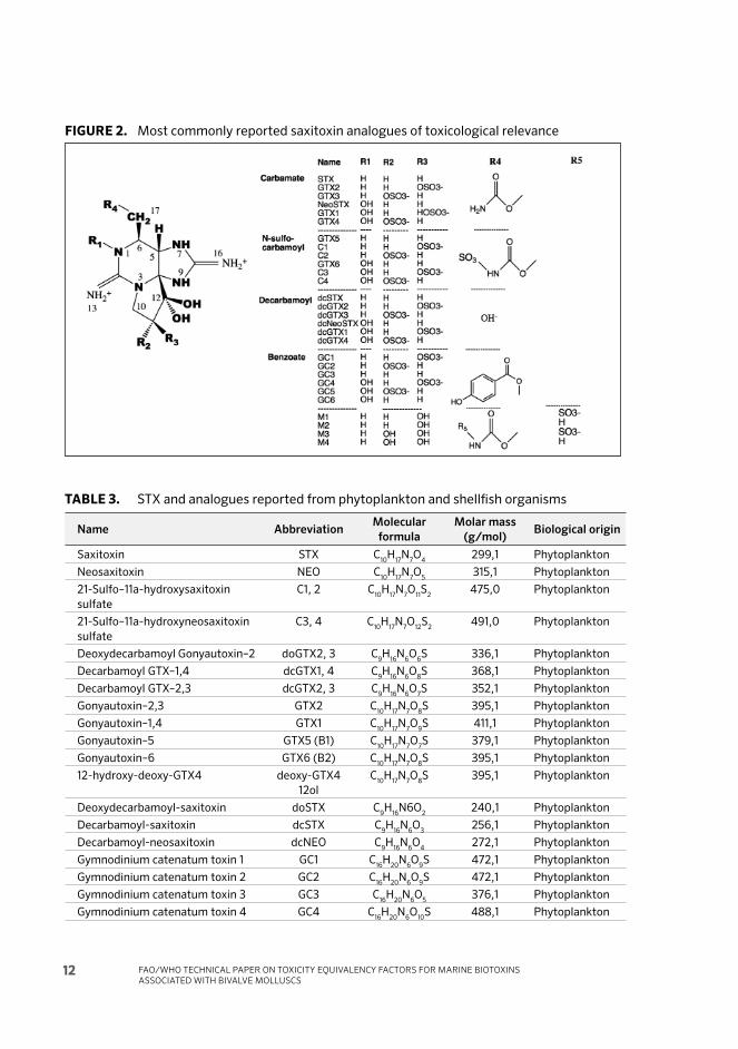
2.1.1 Saxitoxin and analogues
Saxitoxin (STX)-group toxins are mainly produced by dinofl agellates belonging to the genus Alexandrium, e.g. A. tamarensis, A. minutum (syn. A. excavata), A. catenella, A. fraterculus, A. fundyense and A. cohorticula. Also other dinofl agellates such as Pyrodinium bahamense and Gymnodinium catenatum have been identifi ed as sources of STX-group toxins, as well as cyanobacteria of the genera Anabaena, Cylindrospermopsis, Aphanizomenon, Planktothrix and Lyngbia (Wiese et al., 2010).
Th is group of toxin analogues has STX as the reference compound, and they share a common structure of tetrahydropurine. Th ey are soluble in water and thermostable at acidic pH; at alkaline pH they are quickly degraded (Kodama and Sato, 2008). More than 50 compounds have been reported (Table 3) (Dell’Aversano et al., 2008; Wiese et al., 2010) and at least 18 have toxicological relevance (Figure 2). Th is group of toxins is separated into carbamoyl analogues, (STX, neoSTX and gonyautoxins (GTX) 1 to 4), N-sulphocarbamoyl analogues (gonyautoxins 5 and 6, toxins C 1 to 4), and decarbamoyl analogues (dcSTX, dcNeoSTX and dcGTX 1 to 4). Th e diff erent groups of analogues have diff erent relative toxicities, and it is possible that some compounds transform into others, at acidic pH, with an increase in toxicity: GTX5, GTX6, C1, C2, C3, C4 will transform at acidic pH with heat into STX, NeoSTX, GTX2, GTX3, GTX1 and GTX4, respectively (Vale et al., 2008b). Some of these conversions may take place naturally at the acidic pH of the stomach

CHAPTER 2 - CHEMISTRY AND DETECTION METHODS FOR BIOTOXINS 11
(Harada, Oshima and Yasumoto, 1984). At the same time, transformation into a diff erent set of toxins at slightly alkaline pH (7.4–7.5) is also possible with heat: GTX5, GTX6, C1, C2, C3, C4 will transform into dcSTX, dcNeoSTX, dcGTX2, dcGTX3, dcGTX1 and dcGTX4, respectively. Th e toxicity of some of these toxins may vary, since they epimerise until they reach thermodynamic equilibrium and that depends on the storage conditions, i.e. GTX2, 3 reach an equilibrium ratio of 2.7; GTX1 4 of 3.4; C1 2 of 3.5; and dcGTX2, 3 of 4.4.
STX analogues do not exhibit a strong ultraviolet (UV) absorbance or fl uorescence. Th ey are typically stable to heat treatment up to 100°C. Diff erent acid and base treatments will lead to various transformations. In particular, all C11-epimeric pairs (e.g. GTX2 and 3 or GTX1 and 4) will interconvert and equilibrate to a constant ratio at high pH. Similarly, carbamoyl- and sulphocarbamoyl-derivatives will convert to decarbamoyl-analogues through cleavage of the carbamoyl-ester group at high pH (e.g. C1 to dc-GTX2, and C2 to dc-GTX3). Under acidic conditions, the carbamoylester is relatively stable but the sulphate ester will be cleaved to convert sulphocarbamoyl groups into carbamoyl groups (e.g. C1 to GTX2, and C2 to GTX3). Th ese transformations may only occur partially when shellfi sh tissues, human tissues or fl uids contaminated with STXs are exposed to these conditions, as biological tissues typically buff er the pH. Since conversion reactions can result in a several-fold increase in toxicity, a potential danger from these toxins was suggested (Hall and Reichardt, 1984). To examine this phenomenon experimentally, B1 (GTX5) was incubated at conditions simulating the human stomach and analysed by the mouse bioassay. Aft er 5 h incubation at 37°C, a two-fold increase of toxicity, corresponding to 9 percent conversion of toxin, was observed in the artifi cial gastric juice at pH 1.1 and no apparent increase of toxicity was observed in rat gastric juice at pH 2.2 (Harada, Oshima and Yasumoto, 1984).
Th e marine organisms most oft en aff ected are mussels, oysters and clams, but also puff er fi sh and marine snails (e.g. abalone) have been reported to accumulate dangerous concentrations (Pitcher et al., 2001; Harwood et al., 2014). Th e hydrophilic character of the compounds may partially explain the relatively rapid depuration of these toxins from mussels. Th is rapid depuration complicates the regulatory surveillance for these toxins, which is therefore most oft en complemented by observations of the algae responsible for in situ production.
FAO/WHO TECHNICAL PAPER ON TOXICITY EQUIVALENCY FACTORS FOR MARINE BIOTOXINS ASSOCIATED WITH BIVALVE MOLLUSCS
12
FIGURE 2. Most commonly reported saxitoxin analogues of toxicological relevance
TABLE 3. STX and analogues reported from phytoplankton and shellfi sh organisms
Name AbbreviationMolecular
formula
Molar mass
(g/mol)Biological origin
Saxitoxin STX C10
H17
N7O
4299,1 Phytoplankton
Neosaxitoxin NEO C10
H17
N7O
5315,1 Phytoplankton
21-Sulfo–11a-hydroxysaxitoxin
sulfate
C1, 2 C10
H17
N7O
11S
2475,0 Phytoplankton
21-Sulfo–11a-hydroxyneosaxitoxin
sulfate
C3, 4 C10
H17
N7O
12S
2491,0 Phytoplankton
Deoxydecarbamoyl Gonyautoxin–2 doGTX2, 3 C9H
16N
6O
6S 336,1 Phytoplankton
Decarbamoyl GTX–1,4 dcGTX1, 4 C9H
16N
6O
8S 368,1 Phytoplankton
Decarbamoyl GTX–2,3 dcGTX2, 3 C9H
16N
6O
7S 352,1 Phytoplankton
Gonyautoxin–2,3 GTX2 C10
H17
N7O
8S 395,1 Phytoplankton
Gonyautoxin–1,4 GTX1 C10
H17
N7O
9S 411,1 Phytoplankton
Gonyautoxin–5 GTX5 (B1) C10
H17
N7O
7S 379,1 Phytoplankton
Gonyautoxin–6 GTX6 (B2) C10
H17
N7O
8S 395,1 Phytoplankton
12-hydroxy-deoxy-GTX4 deoxy-GTX4
12ol
C10
H17
N7O
8S 395,1 Phytoplankton
Deoxydecarbamoyl-saxitoxin doSTX C9H
16N6O
2240,1 Phytoplankton
Decarbamoyl-saxitoxin dcSTX C9H
16N
6O
3256,1 Phytoplankton
Decarbamoyl-neosaxitoxin dcNEO C9H
16N
6O
4272,1 Phytoplankton
Gymnodinium catenatum toxin 1 GC1 C16
H20
N6O
9S 472,1 Phytoplankton
Gymnodinium catenatum toxin 2 GC2 C16
H20
N6O
9S 472,1 Phytoplankton
Gymnodinium catenatum toxin 3 GC3 C16
H20
N6O
5376,1 Phytoplankton
Gymnodinium catenatum toxin 4 GC4 C16
H20
N6O
10S 488,1 Phytoplankton

CHAPTER 2 - CHEMISTRY AND DETECTION METHODS FOR BIOTOXINS 13
2.1.2 Okadaic acid and analogues
Okadaic acid (OA) was originally isolated from the sponge Halichondria okadaii (Tachibana et al., 1981) but was later identifi ed as a shellfi sh contaminant following a series of poisoning events in 1976 (Yasumoto, Oshima and Yamaguchi, 1978) (Figure 3). Subsequently, it was clearly demonstrated that diarrhetic shellfi sh poisoning was associated with blooms of Dinophysis fortii, a dinofl agellate from which OA and an analogue of OA, dinophysistoxin–1 (DTX1) were isolated (Yasumoto et al., 1980). Th e same class of compounds was promptly discovered to
Name AbbreviationMolecular
formula
Molar mass
(g/mol)Biological origin
Gymnodinium catenatum toxin 5 GC5 C16H20
N6O
10S 488,1 Phytoplankton
Gymnodinium catenatum toxin 6 GC6 C16
H20
N6O
6393,1 Phytoplankton
hydroxy-GC1 GC1a C10
H17
N7O
8S 488,1 Phytoplankton
hydroxy-GC2 GC2a C16
H20
N6O
10S 488,1 Phytoplankton
hydroxy-GC3 GC3a C16
H20
N6O
10S 392,1 Phytoplankton
hydroxy-GC4 GC4a C16
H20
N6O
6504,1 Phytoplankton
hydroxy-GC5 GC5a C16
H20
N6O
11S 504,1 Phytoplankton
hydroxy-GC6 GC6a C16
H20
N6O
11S 409,1 Phytoplankton
sulfo-GC1 GC1b C16
H20
N6O
11S
2536,1 Phytoplankton
sulfo-GC2 GC2b C16
H20
N6O
11S
2536,1 Phytoplankton
sulfo-GC3 GC3b C16
H20
N6O
7S 440,1 Phytoplankton
sulfo-GC4 GC4b C16
H20
N6O
12S
2552,1 Phytoplankton
sulfo-GC5 GC5b C16
H20
N6O
12S
2552,1 Phytoplankton
sulfo-GC6 GC6b C16
H20
N6O
8S 457,1 Phytoplankton
Lyngbia wollei toxin 1 LWTX1 C11H
18N
6O
7S 378,1 Phytoplankton
Lyngbia wollei toxin 2 LWTX2 C11H
18N
6O
8S 394,1 Phytoplankton
Lyngbia wollei toxin 3 LWTX3 C11H
18N
6O
8S 394,1 Phytoplankton
Lyngbia wollei toxin 4 LWTX4 C9H
16N
6O
2240,1 Phytoplankton
Lyngbia wollei toxin 5 LWTX5 C11H
18N
6O
4298,1 Phytoplankton
Lyngbia wollei toxin 6 LWTX6 C11H
18N
6O
3282,1 Phytoplankton
shellfish metabolite 1 M1α C10
H17
N7O
8S 395,1 Shellfish
Shellfish metabolite 1 M1β C10
H17
N7O
8S 395,1 Shellfish
Shellfish metabolite 2 M2α C10
H17
N7O
5315,1 Shellfish
Shellfish metabolite 2 M2β C10
H17
N7O
5315,1 Shellfish
Shellfish metabolite 3 M3 C10
H17
N7O
9S 411,1 Shellfish
Shellfish metabolite 4 M4 C10
H17
N7O
6331,1 Shellfish
Shellfish metabolite 5 M5 C10
H17
N7O
8S 395,1 Shellfish
Shellfish metabolite 6 M6 C10
H17
N7O
5315,1 Shellfish
Shellfish metabolite 8 M8 C10
H17
N7O
6331,1 Shellfish
Shellfish metabolite 10 M10 C10
H17
N7O
7S 347,1 Shellfish
11-saxitoxinethanoic acid SEA C12
H16
N7O
6431,1 Shellfish
Source: Adapted from Wiese et al., 2010.

FAO/WHO TECHNICAL PAPER ON TOXICITY EQUIVALENCY FACTORS FOR MARINE BIOTOXINS ASSOCIATED WITH BIVALVE MOLLUSCS
14
be the causative agents of diarrhetic shellfi sh poisoning in Europe (Kumagai et al., 1986). Dinophysistoxin–2 (DTX2) was discovered to be the third main analogue Dinophysistoxin–2 (DTX2) was discovered to be the third main analogue (Hu et al., 1992b), explaining diarrhetic activity found in Irish mussels. OA and DTXs are produced by a variety of diff erent dinofl agellates from the Dinophysis and Prorocentrum genera, including D. acuta and D. acuminata, as well as P. lima and P. belizeanum. Although the toxins of the OA group have been mainly reported from Japan and Europe, it has recently been shown that Dinophysis in the Gulf of Mexico may also produce DSP under appropriate environmental conditions (Swanson et al., 2010). Also Chinese and South American Dinophysis strains have been shown to produce okadaic acid (Fux et al., 2011; Mafra, Tavares and Schramm, 2014; Li et al., 2015) and shellfi sh from all over the world have been shown to be contaminated with these toxins (Zhao et al., 1993; Suzuki et al., 2004; Villar-Gonzalez et al., 2007; Li et al., 2012). Th erefore, a global distribution of these toxins is now widely accepted.
Chemically, OA is one of the many polyether toxins among phycotoxins. Its structure is characterized by a carboxylic acid group and three spiro-keto ring assemblies, one of which connects a fi ve with a six-membered ring (Figure 3). OA, DTX1 and DTX2 withstand a wide pH range from mildly acidic to strongly basic, e.g. no degradation is found for up to 40 minutes at 76°C in 0.3 molar methanolic NaOH solution. Treatment with strong mineral acids, e.g. HCl, leads to rapid degradation: OA and DTX1 are completely destroyed within 20 min at 76°C in 0.3 molar methanolic HCl, even in the presence of shellfi sh matrix in the extract (Yasumoto et al., 1985). However, without the addition of acid, the compounds are stable to heat. Also, recent work on stomach simulation experiments in the laboratory suggests that the food itself has a buff ering capacity on the acid and the toxins may not be destroyed signifi cantly in the gastric juice. In normal cooking procedures the toxins are not destroyed, although the coagulation of proteins in shellfi sh tissues may lead to redistribution within the organs of shellfi sh and some toxins may be released into the cooking fl uids (McCarron, Kilcoyne and Hess, 2008; Blanco et al., 2015).
Diff erent types of esters of OA and DTXs have been reported. Initially, the palmitoyl ester of DTX1 was isolated and named DTX3 (Yasumoto et al., 1985) with other ester analogues shown to be present; subsequently, all fatty acid esters of variable chain length of OA, DTX1 and DTX2 have been referred to as DTX3 (in a simplifying fashion). In micro-algae (so far mainly P. lima and P. belizeanum), esters of allylic diols with the carboxylic acid at C1 of OA and DTXs have been reported (Yasumoto et al., 1989; Hu et al., 1992a, 1995a); these esters were named DTX4, DTX5, etc. When the algae enter shellfi sh through natural fi lter-feeding, it

CHAPTER 2 - CHEMISTRY AND DETECTION METHODS FOR BIOTOXINS 15
is believed that these esters are rapidly degraded (Vale, 2007). Th e shellfi sh then further metabolize OA and its analogues to form esters of OA and DTXs with fatty acids (at the C7-OH group); these esters were initially identifi ed for DTX1 as shellfi sh derivatives (Yasumoto et al., 1985) and their toxicity has been described to be similar to the parent compounds, although the onset appears later in the i.p. mouse model (Yanagi et al., 1989). A further fatty acid ester of DTX1 at the C27-OH group has been reported in a sponge (Britton et al., 2003), and most recently, Torgersen et al. (2008b) also reported mixed esters of diols (at the C1 carboxyl-end) and fatty acids (at the C7-OH position) in shellfi sh, suggesting that partial degradation and simultaneous metabolism may co-occur during digestion of algae by shellfi sh.
Th e multitude of compounds potentially present in shellfi sh (free toxins, diol esters and their derivatives, fatty acids and mixtures of diol- and fatty acid esters) complicates the determination of the complete toxin content in shellfi sh samples. Th is complexity has added to the diffi culties in estimating the potency of these toxins and evaluation of the risk they present. Th e ester-bond has not shown any degradation in long-term stability studies, although fatty acids have been reported to oxidise easily if they contain double bonds. All of the esters discussed above (either at the C1-carboxy or at the C7-OH) group are quantitatively cleaved through treatment with strong base, e.g. 0.3 molar methanolic NaOH at 76°C for 10 to 40 minutes. Th is characteristic, in combination with the stability of the parent compounds (OA, DTX1 and DTX2) to base treatment, has been extensively used to quantify the equivalent of the parent compound present in any given shellfi sh sample (Lee et al., 1989).
FIGURE 3. Okadaic acid and analogues dinophysistoxins–1 and –2

FAO/WHO TECHNICAL PAPER ON TOXICITY EQUIVALENCY FACTORS FOR MARINE BIOTOXINS ASSOCIATED WITH BIVALVE MOLLUSCS
16
2 .1.3 Domoic acid and analogues
Domoic acid (DA) is a cyclic amino acid (311 Da) with three carboxylic acid groups which are responsible for its water solubility and its high polarity (Quilliam et al., 1989d) (Figure 4). DA has been known for a long time to be the active constituent of the marine red algae of the genus Chondria and this has also been recently confi rmed by culture experiments of Chondria armata (Daigo, 1959; Jiang et al., 2014). Th e production of DA in the genus Pseudo-nitzschia is widely known since the 1987 incident of ASP in Canada (Quilliam, and Wright, 1989a). Its widespread occurrence throughout the genus Pseudo-nitzschia has recently been reviewed (Trainer et al., 2012). Since the earlier studies, DA has now also been detected in other diatom species, including Nitzschia navis-varingica (Romero et al., 2011) and Nitzschia bizertensis (Smida et al., 2014).
Structurally, DA is very similar to another known toxin, kainic acid. For the three carboxylic acids the acid constant values (pKa values) are 2.10–4.97 and for the cyclic amino group, 9.82 (Piñeiro et al., 1999), and hence DA can exist in diff erent charged states depending on pH (Jeff ery et al., 2004). Although several isomers of DA epi-domoic acid (epi-DA) (domoic acid C5’-diastereomer) and isodomoic acids A, B, C, D, E, F, G and H (iso-DA A–H)) have been reported (Maeda et al., 1986, 1987; Wright et al., 1990a; Zaman et al., 1997; Holland et al., 2005), so far only DA and epi-DA have been shown to be of toxicological relevance (Ramsdell, 2007). DA transforms into epi-DA through long-term storage (Quilliam et al., 1989b, Quilliam, Xie and Hardstaff .1995) and degrades and transforms to epi-DA and isodomoic acids through exposure to ultra violet light (Wright et al., 1990a; Djaoued, Balaji and Priya, 2007). Epimerization is also accelerated with warming (Quilliam, 2003). It has been suggested that DA may be a biosynthetic product, which is subsequently converted to isodomoic acids in suitable environmental conditions (Wright et al., 1990b).
Due to the conjugated double bond in the aliphatic side chain, DA absorbs ultra violet (UV) light and is light-sensitive. Th e conjugated double bond is also the cause of radical-mediated oxidative metabolism. DA does not degrade at ambient temperature or when it is exposed to light in sterile saline solution (Johannessen, 2000), but in acidic conditions DA has been shown to decompose. At pH 3 a loss of 50 percent of DA was observed in one week (Quilliam et al., 1989b). As a contaminant in shellfi sh tissues, DA is heat stable and cooking does not typically destroy the toxin. Its stability under various conditions has been studied, and storage of raw or autoclaved tissues only resulted in approximately 50 percent degradation of the toxin aft er 5 months (McCarron, Burrell and Hess, 2007b). DA rapidly degrades under the infl uence of gamma irradiation, either in solution or in shellfi sh tissue, but can be stabilized in shellfi sh tissue by freeze-drying the tissue (McCarron, Emteborg and Hess, 2007a; McCarron et al., 2007c).

CHAPTER 2 - CHEMISTRY AND DETECTION METHODS FOR BIOTOXINS 17
FIGURE 4. Domoic acid and its isomers
Nota bene: isomers A – C do not have conjugated double bonds and are not easily detected using HPLC-UV.

FAO/WHO TECHNICAL PAPER ON TOXICITY EQUIVALENCY FACTORS FOR MARINE BIOTOXINS ASSOCIATED WITH BIVALVE MOLLUSCS
18
2.1.4 Azaspiracids and analogues
Azaspiracids (AZAs) are marine algal toxins produced by the dinofl agellate genera Azadinium (Krock et al., 2009; Tillmann et al., 2009) and Amphidoma (Tillmann et al., 2012, 2014). Th is class of toxins was fi rst identifi ed in the 1990s following an outbreak of human illness in the Netherlands associated with consumption of contaminated mussels from Killary Harbour, Ireland (McMahon et al., 1996). Although the symptoms were typical of DSP toxins, i.e. okadaic acid (OA) and dinophysistoxins (DTX), the levels of DSP toxins in these mussels were well below the regulatory level. Subsequently, it was established that the shellfi sh were contaminated with a novel marine toxin, originally named “Killary-toxin” or KT–3 (Satake et al., 1998a). Shortly thereaft er, the toxin was renamed as azaspiracid (AZA) to more appropriately refl ect its chemical structure: a cyclic amine, or aza group, with a tri-spiro- assembly and carboxylic acid group (Satake et al., 1998a, b).
To date, over 30 AZA analogues have been identifi ed in phytoplankton and shellfi sh (Satake et al., 1998b; Ofuji et al., 1999, 2001; Lehane et al., 2002; James et al., 2003a; Rehmann, Hess and Quilliam, 2008; McCarron et al., 2009; Jauff rais et al., 2012a; Hess et al., 2014).
Over the last 15 years, AZAs have been reported in shellfi sh from many coastal regions of Western Europe (James et al., 2001, 2002; Braña Magdalena et al., 2003; Furey et al., 2003; Hess et al., 2007b; Amzil et al., 2008; Twiner et al., 2008b), Northern Africa (Taleb et al., 2006; Elgarch et al., 2008), South America (Alvarez et al., 2010) and North America (M. Quilliam, pers. comm.; A. Robertson, pers. comm.).
In addition, AZAs have been found in Japanese sponges (Ueoka et al., 2009) and Scandinavian crabs (Torgersen et al., 2008a). Not surprisingly, the global distribution of AZAs appears to correspond to the apparent wide spread occurrence of Azadinium (Tillmann et al., 2010, 2011, 2014; Akselman and Negri, 2012). Empirical evidence is now available that unambiguously demonstrates the accumulation of AZAs in shellfi sh via direct feeding on AZA-producing A. spinosum (Salas et al., 2011; Jauff rais et al., 2012a).
Whereas extensive study of this toxin class has been historically constrained by limited availability of purifi ed material, these restraints are now less of an impediment due to advances in isolation and purifi cation of AZAs from naturally contaminated shellfi sh (Kilcoyne et al., 2012) and the identifi cation of the toxigenic organism A. spinosum coupled with its mass culture in bioreactors (Jauff rais et al., 2012c). As such, certifi ed reference materials of naturally produced AZA1–3 are now commercially available (Perez et al., 2010). Although not yet realized for commercial purposes, limits on toxin supply may be further mitigated by advances

CHAPTER 2 - CHEMISTRY AND DETECTION METHODS FOR BIOTOXINS 19
in the organic total synthesis of AZA1 (Nicolaou et al., 2004a, 2004b) and AZA3 (C. Forsyth, pers. comm.). Accessibility to purifi ed AZAs has led to rapid progress with respect to understanding AZA toxicology over the last few years.
AZAs have sporadically been detected in plankton (James et al., 2003b; Krock et al., 2008) and sea water (Rundberget et al., 2007; Fux, Buré and Hess, 2009), but no progenitor of these toxins could be assigned until 2007 (Krock et al., 2009), when a small dinofl agellate, later named Azadinium spinosum (Krock et al., 2009), was unambiguously identifi ed as an AZA1 and –2 producing organism (ca. 20 and 7 fg cell-1, respectively). Later, additional AZAs with molecular masses of 715 Da (ca. 7 fg cell-1) and 816 Da (AZA33 and –34) have been found in environmental samples and cultures of A. spinosum (Kilcoyne et al., 2014b) (Table 4).
TABLE 4. Structural variants of AZAs, their protonated masses, fragment type
and origin
CompoundProtonated mass
[M+H]+ Type Origin Status
AZA1 842.5 362 A. spinosum phycotoxin
37-epi-AZA1 842.5 362 A. spinosum artefact
AZA2 856.5 362 A. spinosum phycotoxin
AZA3 828.5 362 shellfish metabolite
AZA4 844.5 362 shellfish metabolite
AZA5 844.5 362 shellfish metabolite
AZA6 842.5 362 shellfish metabolite
AZA7 858.5 362 shellfish metabolite
AZA8 858.5 362 shellfish metabolite
AZA9 858.5 362 shellfish metabolite
AZA10 858.5 362 shellfish metabolite
AZA11 872.5 362 shellfish metabolite
AZA12 872.5 362 shellfish metabolite
AZA13 860.5 362 shellfish metabolite
AZA14 874.5 362 shellfish metabolite
AZA15 874.5 362 shellfish metabolite
AZA16 888.5 362 shellfish metabolite
AZA17 872.5 362 shellfish metabolite
AZA19 886.5 362 shellfish metabolite
AZA21 888.5 362 shellfish metabolite
AZA23 902.5 362 shellfish metabolite
AZA26 824.5 362 shellfish metabolite
AZA28 838.5 362 shellfish metabolite
AZA29 842.5 362 shellfish artefact
AZA30 856.5 362 A. spinosum artefact
AZA32 870.5 362 A. spinosum artefact
AZA33 716.5 362 A. spinosum phycotoxin

FAO/WHO TECHNICAL PAPER ON TOXICITY EQUIVALENCY FACTORS FOR MARINE BIOTOXINS ASSOCIATED WITH BIVALVE MOLLUSCS
20
CompoundProtonated mass
[M+H]+ Type Origin Status
AZA34 816.5 362 A. spinosum phycotoxin
AZA35 830.5 362 A. spinosum phycotoxin
AZA36 846.5 348 A. poporum phycotoxin
AZA37 858.5 348 A. poporum phycotoxin
AZA38 830.5 348 A. languida phycotoxin
AZA39 816.5 348 A. languida phycotoxin
AZA40 842.5 348 A. poporum phycotoxin
AZA41 854.5 360 A. poporum phycotoxin
Notes: AZAs highlighted in grey have been isolated and fully characterized (LC-MS and NMR).
Recently, two strains of Azadinium poporum, a species previously reported to be non-toxigenic (Tillmann et al., 2011; Potvin et al., 2012), were proven to be the producers of two previously unknown AZAs (Krock et al., 2012). AZA37 from a North Sea isolate of A. poporum (Tillmann et al., 2011) with a molecular mass of 845 Da (ca. 10 fg cell-1) was determined as 39-desmethyl–7,8-dihydro-3-hydroxy-AZA1 by nuclear magnetic resonance (NMR) spectroscopy (Table 4) (Krock et al., 2015). Th e other strain of A. poporum, from Shiwha Bay, Republic of Korea (Potvin et al., 2012) produced AZA36 with a molecular mass of 857 Da (ca. 2 fg cell-1), which was determined as 39-desmethyl-3-hydroxy-AZA2 (Table 4). Both A. poporum-derived AZAs have a 3-hydroxy substitution and a 39-desmethyl moiety in common.
Whereas the 3-hydroxy function is also found in shellfi sh metabolites of AZA1 and –2, e.g. AZA4 and –9 (Kilcoyne et al., 2015), the 39-desmethyl moiety is unique to a new class of dinofl agellate AZAs. Th is new class of 39-desmethyl AZAs is easily recognized in tandem mass spectrometry by a characteristic m/z 348 fragment, whereas all other AZAs have a m/z 362 fragment. Two additional AZAs with m/z 348 fragments and molecular masses of 815 Da and 829 Da (ca. 11 and 6 fg cell-1 respectively) 18 AZAs were also identifi ed in a strain of Amphidoma languida (Tillmann et al., 2012).
AZAs were also detected in isolates of A. poporum from Chinese coastal waters (Gu et al., 2013). Whereas one strain did not produce any AZAs, three other strains produced exclusively AZA2 at cell quotas ranging from 1.8 to 23 fg cell-1. In addition, new AZAs with the m/z 348 fragment were detected in a fi ft h strain, which also produced AZA36 (1.4 fg cell-1). In contrast to the Korean strains, this Chinese strain produced AZAs (with the m/z 348 fragment) with molecular masses of 919 and 927 Da (ca. 0.02 and 0.14 fg cell-1, respectively). A sixth strain of A. poporum from China produced an AZA with a molecular mass of 871 Da (0.9 to 1.9 fg cell-1) that was tentatively identifi ed as AZA11 by comparison of

CHAPTER 2 - CHEMISTRY AND DETECTION METHODS FOR BIOTOXINS 21
retention times and Collusion induced dissociation (CID) spectra (Gu et al., 2013). Whereas 3-hydroxylated AZAs, such as AZA36 and AZA37, seem to be biosynthesized by several strains of A. poporum, AZA11 may be the fi rst case of an AZA being independently produced by dinofl agellate biosynthesis as well as through shellfi sh metabolic activity.
It is likely that several more analogues of this toxin group will be reported soon, as there are not yet many strains or species isolated from most regions. For instance, Azadinium dexteroporum, a new species recently isolated from the Mediterranean Sea (Italy) has also been reported to produce some novel analogues which have not yet been fully characterized (Percopo et al., 2013).
As the exact nomenclature of AZAs, according to the rules of the International Union of Pure and Applied Chemistry (IUPAC), are long and complicated, AZAs have been named by numbering in the chronological order of their detection or postulation. All AZAs up to AZA23 were originally identifi ed in or postulated from shellfi sh, however, AZA1 and –2 are of dinofl agellate origin, whereas AZA3 to AZA23 have not been detected in planktonic samples and have been shown to be shellfi sh metabolites, with the exception of AZA11 (Rehmann, Hess and Quilliam, 2008; Gu et al., 2013).
Shellfi sh are known to transform AZAs by two diff erent types of reactions: (1) hydroxylation at C3 and C23, and (2) carboxylation and subsequent decarboxylation (James et al., 2003a; Rehmann, Hess and Quilliam, 2008; McCarron et al., 2009; Kittler et al., 2010; O’Driscoll et al., 2011; Jauff rais et al., 2012b). Recent investigations with feeding experiments (Salas et al., 2011; Jauff rais et al., 2012a) revealed that blue mussels (M. edulis) metabolize AZAs quickly (Figure 5). AZA17 and –19 were the most abundant metabolites of AZA1 and –2, respectively, suggesting that carboxylation of the methyl group at C22 is a preferred metabolic pathway (Jauff rais et al., 2012b). Hydroxylation and decarboxylation seem to be secondary degradation routes (McCarron et al., 2009).

FAO/WHO TECHNICAL PAPER ON TOXICITY EQUIVALENCY FACTORS FOR MARINE BIOTOXINS ASSOCIATED WITH BIVALVE MOLLUSCS
22
FIGURE 5. Conversion of algal metabolites into shellfi sh metabolites
Hydroxylations of AZAs by shellfi sh metabolism occur at C3 on the carboxylic acid side chain to form 3-hydroxy-AZAs (e.g. AZA7, –11) as well as at C23 at the E-ring of the molecule, resulting in 23-hydroxy-AZAs (AZA8, –12) (Table 4). Furthermore, the methyl group at C22 can be oxidized to 22-carboxy-AZAs (AZA17, –19), which are subsequently decarboxylated to form the 22-desmethyl-AZAs (AZA3, –6) (McCarron et al., 2009; O’Driscoll et al., 2011). AZA 22-decarboxylation may be a shellfi sh metabolic activity, but this reaction occurs rapidly during heating of shellfi sh meat and slowly in extracts stored at ambient temperature (McCarron et al., 2009). In addition, combinations of these processes are possible, to produce many of the remaining AZAs (AZA4, –5, –9, –10, –13, –14, –15, –16, –21 and –23). Some of the other AZAs originally detected without structural elucidation (AZA25, –29, –30 and –32) 23 AZAs were later identifi ed as extraction artefacts (AZA29, –30 and –32) (Jauff rais et al., 2012a). In contrast to shellfi sh metabolism, phase I metabolites of AZA1 using rat liver microsomes (S9-mix) included an oxidation of the F-ring of the molecule, which is not observed in shellfi sh metabolites. Glucuronides were found as the only phase II metabolites of AZA1, and via precursor ion experiments it could be proven that glucuronic acid is bound to AZA1 at C1 via an ester linkage (Kittler et al., 2010). One study reported on binding of AZAs to proteins (Nzoughet et al., 2008) found that AZAs in mussel hepatopancreas bind to as yet unidentifi ed proteins with molecular masses of 21.8 and 45.3 kDa.
2.1.5 Tetrodotoxin and analogues
Tetrodotoxins (TTXs) are traditionally known as the main toxins found in
Note: Currently only AZA1, AZA2 and AZA3 are regulated in some legislative frameworks. AZA17, AZA19 and AZA6 are also
relevant for assessing public safety of a shellfi sh sample.

CHAPTER 2 - CHEMISTRY AND DETECTION METHODS FOR BIOTOXINS 23
puff er fi sh, and pure TTX was fi rst isolated as a crystalline prism from puff er fi sh by Yokoo (1950). TTX is also frequently found in other marine animals, including some species of gobies, octopuses, sea stars, crabs and gastropods (Hashimoto, Noguchi and Watabe, 1990; McNabb et al., 2010). Recent reports also confi rmed tetrodotoxins in European shellfi sh in both the English Channel and the Mediterranean Sea (Turner et al., 2015a; Vlamis et al., 2015). TTXs are also produced by bacteria that reside within blue-ringed octopuses (Hwang et al., 1989b). Th e most common bacteria associated with TTX production are Vibrio spp., with Vibrio alginolyticus being the most common species (Hwang et al., 1989a; McNabb et al., 2010). although the source of these toxins in marine animals has not yet been conclusively identifi ed. TTX is of low molecular weight and positively charged in neutral solutions. Th e toxin is odourless and heat stable but unstable at pH levels above 8.5 and below 3.0. A recent review gives an overview of the chemistry and structures (Figure 6) of the 30 known analogues of TTX (Bane et al., 2014).
FIGURE 6. Most common TXX analogues in aquatic organisms
R1
R2
R3
R4
R5
TTX H OH OH CH2OH OH
4-epi-TTX OH H OH CH2OH OH
6-epi-TTX H OH CH2OH OH OH
11-deoxy-TTX H OH OH CH3
OH
6,11-dideoxy-TTX H OH H CH3
OH
8,11-dideoxy-TTX H OH OH CH3
H
11-oxo-TTX H OH OH CH(OH)2
OH
11-nor-TTX–6,6-diol H OH OH OH OH
11-nor-TTX–6(R)-ol H OH H OH OH
11-nor-TTX–6(S)-ol H OH OH H OH
TTX–8-O-hemisuccinate H OH OH CH2OH OOC(CH
2)2COO–
TTX–11-carboxylic acid H OH OH COO– OH
Source: Structure adapted from Nishikawa and Isobe ,2013: analogues from Bane et al., 2014.
FAO/WHO TECHNICAL PAPER ON TOXICITY EQUIVALENCY FACTORS FOR MARINE BIOTOXINS ASSOCIATED WITH BIVALVE MOLLUSCS
24
Profi les in the shellfi sh included TTX, 4-epi-TTX, 4,9-anhydro-TTX and 5,6,11-tri-deoxy-TTX.
2.1.6 Brevetoxin and analogues
A group of lipophilic cyclic polyether natural products comprise the brevetoxins (BTXs). Th ese toxins are produced by certain species of dinofl agellates belonging to the genus Karenia, notably K. brevis (Lin et al., 1981). Recent evidence indicates that BTX-like analogues are also produced by some raphidophytes, including Chattonella marina (Haque and Onoue, 2002; Band-Schmidt et al., 2012), C. antiqua (Haque and Onoue, 2002) and Fibrocapsa japonica (de Boer et al., 2009; Band-Schmidt et al., 2012). Brevetoxins, along with their metabolites, are responsible for neurotoxic shellfi sh poisoning (NSP) through oral exposure when contaminated shellfi sh are consumed (Poli et al., 2000; Watkins et al., 2008).
Brevetoxins are tasteless, odourless, and heat-stable. Th ese cyclic polyethers (Figure 7) as isolated from K. brevis, fall into two structural backbone types: A-type and B-type (Lin et al. 1981; Shimizu et al. 1986). PBX1 is an A-type analogue, which has ten trans-fused ether rings; PBX2, a B-type analogue, has eleven trans-fused ether rings. PBX7 and PBX10 are also A-types; PBX3, PBX5, PBX6, PBX8, and PBX9 are B-type. PBX1 is the most potent of the BTXs, whereas PBX2 is the most abundant analogue produced by K. brevis. While PBX1 and PBX2 have the same side chains, variations in side chains (tail regions) account for additional BTX analogues. Baden et al. (2005) hypothesized that PxTx1 and PBX2 are the parent analogues from which the others are derived. Additional analogues are products of reduction, oxidation and hydrolysis.
Following an NSP outbreak in New Zealand in 1992–1993, it was found that BTXs are extensively metabolized in shellfi sh (Ishida et al., 1995). Subsequent investigations in the United States of America found the same (Dickey et al., 1999; Poli et al., 2000). Oxidation, reduction, hydrolysis and conjugation are the pathways that lead to BTX metabolites. Metabolism largely occurs via modifi cations of the α,β–unsaturated aldehyde side chains of PBX1 and PBX2 (Plakas and Dickey, 2010). Th ese metabolites vary in polarity and hydrophobicity, contributing to species-specifi c diff erences in assimilation in, and elimination from, shellfi sh tissues.

CHAPTER 2 - CHEMISTRY AND DETECTION METHODS FOR BIOTOXINS 25
FIGURE 7. Chemical structures of brevetoxins PBX1 (A-type) and PBX2 (B-type)
2.2 DETECTION METHODS Many diff erent detection methods exist for marine biotoxins (Botana et al., 2014a). However, this review focuses on the methods that have undergone collaborative validation trials, as these are the most commonly accepted methods across trade blocks (Table 5).
Source: Plakas and Dickey, 2010.
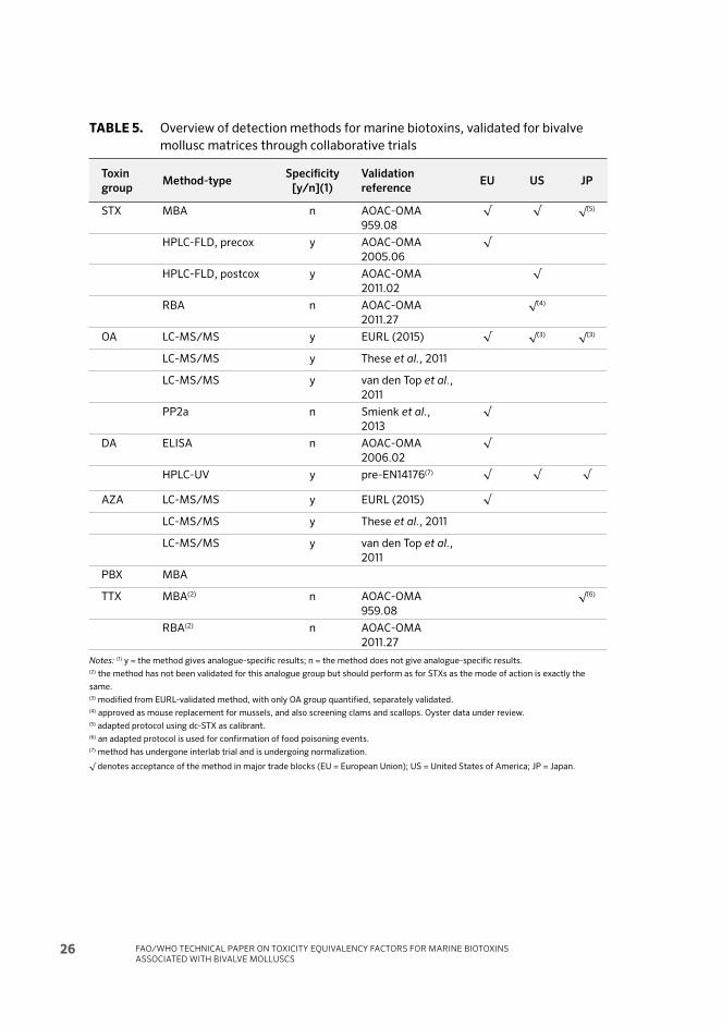
FAO/WHO TECHNICAL PAPER ON TOXICITY EQUIVALENCY FACTORS FOR MARINE BIOTOXINS ASSOCIATED WITH BIVALVE MOLLUSCS
26
TABLE 5. Overview of detection methods for marine biotoxins, validated for bivalve
mollusc matrices through collaborative trials
Toxin
groupMethod-type
Specifi city
[y/n](1)
Validation
referenceEU US JP
STX MBA n AOAC-OMA
959.08
√ √ √(5)
HPLC-FLD, precox y AOAC-OMA
2005.06
√
HPLC-FLD, postcox y AOAC-OMA
2011.02
√
RBA n AOAC-OMA
2011.27
√(4)
OA LC-MS/MS y EURL (2015) √ √(3) √(3)
LC-MS/MS y These et al., 2011
LC-MS/MS y van den Top et al.,
2011
PP2a n Smienk et al.,
2013
√
DA ELISA n AOAC-OMA
2006.02
√
HPLC-UV y pre-EN14176(7) √ √ √
AZA LC-MS/MS y EURL (2015) √
LC-MS/MS y These et al., 2011
LC-MS/MS y van den Top et al.,
2011
PBX MBA
TTX MBA(2) n AOAC-OMA
959.08
√(6)
RBA(2) n AOAC-OMA
2011.27
Notes: (1) y = the method gives analogue-specific results; n = the method does not give analogue-specific results. (2) the method has not been validated for this analogue group but should perform as for STXs as the mode of action is exactly the
same. (3) modified from EURL-validated method, with only OA group quantified, separately validated. (4) approved as mouse replacement for mussels, and also screening clams and scallops. Oyster data under review. (5) adapted protocol using dc-STX as calibrant. (6) an adapted protocol is used for confirmation of food poisoning events.(7) method has undergone interlab trial and is undergoing normalization.
√ denotes acceptance of the method in major trade blocks (EU = European Union); US = United States of America; JP = Japan.
CHAPTER 2 - CHEMISTRY AND DETECTION METHODS FOR BIOTOXINS 27
2.2.1 Saxitoxins
As STXs are the most lethal group of algal toxins, mouse bioassay was developed relatively early (Sommer and Meyer, 1937a). Th e PSP mouse bioassay (MBA) was formally validated in an interlaboratory trial and led to a standardized AOAC method (OMA 959.08) (McFarren, 1959). Despite some ethical and technical issues (Hess et al., 2006), the test is rapid, quantitative and has relatively few interferences. Th e test is thus still used in many countries (DeGrasse and Martinez-Diaz, 2012; Hess, 2012; Suzuki and Watanabe, 2012)(Table 5). Th e MBA does not provide analogue-specifi c data but gives a result in equivalents of STX (in European Union and United States of America protocols) or in equivalents of dc-STX (in Japan).
Th e receptor-binding assay (RBA) using tritiated (i.e. radio-labelled) STX is also an assay that gives a sum value for STX-equivalents (van Dolah et al., 1995; Doucette et al., 1997). Aft er successful validation in a single laboratory (Van Dolah et al., 2009), the test has also undergone formal interlaboratory validation (Van Dolah et al., 2012) and has reached a good level of acceptance in the United States of America (Table 5), and some other countries. In the European Union, a restriction for the use of radiolabelled material typically prevents use of the assay since other methods that do not require the use of radioactive material are available.
Th e above two methods (MBA and RBA) are based on the mode of action of STXs and can thus also be used for TTXs as the latter share their mode of action with STXs. However, it should be noted that the assay has not been formally validated for this compound group.
Analogue-specifi c methods for STXs are based on their fl uorescence aft er oxidation and were developed in the 1990s (Lawrence et al., 1991b; Oshima, 1995). Subsequently, these methods have been standardized and have undergone single laboratory and collaborative trials for formal validation (Lawrence, Niedzwiadek and Menard, 2004; Van de Riet et al., 2009, 2011; Ben-Gigirey, Rodriguez-Velasco and Gago-Martinez, 2012a; Ben-Gigirey et al., 2012b). Th e protocols diff er slightly in that one approach uses an oxidation step prior to chromatographic separation of analogues while the other approach separates compounds fi rst and oxidises them aft er separation on a HPLC column. However, both methods have in common that they are analogue-specifi c, are not suitable for the detection of TTXs and require several injections per sample in the case of complex natural toxin profi les.
2.2.2 Okadaic acid and Azaspiracid and their analogues
OA and AZA and their analogues may all be detected by liquid chromatography coupled to tandem mass spectrometry (LC-MS/MS). Th ree major validation exercises with collaborative trials have recently been undertaken to achieve

FAO/WHO TECHNICAL PAPER ON TOXICITY EQUIVALENCY FACTORS FOR MARINE BIOTOXINS ASSOCIATED WITH BIVALVE MOLLUSCS
28
widespread acceptance of this methodology (Th ese et al., 2011; van den Top et al., 2011; Brana-Magdalena et al., 2014).
Th is analogue-specifi c methodology has now replaced the lipophilic MBA in Europe as a routine method for testing for lipophilic toxins (EU, 2011). MBAs according to diff erent protocols developed by Yasumoto, Oshima and Yamaguchi (1978c) or Yasumoto et al. (1984) are still being used in other parts of the world for practical reasons. However, the MBA assay has never been formally validated for lipophilic marine biotoxins.
For the OA group of toxins only, there is also an enzyme-based assay available, based on the inhibition of phosphoprotein-phosphatase 2a (PP2a). Th is assay has also been recently validated and is accepted in Europe for the OA-group toxins (Smienk et al., 2013). While the LC-MS/MS methodology provides analogue-specifi c information, i.e. concentrations per analogue, the PP2a-assay provides a sum of OA-equivalent material present in a sample.
As mentioned earlier, the chemical resistance of OA and its two major analogues DTX1 and DTX2 towards strong bases allows for conversion of the various ester derivatives to these three analogues for ease of analysis. Th is methodological approach appears justifi ed by the fact that the esters could also be converted in the human digestive system, and the esters appear to have similar toxicity as the parent compounds. Th e base hydrolysis step should therefore be added for any of the methods, LC-MS/MS or PP2a assay.
2.2.3 Domoic acid and analogues
While initial eff orts in method validation focused on using the extraction protocol for the PSP MBA (Lawrence et al., 1991a), this method has been superseded by the knowledge that DA is not very stable in strongly acidic conditions. Hence, methods using an extraction protocol based on aqueous methanol with HPLC-UV detection (Quilliam, Xie and Hardstaff , 1995a) are now generally preferred. Such methods have undergone collaborative trials for validation and are currently undergoing standardization in the frame of the European Normalisation Committee (CEN; EN14176).
As the DA group is relatively simple, with only one major analogue and an epimer (DA and epi-DA), an ELISA has also been developed and a collaborative trial (Kleivdal et al., 2007) permitted interlaboratory validation and formal standardization as AOAC method OMA 2006.02. Th is ELISA has strong approval by stakeholders in the scallop processing industry in Scotland, and the assay is also approved for use in offi cial control in the European Union.

CHAPTER 2 - CHEMISTRY AND DETECTION METHODS FOR BIOTOXINS 29
2.2.4 Brevetoxin and analogues
Th e mouse bioassay (MBA) has been used conventionally for detecting BTXs (APHA, 1970). It is the only method approved in the United States of America under the National Shellfi sh Sanitation Program (NSSP) for use in making regulatory decisions regarding the harvest of bivalves due to BTXs (FDA, 2013). Th e current CODEX Standard for BTXs also involves the use of MBA. Th e MBA involves the intraperitoneal administration of the sample extract into mice. Th e time of death of the animals is correlated with the BTX content of the sample. Results are reported in mouse units (MU) per 100 g shellfi sh tissue, a MU being defi ned as the amount of crude extract that would kill 50% of the test animals in 930 minutes. Th e MBA has been criticized not only for its use of animals, but also because of assay variability, lack of specifi city, and the limitations of the MBA extraction to account for all potential BTX analogues and metabolites (e.g. Dickey et al. 1999; Plakas and Dickey., 2010). One major advantage of the MBA is that it has demonstrated its effi cacy in public health protection. Th e action level for NSP toxins established by the United States Food and Drug Administration (US FDA) is 20 MU per 100 g tissue or 0.8 ppm PBX2 equivalents (FDA. 2011). When toxicity exceeds this level as determined by the MBA, growing areas are closed to the harvest of bivalve molluscs.
While the MBA and a radioimmunoassay were used to investigate the NSP outbreak in New Zealand in 1993 (Temple, 1995), an internally validated liquid chromatography mass spectrometry (LC-MS) method is now used for monitoring BTXs in New Zealand (McNabb et al. 2012). An advantage of the LC-MS method is that it allows for the identifi cation of individual BTX analogues and metabolites. However, the limitation regarding an extraction procedure that recovers all relevant forms also applies. Another challenge is that standards for all of the BTX analogues and metabolites are not readily available. Further, if applying the action level of 0.8 ppm PBX2 equivalents established in the United States of America, a level of 0.8 mg/kg is used, yet TEFs are needed to present the results in PBX2 equivalents.
Additional methods for the determination of BTXs have been developed and are in various stages of validation and use. Th ese include a neuroblastoma assay (Manger et al., 1993), enzyme linked immunoassays (ELISAs) (Naar et al., 2002), and a receptor binding assay (Trainer and Poli, 2000). None of these methods detects individual BTX analogues or metabolites, but are indicative of overall toxicity.

FAO/WHO TECHNICAL PAPER ON TOXICITY EQUIVALENCY FACTORS FOR MARINE BIOTOXINS ASSOCIATED WITH BIVALVE MOLLUSCS
30
3Toxicity Equivalency Factors (TEFs) for specifi c biotoxin groups
CONSIDERATIONS FOR THE ESTABLISHMENT OF TEFSTh e following approach was taken by the Expert Group when considering the establishment of TEFs:
• Data consideration in order of importance/relevance:1. Data from human cases (outbreaks);2. oral LD50 /toxicity data in animals;3. ip LD50 /toxicity data in animals;4. Mouse bioassay;5. In vitro data.
• When TEFs are below 0.1, an increment of 0.05 is used.• TEF should not be established for combinations of diff erent analogues
because they will be dealt with by individual TEFs.
3.1 SAXITOXIN AND ANALOGUES In 1937, Sommer and Mayer reported a quantitative assay for STX, based on the dose-death time relationship in mice dosed intraperitoneally with this substance. Th ey noted that the dose-death time curve was relatively linear between 5 and 7 minutes, and the assay, which is now an approved AOAC method (Hungerford, 1995), involves determining the amount of STX required to cause death in this time interval. Although initially set up for STX, it is now used for estimating the toxicity of mixtures of STX analogues expressed as the specifi c activity (Mouse

CHAPTER 3 - TOXICITY EQUIVALENCY FACTORS (TEFS) FOR SPECIFIC BIOTOXIN GROUPS 31
Units per micromole). Th is assumes that the dose-death time curves for all the analogues are identical, (Munday et al., 2013), calling into question the validity of this assay for comparing analogues.
Th e MBA has been widely used for comparing the toxicity of STX analogues. Again, this depends on the assumption that the dose-death time relationship is the same for all analogues. It was, however, used when only small amounts of material were available, and this assay has been widely used for estimating TEFs (Oshima, 1995).
Th e relative potencies determined by the MBA are shown in Table 6.
TABLE 6. Relative potency of STX derivatives as indicated by the MBA
Compound Relative specifi c activity in the MBA
Relative
LD50
by i.p.
injection(1)
Saxitoxin 1.0 1.00
NeoSTX 0.50, 0.75(2), 0.90, 0.90, 1.0, 1.16(1), 1.2 3.12
GTX–1 0.80, 1.0
GTX–4 0.30, 0.70
GTX–1&4 0.70, 1.02(1), 0.65(2) 1.90
GTX–2 0.40, 0.40
GTX–3 0.60, 1.1
GTX–2&3 0.60, 0.60(1), 0.52(2) 0.757
GTX–5 0.10, 0.10, 0.20 0.222
GTX–6 0.10 0.122
C–1 0.02, 0.00
C–2 0.10, 0.17
C–3 0.0, 0.01
C–4 0.0, 0.10
dcSTX 0.40,0.48(3), 0.50, 0.50, 0.60, 0.64(1), 1.0, 1.02(2) 0.785
dcNeoSTX 0.40, 0.020(4) 0.058
dc-GTX–1 0.5
dc-GTX–2 0.20, 0.20, 0.30
dc-GTX–3 0.20, 0.40, 0.50
dc-GTX–4 0.50
dc-GTX–2&3 0.20, 0.19(2) 0.695
11α-Hydroxy-STX 0.60
11β-hydroxy-STX 0.70
Sources: (1) Munday et al., 2013; (2) Vale et al., 2008b; (3) Suzuki and Machii, 2014; (4) Munday, MS in preparation.
All other data are from Table 13 of the 2009 ESFA report on saxitoxin group toxins (EFSA, 2009b).
As seen in Table 6, there are wide variations in estimates of the relative potencies. While such diff erences most likely refl ect the use of impure compounds, this cannot

FAO/WHO TECHNICAL PAPER ON TOXICITY EQUIVALENCY FACTORS FOR MARINE BIOTOXINS ASSOCIATED WITH BIVALVE MOLLUSCS
32
be the whole answer, since, for example, the estimates for NeoSTX reported in references (Munday et al., 2013) and (Vale et al., 2008b) are signifi cantly diff erent, even though both were conducted using certifi ed materials. Th ere are also errors in the relative potencies given in the EFSA report (EFSA, 2009b). Th e fi gure of 0.40 given for dcNeoSTX was referenced to Sullivan et al., (1983, 1985). However, these authors did not conduct a MBA. Th ey assumed that the specifi c activity of dcNeoSTX was the same as that of dcSTX. Th e actual fi gure for the specifi c activity of dcNeoSTX, measured on a pure sample of this substance, is 0.020. Th e fi gures given for the isomeric 11-hydroxysaxitoxins are also incorrect. Th e reference given for these data in Table 13 of the EFSA report is Schantz (1986). Th e material tested by Schantz was not, however, the isomers of 11-hydroxySTX but the isomers of 11-hydroxySTX sulphate, better known as GTX–2 and GTX–3.
It should also be noted that there are limitations to interpreting the potential impact to human health from the specifi c activities of the STX analogues determined in the mouse bioassay in relation to the measured endpoint (fatality). As shown in Table 6, while there is a correlation between the relative specifi c activity and relative toxicity by intraperitoneal injection with some saxitoxin derivatives, with others, particularly NeoSTX, GTX–1&4 and dcGTX–2&3, there is no correlation. Th is is attributable to diff erences in the dose-death time relationship (Munday et al., 2013).
Because the toxic eff ect of the analogues was originally obtained with an i.p. injection, Munday et al., (2013) re-evaluated, with certifi ed materials, the relative potencies for some of the toxins using oral administration (gavage or feeding) of the toxins. Th e results provided similar results to the i.p. administration, although the relative potency was diff erent for dcSTX (0.37 by feeding, 0.46 by gavage, versus 0.64 by i.p. route), and similar to the value obtained in an i.p. study of STX versus dcSTX specifi cally performed to validate dcSTX for bioassay, with a relative potency for dcSTX of 0.48 (Suzuki and Machii, 2014). Another toxin that provided a rather diff erent value was NeoSTX, with a relative potency of 1.7 by gavage, 2.54 by feeding and 1.16 by i.p. injection (Munday et al., 2013).
Th e shortage of materials also led to the development of in vitro methods for comparing the eff ects of STX with those of its congeners. Although STX has been reported to potentially target many receptors, specially sodium, potassium and calcium channels (Su et al., 2004; Llewellyn, 2006a; Zakon, 2012; Cusick and Sayler, 2013), there is strong evidence that STX and derivatives exert their toxic eff ects in animals by binding to the voltage-gated sodium channel (Nav) (Payandeh et al., 2011). Th is channel contains one alpha subunit and one to three small beta subunits. Th ere are 9 alpha subunits of the Nav channel (Nav 1 to 9) (Wingerd, Vetter and Lewis, 2012), and originally they were divided into tetrodotoxin

CHAPTER 3 - TOXICITY EQUIVALENCY FACTORS (TEFS) FOR SPECIFIC BIOTOXIN GROUPS 33
(TTX)-sensitive (Nav 1, 2, 3 and 7) and TTX insensitive. Th e alpha subunits contain 4 homologous domains, each with 6 hydrophobic transmembrane segments. Th ere are 6 binding sites identifi ed that are the target for many toxins, including several phycotoxin groups. Site 1 is the receptor for TTX and STXs; site 5 is the receptor for ciguatoxins and BTXs (Hartshorne and Catterall, 1981). Th e major molecular mechanism of toxicity of both TTX and STX is to block the channel pore with a ratio 1:1, hence inhibiting the conductance of the channel and the transmission of electrical action potentials generated by the infl ux of sodium ions into the cell. Th is mechanism is responsible for muscle paralysis, potentially leading to paralysis of the diaphragm and death.
With the improved availability of pure and certifi ed PSP materials, a more defi ned list of TEFs was proposed by EFSA, based on the study of the eff ect of certifi ed toxins on sodium channels of neuronal cultures by means of fl uorescent dyes sensitive to membrane potential (Vale et al., 2008b; EFSA, 2009b). Because this study did not cover the same toxins studied by Oshima (1995), the EFSA list provided a combined set of results that were basically similar, with the exception of dcSTX (reported now as more toxic) and GTX1–4 as Oshima provided a higher value for GTX1, and all the values reported by Oshima were with pure GTX1 to 4. Th ese TEF values were later re-evaluated by means of a more precise electrophysiological patch clamp recording method in primary cultured neurons, and the ratio results obtained were equivalent, but with a slightly lower toxicity for GTX1–4 (Perez et al., 2011) and a potency of NeoSTX of 1.02, similar to STX (Perez et al., 2011).
Since the mechanism of toxicity of these substances was known to involve voltage-gated sodium channels, attention was focused on these channels, with ever increasing levels of sophistication. Since each Nav channel subtype has diff erent affi nities for STX and derivatives, an automated electrophysiology platform was used to determine the relative potencies of the analogues in human Nav subtypes 1.1 to 1.7, transfected in HEK–293 cells (Alonso et al., 2016). Th is information indicates direct binding in transfected human receptors. Th e results show that the subtype that best matches the results from previous works is obtained with Nav subtypes 1.2 and specially 1.6, although the toxicity of the epimer mixture GTX1–4 is higher than previously reported (relative potency of 1.4 for Nav 1.6).
Again, there are wide variations among the diff erent assays. Th e results with various methodologies are summarized in Table 7. Comparing the relative potencies based on in vitro studies on sodium channels with those based on median lethal doses by consumption in mice, it is interesting to note that assays involving the frog sciatic nerve (method 3) and the rat muscleplasma membrane (method 5) indicate a relatively high activity of NeoSTX, in accord with the acute toxicity data in mice.

FAO/WHO TECHNICAL PAPER ON TOXICITY EQUIVALENCY FACTORS FOR MARINE BIOTOXINS ASSOCIATED WITH BIVALVE MOLLUSCS
34
TA
BL
E 7
.
Re
lati
ve
to
xic
itie
s o
f S
TX
an
d d
eri
va
tiv
es
in m
ice
(M
BA
) a
nd
re
lati
ve
re
act
ivit
ies
tow
ard
so
diu
m c
ha
nn
els
in v
itro
Co
mp
ou
nd
[9]
Re
lati
ve
ac
tiv
ity
to
wa
rd s
od
ium
ch
an
ne
ls i
n v
itro
by
ty
pe
of
as
sa
y m
eth
od
(1)
[1]
[2]
[3]
[4]
[5]
[6]
[7]
[8,
Na
v 1,
6]
[8,
Na
v 1,
2]
ST
X1.
00
1.0
1.0
1.0
1.0
1.0
1.0
1.0
1.0
1.0
ne
oS
TX
2.5
4/
1.7
–0
.69
4.5
1.0
3.6
/3
.70
.82
1.0
21.
22
.0
GT
X 1
––
––
0.2
8–
–
GT
X 1
&4
0.9
36
/0
.73
9–
0.9
8–
––
0.5
30
.50
1.4
0.5
4
GT
X 2
0.2
0.2
20
.15
/0
.16
GT
X 2
&3
0.5
72
/0
.53
3–
0.3
2–
––
0.3
80
.28
0.1
50
.4
GT
X 3
0.4
2–
1.4
–0
.96
GT
X 4
––
––
GT
X 5
0.0
64
/0
.05
–0
.03
1/0
.03
9–
–0
.02
40
.09
0.0
90
.11
0.0
1
GT
X 6
< 0
.017
/0
.03
8–
––
––
dcS
TX
0.3
68
/0
.45
7–
0.0
97
/0
.29
–0
.20
.44
0.8
41.
00
0.9
60
.25
dcN
eo
ST
X0
.22
4/
0.2
16–
––
0.0
04
–0
.48
0.4
40
.25
0.1
dcG
TX
2,3
0.1
08
/0
.16
70
.02
0.0
5
C1
––
––
–0
.00
17/
0.0
02
8
C2
––
––
0.0
29
C1,
20
.04
3/
0.0
34
0.0
90
.01
C3
––
––
–0
.00
2
Ke
y t
o a
ssa
y m
eth
od
s: [
1] R
ela
tiv
e b
lock
ad
e o
f so
diu
m c
ha
nn
els
in t
he
sq
uid
gia
nt
ax
on
(K
ao
et
al.
, 19
85
b).
[2]
Re
lati
ve
bin
din
g t
o s
od
ium
re
cep
tors
of
the
ra
t ce
reb
ral c
ort
ex
(U
sup
et
al.
, 2
00
4;
Lle
we
lly
n,
20
06
b).
[3]
Re
lati
ve
blo
cka
de
of
imp
uls
es
in f
rog
sci
ati
c n
erv
e (
Str
ich
art
z,
198
4).
[4]
Re
lati
ve
blo
cka
de
of
sod
ium
cu
rre
nt
in f
rog
sk
ele
tal m
usc
le fi
bre
(K
ao
et
al.
, 19
82
; Y
an
g e
t a
l.,
199
2b
).
[5]
Re
lati
ve
blo
cka
de
of
sod
ium
ch
an
ne
ls f
rom
ra
t m
usc
le p
lasm
a m
em
bra
ne
(M
ocz
yd
low
ski e
t a
l.,
198
4;
Gu
o e
t a
l.,
198
7).
[6]
Blo
cka
de
of
ve
ratr
idin
e-i
nd
uce
d c
ha
ng
es
in m
em
bra
ne
po
ten
tia
l in
cu
ltu
red
ne
uro
ns
(Va
le e
t a
l.,
20
08
b).
[7]
So
diu
m c
urr
en
ts v
olt
ag
e-d
ep
en
de
nt
inh
ibit
ion
in p
rim
ary
cu
ltu
res
of
cere
be
lla
r n
eu
ron
s (P
ere
z e
t a
l.,
20
11).
[8]
Hig
h-t
hro
ug
hp
ut
ele
ctro
ph
ysi
olo
gy
sy
ste
m,
in c
ell
s st
ab
ly t
ran
sfe
cte
d w
ith
sp
eci
fi c
sub
un
its
of
sod
ium
ch
an
ne
ls (
Alo
nso
et
al.
, 2
016
).
[9]
Re
lati
ve
to
xic
ity
by
vo
lun
tary
fe
ed
ing
/g
av
ag
e (
Mu
nd
ay
et
al.
, 2
013
).

CHAPTER 3 - TOXICITY EQUIVALENCY FACTORS (TEFS) FOR SPECIFIC BIOTOXIN GROUPS 35
3.1.1 Toxicity of epimers and infl uence of hydrolysis
Some of the toxins are epimers due to the possible double position on the radical 11-hydroxysulfate. Th erefore the pairs GTX1,4, GTX2,3, C3,4 and C1,2 are epimers. Th is process of 11-hydroxysulfate epimerization is spontaneous and the thermodynamic equilibrium is achieved at a higher pH above acidic stability. Th e ratios, depending on the conditions (temperature, ionic strength, pH), may vary between 2 and 4.5 (Hall et al., 1990; Reeves et al., 2003). For this reason, it is diffi cult to implement a TEF for the mixture of epimers because the ratio may be diff erent, depending on the source of the standard. Th e ratio of GTX2,3 and GTX1,4 in Munday et al. (2013) was 2.2 and 4.06, respectively, while in Vale et al. (2008b) it was 3 for both mixtures. Th e supply of pure epimers is diffi cult to obtain because of the spontaneous process of epimerization, therefore it is useful to have the TEF of the mixture, even with a range of results, as there are no two mixtures with the exact same proportion, and the toxicity of both epimers, especially GTX1 and GTX4, is very diff erent, GTX1 being more potent. Although the TEF is to be applied to an analytical result, this possible conversion of epimers may modify the fi nal calculated toxicity of a sample, depending on the storage conditions.
Another important source of possible toxicity change is the modifi cation of the toxicity of a sample aft er hydrolysis by boiling at acidic pH, which may cause a 10-fold increase in the toxicity of a sample by converting C toxins into GTXs (Vale et al., 2008b). Even though the ratio value of the toxicity of each compound is the same, the fi nal toxicity of a sample may change notably. For this reason, the application of a TEF has to be done with a thorough understanding of the chemical profi le of the sample, aft er all possible conversions have taken place.
3.1.2 Conclusion
Although the absolute LD50 values of STX and its analogues by gavage and by voluntary consumption were diff erent, the relative toxicities by these routes, as indicated by the TEFs, were similar. Th e EFSA TEFs for GTX–1&4, GTX–2&3 and C1,2 are consistent with those determined by oral administration. In contrast, and based on oral toxicity to mice, the TEF for NeoSTX was signifi cantly higher than that proposed by EFSA, while the EFSA TEFs for GTX–5, GTX–6, dcSTX, dcNeoSTX were lower.
Th ere are two toxins that require further clarifi cation: dcSTX was recently reported by some authors to be less toxic than STX, with a TEF of 0.8 (Vale et al., 2008b), 0.64 (Munday et al., 2013), 0.478 (Suzuki and Machii. 2014) and 0.37 (Suzuki and Machii, 2014), although the TEF adopted by EFSA was 1. With regard to dcSTX, the application of EFSA TEFs in samples with high levels of dcSTX, such as surf clams, results in a signifi cant improvement in the agreement between HPLC and

FAO/WHO TECHNICAL PAPER ON TOXICITY EQUIVALENCY FACTORS FOR MARINE BIOTOXINS ASSOCIATED WITH BIVALVE MOLLUSCS
36
the mouse assay (Turner et al., 2011). Another toxin that requires clarifi cation is NeoSTX, as the range of toxicity values is very large, from 1 (EFSA 2009b, Alonso et al., 2016) to 2.54 (Munday et al., 2013). If TEFs were to be based on oral toxicity, the EFSA TEFs for GTX–5, GTX–6, dcSTX, dcNeoSTX should be revised downwards, while the TEF for NeoSTX should be increased. It is interesting to note that there is a better match between the results obtained with Nav subtype 1.2 (Alonso et al., 2016) and the results obtained with oral administration to mice (Munday et al., 2013). Th e TEFs recommended by the Expert Group are indicated in Table 8.

CHAPTER 3 - TOXICITY EQUIVALENCY FACTORS (TEFS) FOR SPECIFIC BIOTOXIN GROUPS 37
Ta
ble
8.
T
EF
s re
com
me
nd
ed
by
th
e E
xp
ert
Gro
up
Co
mp
ou
nd
Os
him
a
Re
lati
ve
To
xic
ity
va
lue
s
(MU
/
μm
ole
)
Mo
us
e
LD
50
(i.p
.)
TE
F
ba
se
d o
n
LD
50 b
y
ga
va
ge
TE
F b
as
ed
on
LD
50 b
y
vo
lun
tary
co
ns
um
pti
on
EF
SA
pro
po
se
d
TE
F
Re
co
mm
en
de
d
TE
FR
ati
on
ale
Sa
xit
ox
in1
1.0
01.
00
1.0
01.
01.
0
Ne
oS
TX
0.9
23
.12
1.7
02
.54
1.0
2.0
Bo
th o
ral
stu
die
s su
pp
ort
hig
he
r to
xic
ity
th
an
ST
X.
A
va
lue
of
2.0
is
reco
mm
en
de
d a
nd
su
pp
ort
ed
by
so
me
Na
ch
an
ne
l in
vit
ro r
esu
lts
(ra
ng
e o
f R
P 0
.7–
3.7
).
GT
X1
0.9
91.
01.
0N
o n
ew
da
ta1
GT
X2
0.3
60
.40
.4N
o n
ew
da
ta
GT
X3
0.6
40
.60
.6N
o n
ew
da
ta
GT
X4
0.7
30
.70
.7N
o n
ew
da
ta
GT
X5
0.0
64
0.2
22
0.0
63
0.0
50
0.1
0.1
Re
lati
ve
po
ten
cy v
alu
es
fro
m o
ral
LD
50
stu
die
s
sug
ge
st a
lo
we
r T
EF
th
an
fro
m L
D5
0 i
.p.
0.1
TE
F a
lso
in
ag
ree
me
nt
wit
h o
rig
ina
l O
shim
a T
EF.
As
wit
h N
eo
ST
X,
a n
um
be
r o
f in
vit
ro N
a c
ha
nn
el
ass
ay
s a
lso
su
pp
ort
a
TE
F o
f 0
.1.
GT
X6
0.1
22
0.0
38
0.1
0.0
5N
ew
ora
l d
ata
sh
ow
lo
we
r th
an
0.1
.
C1
0.0
06
0.0
1N
o n
ew
da
ta (
rou
nd
ed
up
)
C2
0.0
96
0.1
0.1
No
ne
w d
ata
C3
0.0
130
.01
No
ne
w d
ata
C4
0.0
58
0.1
0.1
No
ne
w d
ata
dcS
TX
0.5
10
.78
50
.45
70
.36
81.
00
.5F
rom
re
cen
t O
ral
da
ta (
mo
re w
eig
ht
on
ora
l d
ata
, a
lso
sup
po
rte
d b
y i
.p.
tox
icit
y d
ata
)
dcN
eo
ST
X0
.05
80
.216
0.2
24
0.4
0.2
Fro
m r
ece
nt
Ora
l d
ata
(m
ore
we
igh
t o
n o
ral
da
ta,
als
o
sup
po
rte
d b
y i
n v
itro
da
ta)
dcG
TX
20
.15
0.2
0.2
No
ne
w d
ata
dcG
TX
30
.38
0.4
0.4
No
ne
w d
ata
1 In
th
e c
ase
of
sax
ito
xin
an
alo
gu
es,
fo
r w
hic
h n
o o
ral t
ox
icit
y d
ata
we
re a
va
ila
ble
, T
EF
s re
com
me
nd
ed
are
ba
sed
on
intr
ap
eri
ton
ea
l to
xic
ity
da
ta o
f O
shim
a (
Ap
pe
nd
ix I
).

FAO/WHO TECHNICAL PAPER ON TOXICITY EQUIVALENCY FACTORS FOR MARINE BIOTOXINS ASSOCIATED WITH BIVALVE MOLLUSCS
38
3.2 OKADAIC ACID AND ANALOGUES OA and DTXs are polyether compounds that exist in the algae that produce them (Dinophysis, Prorocentrum) (Reguera et al., 2008) as unesterifi ed compounds (OA, DTX1, DTX2) and as esters (diol esters and DTX4, –5 and –6), and in shellfi sh also as DTX3, which is a mixture of esters of several fatty acids with acyl moieties of C14 to C22 and with up to 6 unsaturated bonds, the most common being palmitic esters of OA, DTX1 or DTX2 (Yanagi et al., 1989).
From a toxicological point of view, the most relevant compounds, due to their known involvement in toxic episodes, are OA, DTX1, DTX2 and the DTX3 ester forms. Although DTX3 esters per se are non toxic, they may be hydrolysed aft er ingestion to release the parent compound(s) (OA, DTX1, DTX2) in the gastrointestinal tract.
Th e historical record of intoxications caused by these toxins has many inconsistencies most likely due to variation due to the quantifi cation errors caused by the use of non-certifi ed or non-quality-controlled materials, and the poor traceability of the intoxications (EFSA, 2008c). Many cases of human intoxication have been reported; however, an acute reference dose (ARfD) of 0.3 μg/kg body weight (b.w.) was proposed in 2007 by the EFSA Panel on marine toxins, based on eff ects in consumers (EFSA, 2008c). Th is ARfD is consistent with the provisional value of 0.33 μg/kg b.w. set in 2004 by the Joint FAO/IOC/WHO ad hoc Expert Consultation on Biotoxins in Bivalve Molluscs (CODEX, 2006). Both panels concluded that no tolerable daily intake could be established because of the lack of data on the chronic toxicity of OA.
A well known eff ect of OA and analogues is the inhibition of cytosolic phosphatases, especially protein-phosphatase 2A (PP2A) as the main target (ID50 1.2 nM) and, as secondary targets, PP1 (ID50 315 nM) and PP2B (ID50 4530 nM) (Takai et al., 1987; Bialojan and Takai, 1988). Th ere is no evidence of a toxic mechanism linked to any other type of phosphatase. Th ese toxins are lethal in rodents above a certain value, although large ranges in estimates of toxicity have been reported, again attributable to the lack of use of certifi ed materials that guarantee defi ned quantities. In general, 200 μg/kg b.w. body weight is a lethal i.p. dose in mice (Munday, 2014), with some variations in the time to death, depending on the strain (Suzuki, 2012).
A recent review of poisoning events suggests that esters of OA and DTXs have very similar toxicity to the parent compounds in human poisoning (EFSA, 2008c). OA and DTX1 are considered to be of approximately equal toxicity when injected intraperitoneally into mice, while DTX2 has been reported to have only 50 to 60 percent of the toxicity of OA (Aune et al., 2007), both by i.p. injection into mice

CHAPTER 3 - TOXICITY EQUIVALENCY FACTORS (TEFS) FOR SPECIFIC BIOTOXIN GROUPS 39
and by in vitro assessment of their inhibitory character towards phosphoprotein phosphatases.
3.2.1 Acute toxicity
Th ese toxins are lipophilic and they have good oral bioavailability due to their lipophilic nature. Th ere are several studies of the toxic eff ect of OA and analogues in the intestine, the greatest damaging eff ects being seen aft er i.p. administration of OA, for which the lethal dose is consistently around 200 μg/kg b.w. (Aune et al., 2007; Munday, 2014). Th ere is very limited information on the toxicity of OA analogues (Table 9). DTX1 is reported to be more potent than OA, with a lethal dose of 160 μg/kg b.w. (Tubaro et al., 2008a) for DTX1 versus 200 μg/kg b.w. for OA (Ogino et al., 1997; Tubaro et al., 2008a). DTX2 is 40 percent less potent than OA, as determined by i.p. toxicity in mice (352 μg/kg b.w. for DTX2 and 204 for OA) (Aune et al., 2007). Even though the i.p. route of administration is interesting for the purpose of toxicological studies, there is no clear agreement about its use for the defi nition of a TEF, as the natural route is oral administration (Botana, 2012), although, as stated above, for biotoxins with similar bioavailability, i.p. administration can still provide useful toxicity comparison information.
TABLE 9. Acute toxicities of OA and its analogues by i.p. injection
Compound Parameter Acute toxicity (μg/kg b.w.) and source
OA LD50
192 (Tachibana et al., 1981)
OA LD50
200 (pers. comm. T. Yasumoto, 1991)
OA No death 200 (Ito and Terao, 1994)
OA LD50
204 (Aune et al., 2012)
OA LD50
210 (Dickey et al., 1990)
OA LD50
225 (Tubaro et al., 2003)
OA LD40
to LD100
mean 227, range 216–242 (Suzuki, 2012)
OA LD100
375 (Ito and Terao, 1994)
DTX1 MLD 160 (Murata et al., 1982; Yasumoto and Murato, 1990)
DTX1 LD50
160 (pers. comm. T. Yasumoto, 1991; Dominguez et al., 2010)
DTX1 LD100
375 (Ito and Terao, 1994)
DTX2 LD50
352 (Aune et al., 2007)
DTX3 LD100
375 (Ito and Terao, 1994)
DTX3 MLD 500 (Yasumoto et al.,1985)
DTX4 LD50
610 (Hu et al.,1995a)

FAO/WHO TECHNICAL PAPER ON TOXICITY EQUIVALENCY FACTORS FOR MARINE BIOTOXINS ASSOCIATED WITH BIVALVE MOLLUSCS
40
From these data, it would appear that DTX1 is slightly more toxic than OA, although it must be noted that the toxicity parameter for the former compound was an MLD, not an LD50. DTX2, –3 and –4 were less toxic than OA when administered by this route.
Th ere are several reports of oral administration of OA and analogues, but only few of them report concentrations checked against a certifi ed material (Aune et al., 2012; Vieira et al., 2013). Th is may explain the large discrepancy in reported lethal doses, ranging from 2 000 μg/kg b.w. (Tubaro et al., 2003), to 400 μg/kg b.w. (Ito et al., 2002a), and to death with 575 μg/kg b.w. and no death with 610 μg/kg b.w. in two diff erent experiments in the same report (Le Hegarat et al., 2006). An oral dose of 1 000 μg/kg b.w. in rats was not lethal, again with a commercial supply of OA that was not quality controlled (Berven et al., 2001). Th ose oral studies where the concentration has been checked against certifi ed standards show a consistent lethal dose (LD50) of 880 μg/kg b.w. in 18–22 g NMRI (Naval Medical Research Institute) female mice (Aune et al., 2012) and 1 000 μg/kg b.w. in 18–23 g CD–1 (Cluster Diff erentiation 1) female mice (Vieira et al., 2013). Even though mouse sex and strain show some diff erences, in general this is not a key factor for dose variability (Suzuki, 2012, 2013). A study of susceptibility of diff erent mouse strains to OA (Suzuki, 2012) found that, depending on the strain, 40 to 100 percent of the mice died aft er an i.p. injection of 4 μg OA (mean 227 μg/kg b.w., range 216–242 μg/kg b.w.). No diff erence with regard to the sex of the animal was observed (Suzuki, 2013).
Th e oral administration of DTX1 shows similar toxic eff ects to i.p. administration, with fl uid accumulation being observed with 0.4 and 0.32 μg/mouse for OA and DTX1, respectively (Tubaro et al., 2008a). Th e lethal dose of DTX1 aft er oral administration was reported to be above 750 μg/kg b.w. (no death) and below 300 μg/kg b.w. (all animals dead) in fasted animals (Ogino, Kumagai and Yasumoto, 1997; Munday, 2014). According to Ogino, Kumagai and Yasumoto (1997), the oral LD50 in mice is around 300 μg/kg b.w. However, in other studies, death was not reported in mice or rats given DTX1 orally at 750 mg/kg b.w (Terao et al., 1993; Ito and Terao, 1994).
No published report about the oral toxicity of DTX2 is available, although a work not yet published (Louzao et al, manuscript in preparation, an up-and-down protocol using certifi ed DTX2) has concluded that the oral LD50 is 2 150 μg/kg b.w. (death at 24 h, mice fasted for 12 h, administration by gavage); the same study concluded that the LD100 is 3 000 μg/kg b.w., all animals showing death before 5 h.

CHAPTER 3 - TOXICITY EQUIVALENCY FACTORS (TEFS) FOR SPECIFIC BIOTOXIN GROUPS 41
Th e minimum toxic dose of DTX3 (no death) was reported aft er an oral administration of 750 μg/kg b.w. (Ito and Terao, 1994). Such toxicity is probably related to hydrolysis in the digestive system, hence it is more reliable to refer the toxicity to the toxin obtained aft er hydrolysis (AO, DTX1 or DTX2). DTX3 actually represents a mixture of 7-O-acyl ester derivatives of OA, DTX1 and DTX2. DTX3 is formed as metabolites of free OA, DTX1 and DTX2 in bivalves that have consumed toxic dinofl agellates. During analysis by LC-MS/MS techniques, an alkaline hydrolysis step during extraction yields the parental toxins and therefore no TEF specifi c to DTX3 was considered necessary by the Expert Group.
Th e toxicity of DTX1 by gavage appears to be higher than that of OA, but with the variability in published values for the latter compound, an estimate of TEFs based on oral toxicity is not possible. A recent study on the cardiotoxic eff ects of OA (20 μg/kg b.w.) and DTX1 (16 μg/kg b.w.) in rats shows no cardiotoxic eff ects of these compounds in acute experiments as assessed either by the electrocardiogram or by biomarkers (Ferreiro et al., 2015).
3.2.2 Toxicity of OA and analogues to cells in vitro
Th e toxicities of OA, DTX1 and DTX2 have been recently evaluated in a number of cell lines in vitro (See Table 10).
TABLE 10. Relative toxicities of OA, DTX1 and DTX2 in cells in vitro
Compound Relative toxicity in the specifi ed cell line
SH-SY5Y (1)
Neuro–2a (1)
NG108–15 (1)
MCF–7 (1)
Caco–2 (2)
HT29-MTX (2)
OA 1.0 1.0 1.0 1.0 1.0 1.0
DTX1 4.4 2.1 2.4 3.8 2.2 3.4
DTX2 0.52 0.52 0.73 0.47 0.35
Sources: (1) Louzao et al., 2015. (2) Ferron et al., 2014.
Th e end points used in the sources for Table 10 were: transepithelial electrical resistance and cell viability assessed by the trypan blue exclusion assay in SH-SY5Y (Louzao et al., 2015); colorimetric [3-(4,5-dimethylthiazol-2-yl)-2, 5-diphenyltetrazolium], MTT assay in Neuro–2a (Solino, Sureda and Diogene, 2015); NG108–15 (Solino, Sureda and Diogene, 2015); MCF–7 (Solino, Sureda and Diogene, 2015); and uptake of neutral red in Caco–2 (Ferron et al., 2014) and HT29-MTX (Ferron et al., 2014).
Th ere is remarkable consistency among the cell lines tested, with DTX1 showing a 2–4 fold greater activity than OA, and DTX2 showing less toxicity than OA, by a factor of between 0.35 and 0.73.

FAO/WHO TECHNICAL PAPER ON TOXICITY EQUIVALENCY FACTORS FOR MARINE BIOTOXINS ASSOCIATED WITH BIVALVE MOLLUSCS
42
Taking the averages of these fi gures, the relative potencies based on cytotoxicity for OA, DTX1 and DTX2 would be 1.0, 3.1 and 0.52 respectively.
3.2.3 Diarrhetic eff ect
Th e main eff ect of OA and analogues, which was used as a tag to the group, is diarrhoea, hence the common designation of this group of compounds as diarrhetic shellfi sh poisons or DSP aft er the fi rst description of the intoxication by Yasumoto in 1978 (Yasumoto, Oshima and Yamaguchi, 1978a, b).
Th e association of diarrhoea with phosphatase inhibitors is not established. Th ere are several non-marine substances that are also potent phosphatase inhibitors, such as microcystins, nodularins, tautomycin, calyculin A or cantharidin (MacKintosh and Klumpp, 1990; Laidley, Cohen and Casida, 1997; Craig and Holmes, 2000). Th eir eff ect is mostly hepatotoxicity (Craig and Holmes, 2000), while the most evident short-term eff ect of OA is diarrhoea. Th e diarrhoea caused by OA and DTXs has been proposed to be caused by an increase in intestinal epithelial paracellular permeability (Tripuraneni et al., 1997) as indicated by transepithelial ion and mannitol fl uxes in human intestinal epithelial T84 cell monolayers. Th e purity of the OA employed in this study was not, however, checked against a certifi ed standard. Th is study proposed that OA does not stimulate intestinal secretion directly, but increases paracellular permeability. Th e fact that other phosphatase inhibitors did not show this same diarrhetic eff ect suggest that other factors are involved in the mechanism of toxicity of OA and DTXs.
Th ere is a no clear relationship between reported eff ects of OA and DTXs on the small intestine in the presence of diarrhetic symptoms and the doses tested, probably attributable to uncertainties on the fi nal amount of OA used as mentioned above. Th e same group reported damage to the intestinal mucosa in one study (Tubaro et al., 2003) but none in another study, although similar doses were used (Tubaro et al., 2004). Other studies reported damage to the mucosa with 750 μg/kg b.w. (Wang et al., 2012), or with very low concentrations of 75–250 μg/kg b.w., the lethal oral dose in this case being 400 μg/kg b.w. (Ito et al., 2002a).
Other studies in fasted and fed animals report little or no damage to the intestinal mucosa at a dose of 880 μg/kg b.w. (Aune et al., 2012) and none at a dose of 1 000 μg/kg b.w. (Tubaro et al., 2004; Vieira et al., 2013). Although no damage to the mucosa was observed at these doses, severe diarrhoea was induced, and OA was detected in several organs, such as the liver and kidneys (Vieira et al., 2013). Doses that do not induce damage to the intestinal mucosa and other organs (stomach, lungs, kidney, brain) do induce alterations in the liver and a decrease of its antioxidant and detoxifying capacity (Vieira et al., 2013).

CHAPTER 3 - TOXICITY EQUIVALENCY FACTORS (TEFS) FOR SPECIFIC BIOTOXIN GROUPS 43
NMR structural studies of DTX1 and DTX2 obtained from natural sources showed that DTX1 has an equatorial 35-methyl group and that DTX2 has an axial 35-methyl group. Molecular modelling studies indicated that an axial 35-methyl could exhibit unfavorable interactions in the PP2A binding site, and this has been proposed as the reason for the reduced toxicity of DTX2. Concerning PP1, results suggest that the inhibitory potency of DTX2 should be similar to those of OA and DTX1 (Larsen et al., 2007; Huhn et al., 2009).
Compared with OA and DTX1, DTX3 investigated as 7-O-palmitoyl-OA and 7-O-palmitoyl-DTX1 were found to be weak inhibitors of PP2A and PP1 (Nishiwaki et al., 1990; Takai et al., 1992a, b). It is concluded that the toxic potency of the OA-toxins group is highly associated with the presence of the free carboxylic acid group. Th erefore, OA-group toxins where the carboxylic acid is acylated are assumed to be less toxic. However, hydrolysis to free the corresponding unesterifi ed parent toxins (OA, DTX1 or DTX2) will most likely be a rate limiting step for exerting the toxic eff ects (EFSA, 2008c).
For the sulphate derivatives and OA-diol esters, although they demonstrate weak potential to inhibit PP1 and PP2A in vitro, it is presumed that, similar to DTX3, they can be hydrolysed to free the parent toxin in vivo (Doucet, Ross and Quilliam, 2007; Torgersen et al., 2008b).
Th e inhibitory eff ect of OA and DTXs on phosphatases in vitro, especially PP1 over PP2A shows a ratio of 4800 for OA and 8700 for DTX1 (Bialojan and Takai, 1988; Takai et al., 1992b). Th erefore, OA is 4800 times more eff ective on PP2A than on PP1 (Ki OA for PP2A is 30 pM, for PP1 is 145 nM), and DTX1 is 8 700 times more eff ective on PP2A (Ki DTX1 for PP2A is 19 pM; for PP1 it is 165 nM) (Takai et al., 1992b). Th erefore, the inhibition equivalent factor of DTX1 to OA for PP2A is 0.63, which means that DTX1 is about 37 percent more potent than OA according to PP2A phosphatase inhibition. In contrast, DTX2 is 47 percent less potent than OA, with an IC50 value in PP2A of 5.94 ng/ml for DTX2 and 2.81 ng/ml for OA (Huhn et al., 2009; Pang et al., 2011).1
1 DTX1 inhibited PP1 as potently as OA (Holmes et al., 1990; Takai et al., 1992b). With respect to PP2A, confl icting results have been found in the literature when using recombinant or stabilized phosphatases for the purposes of biochemical detection. Th e reason of these results is the enzyme quaternary structure that can change during purifi cation, and diff ers between diff erent tissues and sources (Rubiolo et al., 2012, 2013). Th e use of the whole enzyme, the catalytic subunit, and the type of isoform may greatly modify the results (Rubiolo et al., 2012, 2013). DTX1 has been reported to be less potent than OA by a factor of 3 (Holmes et al., 1990), 1.6 (Mountfort, Suzuki and Truman, 2001), 1.33 (Smienk et al., 2012) or 1.1 (Ikehara et al., 2010). In contrast, DTX1 was also reported to be more potent than OA by a factor of 1.6 (Takai et al., 1992b) or 2.4 (using PP2A from mussel) (Rivas et al., 2000). Th ere is a report of inhibition equivalency factors (IEFs) of 1.1 and 0.9 for DTX1 compared with OA in recombinant and wild PP2A (Garibo et al., 2013). DTX–2 exerts a lower inhibitory potency toward PP2A compared with OA and DTX1, with a ratio of OA/DTX–2 ranging from 0.4 to 0.6 (Aune et al., 2007; Pang et al., 2011; Garibo et al., 2013), although a similar potency has also been reported (Smienk et al., 2012).

FAO/WHO TECHNICAL PAPER ON TOXICITY EQUIVALENCY FACTORS FOR MARINE BIOTOXINS ASSOCIATED WITH BIVALVE MOLLUSCS
44
Th e relative potencies based on PP2A inhibition for OA, DTX1 and DTX2 would be 1.0, 1.6 and 0.5 respectively.
3.2.4 Intestinal mucosa eff ect
Although the eff ects of these toxins on protein phosphatase are well established, the in vivo diarrhetic eff ects do not relate directly to PP inhibition. Several lines of evidence support this conclusion. Th e eff ect of DSP toxins is cell line-dependent, and the inhibitory potency on phosphatases does not match their eff ect in some cell types, i.e. OA and DTX2 are equipotent as cytotoxic compounds in hepatic Clone 9 cells, but OA is more potent than DTX2 in HepG2 cells, with IC50 (77 and 124 nM for OA and DTX2, respectively) that match the eff ect of PP2A (Rubiolo et al., 2011). A similar observation was earlier reported in cerebellar neurons, DTX2 being less potent than OA (Perez-Gomez et al., 2004). Th e toxicity of DTX4 in mice is 0.3-fold the value of OA (Hu et al., 1995a), but the eff ect on PP2A is 0.002-fold (Hu et al., 1995b; Munday, 2013). As discussed by Munday (2013), there is no direct demonstration that links an alteration of paracellular permeability due to alterations of tight junction integrity with phosphatase inhibition by OA or DTXs. Methyl okadaate, which does not inhibit PP2A or PP1, has a higher potency than OA to disrupt F-actin (Espina et al., 2010), which is a common eff ect for OA and analogues (Vale and Bontana, 2008a). Th erefore, there is no clear link between increased paracellular permeability and phosphatase inhibition. Also, the eff ect of these toxins on the small intestine involves changes in at least 58 proteins, showing that the process is very complex (Wang et al., 2012). Th e threshold for organ damage, at a microscopic level has been proposed to be 500 μg/kg b.w. by oral administration, with 99 percent of the toxin being absorbed aft er one hour (Vieira et al., 2013).
A recent article proposed a new theory for OA eff ect on the intestine by means of inhibition of the level of neuropeptide Y (Louzao et al., 2015). Although the work has been done in an in vitro Caco–2 model, it suggests a physiological reason for OA to induce diarrhoea, as neuropeptide Y plays a role in protecting against diarrhoea by inhibition of intestinal motility and water and electrolyte secretion (Holzer, Reichmann and Farzi, 2012). According to this, DSP toxins would cross the intestinal wall to act on enteric neurons, primary aff erent neurons, sympathetic and neuronal pathways through the brain, all of them related to neuropeptide Y (Tough et al., 2011; Camilleri, Nullens and Nelsen, 2012). Th e results show a 4–5-fold higher potency of DTX1 over OA on cytotoxicity (IC50 of 38.4 and 9.3 nM at 24 h, and 46.1 and 9.3 nM at 12 h for OA and DTX1, respectively), although the results show a potency of DTX1 15 fold higher than OA on transepithelial electrical resistance. Similar IC50 results were reported in Caco–2 cells in an earlier study, with IC50 of 49.7, 22.5 and 106 nM for OA, DTX1 and DTX2, respectively

CHAPTER 3 - TOXICITY EQUIVALENCY FACTORS (TEFS) FOR SPECIFIC BIOTOXIN GROUPS 45
(Ferron et al., 2014). In both cases the toxin concentrations were certifi ed. Th ere is agreement on the higher oral toxicity of DTX1, its higher PP2A potency, and its higher alteration of paracellular permeability (Fernandez et al., 2014). Although the study does not show a dose-response, it can be concluded that DTX1 is at least twice as potent as OA or DTX2 in modifying occluding integrity or intestinal mucosa integrity.
3.2.5 Conclusion
In conclusion, there seems to be a certain correlation between PP2A inhibition, diarrhoea induction, and eff ect on the intestinal mucosa caused by paracellular permeability, neuron eff ect or some other factor not necessarily related to a PP2A eff ect. Th is diarrhetic eff ect is not linked to animal survival: the diarrhoea eff ect is very fast (Vieira et al., 2013) in animals that do not die or even do not suff er damage to the intestinal mucosa.
Th e potency to consider for the main toxins OA, DTX1 and DTX2, depends on the parameter to use as a reference, therefore a TEF approach would be:
Th e TEFs recommended by the EFSA were 1.0, 1.0 and 0.6 for OA, DTX1 and DTX2 respectively, based on toxicity by i.p. injection and relative inhibitory eff ect on PP2A (EFSA, 2008c). Recent results based on oral LD50 suggest a TEF for DTX2 of 0.3.
Based on cytotoxicity in a variety of cell lines, the relative potencies are 1.0, 3.1 and 0.52 for OA, DTX1 and DTX2, respectively.
Th e relative potencies based on PP2A inhibition are 1.0, 1.6 and 0.5 for OA, DTX1 and DTX2 respectively
Based on changes in membrane paracellular permeability, the relative potencies are 1.0, 2 to 15, and 0.6 for OA, DTX1 and DTX2, respectively.
Th e data for DTX2 is reasonably consistent among the diff erent assays and a TEF of 0.5 appears reasonable.
Th ere is, however, marked variation among the assays with regard to DTX1, with factors of 1.0, 1.6, 3.1 and between 2 and 15.
In the case of the OA group of toxins, several case reports from human intoxication are available, and these reports are analogue-specifi c. As outlined in the recent risk assessment by EFSA, a human poisoning event with DTX1 as main contaminant in Japan suggested a LOAEL of 48 μg DTX1 per person. Th is dose is similar to that reported to produce toxic eff ects in poisoning events in Sweden, Norway, UK and Portugal. Th us, it is not surprising that the currently used TEF of 1.0 in some countries appears protective for public health. Still, it should be noted that multiple in vitro studies suggest that the intrinsic potency of DTX1 could be higher than

FAO/WHO TECHNICAL PAPER ON TOXICITY EQUIVALENCY FACTORS FOR MARINE BIOTOXINS ASSOCIATED WITH BIVALVE MOLLUSCS
46
that of OA. However, large uncertainty is associated with these studies (a factor of 10 diff erence between results depending on the cell line used).
Th e other toxin in the OA group is referred to as DTX3. DTX3 actually represents a mixture of 7-O-acyl ester derivatives of OA, DTX1 and DTX2. DTX3-group toxin is formed as metabolites of free OA, DTX1 and DTX2 in bivalves that have consumed toxic dinofl agellates. During analysis by LC-MS/MS techniques, an alkaline hydrolysis step during extraction yields the parental toxins and therefore no TEF specifi c to DTX3 was considered necessary by the Expert Group.2
2 Toxicity was seen with DTX3 aft er oral administration to mice. Relatively little hydrolysis may be expected in the mouse stomach due to the high pH in this organ. Under the highly acidic conditions pertaining in the human stomach, extensive hydrolysis may be anticipated.
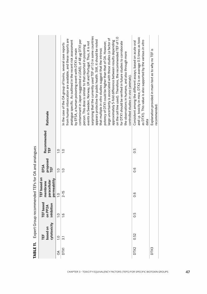
CHAPTER 3 - TOXICITY EQUIVALENCY FACTORS (TEFS) FOR SPECIFIC BIOTOXIN GROUPS 47
TA
BL
E 1
1.
Ex
pe
rt G
rou
p r
eco
mm
en
de
d T
EF
s fo
r O
A a
nd
an
alo
gu
es
TE
F
ba
se
d o
n
cy
toto
xic
ity
TE
F b
as
ed
on
PP
2A
inh
ibit
ion
TE
F b
as
ed
on
me
mb
ran
e
pa
rac
ell
ula
r
pe
rme
ab
ilit
y
EF
SA
pro
po
se
d
TE
F
Re
co
mm
en
de
d
TE
FR
ati
on
ale
OA
1.0
1.0
1.0
1.0
1.0
DT
X1
3.1
1.6
2–
151.
01.
0In
th
e c
ase
of
the
OA
gro
up
of
tox
ins,
se
ve
ral
case
re
po
rts
fro
m h
um
an
in
tox
ica
tio
n a
re a
va
ila
ble
, a
nd
th
ese
re
po
rts
are
an
alo
gu
e s
pe
cifi
c. A
s o
utl
ine
d i
n t
he
re
cen
t ri
sk a
sse
ssm
en
t
by
EF
SA
, a
hu
ma
n p
ois
on
ing
ev
en
t w
ith
DT
X1
as
ma
in
con
tam
ina
nt
in J
ap
an
su
gg
est
ed
a L
OA
EL
of
48
μg
DT
X1
pe
r
pe
rso
n.
Th
is d
ose
is
sim
ila
r to
th
at
rep
ort
ed
in
po
iso
nin
g
ev
en
ts i
n S
we
de
n,
No
rwa
y, U
K a
nd
Po
rtu
ga
l. T
hu
s, i
t is
no
t
surp
risi
ng
th
at
the
cu
rre
ntl
y u
sed
TE
F o
f 1.
0 i
n s
om
e c
ou
ntr
ies
ap
pe
ars
pro
tect
ive
fo
r p
ub
lic
he
alt
h.
Sti
ll,
it s
ho
uld
be
no
ted
tha
t m
ult
iple
in
vit
ro s
tud
ies
sug
ge
st t
ha
t th
e i
ntr
insi
c
po
ten
cy o
f D
TX
1 co
uld
be
hig
he
r th
an
th
at
of
OA
. H
ow
ev
er,
larg
e u
nce
rta
inty
is
ass
oci
ate
d w
ith
th
ese
stu
die
s (a
fa
cto
r o
f
ap
pro
xim
ate
ly 5
-fo
ld d
iffe
ren
ce b
etw
ee
n r
esu
lts
de
pe
nd
ing
on
th
e c
ell
lin
e u
sed
). T
he
refo
re,
the
re
com
me
nd
ed
TE
F o
f 1.
0
for
DT
X1
sho
uld
be
ve
rifi
ed
in
fu
ture
stu
die
s to
co
rro
bo
rate
the
ob
serv
ati
on
s in
hu
ma
ns,
an
d a
lso
th
rou
gh
mo
re
con
tro
lle
d s
tud
ies
in v
ivo
(a
nim
als
).
DT
X2
0.5
20
.50
.60
.60
.5C
on
sist
en
t a
mo
ng
th
e d
iffe
ren
t a
ssa
ys;
ba
sed
on
acu
te o
ral
an
d i
.p.
tox
icit
y i
n m
ice
, D
TX
2 i
s o
n a
ve
rag
e 0
.5 t
ime
s a
s to
xic
as
DT
X1)
. T
his
va
lue
is
als
o s
up
po
rte
d b
y t
he
va
rio
us
in v
itro
da
ta
DT
X3
E
xp
lan
ati
on
pro
vid
ed
in
ma
in t
ex
t a
s to
wh
y n
o T
EF
is
reco
mm
en
de
d.
FAO/WHO TECHNICAL PAPER ON TOXICITY EQUIVALENCY FACTORS FOR MARINE BIOTOXINS ASSOCIATED WITH BIVALVE MOLLUSCS
48
3.3 DOMOIC ACID AND ANALOGUES DA is an excitotoxin produced by the red macro-alga Chondria armata (Takemoto and Daigo, 1958; Takemoto et al., 1966), or by diatoms of the genera Nitzschia, Pseudo-nitzschia (Bates et al., 1989; Wright et al., 1989, 1990a, b; Lundholm et al., 1994) and Amphora (Dhar et al., 2015), and is distributed worldwide.
DA is an acidic crystalline water-soluble amino acid with three carboxyl groups (Takemoto and Daigo, 1958), very similar to the glutamatergic agonist kainate and the amino acid glutamate (Ramsdell, 2007; Vale, 2014).
3.3.1 Toxicity
Th e mechanism of toxicity is well understood, as DA activates alphaamino-5-methyl-3-hydroxyisoxazolone-4-propionate (AMPA)/kainate receptors, thereby increasing intracellular calcium levels, causing glutamate release and subsequent activation of N-methyl-D-aspartate (NMDA) receptors. Th e neurotoxicity is due to the activation of both AMPA/kainate and NMDA receptors (Vale, 2014). At high doses (10 μM), DA releases glutamate and causes neurotoxicity through NMDA receptor activation (Hogberg and Bal-Price, 2011). However, at low doses there is an apoptotic eff ect in neurons through AMPA/kainate that triggers oxidative stress and caspase 3 activation (Giordano et al., 2007).
DA is a partial agonist of kainate receptors, with an affi nity in the low nM range (Crawford et al., 1999). It binds with high affi nity to GluK1 and GluK2 kainate receptors, and with lower affi nity to GluK4 and GluK5 (Vale, 2014). Th e symptoms of toxicity (nausea, vomiting, abdominal cramps, diarrhoea, headache, memory loss) include serious neurological damage, which is well known as a consequence of the deadly human intoxications that occurred in Quebec in 1987 (Perl et al., 1990; Teitelbaum et al., 1990; Doucette and Tasker, 2008). Th e damage aff ected in particular the hippocampus, amygdaloid nucleus and subfrontal cortex. Th e LOAEL in humans is estimated as 0.9 mg/kg, and the EFSA Contam Panel established an ARfD of 30 μg/kg b.w. (EFSA, 2009a). Th e Joint FAO/IOC/WHO ad hoc Expert Consultation proposed a provisional ARfD of 100 μg/kg b.w. and

CHAPTER 3 - TOXICITY EQUIVALENCY FACTORS (TEFS) FOR SPECIFIC BIOTOXIN GROUPS 49
concluded that this dose may also be considered as a provisional TDI (CODEX, 2006).3
DA is especially toxic in young neurons in newborn mice (Xi, Peng and Ramsdell, 1997; Mayer, 2000; Vale, 2014). Recent studies also show that DA may also have endocrine eff ects that relate to water balance, as it has been reported to accumulate in hypophysis (adeno- and neurohypophysis), and not in the pars intermedia (Crespo et al., 2015). A recent article reports a new toxic eff ect not described before, a carditotoxic eff ect of 2.5 mg/kg DA in rats 30 days aft er an i.p. dose (Vieira et al., 2016).
Toxicologic DA studies are conditioned by the quality of the test chemical used, as there are potentially very large diff erences in the toxicity unless it is certifi ed (Crespo et al., 2015). Th erefore, the doses reported in the literature must be treated with caution unless thoroughly checked for the real concentration. Rats and mice are rather diff erent in sensitivity to DA. with i.p. lethal doses of 2.5 mg/kg for rats (Crespo et al., 2015) and 6 mg/kg for mice (Munday et al., 2008b; Crespo et al., 2015). Th ere are diff erences in the oral bioavailability of DA in rats and mice, as mice were reported to have a higher sensitivity than rats (Iverson et al., 1989).
Samples of domoic acid and isodomoic acids A, B and C, of 98–99% purity, were compared with regard to behavioural changes in mice aft er i.p. injection. At a dose of 5 mg/kg, domoic acid induced severe toxic changes, as indicated by hypoactivity, sedation, limb rigidity, stereotypic responses, loss of postural control, and forelimb tremors. In contrast, the isodomoic acid had little or no eff ect at this dose, and even at 20 mg/kg, isodomoic acid induced little change in the behaviour of the animals (Munday et al., 2008b).
3 Th e reason for the discrepancy between EFSA and FAO/IOC/WHO lies in the use of diff erent uncertainty factors by the two expert committees. Th e EFSA Panel used a Lowest Observed Adverse Eff ect Level (LOAEL) of 0.9 mg/kg for neurotoxicity in humans. Th ey applied a factor of 3 for extrapolation from a LOAEL to a No Observed Adverse Eff ect Level (NOAEL) and then applied a further factor of 10 “to allow for human variability and also for the fact that sensitive methods for detection of neurotoxic eff ects had not been used in the investigation of aff ected individuals”. Th ey therefore proposed an ARfD of 30 micrograms/kg b.w. Th e FAO/IOC/WHO Panel used a LOAEL of 1.0 mg/kg, and then applied a safety factor of 10 to give a provisional ARfD of 100 μg/kg b.w. It was argued that this value was reasonable, since one individual who consumed 0.33 mg/kg b.w. did not become ill. It was also argued that cumulative eff ects of low doses of DA are unlikely, and on this basis it was concluded that the ARfD could be considered as a provisional TDI.

FAO/WHO TECHNICAL PAPER ON TOXICITY EQUIVALENCY FACTORS FOR MARINE BIOTOXINS ASSOCIATED WITH BIVALVE MOLLUSCS
50
Of all the analogues of DA, only isodomoic acid A is relevant from a toxicity point of view, since all others have very low toxicity, with nM inhibition constants in the kainate receptor of 2.4, 4.4, 4990, 171, 600, 600 and 67 for DA and Isodomoic acids A, B, C, D, E, F, respectively (Vale, 2014). Only the analogues D, E and F are found in shellfi sh, and they are currently considered not toxic. DA transforms to epi-DA in storage or by ultraviolet light, and in general DA and epi-DA (the C5´-diastereomer) are considered as one toxin (Quilliam et al., 1989c; Wright et al., 1990b; EFSA, 2009a).
DA has a low bioavailability and it is rapidly eliminated in the kidney, with only 6 percent of an i.v. dose reaching the brain in rats (Maucher Fuquay et al., 2012). Th e i.p. LD50 in mice was reported to be 6.2 mg/kg (Iverson et al., 1989), 6 (Munday et al., 2008b; Crespo et al., 2015) and close to 4 (Grimmelt et al., 1990; Peng and Ramsdell, 1996; Nagatomo et al., 1999). Th e i.p. LD50 in rats is lower than in mice, as mentioned before, with reported values of 2.5 mg/kg (Crespo et al., 2015), although higher values were also reported, e.g. 4 (Iverson et al., 1989) and 7.5 (Tryphonas et al., 1990); in young rats, toxicity is higher, with LD50 in 2-day-old animals of 0.25 mg/kg, and 0.7 mg/kg in 10-day-old animals (Xi, Peng and Ramsdell, 1997).
Th e administration of low doses (1.2 mg/kg) of DA to pregnant rats on day 13 causes persisting behavioural changes in newborn animals (Levin et al., 2005).
Th e oral LD50 is much lower than the i.p. LD50, with reported values of 71–83 mg/kg in mice and 80–82 mg/kg in rats (Iverson et al., 1989).
3.3.2 Conclusion
No TEF is necessary as only DA is found in foods (sum of DA and epi-DA expressed as DA).
3.4 AZASPIRACID AND ANALOGUES AZAs, produced by the genera Azadinium and Amphidoma (Tillmann et al., 2009, 2014; Krock et al., 2012), are a food safety problem fi rst recognized in 1996 (McMahon and Silke, 1996) and later identifi ed chemically (Satake et al., 1998b). Th eir chemical structure is unusual, and the mechanism whereby they exert their toxic eff ects is at present unknown (Botana et al., 2014b).
3.4.1 Toxicity
AZAs are easily absorbed orally (Aasen et al., 2010, 2011; Aune et al., 2012), with blood levels peaking within 30 minutes aft er oral administration and a half-life of 24 h in mice (Twiner, Hess and Doucette, 2014). Th e symptoms of intoxication
CHAPTER 3 - TOXICITY EQUIVALENCY FACTORS (TEFS) FOR SPECIFIC BIOTOXIN GROUPS 51
in humans are vomiting, nausea, diarrhoea and stomach cramps, which appear aft er a few hours and last for up to two days (Klontz et al., 2009). No deaths from AZA ingestion have been reported. Human intoxications observed in the United Kingdom, United States of America, Italy and France permitted evaluation of the risk associated with the consumption of these compounds, and the EFSA Panel established an ARfD of 0.2 μg AZA equivalents/kg b.w. (EFSA, 2008a). Th e Joint FAO/IOC/WHO ad hoc Expert Consultation established a provisional ARfD of 0.04 μg/kg b.w. body weight, but they were unable to establish a TDI (FAO/IOC/WHO, 2004).
In mice, AZAs cause multiple organ damage aft er oral administration, with important injuries in intestinal epithelium, lamina propria and villi, and a lethal oral dose of 700 μg/kg b.w. (Ito et al., 2000, 2002b). Prolonged administration (4 months) at doses of 1 to 50 μg/kg b.w. showed organ damage, with lung tumours and malignant lymphomas at the highest doses (Ito et al., 2002b), although the relevance of these studies is far from conclusive due to extensive and severe toxicity noted at the higher doses (EFSA, 2008a).
According to the few data available so far, the simultaneous eff ect of AZAs with other lipophilic toxins is not functionally relevant (pectenotoxin, yessotoxin or OA), since it does not increase the toxicity of the other toxin groups. It has been shown, however, that the bioavailabilities of pectenotoxin 2 and OA are slightly increased (Aune, 2009) and slightly decreased (Aune et al., 2012), respectively, by simultaneous administration of AZA.
AZAs were reported to be cytotoxic by targeting several receptors (Roman et al., 2002) that participate in the apoptosis process (Twiner et al., 2005; Botana et al., 2014b), such as caspase activation and cytoskeletal disruption (Vilarino et al., 2006, 2007), cytochrome release and DNA fragmentation (Twiner et al., 2012b), increase in c-jun-N-terminal protein kinase (JNK) phosphorylation and inhibition of calcium oscillations (Vale et al., 2007, 2008c; Cao et al., 2010), and eff ects at the gene level in several fatty acid biosynthesis routes (Twiner et al., 2008a). Th e eff ect of AZAs in ion channels may be linked to some of its toxic eff ects, as they decrease cell volume mediated by potassium and chloride effl ux (Vale et al., 2010), deplete ATP (Kellmann et al., 2009), inhibit endocytosis (Bellocci et al., 2010) and decrease procathepsin pools in endocytosis (Sala et al., 2013).
An intriguing observation is that AZAs are only present in mussel samples with high levels of glutaric acid, and the combined eff ect of both AZA1 (50 nM) and glutaric acid (1 mM) increases the blockade of voltage dependent sodium channels (Chevallier et al., 2015). Th is combined eff ect might explain the reported neurological symptoms described in AZA toxicity (Twiner, Hess and Doucette, 2014).

FAO/WHO TECHNICAL PAPER ON TOXICITY EQUIVALENCY FACTORS FOR MARINE BIOTOXINS ASSOCIATED WITH BIVALVE MOLLUSCS
52
Data on the acute toxicity of AZAs to mice by i.p. injection are shown in Table 12.
TABLE 12. Acute toxicities of AZAs by i.p. injection
Compound ParameterAcute toxicity
(μg/kg b.w.) Reference
AZA1 Lethality 200 Munday, 2014
AZA1 MLD 150 Satake et al., 1998a
AZA1 LD50
74 Marine-Institute, 2014
AZA1 LD50
>10 and <55 in rats Ferreiro et al., 2016
AZA2 Lethality Approximately 110 Munday, 2014
AZA2 LD50
117 Marine-Institute, 2014
AZA2 LD50
(i.v.) 11 in rats Ferreiro et al., 2014
AZA3 Lethality Approximately 140 Munday, 2014
AZA3 LD50
164 Marine-Institute, 2014
AZA4 Lethality Approximately 470 Munday, 2014
AZA5 Lethality <1 000 Munday, 2014
AZA6 LD50
100 Marine-Institute, 2014
Data on the acute oral toxicity of AZAs are shown in Table 3.8.
TABLE 13. Acute toxicities of AZAs by oral administration
Compound ParameterAcute toxicity
(μg/kg b.w.)Reference
AZA1 Lethality > 700 Ito, 2008
AZA1 LD50
775 Aune et al., 2012
AZA1 LD50
443 Marine-Institute, 2014
AZA2 LD50
626 Marine-Institute, 2014
AZA3 LD50
875 Marine-Institute, 2014
Repeated oral administration of AZA1 to mice at 50 μg/kg b.w. twice weekly over a period of up to 20 weeks caused death of all the animals, and at 20 μg/kg b.w. it killed 30 percent of the mice (Ito et al., 2002b). Th ere are confl icting reports as to what organs are aff ected, from general damage to liver, spleen or thymus with doses of 500 μg/kg b.w. (Ito et al., 2000) to very little damage in these organs and mostly lesions in the stomach and intestine (Aasen et al., 2010; Aune et al., 2012). Probably a more important observation is that AZAs are direct blockers of the open state of hERG potassium channels (Twiner et al., 2012c), and this translates into the in vivo cardiotoxicity of AZAs through hERG channels in rats (Ferreiro et al., 2014), causing a prolongation of the PR electrocardiogram interval and arrhythmias with 11 μg/kg b.w. AZA2 i.v. Th is i.v. value is only 5 times higher than the LOAEL proposed by the EFSA working group in humans of 1.9 μg/kg b.w. (EFSA, 2008a). Th e i.v. bioavailability is 100 percent, and oral availability of AZAs is lower, with only 2 percent of an administered gavage dose in internal

CHAPTER 3 - TOXICITY EQUIVALENCY FACTORS (TEFS) FOR SPECIFIC BIOTOXIN GROUPS 53
mice organs aft er 24 hours (Aasen et al., 2010). Considering oral bioavailability for AZA in humans as 20 percent and with cardiotoxicity potential in humans similar to that in rats, the derived LOAEL in humans could be potentially cardiotoxic (Ferreiro et al., 2014, 2016).
Th e lethal dose of AZA2 in rats is below 55 μg/kg b.w. i.v. and close to 11 μg/kg b.w., since this dose caused the death of 50 percent of the animals; also, 11 μg/kg b.w. i.v. is enough to induce potentially lethal extrasystoles in 50 percent of the animals (Ferreiro et al., 2014). A continuation of this study has shown that, in rats, 10 μg/kg b.w. i.p. aft er 4 doses in 15 days (one dose every 4 days) induces ascites, ultrastuctural damage in the heart. Hypotension is observed with only 1 μg/kg b.w. i.p. It is remarkable that this study, which was performed with AZA1, did not show any death with 10 μg/kg b.w., and all animals died with the fi rst administrations of 55 μg/kg b.w. AZA1 (Ferreiro et al., 2016). Th is suggests that AZA1 i.p. is about 5 times less toxic in rats than AZA2 (which was administered i.v.).
Based on this, the cardiotoxicity results suggest that the toxicity of AZAs might be higher than currently estimated.
3.4.2 In vitro relative potency
Th e recent in vitro studies of several recently identifi ed analogues support the conclusion that relative potencies based on in vitro studies do not match the relative potencies based on in vivo eff ects, i.e. i.p. LD50 in mice (Table 14). Examples of this is the relative cytotoxicity in Jurkat T lymphocytes, being 1, 0.22 and 5.5 for AZA1, AZA33 and AZA34, respectively (Kilcoyne et al., 2014a); 5.1 for 37-epi-AZA1 (Kilcoyne et al., 2014a); 0.6, 0.4, 7, 4.5, 0.4 and 0.2 for AZA4, AZA5, AZA6, AZA8, AZA9, AZA10, respectively (Kilcoyne et al., 2015); 1, 8.3 and 4.5 for AZA1, AZA2 and AZA3, respectively (Twiner et al., 2012a); 1.3 and 1 for AZA2 and AZA3 using patch clamp-on voltage clamped hERK channels in HEK 293 cells (Twiner et al., 2012c); 1.89 for AZA2 in 24 h treated 2–3 days in vitro cultured mice neurons (Vale et al., 2008c), 1.36 and 3.22 for AZA2 and AZA3 in spontaneous calcium oscillations; and 0.89 and 4.32 for AZA2 and AZA3 in LDH release in mice neocortical neurons (Cao et al., 2010). Th is variation in in vitro relative potencies is probably linked to the potential diff erent responses of the diff erent analogues in each cell model and each transduction signal (Roman et al., 2004; Vale et al., 2007, 2008c, 2010; Alfonso et al., 2008; Botana et al., 2014b).

FAO/WHO TECHNICAL PAPER ON TOXICITY EQUIVALENCY FACTORS FOR MARINE BIOTOXINS ASSOCIATED WITH BIVALVE MOLLUSCS
54
TABLE 14. Relative in vitro toxicity of AZAs
Compound
Cell type
Jurkat T
(cytotoxicity)
HEK 293
(hERK
current)
2–3 DIV mice
neurons
(cytotoxicity)
Neocortical neurons
(LDH release) (calcium
oscillations)
AZA1 1 1 1 1 1
AZA2 8.3 1.2 1.89 0.89 1.36
AZA3 4.5 1 4.32 3.22
AZA4 0.6
AZA5 0.4
AZA6 7
AZA8 4.5
AZA9 0.4
AZA10 0.2
AZA33 0.22
AZA34 5.5
37-epi-AZA1 5.1
3.4.3 Conclusion
Th e identifi cation of TEFs for each AZA analogue involves several variables that make it diffi cult to defi ne criteria, especially taking into account that the toxicological target of AZAs is not yet defi ned. Based on the diff erent toxicities, there is some consistency between cardiotoxicity in rats and other studies in mice, which along with the conclusions of the EFSA Panel, allowed the expert meeting to propose the TEFs as shown in Table 15.
TABLE 15. TEFs Recommended by the Expert Group
Compound
TEF based
on i.p.
toxicity
TEF based
on oral
toxicity
EFSA
proposed
TEF
Recommended
TEFRationale
AZA1 1.0 1.0 1 1.0
AZA2 0.6 0.7 1.8 0.7 Based on recent oral
data. (also consistent
with recent i.p. data)
AZA3 0.45 0.51 1.4 0.5 Based on recent oral
data. (also consistent
with recent i.p. data)
AZA4 – Cannot determine TEF
due to lack of data.
AZA5 – Cannot determine TEF
due to lack of data.
AZA6 0.7 – 0.7 No oral data, based on
recent i.p. data.

CHAPTER 3 - TOXICITY EQUIVALENCY FACTORS (TEFS) FOR SPECIFIC BIOTOXIN GROUPS 55
3.5 BREVETOXINSBrevetoxins target the neurotoxin receptor site 5 of domain IV of the alpha subunit of voltage-gated sodium channels, resulting in membrane depolarization, repetitive fi ring, and increases in sodium currents (Murrell and Gibson, 2009). BTXs have been found to activate voltage-gated sodium channels in the following manner: prolonging the mean open time, preventing inactivation of the channel, and making the channel activation more negative in value (Jeglitsch et al. 1998; Schreibmayer and Jeglitsch, 1992; Catterall, 1992). Rapid reductions in respiratory rate, cardiac conduction disturbances, and reduced body temperatures are in vivo eff ects produced by BTXs (Poli et al., 1990; Templeton, Poli and Solow, 1989; van Apeldoorn, van Egmond and Speijers, 2001). Tremors, muscular contractions or fasciculations, Straub tail phenomenon, laboured breathing, and death have been observed in orally dosed mice. Mice injected with PBX3 presented salivation, lacrimation, urination and defecation, with hypersalivation being the most pronounced. At high doses, compulsive chewing and excessive mucous in the nasal cavity were reported. Morohashi et al. (1999) documented that BTX-B2 and BTX-B4 caused paralysis of hind limbs, diarrhoea, dyspnea and convulsion following i.p. injection. I.p. injection of BTX-B1 led to irritability, hind and/or hind-quarter paralysis, severe dyspnea and convulsions before death due to respiratory paralysis (Ishida et al. 1995; 1996).
Much of what is known about the acute toxicity of BTX analogues and metabolites has been determined in mice, mostly via i.p. administration (Table 16). Shimizu et al. (1986) reported PBX1 to be the most toxic of the BTX analogues. PBX2 was determined to be less toxic by oral ingestion compared with i.v. or i.p. administration. Of the metabolites identifi ed in shellfi sh from New Zealand, BTX-B1 was found to be the most toxic (Ishida et al., 1995). BTX-B4 was threefold more toxic than BTX-B2 and comparable to the toxicity of PBX3 (Baden et al., 1982; Murata et al., 1998; Morohashi et al., 1999). In vitro studies using Jurkat calls demonstrated that PBX2 and PBX6, in comparison with PBX3, led to greater cellular eff ects, including apoptosis, as measured by caspase–3 activity (Walsh et al., 2008). Recent studies have employed Caco–2 permeability assays to assess BTX metabolite toxicity (Henri et al., 2014).
3.5.1 Conclusions
Given the limited data currently available for establishing TEFs, notably lacking human oral potency data, and since the Codex Standard method for BTXs is currently the MBA and the limit is expressed as mouse units, TEFs for BTXs are not proposed at this time. Rather the topic is covered in the section on data gaps.

FAO/WHO TECHNICAL PAPER ON TOXICITY EQUIVALENCY FACTORS FOR MARINE BIOTOXINS ASSOCIATED WITH BIVALVE MOLLUSCS
56
TABLE 16. Acute toxicity of BTX analogues in mice
BrevetoxinObservation/
Route
Toxicity
μg/kg b.w.Original source
PBX1 LD50
, 24-h, i.p. >100 Dechraoui et al., 1999
PBX2 LD50
, 24-h, i.v. 200 Baden et al., 1982
LD50
, 24-h, i.p. 200 Baden et al., 1982
LD50
, 24-h, oral 6600 Baden et al., 1982
PBX3 LD50
, 24-h, i.v. 94 Baden et al., 1982
LD50
, 24-h, i.p. 170 Baden et al., 1982
LD50
, 24-h, oral 520 Baden et al., 1982
LD50
, 24-h, i.p. 250 Selwood et al., 2008
BTX-B2 LD50
, 24-h, i.p. 400 Selwood et al., 2008
S-desoxy-BTX-B2 LD50
, 24-h, i.p. 211 Selwood et al., 2008
BTX-B1 MLD(1), <2 h, i.p. 50 Ishida et al., 1995
BTX-B2 MLD, <1 h, i.p. 306 Murata et al., 1998
BTX-B3 MLD, 24 h, i.p. >300(2) Morohashi et al., 1995
BTX-B4 MLD, 6–24 h, i.p. 100 Morohashi et al., 1999
BTX-B5 MLD(3), i.p. 300–500 Ishida et al., 2004
Notes: (1) Minimum lethal dose, (2) No deaths recorded at 300 μg/kg. (3) Time to death not reported. Adapted from Plakas
and Dickey, 2010, and Dickey et al., 2011.
57
4Data gaps and recommendations for research
4.1 DATA GAPS FOR EACH GROUP FOR WHICH TEFS WERE CONSIDERED4.1.1 STX
• No information available about pharmacokinetics of each analogue, and limited information on the oral toxicity of saxitoxin analogues, especially those that are commonly found in seafood or that are found only rarely but at relatively high levels.
• Need for further information of specifi c TEFs of all analogues in pure and certifi ed form.
• Need for toxicity studies with benzoate derivatives.• Need for TEFs for TTX analogues commonly found in bivalves. • Need for comparative studies on the oral toxicity of tetrodotoxin congeners.
4.1.2 OA
• Th ere is no comparative data on analogues in a single study by oral administration with the same methodology. Th ese data are required.
• Th ere is little information clarifying the links between phosphatase inhibition and the physiological mechanisms that regulate intestinal physiology.
• As recommended by the Joint FAO/IOC/WHO ad hoc Expert Consultation (CODEX, 2006), information on the chronic toxicity of DSPs is needed.
CHAPTER 4 - DATA GAPS AND RECOMMENDATIONS FOR RESEARCH

JOINT FAO/WHO EXPERT MEETING ON PUBLIC HEALTH RISKS OF HISTAMINE AND OTHER BIOGENIC AMINES FROM FISH AND FISHERY PRODUCTS
58
• Consideration should be given to the determination of TEFs for DSPs by diarrhetic activity.
• Th e recommended TEF of 1.0 for DTX1 should be verifi ed in future studies to corroborate the observations in humans, also through more controlled studies in vivo (in animals).
4.1.3 Domoic Acid
• Based on the information on chronic and low-dose neurotoxic eff ects, especially related to exposure of prenatal and very young individuals, more information is needed.
• Th e relevance of potential endocrine eff ects needs to be further investigated. • Chronic and low-dose eff ects, especially related to pregnancy and in very
young individuals, and potential endocrine eff ects, need serious attention with regard to DA toxicity.
• Studies needed on the toxicity of DA isomers present in food.
4.1.4 AZA
• Comparative study of all analogues with the same methodology is needed, e.g. for AZA4 and –5, for which oral toxicity data are not currently available.
• Detailed study is needed of oral toxicity, with special attention to cardiotoxicity; and possible interaction with YTXs should be investigated.
• Defi ne the exact mechanisms of chronic and acute toxicity of AZAs (for cardiotoxicity, and GI tract symptoms). Th e relevance of lung toxicity observed for the AZAs for humans is currently unclear.
• In view of the deaths seen aft er repeated oral dosing of AZA1 to mice at levels well below the LD50 (Ito et al., 2002b) and the possible induction of lung cancer by this substance (Ito, 2008), a long-term feeding study, conducted according to OECD guidelines, should be conducted either with AZA1 itself or a mixture of AZAs of known composition. In view of the high spontaneous incidence of lung cancer in mice, rats should be employed in such an experiment. Rats are much less susceptible to spontaneous pulmonary carcinogenesis than mice (Munday, 2014).
4.2 CONSIDERATIONS FOR OTHER BIOTOXINS 4.2.1 The special case of tetrodotoxin (TTX)
TTX is a marine toxin of bacterial origin and is produced, amongst others, by Pseudomonas spp. and Vibrio spp. (Bane et al., 2014). It is becoming a concern in Europe given its presence in gastropods (Rodriguez et al., 2008; Silva et al., 2012) and in shellfi sh (Turner et al., 2015a; Vlamis et al., 2015). Because TTX in shellfi sh is a newly discovered phenomenon, there is no surveillance programme in place per se. Nevertheless, monitoring programmes that employ MBA for STX would
CHAPTER 4 - DATA GAPS AND RECOMMENDATIONS FOR RESEARCH 59
also detect the presence of TTX. While the MBA does not distinguish between TTX and STX, analytical methods for these two toxins diff er. Th us, TTX can be detected as a diff erent chemical entity and a TEF could be applied.
While the mode of action is similar to STX, the main diff erence between both toxin groups is the subtypes of Nav with which they bind preferentially. In the case of TTX, the Nav 1.7 is the main target (Walker et al., 2012; Alonso et al., 2016), although TTX can bind with less affi nity to other Nav subtypes. TTX binds to human Nav 1.7 with 38-fold more potency than STX (Walker et al., 2012). As for STX, TTX may target other receptors but they are not critical to explain its toxicity (Guatimosim et al., 2001).
Th e toxicity of TTX is high, with 2 mg being a lethal dose to humans (Noguchi, Onuki and Arakawa, 2011). A mouse unit is 0.2 μg, which kills a 20 g mouse in 30 minutes. Based on the i.p. toxicity to mice, relative toxicities of TTX analogues described in the literature are 10 μg/kg b.w. for TTX (Noguchi, Onuki and Arakawa, 2011), 70 μg/kg b.w. for 11-deoxy-TTX (Bane et al., 2014), 420 μg/kg b.w. for 6,11-dideoxy TTX (Jang and Yotsu-Yamashita, 2007), 16 μg/kg b.w. for 11-oxo-TTX (Nakamura and Yasumoto, 1985; Yotsu-Yamashita et al., 2003), 64 μg/kg b.w. for 4-epi-TTX (Nakamura and Yasumoto, 1985; Munday, 2014), 60 μg/kg b.w. for 6-epi-TTX (Yasumoto et al., 1988b), 490 μg/kg b.w. for 4,9-Anhydro-TTX (Nakamura and Yasumoto, 1985), 54 μg/kg b.w. for 11-nor-TTX–6(S)-ol (Yotsu et al., 2014), and 70 μg/kg b.w. for 11-nor-TTX–6(R)-ol (Endo et al., 1988).
Since a mouse unit for STX is 0.183 μg (9.15 μg/kg b.w.) (Schantz et al., 1958; AOAC, 2005), then the potency of STX is 10 percent higher than TTX. With these fi gures, it is possible to construct a basic TEF list for TTX compared with STX: 1 for STX; 1.1 for TTX; 7.7 for 11-deoxy-TTX; 46 for 6,11-dideoxy TTX; 1.7 for 11-oxo-TTX; 7 for 4-epi-TTX; 6.6 for 6-epi-TTX (Yasumoto et al., 1988b); 53.6 for 4,9-Anhydro-TTX; 5.9 for 11-nor-TTX–6(S)-ol; and 7.6 for 11-nor-TTX–6(R)-ol. Further information is needed for each TTX derivative.
Th e relative potency of TTX analogues, as reported as IC50 in in vitro systems, are 4.1 nM (frog muscle (Yang, Kao and Oshima, 1992a)) or 5.2 nM (squid axon; Kao and Yasumoto, 1985a) for TTX; 13.2 nM for 4-epi TTX (Kao and Yasumoto, 1985a); 96 nM for 6-epi TTX (Yang, Kao and Oshima, 1992a); 445 nM for 11-deoxy TTX (Yang et al., 1992a); and 298 nM for 4,9-anhydro TTX (Kao and Yasumoto, 1985a).
JOINT FAO/WHO EXPERT MEETING ON PUBLIC HEALTH RISKS OF HISTAMINE AND OTHER BIOGENIC AMINES FROM FISH AND FISHERY PRODUCTS
60
Th e lethality of TTX decreases with the route of administration, from 10 μg/kg b.w. i.p., to 16 μg/kg b.w. subcutaneous, and 332 μg/kg b.w. oral (Kao, 1966; Moczydlowski, 2013).
While, traditionally, TTX contamination has been associated with puff er fi sh, the occurrence in bivalves is emerging, and as such the Expert Group recommended that this should be monitored. Th e Expert Group also noted the:
• Need to establish TEFs for TTX analogues commonly found in bivalves. • Need for comparative studies on the oral toxicity of tetrodotoxin congeners.
4.2.2 Yessotoxin and analogues
Yessotoxins (YTX) are a group of ladder shaped compounds produced by the dinofl agellate Protoceratium reticulatum (Satake, MacKenzie and Yasumoto, 1997a; Miles et al., 2005), which were fi rst isolated from digestive glands of scallops Patinopecten yessoensis in Japan (Murata et al., 1987; Satake et al., 1997b). Th eir chemical identifi cation was fi rst reported by Murata et al. (1987) in Japan, and several analogues were fi rst identifi ed from shellfi sh harvested in Italy (Ciminiello et al., 1997; Satake et al., 1997b), Norway (Lee et al., 1988), New Zealand and the Adriatic Sea (for this reason one of the analogues was named adriatoxin (Ciminiello et al., 1998)). Th ey can also be produced by Lingulodinium polyedrum (Draisci et al., 1999) or Gonyaulax spinifera (Pistocchi et al., 2012), and in total there are about 90 chemical derivatives, very few of them being abundant in nature (yessotoxin, 1a-homoYTX, 45-hydroxyYTX, 45-hydroxy–1ahomoYTX, carboxy-YTX) (Miles et al., 2004a, b; Finch et al., 2005). Th ey have been reported in coastal areas worlwide (Vlamis and Katikou, 2014). Although YTX is rather toxic to mice aft er i.p. injection, these compounds do not show clear signs of toxicity aft er oral administration, even at high doses (Munday et al., 2008a; EFSA, 2008b) – they were identifi ed as toxins mostly because of their eff ect on the mouse bioassay. Th eir toxicology is now better known (Aune et al., 2002; Aasen et al., 2011), they do not produce diarrhoea, and the scientifi c debate today is on whether or not they are capable of causing harm in humans (Tubaro et al., 2003; Tubaro et al., 2004; Munday, 2014).
Th e mechanism of action of YTX remains to be fully elucidated. However, like AZAs that may have several targets, YTX and analogues are compounds that target several apoptotic pathways (Korsnes et al., 2011, 2013, 2014; Tobio et al., 2012) and induce autophagy triggered by stress (Rubiolo et al., 2014). Even though many eff ects are derived from this eff ect, the fi nal eff ect of YTX is an apoptotic eff ect especially intense in tumour cell lines (which makes YTX a potential anticancer drug lead) (Botana et al., 2014b), and a rather mild eff ect in non-tumour cell lines (Fernandez-Araujo et al., 2015b). Although not the only target, phosphodiesterase

CHAPTER 4 - DATA GAPS AND RECOMMENDATIONS FOR RESEARCH 61
4A seems to play a key role in the mode of action of YTX (Alfonso et al., 2003; Fernandez-Araujo et al., 2015a).
Th e EFSA Panel proposed an acute reference dose (ARfD) of 25 μg YTX equivalents/kg b.w. (EFSA, 2008b), and the European Union has defi ned a toxin limit of 3.75 mg yessotoxin equivalent/kg shellfi sh meat (EU, 2013). Th is was imposed as a precautionary measure, because there has never been a report of YTX intoxication in humans. Yessotoxin is an exotoxin, released by the producing cells (Hess and Aasen, 2007a). Its ecological role is not known, and its inclusion in the list of marine toxins is due to the fact that it co-exists with phosphatase inhibitors (OA and dinophysistoxins) and causes mice death in the biological mouse bioassay (Aune et al., 2002).
Since there are no reports of eff ects in humans, all actual toxicology comes from rodent experiments. Th e acute i.p. toxicity of YTX has been reported with a large variation, from 80 (Ogino, Kumagai and Yasumoto, 1997) to 750 μg/kg b.w. (Aasen et al., 2011). Several LD50 studies in diff erent mice strains and in both sexes (Aune et al., 2008) show no discrepancies with regard to both parameters, for YTX and homoYTX (Tubaro et al., 2003, 2004; Aune et al., 2008; Munday, Aune and Rossini, 2008a). Th e reported i.p. LD50 ranges were: 80–100 μg/kg b.w. body weight (Ogino et al., 1997; Munday, 2014); 112–136 μg/kg b.w. (Munday, Aune and Rossini, 2008a); 269–462 μg/kg b.w. (Aune et al., 2008); 286 and 301 μg/kg b.w. for YTX and didesulfated YTX, respectively (Ogino, Kumagai and Yasumoto, 1997); 220 μg/kg b.w. for 45–46–47-Tri-nor-YTX (Satake et al., 1996); 500–750 μg/kg b.w. (Aune et al., 2002; Munday, 2014); and 444 and 530 μg/kg b.w. for homoYTX and YTX, respectively (Tubaro et al., 2003). Some compounds were reported toxic by the mouse bioassay, with lethal doses of 500 μg/kg b.w. for 45-hydroxy-YTX (Satake et al., 1996); carboxy-YTX (Ciminiello et al., 2000a); carboxy-homo-YTX (Ciminiello et al., 2000b); and 1-desulfo-YTX (Daiguji et al., 1998a). Th ere is no report of deaths aft er i.p. administration of other YTX analogues, unless at high concentrations (Munday, 2014).
Acute dosing experiments suggest that YTX is the most toxic of all analogues, and that it is a slow-acting compound, with death aft er injection showing a longer delay with lower doses, from 30 minutes at high doses to 10 hours at doses close to the LD50 (Munday, Aune and Rossini, 2008a). No macroscopic changes were observed in animals aft er YTX injection in some studies, but a few authors have reported some cases of cardiac oedema and vacuolization of cardiac muscle, although some controls reported similar changes (Aune et al., 2002). At the same time, there are studies that have shown no such eff ect aft er lethal doses (Tubaro et al., 2004). Electron microscopy studies revealed some alterations at a cardiac level (Terao et al., 1990), with swollen endothelial cells in the left ventricle, rounded

JOINT FAO/WHO EXPERT MEETING ON PUBLIC HEALTH RISKS OF HISTAMINE AND OTHER BIOGENIC AMINES FROM FISH AND FISHERY PRODUCTS
62
mitochondria, and separated bundles of myofi brils and sarcoplasmic reticulum (Aune et al., 2002). Terao et al. (1990) reported fatty degeneration and pancreatic and hepatic alterations aft er administration of di-desulfo-YTX.
Th e administration of acute oral doses have provided consistent results in several studies (Ogino, Kumagai and Yasumoto, 1997; Aune et al., 2002; Tubaro et al., 2004). Th ey all describe a complete lack of eff ects even at high doses, and most of the toxin is recovered from the lower intestine and faeces (Munday, Aune and Rossini, 2008a). Nevertheless, some structural changes were observed aft er high oral dosing (up to 10 mg/kg), such as heart intracellular oedema or swelling, or pericapillary myocytes, causing the separation of organelles (Aune et al., 2002; Tubaro et al., 2004), while no eff ect was reported at low doses (Terao et al., 1990). Th e signifi cance of these ultrastructural changes are dubious, as some of the changes are reported in control animals, and they are not dose-dependent (Tubaro et al., 2010; Munday, 2014). A recent acute study in rats, where potential electrocardiogram alterations were studied aft er i.v. administration, showed that 10 μg/kg b.w. YTX (and also DTX1 or OA) did not modify the electrical function of the heart (Ferreiro et al., 2015), and the acute eff ects do not modify cardiac biomarkers.
A recent subacute study performed in rats treated for 15 days with four doses of YTX i.p., one every 4 days (days 1, 5, 9 and 13), was designed to study the eff ect of YTX (doses of 50–100 μg/kg b.w.) on electrocardiogram, arterial blood pressure and cardiac biomarkers. Th e results show a clear bradycardia, hypotension, autophagia in the heart tissue, and changes in kidney function (Botana et al., manuscript submitted); there was an ultrastructural cardiomyocyte alteration that was dose-dependent (observed in 1/1 rat treated with 100, in 6/7 rats treated with 70 and in 5/8 rats treated with 50 μg/kg). Th erefore, it is clear that YTX may be cardiotoxic, but the relevance of this to oral administration remains to be confi rmed.
Repeated oral studies in rodents show that yessotoxin does not induce damage in organs or any death for up to 7 days with 2 mg/kg/day, or even 5 mg/kg/day administered seven times in 21 days (Munday, 2014). Ultrastructural analysis of tissues 90 days aft er the treatment show no sign of damage, though Tubaro et al. (2008b) reported some clusters of rounded mitochondria and disorganization of myofi brils in the heart tissue aft er seven days of oral exposure to 1 mg/kg, which may last for 30 days but disappears aft er 90 days. Although very large doses of YTX (5 mg/kg) administered orally give rise to levels of 0.02 percent and 0.01 percent of the total dose in the liver and the kidney, respectively (Aasen et al., 2011), there are no data on the kinetics of YTX excretion in urine, hence a pharmacokinetic model

CHAPTER 4 - DATA GAPS AND RECOMMENDATIONS FOR RESEARCH 63
remains to be defi ned. It is therefore safe to conclude, based on the signifi cant evidence, that YTX is not a toxic compound in mice when given orally, since even doses of 50 mg/kg are not lethal (Munday, 2014).
According to the few studies available, YTX showed no interaction with other co-existing toxins when YTX combines with AZAs (Aasen et al., 2011) or phosphatase inhibitors (Sosa et al., 2013).
4.2.3 Pectenotoxin
Pectenotoxins (PTXs) are macrocyclic polyethers produced by the genus Dinophysis (Miles, 2007; Munday, 2008; Vlamis and Katikou, 2014) and were fi rst isolated from Patinopecten yessoensis, a Japanese scallop (Yasumoto et al., 1985; Yasumoto, 2000). Th ere are several known compounds, but only a few are common in seafood, mostly PTX2. However, PTX2 may be converted to PTX1, PTX3 or PTX6. Th e toxins are not very stable, and they are converted to seco acids or epi-metabolites under alkaline conditions (Sasaki et al., 1997; Sasaki, Satake and Yasimoto, 1998a; Daiguji et al., 1998b; James et al., 1999). Although PTX2 is metabolized by most shellfi sh to a mixture of pectenotoxin–2 seco acid and 7-epi-pectenotoxin–2 seco acid (Miles et al., 2006), PTX11 is not converted to its seco acid derivative (Suzuki et al., 2006).
Th e mechanism of action of PTX is well known, as it binds to actin (Allingham, Miles and Rayment, 2007) and causes cytoskeleton disorganization due to F-actin depolymerization (Leira et al., 2002; Ares et al., 2005, 2007; Espina et al., 2008b). Th erefore, PTXs are apoptotic in vitro (Leira et al., 2002; Espina and Rubiolo, 2008a).
Since PTXs are produced by the same organism that produces OA and analogues, there were initial studies that reported a diarrhetic eff ect of PTXs, but this was later proved to be wrong (Miles et al., 2004c). PTX2 is not toxic in rodent when given orally even at high doses (5 mg/kg) (Miles et al., 2004c). Th ere are no reports of human intoxication by PTXs. Other PTXs were proven to lack diarrhetic eff ect (Suzuki et al., 2006; Ito et al., 2008).
In a similar way to YTX, when given parenterally they cause death in mice. Th e administration of PTXs, even i.p., shows a large excretion in the urine and in the faeces, which suggest a large biliary excretion (Munday, 2014). At the same time, oral absorption is low (Ito et al., 2008; Munday, 2008).
In general, where the presence of contamination with OA has been ruled out, indications are that PTXs are not toxic orally. Th e i.p. lethal doses reported show
JOINT FAO/WHO EXPERT MEETING ON PUBLIC HEALTH RISKS OF HISTAMINE AND OTHER BIOGENIC AMINES FROM FISH AND FISHERY PRODUCTS
64
a lethal dose of 250 μg/kg b.w. for PTX1 (Yasumoto et al., 1985); 219–411 μg/kg b.w. for PTX2 (Yasumoto et al., 1985; Miles et al., 2004c); 350 μg/kg b.w. for PTX3 (Murata et al., 1986); 770 μg/kg b.w. for PTX4 (Yasumoto et al., 1988a); 500 μg/kg b.w. for PTX6 (Ito et al., 2008); and 244 μg/kg b.w. for PTX11 (Suzuki et al., 2006); with all other analogues having no lethality, even at high doses.
Treatment of 7R-PTXs (PTX1, –2, –3, –6 and –11) under acidic conditions (e.g. Trifuoroacetic acid (TFA), pH 2) leads to an equilibrium mixture of spiroketal stereoisomers, 7R-, 7S-, and 6-membered-B-ring-isomers (Sasaki et al., 1998b; Suzuki et al., 2003). PTX2 is completely degraded under alkaline conditions such as those used in hydrolysis of OA, e.g. pH 11, NaOH, 40 minutes at 76°C (EURL, 2015).
PTX2 in bivalves is absorbed from algae and metabolized by two processes. In Japanese scallops, P. yessoensis, PTX2 undergoes step-wise oxidation of the methyl group attached to C–18 (Suzuki et al., 1998). Th us, the 18-methyl group in PTX2 is oxidised to an alcohol (PTX1), aldehyde (PTX3), and fi nally a carboxylic acid (PTX6) group. In many bivalve species, a diff erent process has been demonstrated whereby the lactone moiety of PTX2 undergoes rapid enzymatic hydrolysis to PTX2sa (Suzuki et al., 2001). PTX11 is not enzymatically converted to its seco acid (Suzuki et al., 2006).
PTX2sa has also been reported to form fatty acid esters in shellfi sh (Wilkins et al., 2006).
4.2.4 Palytoxin and analogues
Palytoxin was isolated from Palythoa species (Moore and Scheuer, 1971). It is a unique compound due to its large molecular weight and extreme acute i.p. toxicity (Katikou and Vlamis, 2014), although by voluntary feeding it is not as toxic (below 2500 μg/kg) (Munday, 2014). Th is compound group is receiving attention lately due to its reported presence in bivalves harvested in Europe (Aligizaki et al., 2008), although an intoxication has never been reported in humans that was linked to consumption of bivalves. A fatal human case has been reported with consumption of contaminated crab in Philippines (Alcala et al., 1988) and with consumption of sardines in Madagascar (Onuma et al., 1999). Several analogues of palytoxins produced by Ostreopsis (ovatoxins, ostreocins) were reported to cause respiratory problems on Mediterranean beaches due to aerosols (Ciminiello et al., 2006, 2014), but this has not been linked to seafood. All the analogues share the same mode of action: the inhibition of the Na+-K+ pump (Botana, Vale and Vilariño, 2007). Th e toxicological relevance of this group in bivalves remains to be determined.
CHAPTER 4 - DATA GAPS AND RECOMMENDATIONS FOR RESEARCH 65
4.2.5 Cyclic imines
Th is is a diverse group of compounds, which include spirolides, gymnodimines, pinnatoxins and pteriatoxins (Molgó et al., 2014), which are produced by the genera Alexandrium, Prorocentrum, Karenia and Vulcanodinium. Th ey target the nicotinic receptors (Bourne et al., 2010; Araoz et al., 2011), and to date there is no clear evidence of their potential oral toxicity in either animals or humans (Munday, 2014), although they are orally absorbed and are distributed to the central nervous system (Alonso et al., 2011; Otero et al., 2012).

FAO/WHO TECHNICAL PAPER ON TOXICITY EQUIVALENCY FACTORS FOR MARINE BIOTOXINS ASSOCIATED WITH BIVALVE MOLLUSCS
66
5Recommendations for risk managers
1. In providing recommendations to risk managers, the Expert Group felt that it would be useful to off er some guidance on how to apply TEFs to calculate total potency in a given sample. Th e following hypothetical example of levels of the STX group in a bivalve are given. Th e toxin analogues have been converted to STX equivalents using TEFs. Th e last column indicates the total toxin level in STX equivalent. Th e Codex standard is 80 μg/100 g (0.8 mg/kg). Based on such a result, risk managers can decide whether the sample in question meets the Codex Standard.
Toxin analogue Level (μg/100 g) TEFLevel in saxitoxin
equivalents (μg/100 g)
Saxitoxin 10 1.0 10
Neosaxitoxin 5 2.0 10
GTX1 15 1.0 15
GTX4 15 0.7 10.5
C1 5 0.01 0.05
C2 5 0.1 0.5
Total 46.05
2. Th e Expert Group recommended that the proposed TEF values be reviewed and re-evaluated in a 5–10-year time-frame, refl ecting new data that might be available. Th is process could be facilitated through the establishment of a database on TEF-related data at FAO/WHO.

CHAPTER 5 - RECOMMENDATIONS FOR RISK MANAGERS 67
3. In view of the evolving geographical distribution of marine biotoxins, the Expert Group recommended that national toxin monitoring programmes be updated and expanded to include new and emerging toxins. Also, monitoring should occur during shellfi sh production.
4. Th e Expert Group recommended that the analytical methods and monitoring strategies be strengthened and expanded to include the diff erent range of biotoxin analogues.
5. Th e Expert Group recommended that if methods cannot separate some of the analogues, highest TEF should be used to provide maximum consumer protection.
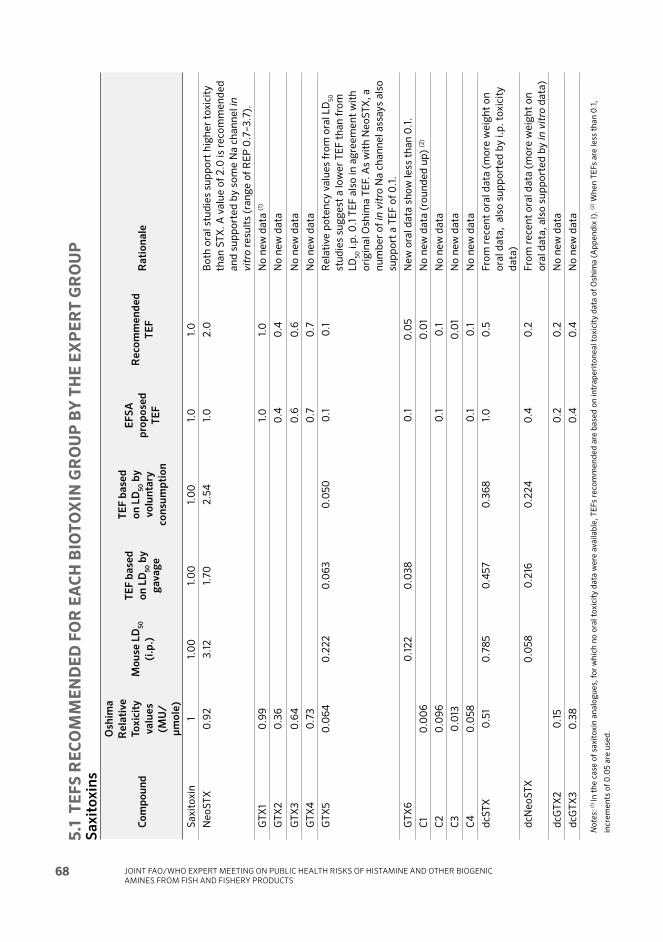
JOINT FAO/WHO EXPERT MEETING ON PUBLIC HEALTH RISKS OF HISTAMINE AND OTHER BIOGENIC AMINES FROM FISH AND FISHERY PRODUCTS
68
5.1
T
EF
S R
EC
OM
ME
ND
ED
FO
R E
AC
H B
IOT
OX
IN G
RO
UP
BY
TH
E E
XP
ER
T G
RO
UP
S
ax
ito
xin
s
Co
mp
ou
nd
Os
him
a
Re
lati
ve
To
xic
ity
va
lue
s
(MU
/
μm
ole
)
Mo
us
e L
D5
0
(i.p
.)
TE
F b
as
ed
on
LD
50 b
y
ga
va
ge
TE
F b
as
ed
on
LD
50 b
y
vo
lun
tary
co
ns
um
pti
on
EF
SA
pro
po
se
d
TE
F
Re
co
mm
en
de
d
TE
FR
ati
on
ale
Sa
xit
ox
in1
1.0
01.
00
1.0
01.
01.
0
Ne
oS
TX
0.9
23
.12
1.7
02
.54
1.0
2.0
Bo
th o
ral
stu
die
s su
pp
ort
hig
he
r to
xic
ity
tha
n S
TX
. A
va
lue
of
2.0
is
reco
mm
en
de
d
an
d s
up
po
rte
d b
y s
om
e N
a c
ha
nn
el
in
vit
ro r
esu
lts
(ra
ng
e o
f R
EP
0.7
–3
.7).
GT
X1
0.9
91.
01.
0N
o n
ew
da
ta (1
)
GT
X2
0.3
60
.40
.4N
o n
ew
da
ta
GT
X3
0.6
40
.60
.6N
o n
ew
da
ta
GT
X4
0.7
30
.70
.7N
o n
ew
da
ta
GT
X5
0.0
64
0.2
22
0.0
63
0.0
50
0.1
0.1
Re
lati
ve
po
ten
cy v
alu
es
fro
m o
ral
LD
50
stu
die
s su
gg
est
a l
ow
er
TE
F t
ha
n f
rom
LD
50 i
.p.
0.1
TE
F a
lso
in
ag
ree
me
nt
wit
h
ori
gin
al
Osh
ima
TE
F. A
s w
ith
Ne
oS
TX
, a
nu
mb
er
of
in v
itro
Na
ch
an
ne
l a
ssa
ys
als
o
sup
po
rt a
TE
F o
f 0
.1.
GT
X6
0.1
22
0.0
38
0.1
0.0
5N
ew
ora
l d
ata
sh
ow
le
ss t
ha
n 0
.1.
C1
0.0
06
0.0
1N
o n
ew
da
ta (
rou
nd
ed
up
) (2
)
C2
0.0
96
0.1
0.1
No
ne
w d
ata
C3
0.0
130
.01
No
ne
w d
ata
C4
0.0
58
0.1
0.1
No
ne
w d
ata
dcS
TX
0.5
10
.78
50
.45
70
.36
81.
00
.5F
rom
re
cen
t o
ral
da
ta (
mo
re w
eig
ht
on
ora
l d
ata
, a
lso
su
pp
ort
ed
by
i.p
. to
xic
ity
da
ta)
dcN
eo
ST
X0
.05
80
.216
0.2
24
0.4
0.2
Fro
m r
ece
nt
ora
l d
ata
(m
ore
we
igh
t o
n
ora
l d
ata
, a
lso
su
pp
ort
ed
by
in
vit
ro d
ata
)
dcG
TX
20
.15
0.2
0.2
No
ne
w d
ata
dcG
TX
30
.38
0.4
0.4
No
ne
w d
ata
No
tes:
(1) In
th
e c
ase
of
sax
ito
xin
an
alo
gu
es,
fo
r w
hic
h n
o o
ral
tox
icit
y d
ata
we
re a
va
ila
ble
, T
EF
s re
com
me
nd
ed
are
ba
sed
on
in
tra
pe
rito
ne
al
tox
icit
y d
ata
of
Osh
ima
(A
pp
en
dix
I).
(2) W
he
n T
EF
s a
re l
ess
th
an
0.1
,
incr
em
en
ts o
f 0
.05
are
use
d.
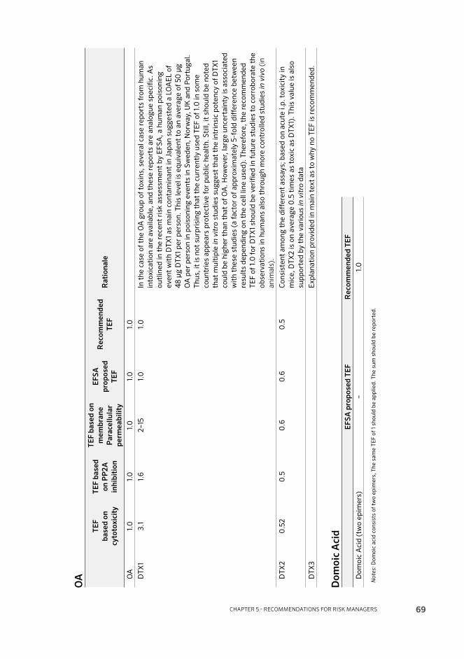
CHAPTER 5 - RECOMMENDATIONS FOR RISK MANAGERS 69
OA
TE
F
ba
se
d o
n
cy
toto
xic
ity
TE
F b
as
ed
on
PP
2A
inh
ibit
ion
TE
F b
as
ed
on
me
mb
ran
e
Pa
rac
ell
ula
r
pe
rme
ab
ilit
y
EF
SA
pro
po
se
d
TE
F
Re
co
mm
en
de
d
TE
FR
ati
on
ale
OA
1.0
1.0
1.0
1.0
1.0
DT
X1
3.1
1.6
2–
151.
01.
0In
th
e c
ase
of
the
OA
gro
up
of
tox
ins,
se
ve
ral
case
re
po
rts
fro
m h
um
an
into
xic
ati
on
are
av
ail
ab
le,
an
d t
he
se r
ep
ort
s a
re a
na
log
ue
sp
eci
fic.
As
ou
tlin
ed
in
th
e r
ece
nt
risk
ass
ess
me
nt
by
EF
SA
, a
hu
ma
n p
ois
on
ing
ev
en
t w
ith
DT
X1
as
ma
in c
on
tam
ina
nt
in J
ap
an
su
gg
est
ed
a L
OA
EL
of
48
μg
DT
X1
pe
r p
ers
on
. T
his
le
ve
l is
eq
uiv
ale
nt
to a
n a
ve
rag
e o
f 5
0 μ
g
OA
pe
r p
ers
on
in
po
iso
nin
g e
ve
nts
in
Sw
ed
en
, N
orw
ay,
UK
an
d P
ort
ug
al.
Th
us,
it
is n
ot
surp
risi
ng
th
at
the
cu
rre
ntl
y u
sed
TE
F o
f 1.
0 i
n s
om
e
cou
ntr
ies
ap
pe
ars
pro
tect
ive
fo
r p
ub
lic
he
alt
h.
Sti
ll,
it s
ho
uld
be
no
ted
tha
t m
ult
iple
in v
itro
stu
die
s su
gg
est
th
at
the
in
trin
sic
po
ten
cy o
f D
TX
1
cou
ld b
e h
igh
er
tha
n t
ha
t o
f O
A.
Ho
we
ve
r, l
arg
e u
nce
rta
inty
is
ass
oci
ate
d
wit
h t
he
se s
tud
ies
(a f
act
or
of
ap
pro
xim
ate
ly 5
-fo
ld d
iffe
ren
ce b
etw
ee
n
resu
lts
de
pe
nd
ing
on
th
e c
ell
lin
e u
sed
). T
he
refo
re,
the
re
com
me
nd
ed
TE
F o
f 1.
0 f
or
DT
X1
sho
uld
be
ve
rifi
ed
in
fu
ture
stu
die
s to
co
rro
bo
rate
th
e
ob
serv
ati
on
s in
hu
ma
ns
als
o t
hro
ug
h m
ore
co
ntr
oll
ed
stu
die
s in
viv
o (
in
an
ima
ls).
DT
X2
0.5
20
.50
.60
.60
.5C
on
sist
en
t a
mo
ng
th
e d
iffe
ren
t a
ssa
ys;
ba
sed
on
acu
te i
.p.
tox
icit
y i
n
mic
e,
DT
X2
is
on
av
era
ge
0.5
tim
es
as
tox
ic a
s D
TX
1).
Th
is v
alu
e i
s a
lso
sup
po
rte
d b
y t
he
va
rio
us
in v
itro
da
ta
DT
X3
E
xp
lan
ati
on
pro
vid
ed
in
ma
in t
ex
t a
s to
wh
y n
o T
EF
is
reco
mm
en
de
d.
Do
mo
ic A
cid
EF
SA
pro
po
se
d T
EF
Re
co
mm
en
de
d T
EF
Do
mo
ic A
cid
(tw
o e
pim
ers
)–
1.0
No
tes:
Do
mo
ic a
cid
co
nsi
sts
of
two
ep
ime
rs,
Th
e s
am
e T
EF
of
1 sh
ou
ld b
e a
pp
lie
d.
Th
e s
um
sh
ou
ld b
e r
ep
ort
ed
.

JOINT FAO/WHO EXPERT MEETING ON PUBLIC HEALTH RISKS OF HISTAMINE AND OTHER BIOGENIC AMINES FROM FISH AND FISHERY PRODUCTS
70
AZ
As
TE
F b
as
ed
on
i.p
. to
xic
ity
TE
F b
as
ed
on
ora
l
tox
icit
y
EF
SA
pro
po
se
d
TE
F
Re
co
mm
en
de
d
TE
FR
ati
on
ale
AZ
A 1
1.0
1.0
11.
0
AZ
A 2
0.6
0.7
1.8
0.7
Ba
sed
on
re
cen
t o
ral
da
ta.
(als
o c
on
sist
en
t w
ith
re
cen
t
i.p
. d
ata
)
AZ
A 3
0.4
50
.51
1.4
0.5
Ba
sed
on
re
cen
t o
ral
da
ta.
(als
o c
on
sist
en
t w
ith
re
cen
t
i.p
. d
ata
)
AZ
A 4
-C
an
no
t d
ete
rmin
e T
EF
du
e t
o l
ack
of
da
ta.
AZ
A 5
-C
an
no
t d
ete
rmin
e T
EF
du
e t
o l
ack
of
da
ta.
AZ
A 6
0.7
-0
.7N
o o
ral
da
ta;
ba
sed
on
re
cen
t i.
p.
da
ta.

71
6References
Aasen, J.A., Espenes, A., Hess, P. & Aune, T. 2010. Sub-lethal dosing of azaspiracid-1 in female NMRI mice. Toxicon, 56(8): 1419–1425.
Aasen, J.A., Espenes, A., Miles, C.O., Samdal, I.A., Hess, P. & Aune, T. 2011. Combined oral toxicity of azaspiracid-1 and yessotoxin in female NMRI mice. Toxicon, 57(6): 909–917.
Akselman, R. & Negri, R.M. 2012. Blooms of Azadinium cf. spinosum Elbrächter et Tillmann (Dinophyceae) in northern shelf waters of Argentina, Southwestern Atlantic. Harmful Algae, 19: 30–38.
Alcala, A.C., Alcala, L. C., Garth, J.S., Yasumura, D and Yasumoto, T. 1988. Human fatality due to ingestion of carb Demania reynaudii that contained a palytoxin-like toxin. Toxicon 26(1): 105-107.
Alfonso, A., de la Rosa, L., Vieytes, M.R., Yasumoto, T. & Botana, L.M. 2003. Yessotoxin, a novel phycotoxin, activates phosphodiesterase activity. Eff ect of yessotoxin on cAMP levels in human lymphocytes. Biochemical Pharmacology, 65(2): 193–208.
Alfonso, C., Rehmann, N., Hess, P., Alfonso, A., Wandscheer, C.B., Abuin, M.,Vale, C., Otero, P., Vieytes, M.R. & Botana, L.M. 2008. Evaluation of various pH and temperature conditions on the stability of azaspiracids and their importance in preparative isolation and toxicological studies. Analytical Chemistry, 80(24): 9672–9680.
Aligizaki, K., Katikou, P., Nikolaidis, G. & Panou, A. 2008. First episode of shellfi sh contamination by palytoxin-like compounds from Ostreopsis species (Aegean Sea, Greece). Toxicon, 51(3): 418–427.
Allingham, J.S., Miles, C.O. & Rayment, I. 2007. A structural basis for regulation of actin polymerization by pectenotoxins. Journal of Molecular Biology, 371(4): 959–970.
Alonso, E., Vale, C., Vieytes, M.R., Laferla, F.M., Gimenez-Llort, L. & Botana, L.M. 2011. 13-Desmethyl spirolide-C is neuroprotective and reduces intracellular Abeta and hy-perphosphorylated tau in vitro. Neurochemistry International, 59(7): 1056–1065.
CHAPTER 6 - REFERENCES

FAO/WHO TECHNICAL PAPER ON TOXICITY EQUIVALENCY FACTORS FOR MARINE BIOTOXINS ASSOCIATED WITH BIVALVE MOLLUSCS
72
Alonso, E., Alfonso, A., Vieytes, M.R. & Botana, L.M. 2016. Evaluation of toxicity equivalent factors of paralytic shellfi sh poisoning toxins in seven human sodium channels types by an automated high throughput electrophysiology system. Archives of Toxicology, 90(2): 479–488.
Alvarez, G., Uribe, E., Avalos, P., Marino, C. & Blanco, J. 2010. First identifi cation of azaspiracid and spirolides in Mesodesma donacium and Mulinia edulis from northern Chile. Toxicon, 55(2-3): 638–641.
Amzil, Z., Sibat, M., Royer, F. & Savar, V. 2008. First report on azaspiracid and yessotoxin groups detection in French shellfi sh. Toxicon, 52(1): 39–48.
AOAC International. 2005. AOAC Offi cial Method 959.08 Paralytic Shellfi sh Poison. Biological Method. First Action 1959. Final Action. Offi cial Methods of Analysis of the AOAC.
AOAC International 2005. AOAC Offi cial Method 2005.06. Paralytic shellfi sh toxins in seafood. In: Offi cial Methods of Analysis of AOAC International. 18th Ed. Gaitherburg (MD). AOAC International, Section 49.10.03.
AOAC International 2011. AOAC Offi cial Method 2011.02. Paralytic shellfi sh toxins in mussels, clams, oysters and scallops. Post-column oxidation (PCOX) Method. In: Offi cial Methods of Analysis of AOAC International. 18th Ed. Gaitherburg (MD). AOAC International, Section 49.10.05.
AOAC International 2011. AOAC Offi cial Method 2011.27. Paralytic shellfi sh toxins (PSTs)in shellfi sh, receptor binding assay. In: Offi cial Methods of Analysis of AOAC International. 18th Ed. Gaitherburg (MD). AOAC International, Section 49.10.06.
APHA [American Public Health Association]. 1970. Subcommittee of laboratory methods for examination of shellfi sh. Method of bioassay of Gymnodinium brevetoxin(s). pp. 1–68, in: Recommended Procedures for the Examination of Seawater and Shellfi sh. 4th ed. APHA, Washington, DC, USA..
Araoz, R., Servent, D., Molgó, J., Iorga, B.I., Fruchart-Gaillard, C., Benoit, E., Gu, Z., Stivala, C. & Zakarian, A. 2011. Total synthesis of pinnatoxins A and G and revision of the mode of action of pinnatoxin A. Journal of the American Chemical Society 133(27): 10499–10511.
Ares, I.R., Louzao, M.C., Vieytes, M.R., Yasumoto, T. & Botana, L.M. 2005. Actin cytoskeleton of rabbit intestinal cells is a target for potent marine phycotoxins. Journal of Experimental Biology, 208(22): 4345–4354.
Ares, I.R., Louzao, M.C., Espina, B., Vieytes, M.R., Miles, C.O., Yasumoto, T. & Botana, L. 2007. Lactone ring of pectenotoxins: a key factor for their activity on cytoskeletal dynamics. Cellular Physiology and Biochemistry, 19(5-6): 283–292.
Aune, T. 2009. Oral toxicity of mixtures of lipophilic marine algal toxins in mice. In: P. Lassus (ed). 7th International Conference on Molluscan Shellfi sh Safety. Nantes, France, June 14–19. Quae Publishing, Versailles, France.
Aune, T., Sorby, R., Yasumoto, T., Ramstad, H. & Landsverk, T. 2002. Comparison of oral and intraperitoneal toxicity of yessotoxin towards mice. Toxicon, 40(1): 77–82.

CHAPTER 6 - REFERENCES 73
Aune, T., Larsen, S., Aasen, J.A., Rehmann, N., Satake, M. & Hess, P. 2007. Relative toxicity of dinophysistoxin-2 (DTX-2) compared with okadaic acid, based on acute intraperitoneal toxicity in mice. Toxicon, 49(1): 1–7.
Aune, T., Aasen, J.A., Miles, C.O. & Larsen, S. 2008. Eff ect of mouse strain and gender on LD50 of yessotoxin. Toxicon, 52(4): 535–540.
Aune, T., Espenes, A., Aasen, J.A., Quilliam, M.A., Hess, P. & Larsen, S. 2012. Study of possible combined toxic eff ects of azaspiracid-1 and okadaic acid in mice via the oral route. Toxicon, 60(5): 895–906.
Baden, D.G., Mende, T.J., Bikhazi, G. & Leung, I. 1982. Bronchoconstriction caused by Florida red tide toxins. Toxicon, 20(5): 929–932.
Baden, D.G. & Mende, T.J. 1982. Toxicity of two toxins from the Florida red tide marine dinofl agellate, Ptychodiscus brevis. Toxicon, 20(2): 457–461.
Baden, D.G., Bourdelais, A.J., Jaycocks, J., Michelliza, S., and Naar, J., 2005. Natural and derivative brevetoxins: historical background, multiplicity, and eff ects. Environmental Health Perspectives, 113(5): 621–625.
Band-Schmidt, C.J., Martinez-Lopez, A., Bustillos-Guzman, J.J., Carreon-Palau, L., Morquecho, L., Olguin-Monroy, N.O., Zenteno-Savin, T., Mendoza-Flores, A., Gonzalez-Acosta, B., Hernandez-Sandoval, F.H. & Tomas, C. 2012. Morphology, biochemistry, and growth of raphidophyte strains from the Gulf of California. Hydrobiologia 693(1): 81–97.
Bane, V., Lehane, M., Dikshit, M., O’Riordan, A. & Furey, A. 2014. Tetrodotoxin: chemistry, toxicity, source, distribution and detection. Toxins (Basel) 6(2): 693–755.
Bates, S.S., Bird, C.J., De Freitas, A.S.W., Foxall, R.A., Gilgan, M., Hanic, L.A., Johnson, G.R., McCulloch, A.W., Odense, P., Pocklington, R., Quilliam, M.A., Sim, P.G., Smith, J.C., Rao, D.V.S., Todd, E.C.D., Walter, J.A. & Wright, J.L.C. 1989. Pennate diatom Nitzschia pungens as the primary source of domoic acid, a toxin in shellfi sh from Eastern Prince Edward Island. Canadian Journal of Fisheries and Aquatic Sciences, 46(7): 1203–1215.
Bellocci, M., Sala, G.L., Callegari, F. & Rossini, G.P. 2010. Azaspiracid-1 inhibits endocytosis of plasma membrane proteins in epithelial cells. Toxicological Sciences, 117(1): 109–121.
Ben-Gigirey, B., Rodriguez-Velasco, M.L. & Gago-Martinez, A. 2012a. Extension of the Validation of AOAC Offi cial Method(SM) 2005.06 for dc-GTX2,3: Interlaboratory Study. Journal of AOAC International, 95(1): 111–121.
Ben-Gigirey, B., Rodriguez-Velasco, M.L., Otero, A., Vieites, J.M. & Cabado, A.G. 2012b. A comparative study for PSP toxins quantifi cation by using MBA and HPLC offi cial methods in shellfi sh. Toxicon, 60(5): 864–873.
Berven, G., Sætre, F., Halvorsen, K. & Seglen, P.O. 2001. Eff ects of the diarrhetic shellfi sh toxin, okadaic acid, on cytoskeletal elements, viability and functionality of rat liver and intestinal cells. Toxicon, 39(2-3): 349–362.

FAO/WHO TECHNICAL PAPER ON TOXICITY EQUIVALENCY FACTORS FOR MARINE BIOTOXINS ASSOCIATED WITH BIVALVE MOLLUSCS
74
Bialojan, C. & Takai, A. 1988. Inhibitory eff ect of a marine-sponge toxin, okadaic acid, on protein phosphatases. Specifi city and kinetics. Biochemical Journal, 256(1): 283–290.
Blanco, J., Arevalo, F., Correa, J., Porro, M.C., Cabado, A.G., Vieites, J.M. & Morono, A. 2015. Eff ect of the industrial steaming on the toxicity, estimated by LC-MS/MS, of mussels exposed for a long time to diarrhetic shellfi sh poisoning (DSP) toxins. Food Chemistry, 177: 240–247.
Botana, L.M. 2012. A perspective on the toxicology of marine toxins. Chemical Research in Toxicology, 25(9): 1800–1804.
Botana, L.M., Vale, C. & Vilariño, N. 2007. Mechanism of action of palytoxin and azaspiracids. pp. 149–156, in: F. Goudey-Perriére, E. Benoit, P. Marchot and M.R. (eds). Toxines émergentes: nouveaux risques, edn. Editions TEC & DOC, Lavoisier, Paris, France.
Botana, L.M., Vilariño, N., Elliott, C.T., Campbell, K., Alfonso, A., Vale, C., Louzao, M.C. & Botana, A.M. 2010. Th e problem of toxicity equivalent factors in developping alternative methods to animal bioassays for marine toxin detection. TrAC – Trends in Analytical Chemistry, 29(11): 1316–1325.
Botana, L.M., Alfonso, A., Vieytes, M.R., Vilariño, N., Botana, A.M. & Louzao, M.C. 2014a. Marine toxins analysis. pp. 153–162, in: L. Nollet and L.S.P. De Gelder (eds). Handbook of Water Analysis. 3rd ed. CRC Press, London, UK.
Botana, L.M., Alfonso, A., Vale, C., Vilariño, N., Rubiolo, J., Alonso, E., Cagide, E. 2014b. Th e mechanistic complexities of phycotoxins: toxicology of azaspiracids and yessotoxins. pp. 1–33, in: Vol. 8 of J.C. Fishbein and J.M. Heiman (eds). Advances in Molecular Toxicology, 1st ed. Elsevier, Oxford, UK.
Bourne, Y., Radic, Z., Araoz, R., Talley, T.T., Benoit, E., Servent, D., Taylor, P., Molgó, J. & Marchot, P. 2010. Structural determinants in phycotoxins and AChBP conferring high affi nity binding and nicotinic AChR antagonism. Proceedings of the National Academy of Sciences of the United States of America, 107(13): 6076–6081.
Braña-Magdalena, A., Lehane, M., Krys, S., Fernandez, M.L., Furey, A. & James, K.J. 2003. Th e fi rst identifi cation of azaspiracids in shellfi sh from France and Spain. Toxicon, 42(1): 105–108.
Braña-Magdalena, A., Leao-Martins, J.M., Glauner, T. & Gago-Martinez, A. 2014. In-tralaboratory validation of a fast and sensitive UHPLC/MS/MS method with fast polarity switching for the analysis of lipophilic shellfi sh toxins. Journal of AOAC International, 97(2): 285–292.
Britton, R., Roberge, M., Brown, C., van Soest, R. & Andersen, R.J. 2003. New okadaic acid analogues from the marine sponge Merriamum oxeato and their eff ect on mitosis. Journal of Natural Products, 66(6): 838–843.
Camilleri, M., Nullens, S. & Nelsen, T. 2012. Enteroendocrine and neuronal mechanisms in pathophysiology of acute infectious diarrhea. Digestive Diseases and Sciences, 57(1): 19–27.

CHAPTER 6 - REFERENCES 75
Cao, Z., LePage, K.T., Frederick, M.O., Nicolaou, K.C. & Murray, T.F. 2010. Involvement of caspase activation in azaspiracid-induced neurotoxicity in neocortical neurons. Toxicological Sciences, 114(2): 323–334.
Catterall, W.A., 1992. Cellular and molecular biology of voltage-gated sodium channels. Physiology Review, 72(4 Suppl.): S15–S48.
CEN [European Committee for Standardization]. 2003. EN 14176. Foodstuff s – Determination of Domoic Acid in Mussels by HPLC. European Committee for Stan-dardization (CEN).
Chevallier, O.P., Graham, S.F., Alonso, E., Duff y, C., Silke, J., Campbell, K., Botana, L.M. & Elliott, C.T. 2015. New insights into the causes of human illness due to consumption of azaspiracid contaminated shellfi sh. Scientifi c Reports, 5: Art. No. 9818.
Ciminiello, P., Fattorusso, E., Forino, M., Magno, S., Poletti, R., Satake, M., Viviani, R. & Yasumoto, T. 1997. Yessotoxin in mussels of the northern Adriatic Sea. Toxicon, 35(2): 177–183.
Ciminiello, P., Fattorusso, E., Forino, M., Magno, S., Poletti, R. & Viviani, R. 1998. Isolation of adriatoxin, a new analogue of yessotoxin from mussels of the Adriatic sea. Tetrahedron Letters, 39(48): 8897–8900.
Ciminiello, P., Fattorusso, E., Forino, M., Poletti, R. & Viviani, R. 2000a. A new analogue of yessotoxin, carboxyyessotoxin, isolated from Adriatic Sea Mussels. European Journal of Organic Chemistry, 2000(2): 291–295.
Ciminiello, P., Fattorusso, E., Forino, M., Poletti, R. & Viviani, R. 2000b. Structure determination of carboxyhomoyessotoxin, a new yessotoxin analogue isolated from adriatic mussels. Chemical Research in Toxicology, 13(8): 770–774.
Ciminiello, P., Dell’Aversano, C., Fattorusso, E., Forino, M., Magno, G.S., Tartaglione, L., Grillo, C. & Melchiorre, N. 2006. Th e Genoa 2005 outbreak. Determination of putative palytoxin in Mediterranean Ostreopsis ovata by a new liquid chromatography tandem mass spectrometry method. Analytical Chemistry, 78(17): 6153–6159.
Ciminiello, P., Dell’Aversano, C., Dello Iacovo, E., Fattorusso, E., Forino, M., Tartaglione, L., Benedettini, G., Onorari, M., Serena, F., Battocchi, C., Casabianca, S. & Penna, A. 2014. First fi nding of Ostreopsis cf. ovata toxins in marine aerosols. Environmental Science & Technology, 48(6): 3532–3540.
CODEX. 2006. Joint Fao/Who Food Standards Programme, Codex Committee on Fish and Fishery Products, Twenty-eighth Session. Beijing, China, 18–22 September 2006. Proposed Draft Standard for Live and Raw Bivalve Molluscs. Report of the Working Group Meeting to Assess the Advice from the Joint FAO/WHO/IOC Ad Hoc Expert Consultation on Biotoxins in Bivalve Molluscs. Doc. no. CX/FFP 06/28/6-Add.1. Available at ft p://ft p.fao.org/codex/Meetings/CCFFP/ccff p28/fp2806ae.pdf Accessed 2016-05-19.
Craig, M. & Holmes, C.F.B. 2000. Freshwater hepatotoxins. Microcystin and Nodularin, mechanisms of toxicity and eff ects on health. pp. 643–672, in: L.M. Botana(ed). Seafood and freshwater toxins: pharmacology, physiology and detection. Marcel Dekker, New York, USA.

FAO/WHO TECHNICAL PAPER ON TOXICITY EQUIVALENCY FACTORS FOR MARINE BIOTOXINS ASSOCIATED WITH BIVALVE MOLLUSCS
76
Crawford, N., Lang, T.K., Kerr, D.S. & de Vries, D.J. 1999. High-affi nity [3H] kainic acid binding to brain membranes: a re-evaluation of ligand potency and selectivity.Journal of Pharmacological and Toxicological Methods, 42(3): 121–125.
Crespo, A., Cifuentes, J.M., Bermúdez, R., Antelo, A., Alemañ, N. & Botana, L.M. 2015. Dose-response and histopathological study, with special attention to the hypophysis, of the diff erential eff ects of domoic acid on rats and mice. Microscopy Research and Technique, 78(5): 396–403.
Cusick, K.D. & Sayler, G.S. 2013. An overview on the marine neurotoxin, Saxitoxin: genetics, molecular targets, methods of detection and ecological functions. Marine Drugs, 11(4): 991–1018.
Daigo, K. 1959. Constituents of Chondria armata. IV. Determination of domoic acid. Yakugaku zasshi : Journal of the Pharmaceutical Society of Japan, 79: 360–364.
Daiguji, M., Satake, M., Ramstad, H., Aune, T., Naoki, H. & Yasumoto, T. 1998a. Structure and fl uorometric HPLC determination of 1–desulfoyessotoxin, a new yessotoxin analog isolated from mussels from Norway. Natural Toxins, 6(6): 235–239.
Daiguji, M., Satake, M., James, K.J., Bishop, A., MacKenzie, L., Naoki, H. & Yasumoto, T. 1998b. Structures of new pectenotoxin analogs, pectenotoxin-2 seco acid and 7-epi-pectenotoxin-2 seco acid, isolated from a dinofl agellate and greenshell mussels. Chemistry Letters, Issue 7: 653–654.
DeBoer, M.K., Tyl, M.R., Fu, M. Kulk, G., Liebezeit, G., Tomas, C.R., Lenzi, A., Naar, J., Vrieling, E.G. & van Rijssel, M. 2009. Haemolytic activity within the species Fibrocapsa japonica (Raphidophyceae). Harmful Algae, 8(5): 699–705.
Dechraoui, M.Y., Naar, J., Pauillac, S. & Legrand, A.M. 1999. Ciguatoxins and brevetoxins, neurotoxic polyether compounds active on sodium channels. Toxicon, 37(1): 125–143.
DeGrasse, S.L. & Martinez-Diaz, K. 2012. Biotoxin control programmes in North, Central and South American countries. In: A.G. Cabado and J.M. Vieites (eds). New Trends in Marine and Freshwater Toxins: Food Safety Concerns. Nova Science Publishers Inc.
Dell’Aversano, C., Walter, J.A., Burton, I.W., Stirling, D.J., Fattorusso, E. & Quilliam, M.A. 2008. Isolation and structure elucidation of new and unusual saxitoxin analogues from mussels. Journal of Natural Products, 71(9): 1518–1523.
Dhar, B.C., Cimarelli, L., Singh, K.S., Brandi, L., Brandi, A., Puccinelli, C., Marcheggiani, S. & Spurio, R. 2015. Molecular detection of a potentially toxic diatom species. International Journal of Environmental Research and Public Health, 12(5): 4921–4941.
Dickey, R.W., Baden, D. & Fleming, L.E. 2011. Brevetoxins. pp. 51–98, in: Assessment and management of biotoxin risks in bivalve molluscs. Edited by J. Lawrence, H. Loreal, H. Toyofuku, P. Hess, I. Karunasagar and L. Ababouch. FAO Fisheries and Aquaculture Technical Paper, no. 551.

CHAPTER 6 - REFERENCES 77
Dickey, R.W., Bobzin, S.C., Faulkner, D.J., Bencsath, F.A. & Andrzejewski, D. 1990. Identifi cation of okadaic acid from a Caribbean dinofl agellate, Prorocentrum concavum. Toxicon, 28(4): 371–377.
Dickey, R., Jester, E., Granade, R., Mowdy, D., Moncrieff , C., Rebarchik, D., Robl, M., Musser, S. & Poli, M. 1999. Monitoring brevetoxins during a Gymnodinium breve red tide: Comparison of sodium channel specifi c cytotoxicity assay and mouse bioassay for determination of neurotoxic shellfi sh toxins in shellfi sh extracts. Natural Toxins, 7(4): 157–165.
Djaoued, Y., Balaji, S. & Priya, S. 2007. Non-resonance micro-Raman spectroscopic studies on crystalline domoic acid and its aqueous solutions. Spectrochimica Acta Part A – Molecular And Biomolecular Spectroscopy, 67(5): 1362–1369.
Dominguez, H.J., Paz, B., Daranas, A.H., Norte, M., Franco, J.M. & Fernandez, J.J. 2010. Dinofl agellate polyether within the yessotoxin, pectenotoxin and okadaic acid toxin groups: characterization, analysis and human health implications. Toxicon, 56(2); special issue): 191–217.
Doucet, E., Ross, N.N. & Quilliam, M.A. 2007. Enzymatic hydrolysis of esterifi ed diarrhetic shellfi sh poisoning toxins and pectenotoxins. Analytical and Bioanalytical Chemistry, 389(1): 335–342
Doucette, G.J., Logan, M.M., Ramsdell, J.S. & Van Dolah, F.M. 1997. Development and preliminary validation of a microtiter plate-based receptor binding assay for paralytic shellfi sh poisoning toxins. Toxicon, 35(5): 625–636.
Doucette, T.A. & Tasker, R.A. 2008. Domoic acid: detection methods, pharmacology, and toxicology. pp. 397–429, in: L.M. Botana (ed). Seafood and Freshwater toxins: Pharmacology, Physiology and Detection, 2nd ed. CRC Press, Boca Raton, FL, USA.
Draisci, R., Ferretti, E., Palleschi, L., Marchiafava, C., Poletti, R., Milandri, A., Ceredi, A. & Pompei, M. 1999. High levels of yessotoxin in mussels and presence of yessotoxin and homoyessotoxin in dinofl agellates of the Adriatic Sea. Toxicon, 37(8): 1187–1193.
EFSA. [European Food Safety Authority]. 2008a. Marine biotoxins in shellfi sh – Azaspiracid group[1] – Scientifi c Opinion of the Panel on Contaminants in the Food chain. Th e EFSA Journal 723: 1–52. DOI 10.2903/j.efsa.2008.723
EFSA. 2008b. Scientifi c Opinion on marine biotoxins in shellfi sh – Yessotoxin group. EFSA Panel on Contaminants in the Food Chain (CONTAM). Th e EFSA Journal, 907: 1–62.
EFSA. 2008c. Opinion of the Scientifi c Panel on Contaminants in the Food chain on a request from the European Commission on marine biotoxins in shellfi sh – okadaic acid and analogues. Th e EFSA Journal 589: 1–62.
EFSA 2008d. Scientifi c Opinion of the Panel on Contaminants in the Food Chain on a request from the European Commission on Polycyclic Aromatic Hydrocarbons in Food. Th e EFSA Journal 724: 1–114. Available at doi 10.2903/j.efsa.2008.589 Accessed 2016-05-20.

FAO/WHO TECHNICAL PAPER ON TOXICITY EQUIVALENCY FACTORS FOR MARINE BIOTOXINS ASSOCIATED WITH BIVALVE MOLLUSCS
78
EFSA. 2009a. Scientifi c Opinion on marine biotoxins in shellfi sh – Domoic acid. EFSA Panel on Contaminants in the Food Chain (CONTAM). Th e EFSA Journal, 1181: 1–61.
EFSA. 2009b. Opinion of the Scientifi c Panel on Contaminants in the Food chain on a request from the European Commission on marine biotoxins in shellfi sh – Saxitoxin group. Th e EFSA Journal, 1019: 1–76.
Elgarch, A., Vale, P., Rifai, S. & Fassouane, A. 2008. Detection of diarrheic shellfi sh poisoning and azaspiracid toxins in Moroccan mussels: comparison of the LC-MS method with the commercial immunoassay kit. Marine Drugs 6(4): 587–594.
Endo, A., Khora, S.S., Murata, M., Naoki, H. & Yasumoto, T. 1988. Isolation of 11-nor-tetrodotoxin-6(R)-ol and other tetrodotoxin derivatives from the puff er Fugu niphobles. Tetrahedron Letters, 29(33): 4127–4128.
Espina, B. & Rubiolo, J.A. 2008a. Marine toxins and the cytoskeleton: pectenotoxins, unusual macrolides that disrupt actin. FEBS J 275: 6082–6088.
Espina, B., Louzao, M.C., Ares, I.R., Cagide, E., Vieytes, M.R., Vega, F.V., Rubiolo, J.A., Miles, C.O., Suzuki, T., Yasumoto, T. & Botana, L.M. 2008b. Cytoskeletal toxicity of pectenotoxins in hepatic cells. British Journal of Pharmacology, 155(6): 934–944.
Espina, B., Louzao, M.C., Cagide, E., Alfonso, A., Vieytes, M.R., Yasumoto, T. & Botana, L.M. 2010. Th e methyl ester of okadaic acid is more potent than okadaic acid in disrupting the actin cytoskeleton and metabolism of primary cultured hepatocytes. British Journal of Pharmacology, 159(2): 337–344.
EU [European Commission]. 2011. Commission Regulation (EU) No 15/2011 of 10 January 2011 amending Regulation (EC) No 2074/2005 as regards recognised testing methods for detecting marine biotoxins in live bivalve molluscs. Offi cial Journal L6: 3–6. Available at http://eur-lex.europa.eu/legal-content/EN/TXT/HTML/?uri=OJ:L:2011:006:FULL&from=EN#TN0001 Accessed 2016-05-19.
EU. 2013. Commission Regulation (EU) No 786/2013 of 16 August 2013 amending Annex III to Regulation (EC) No 853/2004 of the European Parliament and of the Council as regards the permitted limits of yessotoxins in live bivalve molluscs. Offi cial Journal of the European Communities L220: 14.
EURL. 2015. EU Reference Laboratory for Marine Biotoxins, Web site. Available at: http://aesan.msssi.gob.es/en/CRLMB/web/procedimientos_crlmb/ crlmb_standard_operating_procedures.shtml Accessed 2016-05-20.
FAO/IOC/WHO [Food and Agriculture Organization of the United Nations/Intergov-ernmental Oceanographic Commission of UNESCO/World Health Organization]. 2004. Report of the Joint FAO/IOC/WHO ad hoc Expert Consultation on Biotoxins in Bivalve Molluscs. Oslo, Norway, 26–30 September 2004. Available at ft p://ft p.fao.org/es/esn/food/biotoxin_report_en.pdf Accessed 2016-07-01.

CHAPTER 6 - REFERENCES 79
FAO/WHO. 2009. Principles and Methods for the Risk Assessment of Chemicals in Food. Chapter 7 – Risk Characterization, in Environmental Health Criteria 240. A joint publication of the Food and Agriculture Organization of the United Nations and the World Health Organization, published under the joint sponsorship of the United Nations Environment Programme (UNEP), the International Labour Organization (ILO) and the World Health Organization (WHO), and produced within the framework of the Inter-Organization Programme for the Sound Management of Chemicals. Available at http://apps.who.int/iris/bitstream/10665/44065/10/WHO_EHC_240_10_eng_Chapter7.pdf Accessed 2016-05-29.
FDA [U.S. Food and Drug Administration]. 2011. Fish and fi shery products hazards and controls guidance. 4th ed. Available at http://www.fda.gov/Food/Guidance-Regulation/GuidanceDocumentsRegulatoryInformation/Seafood/ucm2018426.htm Accessed 2016-06-29.
FDA, 2013. National Shellfi sh Sanitation Program (NSSP) Guide for the Control of Molluscan Shellfi sh. 2011 Revision. Available at http://www.fda.gov/downloads/Food/GuidanceRegulation/FederalStateFoodPrograms/UCM350344.pdf Accessed 2016-06-29.
Fernandez, D.A., Louzao, M.C., Fraga, M., Vilarino, N., Vieytes, M.R. Botana, L.M. 2014. Experimental basis for the high oral toxicity of dinophysistoxin 1: a comparative study of DSP. Toxins (Basel), 6(1): 211–228.
Fernandez-Araujo, A., Alfonso, A., Vieytes, M.R. & Botana, L.M. 2015a. Key role of phosphodiesterase 4A (PDE4A) in autophagy triggered by yessotoxin. Toxicology, 329: 60–72.
Fernandez-Araujo, A., Sanchez, J.A., Alfonso, A., Vieytes, M.R. & Botana, L.M. 2015b. Diff erent toxic eff ects of YTX in tumor K-562 and lymphoblastoid cell lines. Frontiers in Pharmacology, 6: Art. no. 124.
Ferreiro, S.F., Carrera, C., Vilarino, N., Louzao, M.C., Santamarina, G., Cantalapiedra, A.G. & Botana, L.M. 2015. Acute cardiotoxicity evaluation of the marine biotoxins OA, DTX-1 and YTX. Toxins (Basel), 7(4): 1030–1047.
Ferreiro, S.F., Vilarino, N., Carrera, C., Louzao, M.C., Cantalapiedra, A.G., Santamarina, G., Cifuentes, J.M., Vieira, A.C. & Botana, L.M. 2016. Subacute cardiovascular toxicity of the marine phycotoxin Azaspiracid-1 in Rats. Toxicological Sciences, 151(1): 104–114.
Ferreiro, S.F., Vilarino, N., Carrera, C., Louzao, M.C., Santamarina, G., Cantalapiedra, A.G., Rodriguez, L.P., Cifuentes, J.M., Vieira, A.C., Nicolaou, K.C., Frederick, M.O. & Botana, L.M. 2014. In vivo arrhythmogenicity of the marine biotoxin azaspiracid-2 in rats. Archives of Toxicology, 88(2): 425–434.
Ferron, P.J., Hogeveen, K., Fessard, V. & Le Hegarat, L. 2014. Comparative analysis of the cytotoxic eff ects of okadaic acid-group toxins on human intestinal cell lines. Marine Drugs 12(8): 4616–4634.

FAO/WHO TECHNICAL PAPER ON TOXICITY EQUIVALENCY FACTORS FOR MARINE BIOTOXINS ASSOCIATED WITH BIVALVE MOLLUSCS
80
Finch, S.C., Wilkins, A.L., Hawkes, A.D., Jensen, D.J., MacKenzie, A.L., Beuzenberg, V., Quilliam, M.A., Olseng, C.D., Samdal, I.A., Aasen, J., Selwood, A.I. Cooney, J.M., Sandvik, M. & Miles, C.O. 2005. Isolation and identifi cation of (44-R,S)-44,55-dihy-droxyyessotoxin from Protoceratium reticulatum, and its occurrence in extracts of shellfi sh from New Zealand, Norway and Canada. Toxicon, 46(2): 160–170.
Furey, A., Moroney, C., Braña Magdalena, A., Saez, M.J.F., Lehane, M. & James, K.J. 2003. Geographical, temporal, and species variation of the polyether toxins, azaspiracids, in shellfi sh. Environmental Science & Technology, 37(14): 3078–3084.
Fux, E., Biré, R. & Hess, P. 2009. Comparative accumulation and composition of lipophilic marine biotoxins in passive samplers and in mussels (M. edulis) on the West Coast of Ireland. Harmful Algae, 8(3): 523–537.
Fux, E., Smith, J.L., Tong, M.M., Guzman, L. & Anderson, D.M. 2011. Toxin profi les of fi ve geographical isolates of Dinophysis spp. from North and South America. Toxicon, 57(2): 275–287.
Garibo, D., de la Iglesia, P., Diogene, J. & Campas, M. 2013. Inhibition equivalency factors for dinophysistoxin-1 and dinophysistoxin-2 in protein phosphatase assays: applicability to the analysis of shellfi sh samples and comparison with LC-MS/MS. Journal of Agricultural and Food Chemistry, 61(10): 2572–2579.
Giordano, G., White, C.C., Mohar, I., Kavanagh, T.J. & Costa, L.G. 2007. Glutathione levels modulate domoic acid induced apoptosis in mouse cerebellar granule cells. Toxicological Sciences, 100(2): 433–444.
Griffi th, A.W., Shumway, S.E., Volety, A.K., 2013. Bioaccumulation and depuration of brevetoxins in the eastern oyster (Crassostrea virginica) and the northern quahog (= hard clam, Mercenaria mercenaria). Toxicon, 66: 75–81.
Grimmelt, B., Nijjar, M.S., Brown, J., Macnair, N., Wagner, S., Johnson, G.R. & Amend, J.F. 1990. Relationship between domoic acid levels in the blue mussel (Mytilus edulis) and toxicity in mice. Toxicon, 28(5): 501–508.
Gu, H.F., Luo, Z.H., Krock, B., Witt, M. & Tillmann, U. 2013. Morphology, phylogeny and azaspiracid profi le of Azadinium poporum (Dinophyceae) from the China Sea. Harmful Algae, 21-22: 64–75.
Guatimosim, S., Sobie, E.A., Cruz, J.D.S., Martin, L.A. & Lederer, W.J. 2001. Molecular identifi cation of a TTX-sensitive Ca2+ current. American Journal of Physiology – Cell Physiology, 280(5): C1327–C1339.
Guo, X.T., Uehara, A., Ravindran, A., Bryant, S.H., Hall, S. & Moczydlowski, E. 1987. Kinetic basis for insensitivity to tetrodotoxin and saxitoxin in sodium channels of canine heart and denervated rat skeletal muscle. Biochemistry 26(24): 7546–7556.
Hall, S. & Reichardt, P.B. 1984. Cryptic paralytic shellfi sh toxins. pp. 113–124, in: E.P. Ragelis (ed). Seafood Toxins. American Chemical Society, Washington DC, USA.

CHAPTER 6 - REFERENCES 81
Hall, S., Strichartz, G., Moczydlowski, E., Ravindran, A. & Reichardt, P.B. 1990. Th e saxitoxins. Sources, chemistry, and pharmacology. pp. 29–65, in: S. Hall and G. Strichartz (eds). Marine Toxins: origin, structure, and molecular pharmacology. ACS Sympsium Series, Washington DC, USA.
Haque, S.M. & Onoue, Y. 2002. Variation in toxin compositions of two harmful raphidophytes, Chattonella antiqua and Chattonella marina, at diff erent salinities. Environmental Toxicology, 17(2): 113–118.
Harada, T., Oshima, Y. & Yasumoto, T. 1984. Assessment of potential activation of gonyautoxin V in the stomach of mice and rats. Toxicon, 22(3): 476–478.
Hartshorne, R.P. & Catterall, W.A. 1981. Purifi cation of the saxitoxin receptor of the sodium channel from rat brain. Proceedings of the National Academy of Sciences of the United States of America, 78: 4620–4624.
Harwood, D.T., Selwood, A.I., van Ginkel, R., Waugh, C., McNabb, P.S., Munday, R., Hay, B., Th omas, K., Quilliam, M.A., Malhi, N., Dowsett, N. & McLeod, C. 2014. Paralytic shellfi sh toxins, including deoxydecarbamoyl-STX, in wild-caught Tasmanian abalone (Haliotis rubra). Toxicon, 90: 213–225.
Hashimoto, K., Noguchi, T. & Watabe, S. 1990. New aspects of tetrodotoxin. pp. 575–588, in: A. Pohland, V. Dowell Jr., J. Richard, R. Cole, M. Eklund, S. Green, W. Norred III and M. Potter (eds). Microbial Toxins in Foods and Feeds. Springer, USA.
Henri, J., Leighfi eld, T.A., Lanceleur, R., Huguet, A., Ramsdell, J.S. & Fessard, V. 2014. Permeability of dihydro- and cysteine-brevetoxin metabolites across a Caco-2 cell monolayer. Harmful Algae, 32(1): 22–26.
Henri, J., Leighfi eld, T.A., Lanceleur, R., Huguet, A., Ramsdell, J.S., and Fessard, V., 2014. Permeability of dihydro- and cysteine-brevetoxin metabolites across a Caco-2 cell monolayer. Harmful Algae 32, 22-26.
Hess, P. 2012. Phytoplankton and biotoxin monitoring programmes for the safe exploitation of shellfi sh in Europe. In: A.G. Cabado and J.M. Vieites (eds). New Trends in Marine and Freshwater Toxins: Food Safety Concerns. Nova Science Publishers.
Hess, P. & Aasen, J. 2007. Chemistry, origins and distribution of yessotoxin and its analogues. pp. 187–202, in: L.M. Botana (ed). Phytotoxins, Chemistry and Biochemistry. Blackwell Publishing, Ames, Iowa, USA.
Hess, P., Grune, B., Anderson, D.B., Aune, T., Botana, L.M., Caricato, P., van Egmond, H.P., Halder, M., Hall, S., Lawrence, J.F., Moff at, C., Poletti, R., Richmond, J., Rossini, G.P., Seamer, C. & Vilageliu, J.S. 2006. Th ree Rs Approaches in Marine Biotoxin Testing. Th e report and recommendations of a joint ECVAM/DG SANCO workshop (ECVAM workshop 55). ATLA – Alternatives to Laboratory Animals, 34: 193–224.
Hess, P., McCarron, P., Rehmann, N., Kilcoyne, J., McMahon, T., Ryan, G., Ryan, M.P., Twiner, M.J., Doucette, G.J., Satake, M., Ito, E. & Yasumoto, T. 2007. Isolation and purifi cation of AZAs from naturally contaminated materials, and evaluation of their toxicological eff ects. Final project report ASTOX (ST/02/02). Marine Environment & Health Series, no. 28. 129 p. Available at http://hdl.handle.net/10793/268 Accessed 2016-05-21.

FAO/WHO TECHNICAL PAPER ON TOXICITY EQUIVALENCY FACTORS FOR MARINE BIOTOXINS ASSOCIATED WITH BIVALVE MOLLUSCS
82
Hess, P., McCarron, P., Krock, B., Kilcoyne, J. & Miles, C.O. 2014. Azaspiracids: chemistry, biosynthesis, metabolism and detection. pp. 799–822, in: Seafood and Freshwater Toxins. CRC Press, Boca Raton, Florida, USA.
Hogberg, H.T. & Bal-Price, A.K. 2011. Domoic acid-induced neurotoxicity is mainly mediated by the AMPA/KA receptor: comparison between immature and mature primary cultures of neurons and glial cells from rat cerebellum. Journal of Toxicology, 2011: 543512.
Holland, P.T., Selwood, A.I., Mountfort, D.O., Wilkins, A.L., McNabb, P., Rhodes, L.L., Doucette, G.J., Mikulski, C.M. & King, K.L. 2005. Isodomoic acid C, an unusual amnesic shellfi sh poisoning toxin from Pseudo-nitzschia australis. Chemical Research in Toxicology, 18(5): 814–816.
Holmes, C.F., Luu, H.A., Carrier, F. & Schmitz, F.J. 1990. Inhibition of protein phosphatases-1 and -2A with acanthifolicin. Comparison with diarrhetic shellfi sh toxins and identifi cation of a region on okadaic acid important for phosphatase inhibition. FEBS Letters, 270(1-2): 216–218.
Holzer, P., Reichmann, F. & Farzi, A. 2012. Neuropeptide Y, peptide YY and pancreatic polypeptide in the gut-brain axis. Neuropeptides 46(6; SI): 261–274.
Hu, T., Marr, J., DeFreitas, A.S.W., Quilliam, M.A., Walter, J.A. & Wright, J.L.C. 1992a. New diol esters isolated from cultures of the dinofl agellates Prorocentrum lima and Prorocentrum concavum. Journal of Natural Products, 55(11): 1631–1637.
Hu, T., Doyle, J., Jackson, D., Marr, J., Nixon, E., Pleasance, S., Quilliam, M.A., Walter, J.A. & Wright, J.L.C. 1992b. Isolation of a new diarrhetic shellfi sh poison from Irish mussels. Journal of the Chemical Society – Chemical Communications, 1992(1): 39–41.
Hu, T., Curtis, J.M., Walter, J.A. & Wright, J.L.C. 1995a. Identifi cation of DTX-4, a new water-soluble phosphatase inhibitor from the toxic dinofl agellate Prorocentrum lima. Journal of the Chemical Society – Chemical Communications, 1995(5): 597–599.
Hu, T., Curtis, J.M., Walter, J.A., McLachlan, J.L. & Wright, J.L.C. 1995b. Two new water-soluble DSP toxin derivatives from the dinofl agellate Prorocentrum maculosum: possible storage and excretion products. Tetrahedron Letters, 36(51): 9273–9276.
Huhn, J., Jeff rey, P.D., Larsen, K., Rundberget, T., Rise, F., Cox, N.R., Arcus, V., Shi, Y. & Miles, C.O. 2009. A structural basis for the reduced toxicity of dinophysistoxin-2. Chemical Research in Toxicology, 22(11): 1782–1786.
Hungerford, J.M. 1995. 35. Fish and other marine poducts. AOAC offi cial method 959.08 . Paralytic shellfi sh poison. Biological method. Final action. pp. 21–22, in: P. Cunniff (ed). Offi cial methods of analysis of AOAC International. 16th ed. Vol. 2. AOAC, Arlington, VA, USA.
Hwang, D.F., Wang, W.C., Chung, H.M. & Jeng, S.S. 1989a. First identifi cation of acute tetrodotoxin-associated food poisoning in Taiwan. Taiwan yi xue hui za zhi. Journal of the Formosan Medical Association, 88(3): 289–291.

CHAPTER 6 - REFERENCES 83
Hwang, D.F., Arakawa, O., Saito, T., Noguchi, T., Simidu, U., Tsukamoto, K., Shida, Y., Hashimoto, K. 1989b. Tetrodotoxin-producing bacteria from the blue-ringed octopus Octopus maculosus. Marine Biology 100(3): 327–332.
ICCVAM. 2006a. ICCVAM. In vitro cytotoxicity test methods for estimating acute oral systemic toxicity. NIH Publication 07-4518. Available at http://iccvam.niehs.nih.gov/docs/acutetox_docs/BRD_TMER/BRDvol1_Nov2006.pdf..
ICCVAM. 2006b. ICCVAM. Validation study of in vitro cytotoxicity test methods. Available at http://iccvam.niehs.nih.gov/methods/acutetox/inv_nru_announce.htm.
Ikehara, T., Imamura, S., Yoshino, A. & Yasumoto, T. 2010. PP2A inhibition assay using recombinant enzyme for rapid detection of okadaic acid and its analogs in shellfi sh. Toxins (Basel,) 2(1): 195–204.
Ishida, H., Nozawa, A., Totoribe, K., Muramatsu, N., Nukaya, H., Tsuji, K., Yamaguchi, K., Yaasumoto, T., Kaspar, H., Berkett, N. & Kosuge, T. 1995. Brevetoxin B1, a new polyether marine toxin from the New Zealand shellfi sh, Austrovenus stutchburyi. Tetrahedron Letters, 36(5): 725–728.
Ishida, H., Muramatsu, N., Nukaya, H., Kosuge, T. & Tsuji, K. 1996. Study on neurotoxic shellfi sh poisoning involving the oyster, Crassostrea gigas, in New Zealand. Toxicon, 34(9): 1050–1053.
Ishida, H., Nozawa, A., Hamano, H., Naoki, H., Fujita, T., Kaspar, H.F. & Tsuji, K. 2004a. Brevetoxin B5, a new brevetoxin analog isolated from cockle Austrovenus stutchburyi in New Zealand, the marker for monitoring shellfi sh neurotoxicity. Tetrahedron Letters, 45(1): 29–33.
Ishida, H., Nozawa, A., Nukaya, H., Tsuji & K., 2004b. Comparative concentrations of brevetoxins PbTx-2, PbTx-3, BTX-B1 and BTX-B5 in cockle, Austrovenus stutchburyi, greenshell mussel, Perna canaliculus, and Pacifi c oyster, Crassostrea gigas, involved in neurotoxic shellfi sh poisoning in New Zealand. Toxicon, 43(7): 779–789.
Ito, E. 2008. Toxicology of azaspiracid-1: acute and chronic poisoning, tumorigenicity, and chemical structure relationship to toxicity in a mouse model. pp. 775–784, in: L.M. Botana (ed). Seafood and Freshwater toxins: Pharmacology, Physiology and Detection, 2nd ed. CRC Press. Boca Raton, FL, USA.
Ito, E. & Terao, K. 1994. Injury and recovery process of intestine caused by okadaic acid and related compounds. Natural Toxins, 2(6): 371–377.
Ito, E., Satake, M., Ofuji, K., Higashi, M., Harigaya, K., McMahon, T., Yasumoto, T. 2002b. Chronic eff ects in mice caused by oral administration of sublethal doses of azaspiracid, a new marine toxin isolated from mussels. Toxicon, 40(2): 193–203.
Ito, E., Satake, M., Ofuji, K., Kurita, N., McMahon, T., James, K. & Yasumoto, T. 2000. Multiple organ damage caused by a new toxin azaspiracid, isolated from mussels produced in Ireland. Toxicon, 38(7): 917–930.
Ito, E., Suzuki, T., Oshima, Y. & Yasumoto, T. 2008. Studies of diarrhetic activity on pectenotoxin-6 in the mouse and rat. Toxicon, 51(4): 707–716.
FAO/WHO TECHNICAL PAPER ON TOXICITY EQUIVALENCY FACTORS FOR MARINE BIOTOXINS ASSOCIATED WITH BIVALVE MOLLUSCS
84
Ito, E., Yasumoto, T., Takai, A., Imanishi, S. & Harada, K. 2002a. Investigation of the distribution and excretion of okadaic acid in mice using immunostaining method. Toxicon, 40(2): 159–165.
Iverson, F., Truelove, J., Nera, E., Tryphonas, L., Campbell, J. & Lok, E. 1989. Domoic acid poisoning and mussel-associated intoxication: preliminary investigations into the response of mice and rats to toxic mussel extract. Food & Chemical Toxicology, 27(6): 377–384.
James, K.J., Bishop, A.G., Draisci, R., Palleschi, L., Marchiafava, C., Ferretti, E., Satake, M. & Yasumoto, T. 1999. Liquid chromatographic methods for the isolation and identifi cation of new pectenotoxin-2 analogues from marine phytoplankton and shellfi sh. Journal of Chromatography A, 844(1-2): 53–65.
James, K.J., Furey, A., Satake, M. & Yasumoto, T. 2001. Azaspiracid poisoning (AZP): A new shellfi sh toxic syndrome in Europe. pp. 250–253, in: G.M. Hallegraeff , S.I. Blackburn, C.J. Bolch and R.J. Lewis (eds). Harmful Algal Blooms 2000. Intergovern-mental Oceanographic Commission of UNESCO.
James, K.J., Furey, A., Lehane, M., Ramstad, H., Aune, T., Hovgaard, P., Morris, S., Higman, W Satake, M. & Yasumoto, T. 2002. First evidence of an extensive northern European distribution of azaspiracid poisoning (AZP) toxins in shellfi sh. Toxicon, 40(7): 909–915.
James, K.J., Sierra, M.D., Lehane, M., Braña Magdalena, A. & Furey, A. 2003a. Detection of fi ve new hydroxyl analogues of azaspiracids in shellfi sh using multiple tandem mass spectrometry. Toxicon, 41(3): 277–283.
James, K.J., Moroney, C., Roden, C., Satake, M., Yasumoto, T., Lehane, M. & Furey, A. 2003b. Ubiquitous ‘benign’ alga emerges as the cause of shellfi sh contamination responsible for the human toxic syndrome, azaspiracid poisoning. Toxicon, 41(2): 145–151.
Jang, J.H. & Yotsu-Yamashita, M. 2007. 6,11-Dideoxytetrodotoxin from the puff er fi sh, Fugu pardalis. Toxicon, 50(7): 947–951.
Jauff rais, T., Contreras, A., Herrenknecht, C., Truquet, P., Sechet, V., Tillmann, U. & Hess, P. 2012a. Eff ect of Azadinium spinosum on the feeding behaviour and azaspiracid accumulation of Mytilus edulis. Aquatic Toxicology, 124: 179–187.
Jauff rais, T., Marcaillou, C., Herrenknecht, C., Truquet, P., Sechet, V., Nicolau, E., Tillmann, U. & Hess, P. 2012b. Azaspiracid accumulation, detoxifi cation and biotransformation in blue mussels (Mytilus edulis) experimentally fed Azadinium spinosum. Toxicon, 60(4; SI): 582–595.
Jauff rais, T., Kilcoyne, J., Sechet, V., Herrenknecht, C., Truquet, P., Herve, F., Berard, J.B., Nulty, C., Taylor, S., Tillmann, U., Miles, C.O. & Hess, P. 2012c. Production and isolation of azaspiracid-1 and -2 from Azadinium spinosum culture in pilot-scale photobioreactors. Marine Drugs, 10(6): 1360–1382.
Jeff ery, B., Barlow, T., Moizer, K., Paul, S. & Boyle, C. 2004. Amnesic shellfi sh poison. Food and Chemical Toxicology, 42(4): 545–557.
CHAPTER 6 - REFERENCES 85
Jeglitsch, G., Rein, K., Baden, D.G. & Adams, D.G. 1998. Brevetoxin-3 (PbTx-3) and its derivatives modulate single tetrodotoxin-sensitive sodium channels in rat sensory neurons. Journal of Pharmacology and Experimental Th erapeutics, 284(2): 516–525.
Jiang, S.S., Kuwano, K., Ishikawa, N., Yano, M., Takatani, T. & Arakawa, O. 2014. Production of domoic acid by laboratory culture of the red alga Chondria armata. Toxicon, 92: 1–5.
Johannessen, J.N. 2000. Stability of domoic acid in saline dosing solutions. Journal of AOAC International, 83(2): 411–412.
Kao, C.Y. 1966. Tetrodotoxin, saxitoxin and their signifi cance in the study of excitation phenomena. Pharmacological Reviews 18(2): 997–1049.
Kao, C.Y. & Walker, S.E. 1982. Active groups of saxitoxin and tetrodotoxin as deduced from actions of saxitoxin analogues on frog muscle and squid axon. Journal of Physiology (London). 323(Feb.): 619–637.
Kao, C.Y. & Yasumoto, T. 1985. Actions of 4-epitetrodotoxin and anhydrotetrodotoxin on the squid axon. Toxicon, 23(5): 725–729.
Kao, C.Y., Kao, P.N., James, K.M.R., Koehn, F.E., Wichmann, C.F. & Schnoes, H.K. 1985. Actions of epimers of 12-(OH)-reduced saxitoxin and of 11-(OSO3)-saxitoxin on squid axon. Toxicon, 23(4): 647–655.
Katikou, P. & Vlamis, A. (eds). 2014. Palytoxin and analogs: ecobiology and origin, chemistry, and chemical analysis. CRC Press, Boca Ratón, FL, USA.
Kellmann, R., Schaff ner, C.A., Gronset, T.A., Satake, M., Ziegler, M. & Fladmark, K.E. 2009. Proteomic response of human neuroblastoma cells to azaspiracid-1. Journal of Proteomics, 72(4): 695–707.
Kilcoyne, J., Keogh, A., Clancy, G., LeBlanc, P., Burton, I., Quilliam, M.A., Hess, P. & Miles, C.O. 2012. Improved isolation procedure for azaspiracids from shellfi sh, structural elucidation of azaspiracid-6, and stability studies. Journal of Agricultural and Food Chemistry, 60(10): 2447–2455.
Kilcoyne, J., McCarron, P., Twiner, M.J., Nulty, C., Crain, S., Quilliam, M.A., Rise, F., Wilkins, A.L. & Miles, C.O. 2014a. Epimers of azaspiracids: Isolation, structural elucidation, relative LC-MS response, and in vitro toxicity of 37-epi-azaspiracid-1. Chemical Research in Toxicology, 27(4): 587–600.
Kilcoyne, J., Nulty, C., Jauff rais, T., McCarron, P., Herve, F., Foley, B., Rise, F., Crain, S., Wilkins, A.L., Twiner, M.J., Hess, P. & Miles, C.O. 2014b. Isolation, structure elucidation, relative LC-MS response, and in vitro toxicity of azaspiracids from the dinofl agellate Azadinium spinosum. Journal of Natural Products, 77(11): 2465–2474.
Kilcoyne, J., Twiner, M.J., McCarron, P., Crain, S., Giddings, S.D., Foley, B., Rise, F., Hess, P., Willkins, A.L. & Miles, C.O. 2015. Structure elucidation, relative LC-MS response and in vitro toxicity of Azaspiracids 7-10 isolated from mussels (Mytilus edulis). Journal of Agricultural and Food Chemistry, 63(20): 5083–5091.

FAO/WHO TECHNICAL PAPER ON TOXICITY EQUIVALENCY FACTORS FOR MARINE BIOTOXINS ASSOCIATED WITH BIVALVE MOLLUSCS
86
Kittler, K., Preiss-Weigert, A. & Th ese, A. 2010. Identifi cation strategy using combined mass spectrometric techniques for elucidation of phase I and phase II in vitro metabolites of lipophilic marine biotoxins. Analytical Chemistry, 82(22): 9329–9335.
Kleivdal, H., Kristiansen, S.I., Nilsen, M.V., Goksoyr, A., Briggs, L., Holland, P. & McNabb, P. 2007. Determination of domoic acid toxins in shellfi sh by Biosense ASP ELISA – A direct competitive enzyme-linked immunosorbent assay: Collaborative study. Journal of AOAC International, 90(4): 1011–1027.
Klontz, K.C., Abraham, A., Plakas, S.M., Dickey, R.W. 2009. Mussel-associated azaspiracid intoxication in the United States. Annals of Internal Medicine, 150(4): 361–361.
Kodama, M. & Sato, S. 2008. Metabolism of paralytic shellfi sh toxins incorporated into bivalves. pp. 165–175, in: L.M. Botana (ed). Seafood and Freshwater toxins: Pharmacology, Physiology and Detection, 2nd ed. CRC Press, Boca Raton, FL, USA.
Korsnes, M.S. & Espenes, A. 2011. Yessotoxin as an apoptotic inducer. Toxicon, 57(7-8): 947–958.
Korsnes, M.S., Espenes, A., Hermansen, L.C., Loader, J.I. & Miles, C.O. 2013. Cytotoxic responses in BC3H1 myoblast cell lines exposed to 1-desulfoyessotoxin. Toxicol In vitro, 27(6): 1962–1969.
Korsnes, M.S., Roed, S.S., Tranulis, M.A., Espenes, A. & Christophersen, B. 2014. Yessotoxin triggers ribotoxic stress. Toxicol In vitro 28(5): 975–981.
Krock, B., Tillmann, U., John, U. & Cembella, A. 2008. LC-MS-MS aboard ship: tandem mass spectrometry in the search for phycotoxins and novel toxigenic plankton from the North Sea. Analytical and Bioanalytical Chemistry, 392(5): 797–803.
Krock, B., Tillmann, U., John, U. & Cembella, A.D. 2009. Characterization of azaspiracids in plankton size-fractions and isolation of an azaspiracid-producing dinofl agellate from the North Sea. Harmful Alga, 8(2): 254–263.
Krock, B., Tillmann, U., Voss, D., Koch, B.P., Salas, R., Witt, M., Potvin, E. & Jeong, H.J. 2012. New azaspiracids in Amphidomataceae (Dinophyceae). Toxicon, 60(5): 830–839.
Krock, B., Tillmann, U., Potvin, E., Jeong, H.-J., Drebing, W., Kilcoyne, J., Al-Jorani, A., Twiner, M.J., Gothel, Q. & Kock, M. 2015. Structure elucidation and in vitro toxicity of new azaspiracids isolated from the marine dinofl agellate Azadinium poporum. Marine Drugs, 13(11): 6687–6702.
Kumagai, M., Yanagi, T., Murata, M., Yasumoto, T., Kat, M., Lassus, P. & Rodriguez-Vazquez, J.A. 1986. Okadaic acid as the causative toxin of diarrhetic shellfi sh poisoning in Europe. Agriculture and Biological Chemistry, 50(11): 2853–2857.
Laidley, C.W., Cohen, E. & Casida, J.E. 1997. Protein phosphatase in neuroblastoma cells: [3H]cantharidin binding site in relation to cytotoxicity. Journal of Pharmacology and Experimental Th erapeutics, 280(3): 1152–1158.

CHAPTER 6 - REFERENCES 87
Larsen, K., Petersen, D., Wilkins, A.L., Samdal, I.A., Sandvik, M., Rundberget, T., Goldstone, D., Arcus, V., Hovgaard, P., Rise, F., Rehmann, N., Hess, P. & Miles, C.O. 2007. Clarifi cation of the C-35 stereochemistries of dinophysistoxin-1 and dino-physistoxin-2 and its consequences for binding to protein phosphatase. Chemical Research in Toxicology, 20(6): 868–875.
Lawrence, J.F., Charbonneau, C.F. & Menard, C. 1991. Liquid chromatographic determination of domoic acid in mussels, using AOAC paralytic shellfi sh poison extraction procedure: collaborative study. Journal of the Association of Offi cial Analytical Chemists, 74(1): 68–72.
Lawrence, J.F., Niedzwiadek, B. & Menard, C. 2004. Quantitative determination of paralytic shellfi sh poisoning toxins in shellfi sh using prechromatographic oxidation and liquid chromatography with fl uorescence detection: interlaboratory study. Journal of AOAC International, 87(1): 83–100.
Lawrence, J.F., Menard, C., Charbonneau, C.F. & Hall, S. 1991. A study of ten toxins associated with paralytic shellfi sh poison using prechromatographic oxidation and liquid chromatography with fl uorescence detection. Journal of the Association of Offi cial Analytical Chemists, 74(2): 404–409.
Le Hegarat, L., Jacquin, A.G., Bazin, E. & Fessard, V. 2006. Genotoxicity of the marine toxin okadaic acid, in human Caco-2 cells and in mice gut cells. Environmental Toxicology, 21(1): 55–64.
Lee, J.S., Igarashi, T., Fraga, S., Dahl, E., Hovgaard, P. & Yasumoto, T. 1989. Determination of diarrhetic shellfi sh toxins in various dinofl agellate species. Journal of Applied Phycology 1(2): 147–152.
Lee, J.S., Tangen, K., Dahl, E., Hovgaard, P. & Yasumoto, T. 1988. Diarrhetic shellfi sh toxins in Nowegian mussels. Nippon Suisan Gakkaishi, 54(11): 1953–1957.
Lehane, M., Braña Magdalena, A., Moroney, C., Furey, A. & James, K.J. 2002. Liquid chromatography with electrospray ion trap mass spectrometry for the determination of fi ve azaspiracids in shellfi sh. Journal of Chromatography A, 950(1-2): 139–147.
Leira, F., Cabado, A.G., Vieytes, M.R., Roman, Y., Alfonso, A., Botana, L.M., Yasumoto, T., Malaguti, C. & Rossini, G.P. 2002. Characterization of F-actin depolymerization as a major toxic event induced by pectenotoxin-6 in neuroblastoma cells. Biochemical Pharmacology, 63(11): 1979–1988.
Levin, E.D., Pizarro, K., Pang, W.G., Harrison, J. & Ramsdell, J.S. 2005. Persisting behavioral consequences of prenatal domoic acid exposure in rats. Neurotoxicology and Teratology 27(5): 719–725.
Li, A., Ma, J., Cao, J., Wang, Q., Yu, R., Th omas, K. & Quilliam, M.A. 2012. Analysis of paralytic shellfi sh toxins and their metabolites in shellfi sh from the North Yellow Sea of China. Food Additives and Contaminants Part A – Chemistry Analysis Control Exposure & Risk Assessment, 29(9): 1455–1464.
Li, A.F., Sun, G., Qiu, J.B. & Fan, L. 2015. Lipophilic shellfi sh toxins in Dinophysis caudata picked cells and in shellfi sh from the East China Sea. Environmental Science and Pollution Research, 22(4): 3116–3126.

FAO/WHO TECHNICAL PAPER ON TOXICITY EQUIVALENCY FACTORS FOR MARINE BIOTOXINS ASSOCIATED WITH BIVALVE MOLLUSCS
88
Lin, Y.-Y., Risk, M., Ray, S.M., Van Engen, D., Clardy, J., Golik, J., James, C.J. & Nakanishi, K. 1981. Isolation and structure of brevetoxin B from the “red tide” dinofl agellate Ptychodiscus brevis (Gymnodinium breve). Journal of the American Chemical Society, 103(22): 6773–6775.
Llewellyn, L.E. 2006a. Saxitoxin, a toxic marine natural product that targets a multitude of receptors. Natural Products Research, 23(2): 200–222.
Llewellyn, L.E. 2006b. Th e behavior of mixtures of paralytic shellfi sh toxins in competitive binding assays. Chemical Research in Toxicology, 19(5): 661–667.
Louzao, M.C., Fernandez, D.A., Abal, P., Fraga, M., Vilarino, N., Vieytes, M.R. & Botana, L.M. 2015. Diarrhetic eff ect of okadaic acid could be related with its neuronal action: Changes in neuropeptide Y. Toxicology Letters, 237(2): 151–160.
Lundholm, N., Skov, J., Pocklington, R. & Moestrup, Ø. 1994. Domoic acid, the toxic amino acid responsible for amnesic shellfi sh poisoning, now in Pseudo-nitzschia seriata (Bacillariophyceae) in Europe. Phycologia, 33(6): 475–478.
MacKintosh, C. & Klumpp, S. 1990. Tautomycin from the bacterium Streptomyces verticillatus, another potent and specifi c inhibitor of protein phosphatases 1 and 2A. FEBS Letters, 277(1-2): 137–140.
Maeda, M., Kodama, T., Tanaka, T., Yoshizumi, H., Takemoto, T., Nomoto, K. & Fujita, T. 1986. Structures of Isodomoic Acid-A, Acid-B and Acid-C, novel insecticidal amino-acids from the red alga Chondria armata. Chemical & Pharmaceutical Bulletin, 34(11): 4892–4895.
Maeda, M., Kodama, T., Tanaka, T., Yoshizumi, H., Takemoto, T., Nomoto, K. & Fujita, T. 1987. Structures of domoilactone A and B, novel amino acids from the red alga, Chondria armata. Tetrahedron Letters, 28(6): 633–636.
Mafra, L.L., Tavares, C.P.D. & Schramm, M.A. 2014. Diarrheic toxins in fi eld-sampled and cultivated Dinophysis spp. cells from southern Brazil. Journal of Applied Phycology 26(4): 1727–1739.
Manger, R.L., Leja, L.S., Lee, S.Y., Hungerford, J.M. & Wekell, M.M. 1993. Tetra-zolium-based cell bioassay for neurotoxins active on voltage-sensitive sodium channels: semiautomated assay for saxitoxins, brevetoxins & ciguatoxins. Analytical Biochemistry, 214(1): 190–194.
Marine Institute, 2014. Azaspiracids – Toxicological evaluation, Test Methods and identifi cation of source organisms (ASTOX II). www.oar.marine.ie/handle/10793/970.
Maucher Fuquay, J., Muha, N., Wang, Z. & Ramsdell, J.S. 2012. Elimination kinetics of domoic acid from the brain and cerebrospinal fl uid of the pregnant rat. Chemical Research in Toxicology, 25(12): 2805–2809.
Mayer, A.M. 2000. Th e marine toxin domoic acid may aff ect the developing brain by activation of neonatal brain microglia and subsequent neurotoxic mediator generation. Medical Hypotheses, 54(5): 837–841.

CHAPTER 6 - REFERENCES 89
McCarron, P., Burrell, S. & Hess, P. 2007. Eff ect of addition of antibiotics and an antioxidant on the stability of tissue reference materials for domoic acid, the amnesic shellfi sh poison. Analytical and Bioanalytical Chemistry, 387(7): 2495–2502.
McCarron, P., Emteborg, H. & Hess, P. 2007. Freeze-drying for the stabilisation of shellfi sh toxins in mussel tissue (Mytilus edulis) reference materials. Analytical and Bioanalytical Chemistry, 387(7): 2475–2486.
McCarron, P., Kilcoyne, J., Hess, P. 2008. Eff ects of cooking and heat treatment on concentration and tissue distribution of okadaic acid and dinophysistoxin-2 in mussels (Mytilus edulis). Toxicon, 51(6): 1081–1089.
McCarron, P., Kilcoyne, J., Miles, C.O. & Hess, P. 2009. Formation of Azaspiracids-3, -4, -6, and -9 via decarboxylation of carboxyazaspiracid metabolites from shellfi sh. Journal of Agricultural and Food Chemistry, 57(1): 160–169.
McCarron, P., Kotterman, M., de Boer, J., Rehmann, N. & Hess, P. 2007. Feasibility of gamma irradiation as a stabilisation technique in the preparation of tissue reference materials for a range of shellfi sh toxins. Analytical and Bioanalytical Chemistry, 387(7): 2487–2493.
McFarren, E.F. 1959. Report on collaborative studies of the bioassay for paralytic shellfi sh poison. Journal of the Association of Offi cial Analytical Chemists, 42: 263–271.
McMahon, T. & Silke, J. 1996. West coast of Ireland; winter toxicity of unknown aetiology in mussels. Harmful Algae, News, 14: 2.
McNabb, P. Selwood, A.I., Munday, R., Wood, S.A., Taylor, D.I., Mackenzie L. A. van Ginkel R., Rhodes, L.L., Cornelisen, C., Heasman , K., Holland, P.T., King, C. 2010. Detection of tetrodotoxin from the grey side-gilled slug- Pleurobranchaea maculata and associated dog neurotoxicosis on beaches adjecent to the Hauraki Gulf, Auckland, New Zealand. Toxicon. 56 (3) 466-473.
McNabb, P.S., Selwood, A.I., van Ginkel, R., Boundy, M. & Holland, P.T. 2012. Determination of brevetoxins in shellfi sh by LC/MS/MS: Single-laboratory validation. Journal of AOAC International, 95(4): 1097–1105.
Miles, C.O. 2007. Pectenotoxins. pp. 159–186, in: L.M. Botana (ed). Phytotoxins, chemistry and Biochemistry. Blackwell Publishing, Ames, Iowa, USA.
Miles, C.O., Wilkins, A.L., Jensen, D.J., Cooney, J.M., Quilliam, M.A., Aasen, J. & MacKenzie, A.L. 2004a. Isolation of 41a-homoyessotoxin and the identifi cation of 9-methyl-41a-homoyessotoxin and nor-ring A-yessotoxin from Protoceratium reticulatum. Chemical Research in Toxicology, 17(11): 1414–1422.
Miles, C.O., Wilkins, A.L., Hawkes, A.D., Selwood, A., Jensen, D.J., Aasen, J., Munday, R., Samdal, I.A., Briggs, L.R., Beuzenberg, V. & MacKenzie, A.L. 2004b. Isolation of a 1,3-enone isomer of heptanor-41-oxoyessotoxin from Protoceratium reticulatum cultures. Toxicon, 44(3): 325–336.

FAO/WHO TECHNICAL PAPER ON TOXICITY EQUIVALENCY FACTORS FOR MARINE BIOTOXINS ASSOCIATED WITH BIVALVE MOLLUSCS
90
Miles, C.O., Wilkins, A.L., Munday, R., Dines, M.H., Hawkes, A.D., Briggs, L.R., Sandvik, M., Jensen, D.J., Cooney, J.M., Holland, P.T. (Holland, PT); Quilliam, MA (Quilliam, MA); MacKenzie, AL (MacKenzie, AL); Beuzenberg, V. & Towers, N.R. 2004c. Isolation of pectenotoxin-2 from Dinophysis acuta and its conversion to pectenotoxin-2 seco acid, and preliminary assessment of their acute toxicities. Toxicon, 43(1): 1–9.
Miles, C.O., Wilkins, A.L., Hawkes, A.D., Selwood, A.I., Jensen, D.J., Munday, R., Cooney, J.M. & Beuzenberg, V. 2005. Polyhydroxylated amide analogs of yessotoxin from Protoceratium reticulatum. Toxicon, 45(1): 61–71.
Miles, C.O., Wilkins, A.L., Munday, J.S., Munday, R., Hawkes, A.D., Jensen, D.J., Cooney, J.M. & Beuzenberg, V. 2006. Production of 7-epi-pectenotoxin-2 seco acid and assessment of its acute toxicity to mice. Journal of Agricultural and Food Chemistry, 54(4): 1530–1534.
Moczydlowski, E.G. 2013. Th e molecular mystique of tetrodotoxin. Toxicon, 63: 165–183.
Moczydlowski, E., Hall, S., Garber, S.S., Strichartz, G.S. & Miller, C. 1984. Voltage-depen-dent blockade of muscle Na+ channels by guanidinium toxins - eff ect of toxin charge. Journal of General Physiology, 84(5): 687–704.
Molgó, J., Aráoz, R., Benoit, E. & Iorga, B.I. 2014. Cyclic imine toxins: chemistry, origin, metabolism, pharmacology, toxicology and detection. pp. 951–989, in: L.M. Botana (ed). Seafood and Freshwater toxins: Pharmacology, Physiology and Detection, 3rd ed. CRC Press, Boca Raton, FL, USA.
Moore, R.E. & Scheuer, P.J. 1971. Palytoxin: a new marine toxin from a coelenterate. Science 172(3982): 495–498.
Morohashi, A., Satake, M., Murata, K., Naoki, H., Kaspar, H.F. & Yasumoto, T. 1995. Brevetoxin B3, a new brevetoxin analog isolated from the greenshell mussel Perna canaliculus involved in neurotoxic shellfi sh poisoning in New Zealand. Tetrahedron Letters, 36(49): 8995–8998.
Morohashi, A., Satake, M., Naoki, H., Kaspar, H.F., Oshima, Y. & Yasumoto, T. 1999. Brevetoxin B4 isolated from greenshell mussels Perna canaliculus, the major toxin involved in neurotoxic shellfi sh poisoning in New Zealand. Natural Toxins, 7(2): 45–48.
Morris, P.D., Campbell, D.S., Taylor, T.J. & Freeman, J.I., 1991. Clinical and epidemiologi-cal features of neurotoxic shellfi sh poisoning in North Carolina. American Journal of Public Health, 81(4): 471–474.
Mountfort, D.O., Suzuki, T. & Truman, P. 2001. Protein phosphatase inhibition assay adapted for determination of total DSP in contaminated mussels. Toxicon, 39(2-3): 383–390.
Munday, R. 2006. Toxicological requirements for risk assessment of shellfi sh contaminants: a review. Afr. J. Marine Sci., 28, 447-449.

CHAPTER 6 - REFERENCES 91
Munday, R. 2008. Toxicology of the pectenotoxins. pp. 371–380, in: L.M. Botana (ed). Seafood and Freshwater toxins: Pharmacology, Physiology and Detection, 2nd ed. CRC Press, Boca Raton, FL, USA
Munday, R. 2013. Is protein phosphatase inhibition responsible for the toxic eff ects of okadaic acid in animals? Toxins (Basel) 5(2): 267–285.
Munday, R. 2014. Toxicology of seafood toxins: a critical review. pp. 197–290, in: L.M. Botana (ed). Seafood and Freshwater Toxins: Pharmacology, Physiology and Detection, 3rd ed. CRC Press, Boca Ratón, FL, USA.
Munday, R., Aune, T. & Rossini, G.P. 2008. Toxicology of the Yessotoxins. pp. 329–339, in: L.M. Botana (ed). Seafood and Freshwater toxins: Pharmacology, Physiology and Detection, 2nd ed. CRC Press, Boca Raton, FL, USA.
Munday, R., Towers, N.R., Mackenzie, L. Beuzenberg, V., Holland P.T., Miles, C. O. 2004. Acute toxicity of gymnodimine to mice. Toxicon, 44, 173-178
Munday, R., Holland, P.T., McNabb, P., Selwood, A.I. & Rhodes, L.L. 2008. Comparative toxicity to mice of domoic acid and isodomoic acids A, B and C. Toxicon, 52(8): 954–956.
Munday, R., Quilliam, M.A., LeBlanc, P., Lewis, N., Gallant, P., Sperker, S. A., Ewert, H. S., MacKinnon, S. L. 2012a. Investigations into the toxicology of spirolides, a group of marine phycotoxins. Toxins, 4, 1-14; doi:10.3390/toxins4010001
Munday, R., Selwood, A. I., Rhodes, L. 2012b Acute toxicity of pinnatoxins E, F and G to mice. Toxicon, 2012, 60, 995-999.
Munday, R., Th omas, K., Gibbs, R., Murphy, C. & Quilliam, M.A. 2013. Acute toxicities of saxitoxin, neosaxitoxin, decarbamoyl saxitoxin and gonyautoxins 1&4 and 2&3 to mice by various routes of administration. Toxicon, 76: 77–83.
Murata, M., Shimatani, M., Sugitani, H., Oshima, Y. & Yasumoto, T. 1982. Isolation and structural elucidation of the causative toxin of the diarrhetic shellfi sh poisoning. Bulletin of the Japanese Society of Scientifi c Fisheries, 48(4): 549–552.
Murata, M., Sano, M., Iwashita, T., Naoki, H. & Yasumoto, T. 1986. Th e structure of pectenotoxin-3, a new constituent of diarrhetic shellfi sh toxins. Agricultural and Biological Chemistry, 50(10): 2693–2695.
Murata, M., Kumagai, M., Lee, J.S. & Yasumoto, T. 1987. Isolation and structure of yessotoxin, a novel polyether compound implicated in diarrhetic shellfi sh poisoning. Tetrahedron Letters, 28(47): 5869–5872.
Murata, K., Satake, M., Naoki, H., Kaspar, H.F. & Yasumoto, T., 1998. Isolation and structure of a new brevetoxin analog, brevetoxin B2, from greenshell mussels from New Zealand. Tetrahedron 54(5-6): 735–742.
Murrell, R.N. & Gibson, J.E. 2009. Brevetoxins 2, 3, 6 and 9 show variability in potency and cause signifi cant induction of DNA damage and apoptosis in Jurkat E6-1 cells. Archives of Toxicology, 83(11): 1009–1019.
FAO/WHO TECHNICAL PAPER ON TOXICITY EQUIVALENCY FACTORS FOR MARINE BIOTOXINS ASSOCIATED WITH BIVALVE MOLLUSCS
92
Naar, J., Bourdelais, A., Tomas, C., Kubanek, J., Whitney, P.L., Flewelling, L., Steidinger, K., Lancaster, J. & Baden, D.G. 2002. A competitive ELISA to detect brevetoxins from Karenia brevis (formerly Gymnodinium breve) in seawater, shellfi sh, and mammalian body fl uid. Environmental Health Perspectives, 110(2): 179–185.
Nagatomo, I., Akasaki, Y., Uchida, M., Tominaga, M., Kuchiiwa, S., Nakagawa, S. & Takigawa, M. 1999. Kainic and domoic acids diff erentially aff ect NADPH-di-aphorase neurons in the mouse hippocampal formation. Brain Research Bulletin, 48(3): 277–282.
Nakamura, M. & Yasumoto, T. 1985. Tetrodotoxin derivatives in puff er fi sh. Toxicon, 23(2): 271–276.
Nicolaou, K.C., Koft is, T.V., Vyskocil, S., Petrovic, G., Ling, T., Yamada, Y.M.A., Tang, W. & Frederick, M.O. 2004a. Structural revision and total synthesis of azaspiracid-1, Part 2: Defi nition of the ABCD domain and total. Angewandte Chemie – International Edition, 43(33): 4318–4324.
Nicolaou, K.C., Vyskocil, S., Koft is, T.V., Yamada, Y.M., Ling, T., Chen, D.Y., Tang, W., Petrovic, G., Frderick, M.O., Li, Y. and Satake, M. 2004b. Structural revision and total synthesis of azaspiracid-1, Part 1: Intelligence gathering and tentative proposal. Angewandte Chemie – International Edition, 43(33): 4312–4318.
Nishikawa, T. & Isobe, M. 2013. Synthesis of tetrodotoxin, a classic but still fascinating natural product. Chemical Record, 13(3): 286–302.
Nishiwaki, S., Fujiki, H., Suganuma, M., Furuya-Suguri, H., Matsushima, R., Iida, Y., Ojika, M., Yamada, K., Uemura, D., Yasumoto, T., Schmitz, F.J. & Sugimura, T. 1990. Structure-activity relationship within a series of okadaic acid derivatives. Carcinogenesis, 11(10): 1837–1841.
Noguchi, T., Onuki, K. & Arakawa, O. 2011. Tetrodotoxin poisoning due to puff erfi sh and gastropods, and their intoxication mechanism. ISRN toxicology, 2011: Article 276939.
Nzoughet, K.J., Hamilton, J.T., Floyd, S.D., Douglas, A., Nelson, J., Devine, L. & Elliott, C.T. 2008. Azaspiracid: fi rst evidence of protein binding in shellfi sh. Toxicon, 51(7): 1255–1263.
O’Driscoll, D., Škrabáková, Z., O’Halloran, J., van Pelt, F.N.A.M. & James, K.J. 2011. Mussels increase xenobiotic (azaspiracid) toxicity using a unique bioconversion mechanism. Environmental Science and Technology, 45(7): 3102–3108.
OECD [Organisation for Economic Co-operation and Development]. 1998a OECD Guideline for the Testing of Chemicals 407. Repeated Dose 28 day oral toxicity study in rodents. https://ntp.niehs.nih.gov/iccvam/suppdocs/feddocs/oecd/oecdtg407-2008.pdf
OECD. 1998b. Guideline for the Testing of Chemicals 408. Repeated Dose 90 day oral toxicity study in rodents.http://www.oecd-ilibrary.org/environment/test-no-408-re-peated-dose-90-day-oral-toxicity-study-in-rodents_9789264070707-en.

CHAPTER 6 - REFERENCES 93
Ofuji, K., Satake, M., McMahon, T., James, K.J., Naoki, H., Oshima, Y. & Yasumoto, T. 2001. Structures of azaspiracid analogs, azaspiracid-4 and azaspiracid-5, causative toxins of azaspiracid poisoning in Europe. Bioscience Biotechnology and Biochemistry, 65(3): 740–742.
Ofuji, K., Satake, M., McMahon, T., Silke, J., James, K.J., Naoki, H., Oshima, Y. & Yasumoto, T. 1999. Two analogs of azaspiracid isolated from mussels, Mytilus edulis, involved in human intoxication in Ireland. Natural Toxins, 7(3): 99–102.
Ogino, H., Kumagai, M. & Yasumoto, T. 1997. Toxicologic evaluation of yessotoxin. Natural Toxins, 5(6): 255–259.
Onoue, Y., Noguchi, T., Maruyama, J., Hashimoto, K. & Seto, H. 1983. Properties of two toxins newly isolated from oysters. Journal of Agricultural and Food Chemistry, 31(2): 420–423.
Onuma, Y., Satake, M., Ukena, T., Roux, J., Chanteau, S., Rasolofonirina, N. Ratsimaloto, M., Naoki, H. and Yasumoto, T., 1999. Identifi cation of putative palytoxin as the cause of clupitoxism. Toxicon, 37(1): 55-65.
Oshima, Y. 1995. Postcolumn derivatization liquid chromatographic method for paralytic shellfi sh toxins. Journal of AOAC International, 78(2): 528–532.
Otero, P., Alfonso, A., Rodriguez, P., Rubiolo, J.A., Cifuentes, J.M., Bermudez, R., Vieytes, M.R. & Botana, L.M. 2012. Pharmacokinetic and toxicological data of spirolides aft er oral and intraperitoneal administration. Food and Chemical Toxicology, 50(2): 232–237.
Pang, Y., Fang, C., Twiner, M.J., Miles, C.O. & Forsyth, C.J. 2011. Total synthesis of dino-physistoxin-2 and 2-epi-dinophysistoxin-2 and their PPase inhibition. Angewandte Chemie – International Edition, 50(33): 7631–7635.
Payandeh, J., Scheuer, T., Zheng, N. & Catterall, W.A. 2011. Th e crystal structure of a voltage-gated sodium channel. Nature, 475(7356): 353–358.
Peng, Y.G. & Ramsdell, J.S. 1996. Brain Fos induction is a sensitive biomarker for the lowest observed neuroexcitatory eff ects of domoic acid. Fundamental and Applied Toxicology, 31(2): 162–168.
Percopo, I., Siano, R., Rossi, R., Soprano, V., Sarno, D. & Zingone, A. 2013. A new potentially toxic Azadinium species (Dinophyceae) from the Mediterranean Sea, A. dexteroporum sp. nov. Journal of Phycology, 49(5): 950–966.
Perez, R., Rehmann, N., Crain, S., LeBlanc, P., Craft , C., MacKinnon, S., Reeves, K., Burton, I., Walter, J.A., Hess, P., Quilliam, M.A. & Melanson, J.E. 2010. Th e preparation of certifi ed calibration solutions for azaspiracid-1, -2, and -3, potent marine biotoxins found in shellfi sh. Analytical and Bioanalytical Chemistry, 398(5): 2243–2252.
Perez, S., Vale, C., Botana, A.M., Alonso, E., Vieytes, M.R. & Botana, L.M. 2011. Determination of toxicity equivalent factors for paralytic shellfi sh toxins by electro-physiological measurements in cultured neurons. Chemical Research in Toxicology, 24(7): 1153–1157.

FAO/WHO TECHNICAL PAPER ON TOXICITY EQUIVALENCY FACTORS FOR MARINE BIOTOXINS ASSOCIATED WITH BIVALVE MOLLUSCS
94
Perez-Gomez, A., Garcia-Rodriguez, A., James, K.J., Ferrero-Gutierrez, A., Novelli, A. & Fernandez-Sanchez, M.T. 2004. Th e marine toxin dinophysistoxin-2 induces diff erential apoptotic death of rat cerebellar neurons and astrocytes. Toxicological Sciences, 80(1): 74–82.
Perl, T.M., Bedard, L., Kosatsky, T., Hockin, J.C., Todd, E.C. & Remis, R.S. 1990. An outbreak of toxic encephalopathy caused by eating mussels contaminated with domoic acid. New England Journal of Medicine, 322(25): 1775–1780.
Piñeiro, N., Leao, J.M., Gago, A. & Rodriguez, J.A. (1999). Capillary electrophoresis with diode array detection as an alternative analytical method for paralytic and amnesic shellfi sh toxins. Journal of Chromatography A, 847(1-2): 223–232.
Pistocchi, R., Guerrini, F., Pezzolesi, L., Riccardi, M., Vanucci, S., Ciminiello, P., Dell’Aversano, C., Forino, M., Fattorusso, E., Tartaglione, L., Milandri, A., Pompei, M., Cangini, M., Pigozzi, S. & Riccardi, E. 2012. Toxin levels and profi les in microalgae from the north-western Adriatic Sea – 15 years of studies on cultured species. Marine Drugs, 10(1): 140–162.
Pitcher, G.C., Franco, J.M., Doucette, G.J., Powell, C.L. & Mouton, A. 2001. Paralytic shellfi sh poisoning in the abalone Haliotis midae on the west coast of South Africa. Journal of Shellfi sh Research, 20(2): 895–904.
Plakas, S.M. & Dickey, R.W., 2010. Advances in monitoring and toxicity assessment of brevetoxins in molluscan shellfi sh. Toxicon, 56(2; Special Issue): 137–149.
Poli, M.A., Templeton, C.B., Th ompson, W.L. & Hewetson, J.F. 1990. Distribution and elimination of brevetoxin PbTx-3 in rats. Toxicon, 28(8): 903–910.
Poli, M.A., Musser, S.M., Dickey, R.W., Eilers, P.P. & Hall, S. 2000. Neurotoxic shellfi sh poisoning and brevetoxin metabolites: a case study from Florida. Toxicon, 38(7): 981–993.
Potvin, E., Jeong, H.J., Kang, N.S., Tillmann, U. & Krock, B. 2012. First report of the photosynthetic dinofl agellate genus Azadinium in the Pacifi c Ocean: morphology and molecular characterization of Azadinium cf. poporum. Journal of Eukaryotic Microbiology, 59(2): 145–156.
Quilliam, M. 2003. Chemical methods for domoic acid, the amnesic shellfi sh poisoning (ASP) toxin. pp. 247–266, in: G. Hallegraeff , D.M. Anderson and A.D. Cembella (eds). Manual on Harmful Marine Microalgae. UNESCO, Paris, France.
Quilliam, M.A. & Wright, J.L. 1989. Th e amnesic shellfi sh poisoning mystery. Analytical Chemistry, 61(18): 1053A–1060A.
Quilliam, M.A., Xie, M. & Hardstaff , W.R. 1995a. Rapid extraction and cleanup for liquid chromatography determination of domoic acid in unsalted food. Journal of the Assocation of Offi cial Analytical Chemists,18: 543–554.
Quilliam, M.A., Sim, P.G., McCulloch, A.W. & McInnes, A.G. 1989a. High-performance liquid chromatography of domoic acid, a marine neurotoxin, with application to shellfi sh and plankton. International Journal of Environmental Analytical Chemistry, 36(3): 139–154.

CHAPTER 6 - REFERENCES 95
Quilliam, M.A., Th omson, B.A., Scott, G.J. & Siu, K.W. 1989d. Ion-spray mass spectrometry of marine neurotoxins. Rapid Communications in Mass Spectrometry, 3(5): 145–150.
Ramsdell, J.S. 2007. Domoic acid. Th e molecular and integrative basis to domoic acid toxicity. pp. 223–250, in: L.M. Botana (ed). Phytotoxins, Chemistry and Biochemistry, Blackwell, Ames, Iowa. USA.
Reeves, K., Th omas, K. & Quilliam, M.A. 2003. Preparation of a certifi ed reference material for paralytic shellfi sh poisoning toxins. pp. 90–98, in: S.S. Bates (ed). 8th Canadian workshop on harmful marine algae. Moncton, Canada. Canadian Technical Report of Fisheries and Aquatic Sciences.
Reguera, B. & Pizarro, G. 2008. Planktonic dinofl agellates that contain polyether toxins of the old “DSP complex”. pp. 257–284, in: L.M. Botana (ed). Seafood and Freshwater toxins: Pharmacology, Physiology and Detection. 2nd ed. CRC Press, Boca Raton, FL, USA.
Rehmann, N., Hess, P. & Quilliam, M.A. 2008. Discovery of new analogs of the marine biotoxin azaspiracid in blue mussels (Mytilus edulis) by ultra-performance liquid chromatography/tandem mass spectrometry. Rapid Communications in Mass Spectrometry, 22(4): 549–558.
Rivas, M., Garcia, C., Liberona, J.L. & Lagos, N. 2000. Biochemical characterization and inhibitory eff ects of dinophysistoxin-1, okadaic acid and microcystine 1-r on protein phosphatase 2a purifi ed from the mussel Mytilus chilensis. Biological Research, 33(3-4): 197–206.
Rodriguez, P., Alfonso, A., Vale, C., Alfonso, C., Vale, P., Tellez, A. & Botana, L.M. 2008. First toxicity report of tetrodotoxin and 5,6,11-trideoxyTTX in the trumpet shell Charonia lampas lampas in Europe. Analytical Chemistry, 80(14): 5622–5629.
Roman, Y., Alfonso, A., Louzao, M.C., de la Rosa, L.A., Leira, F., Vieites, J.M., Vieytes, M.R., Ofuji, K., Satake, M., Yasumoto, T. & Botana, L.M. 2002. Azaspiracid-1, a potent, non-apoptotic new phycotoxin with several cell targets. Cellular Signalling, 14(8): 703–716.
Roman, Y., Alfonso, A., Vieytes, M.R., Ofuji, K., Satake, M., Yasumoto, T. & Botana, L.M. 2004. Eff ects of Azaspiracids 2 and 3 on intracellular cAMP, [Ca2+] and pH. Chemical Research in Toxicology, 17(10): 1338–1349.
Romero, M.L.J., Kotaki, Y., Lundholm, N., Th oha, H., Ogawa, H., Relox, J.R., Terada, R., Takeda, S., Takata, Y., Haraguchi, K., Endo, T., Lim, P.T., Kodama, M. & Fukuyo, Y. 2011. Unique amnesic shellfi sh toxin composition found in the South East Asian diatom Nitzschia navis-varingica. Harmful Algae, 10(5): 456–462.
Rubiolo, J.A., Lopez-Alonso, H., Vega, F.V., Vieytes, M.R. & Botana, L.M. 2011. Okadaic acid and dinophysis toxin 2 have diff erential toxicological eff ects in hepatic cell lines inducing cell cycle arrest, at G0/G1 or G2/M with aberrant mitosis depending on the cell line. Archives of Toxicology, 85(12): 1541–1550.
Rubiolo, J.A., Lopez-Alonso, H., Alfonso, A., Vega, F.V., Vieytes, M.R. & Botana, L.M. 2012. Characterization and activity determination of the human protein phosphatase
FAO/WHO TECHNICAL PAPER ON TOXICITY EQUIVALENCY FACTORS FOR MARINE BIOTOXINS ASSOCIATED WITH BIVALVE MOLLUSCS
96
2A catalytic subunit alpha expressed in insect larvae. Applied Biochemistry and Biotechnology, 167(4): 918–928.
Rubiolo, J.A., Lopez-Alonso, H., Alfonso, A., Vega, F.V., Vieytes, M.R. & Botana, L.M. 2013. Bioengineered protein phosphatase 2A: update on need. Bioengineered, 4(2): 72–77.
Rubiolo, J.A., Lopez-Alonso, H., Martinez, P., Millan, A., Cagide, E., Vieytes, M.R., Vega, F.V. & Botana, L.M. 2014. Yessotoxin induces ER-stress followed by autophagic cell death in glioma cells mediated by mTOR and BNIP3. Cellular Signalling, 26(2): 419–432.
Rundberget, T., Sandvik, M., Larsen, K., Pizarro, G.M., Reguera, B., Castberg, T., Gustad, E., Loader, J.I. Rise, F., Wilkins, A.L. & Miles, C.O.. 2007. Extraction of micro-algal toxins by large-scale pumping of seawater in Spain and Norway, and isolation of okadaic acid and dinophysistoxin-2. Toxicon, 50(7): 960–970.
Sala, G.L., Bellocci, M., Callegari, F. & Rossini, G.P. 2013. Azaspiracid-1 inhibits the maturation of cathepsin D in mammalian cells. Chemical Research in Toxicology, 26(3): 444–455.
Salas, R., Tillmann, U., John, U., Kilcoyne, J., Burson, A., Cantwell, C., Hess, P., Jauff rais, T. & Silke, J. 2011. Th e role of Azadinium spinosum (Dinophyceae) in the production of azaspiracid shellfi sh poisoning in mussels. Harmful Algae, 10(6): 774–783.
Sasaki, K., Satake, M. & Yasumoto, T. 1997. Identifi cation of the absolute confi guration of pectenotoxin-6, a polyether macrolide compound, by NMR spectroscopic method using a chiral anisotropic reagent, phenylglycine methyl ester. Bioscience Biotechnology and Biochemistry, 61(10): 1783–1785.
Sasaki, K., Wright, J.L.C. & Yasumoto, T. 1998a. Identifi cation and characterization of pectenotoxin (PTX) 4 and PTX7 as spiroketal stereoisomers of two previously reported pectenotoxins. Journal of Organic Chemistry, 63(8): 2475–2480.
Sasaki, K., Wright, J.L. and Yasumoto, T. 1998b. Identifi cation and characterisation of Pectenotoxin (PTX) 4 and PTX7 as spiroketal stereoisomers of previously reported pectenotoxins. J. Org. Chem. 63 (8): 2475-2480.
Satake, M., Terasawa, K., Kadowaki, Y. & Yasumoto, T. 1996. Relative confi guration of yessotoxin and isolation of two new analogs from toxic scallops. Tetrahedron Letters, 37(33): 5955–5958.
Satake, M., MacKenzie, L. & Yasumoto, T. 1997a. Identifi cation of Protoceratium reticulatum as the biogenetic orgin of yessotoxin. Natural Toxins, 5(4): 164–167.
Satake, M., Tubaro, A., Lee, J.S. & Yasumoto, T. 1997b. Two new analogs of yessotoxin, homoyessotoxin and 45-hydroxyhomoyessotoxin, isolated from mussels of the Adriatic Sea. Natural Toxins, 5(3): 107–110.
Satake, M., Ofuji, K., James, K.J., Furey, A. & Yasumoto, T. 1998a. New toxic event caused by Irish mussels. In: B. Reguera, J. Blanco and M.L. Fernandez (eds). Harmful Algae, Proceedings of the VIII International Conference on Harmful Algae, June 1999, Vigo, Spain. Xunta de Galicia, Santiago de Compostela, Spain, and Intergovernmental Oceanographic Commission of UNESCO.

CHAPTER 6 - REFERENCES 97
Satake, M., Ofuji, K., Naoki, H., James, K.J., Furey, A., McMahon, T., Silke, J. & Yasumoto, T. 1998b. Azaspiracid, a new marine toxin having unique spiro ring assemblies, isolated from Irish mussels, Mytilus edulis. Journal of the American Chemical Society, 120(38): 9967–9968.
Schantz, E.J. 1986. Chemistry and biology of saxitoxin and related toxins. Ann. N. Y. Acad. Sci. 479: 15-23.
Schantz, E.J., McFarren, E.F., Schafer, M.L.& Lewis, K.H. 1958. Purifi ed shellfi sh poison for bioassay standardization. Journal of the Association of Offi cial Analytical Chemists, 41: 150–168.
Schreibmayer, W. & Jeglitsch, G. 1992. Th e sodium channel activator Brevetoxin-3 uncovers a multiplicity of diff erent open states of the cardiac sodium channel. Biochimica & Biophysica Acta, 1104(2): 233–242.
Selwood, A.I., van Ginkel, R., Wilkins, A.L., Munday, R., Ramsdell, J.S., Jensen, D.J., Cooney, J.M. & Miles, C.O. 2008. Semisynthesis of S-desoxybrevetoxin-B2 and brevetoxin-B2, and assessment of their acute toxicities. Chemical Research in Toxicology, 21(4): 944–950.
Selwood, A.I., Miles, C.O., Wilkins, A.L., van Ginkel, R., Munday, R., Rise, F., McNabb, P. 2010. Isoltion, structural determination and acute toxicity of pinnatoxins E, F. and G. J. Agric. Food Chem., 2010, 58, 6532-6542.
Shimizu, Y., Chou, H.N., Bando, H., Van Duyne, G. & Clardy, J. 1986. Structure of brevetoxin A (GB-1 toxin), the most potent toxin in the Florida red tide organism Gymnodinium breve (Ptychodiscus brevis). Journal of the American Chemical Society, 108(3): 514–515.
Silva, M., Azevedo, J., Rodriguez, P., Alfonso, A., Botana, L.M. & Vasconcelos, V. 2012. New gastropod vectors and tetrodotoxin potential expansion in temperate waters of the Atlantic Ocean. Marine Drugs 10(4): 712–726.
Smida, D.B., Lundholm, N., Kooistra, W.H.C.F., Sahraoui, I., Ruggiero, M.V., Kotaki, Y., Ellegaard, M., Lambert, C., Mabrouk, H.H. & Hlaili, A.S. 2014. Morphology and molecular phylogeny of Nitzschia bizertensis sp. nov. – A new domoic acid-producer. Harmful Algae, 32: 49–63.
Smienk, H., Dominguez, E., Rodriguez-Velasco, M.L., Clarke, D., Kapp, K., Katikou, P., Cabado, A.G., Otero, A., Vieites, J.M., Razquin, P. & Mata, L. 2013. Quantitative determination of the okadaic acid toxins group by a colorimetric phosphatase inhibition assay: interlaboratory study. Journal of AOAC International, 96(1): 77–85.
Smienk, H.G., Calvo, D., Razquin, P., Dominguez, E. & Mata, L. 2012. Single laboratory validation of a ready-to-use phosphatase inhibition assay for detection of okadaic acid toxins. Toxins (Basel) 4(5): 339–352.
Solino, L., Sureda, F.X. & Diogene, J. 2015. Evaluation of okadaic acid, dinophysistox-in-1 and dinophysistoxin-2 toxicity on Neuro-2a, NG108-15 and MCF-7 cell lines. Toxicology In vitro, 29(1): 59–62.

FAO/WHO TECHNICAL PAPER ON TOXICITY EQUIVALENCY FACTORS FOR MARINE BIOTOXINS ASSOCIATED WITH BIVALVE MOLLUSCS
98
Sosa, S., Ardizzone, M., Beltramo, D., Vita, F., Dell’Ovo, V., Barreras, A., Yasumoto, T. & Tubaro, A. 2013. Repeated oral co-exposure to yessotoxin and okadaic acid: a short term toxicity study in mice. Toxicon, 76: 94–102.
Sommer, H.& Meyer, K.F. 1937. Paralytic shellfi sh poisoning. AMA Archives of Pathology, 24: 560–598.
Sommer, H., Whedon, W.F., Kofoid, C.A. & Stohler, R. 1937. Relation of paralytic shellfi sh poison to certain plankton organisms of the genus Gonyaulax. AMA Archives of Pathology, 24: 537–542.
Strichartz, G. 1984. Structural determinants of the affi nity of saxitoxin for neuronal sodium channels. Electrophysiological studies on frog peripheral nerve. Journal of General Physiology, 84(2): 281–305.
Su, Z., Sheets, M., Ishida, H., Li, F. & Barry, W.H. 2004. Saxitoxin blocks L-type/Ca. Journal of Pharmacology and Experimental Th erapeutics, 308(1): 324–329.
Sullivan, J.J., Iwaoka, W.T. and Liston, J. 1983. Enzymatic transformation of PSP toxins in the littlenck clam (Protothaca staminea). Biochem. Biophys. Res. Commun. 114: 465-472.
Sullivan, J.J., Wekell, M.M., Kentala, L.L. 1985. Application of HPLC for the determination of PSP toxins in shellfi sh. J. Food Sci. 50: 26-29.
Suzuki, H. 2012. Susceptibility of diff erent mice strains to okadaic acid, a diarrhetic shellfi sh poisoning toxin. Food Additives and Contaminants Part A – Chemistry Analysis Control Exposure & Risk Assessment, 29(8): 1307–1310.
Suzuki, H. 2013. Diff erences in susceptibility to okadaic acid, a diarrhetic shellfi sh poisoning toxin, between male and female mice. Toxins (Basel), 5(1): 9–15.
Suzuki, H. & Machii, K. 2014. Comparison of toxicity between saxitoxin and decarbamoyl saxitoxin in the mouse bioassay for paralytic shellfi sh poisoning toxins. Journal of Veterinary Medical Science, 76(11): 1523–1525.
Suzuki, T. & Watanabe, R. 2012. Shellfi sh toxin monitoring system in Japan and some Asian countries. In: A.G. Cabado and J.M. Vieites (eds). New Trends in Marine and Freshwater Toxins: Food Safety Concerns. Nova Science Publishers.
Suzuki, T., Mitsuya, T., Matsubara, H. & Yamasaki, M. 1998. Determination of pectenotoxin-2 aft er solid-phase extraction from seawater and from the dinofl agellate Dinophysis fortii by liquid chromatography with electrospray mass spectrometry and ultraviolet detection: Evidence of oxidation of pectenotoxin-2 to pectenotoxin-6 in scallops. Journal of Chromatography A, 815(1): 155–160.
Suzuki, T., Mackenzie, L., Stirling, D. & Adamson, J. 2001. Pectenotoxin-2 seco acid: a toxin converted from pectenotoxin-2 by the New Zealand Greenshell mussel, Perna canaliculus. Toxicon, 39(4): 507–514.
Suzuki, T., Beuzenberg, V., Mackenzie, L. & Quilliam, M.A. 2003. Liquid chromatogra-phy-mass spectrometry of spiroketal stereoisomers of pectenotoxins and the analysis of novel pectenotoxin isomers in the toxic dinofl agellate Dinophysis acuta from New Zealand. Journal of Chromatography A, 992(1-2): 141–150.

CHAPTER 6 - REFERENCES 99
Suzuki, T., Beuzenberg, V., Mackenzie, L. & Quilliam, M.A. 2004. Discovery of okadaic acid esters in the toxic dinofl agellate Dinophysis acuta from New Zealand using liquid chromatography tandem mass spectrometry. Rapid Communications in Mass Spectrometry, 18(10): 1131–1138.
Suzuki, T., Walter, J.A., LeBlanc, P., MacKinnon, S., Miles, C.O., Wilkins, A.L., Munday, R., Beuzenberg, V., MacKenzie, A.L., Jensen, D.J., Cooney, J.M. & Quilliam, M.A. 2006. Identifi cation of pectenotoxin-11 as 34S-hydroxypectenotoxin-2, a new pectenotoxin analogue in the toxic dinofl agellate Dinophysis acuta from New Zealand. Chemical Research in Toxicology, 19(2): 310–318.
Swanson, K.M., Flewelling, L.J., Byrd, M., Nunez, A. & Villareal, T.A. 2010. Th e 2008 Texas Dinophysis ovum bloom: Distribution and toxicity. Harmful Algae, 9(2): 190–199.
Tachibana, K., Scheuer, P.J., Tsukitani, H., Van Engen, D., Clardy, J., Gopichand, Y. & Schmitz, F.J. 1981. Okadaic acid, a cytotoxic polyether from two marine sponges of the genus Halichondria. Journal of the Americal Chemical Society, 103(9): 2469–2471.
Takai, A., Bialojan, C., Troschka, M. & Ruegg, J.C. 1987. Smooth muscle myosin phosphatase inhibition and force enhancement by black sponge toxin. Febs Letters, 217(1): 81–84.
Takai, A., Ohno, Y., Yasumoto, T. & Mieskes, G. 1992a. Estimation of the rate constants associated with the inhibitory eff ect of okadaic acid on type 2A protein phosphatase by time-course analysis. Biochemical Journal, 287(1): 101–106.
Takai, A., Murata, M., Torigoe, K., Isobe, M., Mieskes, G. & Yasumoto, T. 1992b. Inhibitory eff ect of okadaic acid derivatives on protein phosphatases: a study on structure-affi n-ity relationship. Biochemical Journal, 284(2): 539–544.
Takemoto, T. & Daigo, K. 1958. Constituents of Chondria armata. Chemical Pharmaceutical Bulletin, 6: 578–580.
Takemoto, T., Daigo, K., Kondo, Y. & Kondo, K. 1966. [Studies on the constituents of Chondria armata. 8. On the structure of domoic acid. (1)]. Yakugaku zasshi : Journal of the Pharmaceutical Society of Japan 86(10): 874–877.
Taleb, H., Vale, P., Amanhir, R., Benhadouch, A., Sagou, R. & Chafi k, A. 2006. First detection of azaspiracids in mussels in north west Africa. Journal of Shellfi sh Research, 25(3): 1067–1070.
Teitelbaum, J.S., Zatorre, R.J., Carpenter, S., Gendron, D., Evans, A.C., Gjedde, A. & Cashman, N.R. 1990. Neurologic sequelae of domoic acid intoxication due to the ingestion of contaminated mussels. New England Journal of Medicine, 322(25): 1781–1787.
Temple, W.A. 1995. Overview of the 1993 New Zealand Marine Biotoxin Crisis. Journal of Natural Toxins, 4(2): 181–184.
Templeton, C.B., Poli, M.A. & Solow, R. 1989. Prophylactic and therapeutic use of an anti-brevetoxin (PbTx-2) antibody in conscious rats. Toxicon, 27(12): 1389–1395.

FAO/WHO TECHNICAL PAPER ON TOXICITY EQUIVALENCY FACTORS FOR MARINE BIOTOXINS ASSOCIATED WITH BIVALVE MOLLUSCS
100
Terao, K., Ito, E., Oarada, M., Murata, M. & Yasumoto, T. 1990. Histopathological studies on experimental marine toxin poisoning – 5. Th e eff ects in mice of yessotoxin isolated from Patinopecten yessoensis and of a desulfated derivative. Toxicon, 28(9): 1095–1104.
Terao, K., Ito, E., Ohkusu, M. & Yasumoto, T. 1993. A comparative study of the eff ects of DSP-toxins on mice and rats. pp. 581–586, in: T.J. Smayda and Y. Shimizu (eds). Toxic phytoplankton blooms in the sea.: Elsevier Science, New York, USA.
Th ese, A., Klemm, C., Nausch, I. & Uhlig, S. 2011. Results of a European interlabora-tory method validation study for the quantitative determination of lipophilic marine biotoxins in raw and cooked shellfi sh based on high-performance liquid chromatography–tandem mass spectrometry. Part I: collaborative study. Analytical and Bioanalytical Chemistry 399(3): 1245–1256.
Tillmann, U., Elbrächter, M., Krock, B., John, U. & Cembella, A. 2009. Azadinium spinosum gen. et sp. nov. (Dinophyceae) identifi ed as a primary producer of azaspiracid toxins. European Journal of Phycology, 44(1): 63–79.
Tillmann, U., Elbrächter, M., John, U., Krock, B. & Cembella, A.D. 2010. Azadinium obesum (Dinophyceae), a new non-toxic species in the genus that can produce azaspiracid toxins. Phycologia 49(2): 169–182.
Tillmann, U., Elbrächter, M., John, U. & Krock, B. 2011. A new non-toxic species in the dinofl agellate genus Azadinium: A. poporum sp. nov. European Journal of Phycology, 46(1): 74–87.
Tillmann, U., Salas, R., Gottschling, M., Krock, B., O’Driscoll, D. & Elbrachter, M. 2012. Amphidoma languida sp. nov. (Dinophyceae) reveals a close relationship between Amphidoma and Azadinium. Protist, 163(5): 701–719.
Tillmann, U., Salas, R., Jauff rais, T., Hess, P. & Silke, J. 2014. AZA: Th e producing organ-isms-Biology and trophic transfer. pp. 773–795, in: L.M. Botana (ed). Seafood and Freshwater Toxins: Pharmacology, Physiology and Detection, 3rd ed. CRC Press, Boca Ratón, USA.
Tobio, A., Fernandez-Araujo, A., Alfonso, A. & Botana, L.M. 2012. Role of yessotoxin in calcium and cAMP-crosstalks in primary and K-562 human lymphocytes: the eff ect is mediated by anchor kinase A mitochondrial proteins. Journal of Cellular Biochemistry, 113(12): 3752–3761.
Torgersen, T., Bremnes, N.B., Rundberget, T. & Aune, T. 2008a. Structural confi rmation and occurrence of azaspiracids in Scandinavian brown crabs (Cancer pagurus). Toxicon, 51(1): 93–101.
Torgersen, T., Wilkins, A.L., Rundberget, T. & Miles, C.O. 2008b. Characterization of fatty acid esters of okadaic acid and related toxins in blue mussels (Mytilus edulis) from Norway. Rapid Communications in Mass Spectrometry, 22(8): 1127–1136.
Tough, I.R., Forbes, S., Tolhurst, R., Ellis, M., Herzog, H., Bornstein, J.C. & Cox, H.M. 2011. Endogenous peptide YY and neuropeptide Y inhibit colonic ion transport, contractility and transit diff erentially via Y(1) and Y(2) receptors. British Journal of Pharmacology, 164(2B): 471–484.

CHAPTER 6 - REFERENCES 101
Trainer, V.L. & Poli, M.A. 2000. Assays for dinofl agellate toxins, specifi cally brevetoxin, ciguatoxin and saxitoxin. pp. 1–19, in: H. Rochat and M.F. Martin-Eauclaire (eds.). Animal toxins: facts and protocols. Birkhauser Verlag, Basel, Switzerland.
Trainer, V.L., Bates, S.S., Lundholm, N., Th essen, A.E., Cochlan, W.P., Adams, N.G. & Trick, C.G. 2012. Pseudo-nitzschia physiological ecology, phylogeny, toxicity, monitoring and impacts on ecosystem health. Harmful Algae, 14(Special issue): 271–300.
Tripuraneni, J., Koutsouris, A., Pestic, L., De Lanerolle, P. & Hecht, G. 1997. Th e toxin of diarrheic shellfi sh poisoning, okadaic acid, increases intestinal epithelial paracellular permeability. Gastroenterology 112(1): 100–108.
Tryphonas, L., Truelove, J., Nera, E. & Iverson, F. 1990. Acute neurotoxicity of domoic acid in the rat. Toxicologic Pathology, 18(1: 1–9.
Tubaro, A., Sosa, S., Carbonatto, M., Altinier, G., Vita, F., Melato, M., Satake, M. & Yasumoto, T. 2003. Oral and intraperitoneal acute toxicity studies of yessotoxin and homoyessotoxins in mice. Toxicon, 41(7): 783–792.
Tubaro, A., Sosa, S., Altinier, G., Soranzo, M.R., Satake, M., Della Loggia, R. & Yasumoto, T. 2004. Short-term oral toxicity of homoyessotoxins, yessotoxin and okadaic acid in mice. Toxicon, 43(4): 439–445.
Tubaro, A., Sosa, S., Bornacin, A. & Jungerford, J. 2008a. Pharmacology and toxicology of diarrheic shellifh toxins. pp. 229–253, in: L.M. Botana (ed). Seafood and Freshwater toxins: Pharmacology, Physiology and Detection, 2nd ed. CRC Press, Boca Raton, FL, USA.
Tubaro, A., Giangaspero, A., Ardizzone, M., Soranzo, M.R., Vita, F., Yasumoto, T., Maucher, J.M. & Ramsdell, J.S. 2008b. Ultrastructural damage to heart tissue from repeated oral exposure to yessotoxin resolves in 3 months. Toxicon, 51(7): 1225–1235.
Tubaro, A., Dell’ovo, V., Sosa, S. & Florio, C. 2010. Yessotoxins: a toxicological overview. Toxicon, 56(2; Special Issue): 163–172.
Turner, A., Hatfi eld, H.G., Rapkova, M., Higman, W., Algoet, M., Suarez-Isla, B.A., Cardova, M., Caceres, C., van de Riet, J., Gibs, R., Th omas, K., Quilliam, M. and Lees, D. 2011. Comparison of AOAC 2005.06 LC offi cial method with other methodologies for the quantitation of paralytic shellfi sh poisoning toxins in UK shellfi sh species. Anal. Bioanal. Chem. 399: 1257-1270.
Turner, A., Powell, A., Schofi eld, A., Lees, D. & Baker-Austin, C. 2015a. Detection of the puff erfi sh toxin tetrodotoxin in European bivalves, England, 2013 to 2014. EuroSur-veillance, 20(2): 2–8. Article no. 21009.
Turner, A.D., McNabb, P.S., Harwood, D.T., Selwood, A.I. & Boundy, M.J. 2015b. Single-laboratory validation of a multitoxin ultra-performance LC-hydrophilic interaction LC-MS/MS method for quantitation of paralytic shellfi sh toxins in bivalve shellfi sh. Journal of AOAC International, 98(3): 609–621.
Twiner, M.J., Hess, P. & Doucette, G.J. 2014. Azaspiracids: toxicology, pharmacology and risk assessment. pp. 824–855, in L.M. Botana (ed). Seafood and Freshwater Toxins: Pharmacology, Physiology and Detection, 3rd ed. CRC Press, Boca Ratón, USA.

FAO/WHO TECHNICAL PAPER ON TOXICITY EQUIVALENCY FACTORS FOR MARINE BIOTOXINS ASSOCIATED WITH BIVALVE MOLLUSCS
102
Twiner, M.J., Hess, P., Dechraoui, M.-Y.B., McMahon, T., Samons, M.S., Satake, M., Yasumoto, T., Ramsdell, J.S. & Doucette, G.J. 2005. Cytotoxic and cytoskeletal eff ects of azaspiracid-1 on mammalian cell lines. Toxicon, 45(7): 891–900.
Twiner, M., Ryan, J., Morey, J., Smith, K., Hammad, S., Van Dolah, F., Hess, P., McMahon, T., Satake, M., Yasumoto, T. & Doucette, G.J. 2008a. Transcriptional profi ling and inhibition of cholesterol biosynthesis in human T lymphocyte cells by the marine toxin azaspiracid. Genomics, 91(3): 289–300.
Twiner, M.J., Rehmann, N., Hess, P. & Doucette, G.J. 2008b. Azaspiracid shellfi sh poisoning: a review on the chemistry, ecology, and toxicology with an emphasis on human health impacts. Marine Drugs, 6(2): 39–72.
Twiner, M.J., El-Ladki, R., Kilcoyne, J. & Doucette, G.J. 2012a. Comparative eff ects of the marine algal toxins azaspiracid-1, -2, and -3 on Jurkat T lymphocyte cells. Chemical Research in Toxicology, 25(3): 747–754.
Twiner, M.J., Hanagriff , J.C., Butler, S., Madhkoor, A.K. & Doucette, G.J. 2012b. Induction of apoptosis pathways in several cell lines following exposure to the marine algal toxin azaspiracid. Chemical Research in Toxicology, 25(7): 1493–1501.
Twiner, M.J., Doucette, G.J., Rasky, A., Huang, X.P., Roth, B.L. & Sanguinetti, M.C. 2012c. Marine algal toxin azaspiracid is an open-state blocker of hERG potassium channels. Chemical Research in Toxicology, 25(9): 1975–1984.
Ueoka, R., Ito, A., Izumikawa, M., Maeda, S., Takagi, M., Shin-ya, K., Yoshida, M., van Soest, R.W. & Matsunaga, S. 2009. Isolation of azaspiracid-2 from a marine sponge Echinoclathria sp. as a potent cytotoxin. Toxicon, 53(6): 680–684.
US EPA [United States Environmental Protection Agency]. 2010. Recommended Toxicity Equivalence Factors (TEFs) for Human Health Risk Assessments of 2,3,7,8-Tetra-chlorodibenzo-p-dioxin and Dioxin-Like Compounds. Risk Assessment Forum, Washington, DC. EPA/600/R-10/005.
Usup, G., Leaw, C.P., Cheah, M.Y., Ahmad, A. & Ng, B.K. 2004. Analysis of paralytic shellfi sh poisoning toxin congeners by a sodium channel receptor binding assay. Toxicon, 44(1): 37–43.
Vale, P. 2007. Chemistry of diarrhetic shellfi sh poisoning toxins. pp. 211–221, in: L.M. Botana (ed). Phytotoxins, Chemistry and Biochemistry. Blackwell Publishing, Ames, Iowa, USA.
Vale, C. 2014. Domoic acid: Chemistry and Pharmacology. pp. 875–894, in: L.M. Botana (ed). Seafood and Freshwater toxins: Pharmacology, Physiology and Detection, 3rd ed. CRC Press, Boca Raton, FL, USA.
Vale, C. & Botana, L.M. 2008a. Marine toxins and the cytoskeleton: okadaic acid and dinophysistoxins. FEBS Journal, 275(24): 6060–6066.
Vale, C., Gomez-Limia, B., Nicolaou, K.C., Frederick, M.O., Vieytes, M.R. & Botana, L.M. 2007. Th e c-Jun-N-terminal kinase is involved in the neurotoxic eff ect of azaspiracid-1. Cell Physiology and Biochemistry, 20(6): 957–966.

CHAPTER 6 - REFERENCES 103
Vale, C., Alfonso, A., Vieytes, M.R., Romaris, X.M., Arevalo, F., Botana, A.M. & Botana, L.M. 2008b. In vitro and in vivo evaluation of paralytic shellfi sh poisoning toxin potency and the infl uence of the pH of extraction. Analytical Chemistry, 80(5): 1770–1776.
Vale, C., Wandscheer, C., Nicolaou, K.C., Frederick, M.O., Alfonso, C., Vieytes, M.R. & Botana, L.M. 2008c. Cytotoxic eff ect of azaspiracid-2 and azaspiracid-2-methyl ester in cultured neurons: involvement of the c-Jun N-terminal kinase. Journal of Neuroscience Research, 86(13): 2952–2962.
Vale, C., Nicolaou, K.C., Frederick, M.O., Vieytes, M.R. & Botana, L.M. 2010. Cell volume decrease as a link between azaspiracid-induced cytotoxicity and c-Jun-N-terminal kinase activation in cultured neurons. Toxicological Sciences, 113(1): 158–168.
Van Apeldoorn, M.E., van Egmond, H.P. & Speijers, G.J.A. 2001. Neurotoxic shellfi sh poisoning: A review. RIVM report 388802 023.
van de Riet, J.M., Gibbs, R.S., Chou, F.W., Muggah, P.M., Rourke, W.A., Burns, G., Th omas, K. & Quilliam, M.A. 2009. Liquid chromatographic post–column oxidation method for analysis of paralytic shellfi sh toxins in mussels, clams, scallops, and oysters: single–laboratory validation. Journal of AOAC International, 92(6): 1690–1704.
van de Riet, J., Gibbs, R.S., Muggah, P.M., Rourke, W.A., MacNeil, J.D. & Quilliam, M.A. 2011. Liquid chromatography post-column oxidation (PCOX) method for the determination of paralytic shellfi sh toxins in mussels, clams, oysters, and scallops: collaborative study. Journal of AOAC International, 94(4): 1154–1176.
Van den Berg, M., Birnbaum, L., Boseveld, A.T.C., Brunstrom, B., Cook, P., Feeley, M., Giesy, J.P., Hanberg, A., Hasegawa, R., Kennedy, S.W., Kubiak, T., Larsen, J.C., van Leeuwen, F.X.R., Liem, A.K.D., Nolt, C., Peterson, R.E., Poellinger, L., Safe, S., Shrenk, D., Tillitt, D., Tysklind, Maged, Y., Waern, F., Zacharewski, T. 1998. Toxicity Equivalency Factors (TEFs) for PCBs, PCDDs, PCDFs for Humans and Wildlife. Environmental Health Perspectives 106 (12) 775-792.
Van den Berg, M., Birnbaum, L.S., Denison, M., De Vito, M., Farland, W., Feeley, M., Fiedler, H., Hakansson, H., Hanberg, A., Haws, L., Rose, M., Safe, S., Schrenk, D., Tohyama, C., Tritscher, A., Tuomisto, J., Tysklind, M., Walker, N. & Peterson, R.E. 2006. Th e 2005 World Health Organization Re-evaluation of Human and Mammalian Toxic Equivalency Factors for Dioxins and Dioxin-Like Compounds. Toxicological Sciences, 93(2): 223–241.
Van den Berg, M., Denison, M.S., Birnbaum, L.S., DeVito, M., Fiedler, H., Falandysz, J., Rose, M., Schrenk, D., safe, S., Tohyama, C., Tritscher, A., Tysklind, M., and Peterson, R.E. 2014. Polybrominated Dibenzo-p-dioxins (PBDDs), Dibenzofurans (PBDFs) and Biphenyls (PBBs)- Inclusion in Toxicity Equivalency Factor Concept for Dioxin-like Compounds. Toxicological Sciences. 139 (2) 488-500.

FAO/WHO TECHNICAL PAPER ON TOXICITY EQUIVALENCY FACTORS FOR MARINE BIOTOXINS ASSOCIATED WITH BIVALVE MOLLUSCS
104
van den Top, H.J., Gerssen, A., McCarron, P. & van Egmond, H.P. 2011. Quantitative determination of marine lipophilic toxins in mussels, oysters and cockles using liquid chromatography-mass spectrometry: inter-laboratory validation study. Food Additives and Contaminants Part A – Chemistry Analysis Control Exposure & Risk Assessment, 28(12): 1745–1757.
van Dolah, F.M., Zevotek, N.A., Finley, E.L., Doucette, G.J., Moeller, P.D. & Ramsdell, J.S. 1995. A high throughput domoic acid receptor binding assay utilizing microplate scintillation technology. pp. 365–370, in: P. Lassus, G. Arzul, E. Erard, P. Gentien and C. Marcaillou (eds). Harmful Marine Algal Blooms. Lavoisier Intercept, Nantes, France.
van Dolah, F.M., Leighfi eld, T.A., Doucette, G.J., Bean, L., Niedzwiadek, B. & Rawn, D.F. 2009. Single-laboratory validation of the microplate receptor binding assay for paralytic shellfi sh toxins in shellfi sh. Journal of AOAC International, 92(6): 1705–1713.
van Dolah, F.M., Fire, S.E., Leighfi eld, T.A., Mikulski, C.M. & Doucette, G.J. 2012. Determination of paralytic shellfi sh toxins in shellfi sh by receptor binding assay: collaborative study. Journal of AOAC International, 95(3): 795–812.
Vieira, A.C., Rubiolo, J.A., Lopez-Alonso, H., Cifuentes, J.M., Alfonso, A., Bermudez, R., Otero, P., Vieytes, M.R., Vega, F.V. & Botana, L.M. 2013. Oral toxicity of okadaic acid in mice: study of lethality, organ damage, distribution and eff ects on detoxifying gene expression. Toxins (Basel), 5(11): 2093–2108.
Vieira, A.C., Cifuentes, J.M., Bermúdez, R., Ferreiro, S.F., Román, A. & Botana, L.M. 2016. Heart alterations aft er domoic acid administration in rats. Toxins (Basel), 8(3): in press. Publ. electronically March 2016.
Vilarino, N., Nicolaou, K.C., Frederick, M.O., Cagide, E., Ares, I.R., Louzao, M.C., Vieytes, M.R. & Botana, L.M. 2006. Cell growth inhibition and actin cytoskeleton disorga-nization induced by azaspiracid-1 structure-activity studies. Chemical Research in Toxicology, 19(11): 1459–1466.
Vilarino, N., Nicolaou, K.C., Frederick, M.O., Vieytes, M.R. & Botana, L.M. 2007. Irreversible cytoskeletal disarrangement is independent of caspase activation during in vitro azaspiracid toxicity in human neuroblastoma cells. Biochemical Pharmacology, 74(2): 327–335.
Villar-Gonzalez, A., Rodriguez-Velasco, M.L., Ben-Gigirey, B. & Botana, L.M. 2007. Lipophilic toxin profi le in Galicia (Spain): 2005 toxic episode. Toxicon, 49(8): 1129–1134.
Vlamis, A. & Katikou, P. 2014. Ecobiology and geographical distribution of potentially toxic marine dinofl agellates. 3rd ed. CRC Press, Boca Ratón, USA.
Vlamis, A., Katikou, P., Rodriguez, I., Rey, V., Alfonso, A., Papazachariou, A., Zacharaki, T., Botana, A.M. & Botana, L.M. 2015. First detection of tetrodotoxin in Greek shellfi sh by UPLC-MS/MS potentially linked to the presence of the dinofl agellate Prorocentrum minimum. Toxins (Basel), 7(5): 1779–1807.

CHAPTER 6 - REFERENCES 105
Walker, J.R., Novick, P.A., Parsons, W.H., McGregor, M., Zablocki, J., Pande, V.S. & Du Bois, J. 2012. Marked diff erence in saxitoxin and tetrodotoxin affi nity for the human nociceptive voltage-gated sodium channel (Nav1.7) [corrected]. Proceedings of the National Academy of Sciences of the United States of America, 109(44): 18102–18107.
Walsh, C. J., Leggett, S.R., Strohbehn, K., Pierce, R.H. & Sleasman, J.W. 2008. Eff ects of in vitro brevetoxin exposure on apoptosis and cellular metabolism in a leukemic T cell line (Jurkat). Marine Drugs, 6(2): 291–307.
Wang, J., Wang, Y.Y., Lin, L., Gao, Y., Hong, H.S. & Wang, D.Z. 2012. Quantitative proteomic analysis of okadaic acid treated mouse small intestines reveals diff erentially expressed proteins involved in diarrhetic shellfi sh poisoning. Journal of Proteomics 75(7): 2038–2052.
Watkins, S.M., Reich, A., Fleming, L.E. & Hammond, R. 2008. Neurotoxic shellfi sh poisoning. Marine Drugs, 6(3): 431–455.
Wiese, M., D’Agostino, P.M., Mihali, T.K., Moffi tt, M.C. & Neilan, B.A. 2010. Neurotoxic alkaloids: saxitoxin and its analogs. Marine Drugs, 8(7): 2185–2211.
Wilkins, A.L., Rehmann, N., Torgersen, T., Rundberget, T., Keogh, M., Petersen, D., Hess, P., Rise, F. & Miles, C.O. 2006. Identifi cation of fatty acid esters of pectenotoxin-2 seco acid in blue mussels (Mytilus edulis) from Ireland. Journal of Agricultural and Food Chemistry, 54(15): 5672–5678.
Wingerd, J.S., Vetter, I.& Lewis, R.J. 2012. Voltage-gated sodium channels as therapeutic targets. pp. 63–122, in: L.M. Botana and M.I. Loza (eds). Th erapeutic targets. Modulation, inhibition and activation.: John Wiley, Hoboken, NJ, USA.
Wong, J.L., Oesterlin, R. & Rapoport, H. 1971. Th e structure of saxitoxin. Journal of the American Chemical Society, 93(26): 7344–7345.
Wright, J.L.C. & Quilliam, M.A. 1995. Methods for domoic acid, the amnesic shellfi sh poisoning (ASP) toxin. pp. 115–135, in: G.M. Hallegraeff , D.M. Anderson and A.D. Cembella (eds). Manual on Harmful Marine Microalgae. IOC-UNESCO, Paris, France.
Wright, J.L.C., Boyd, R.K., de Freitas, A.S.W., Falk, M., Foxall, R.A., Jamieson, W.D., Laycock, M.V., McCulloch, A.W., McInnes, A.G., Odense, P., Pathak, V.P., Quilliam, M.A., Ragan, M.A., Sim, P.G., Th ibault, P., Walter, J.A., Gilgan, M., Richard, D.J.A. & Dewar, D. 1989. Identifi cation of domoic acid, a neuroexcitatory amino acid, in toxic mussels from eastern Prince Edward Island. Canadian Journal of Chemistry, 67(3): 481–490.
Wright, J.L., Bird, C.J., de Freitas, A.S., Hampson, D., McDonald, J. & Quilliam, M.A. 1990a. Chemistry, biology, and toxicology of domoic acid and its isomers. Canadian Disease Weekly Report, 16(Suppl. 1E): 21–26.
Wright, J.L.C., Falk, M., McInnes, A.G. & Walter, J.A. 1990b. Identifi cation of isodomoic acid D and two new geometrical isomers of domoic acid in toxic mussels. Canadian Journal of Chemistry, 68(1): 22–25.

FAO/WHO TECHNICAL PAPER ON TOXICITY EQUIVALENCY FACTORS FOR MARINE BIOTOXINS ASSOCIATED WITH BIVALVE MOLLUSCS
106
Xi, D., Peng, Y.G. & Ramsdell, J.S. 1997. Domoic acid is a potent neurotoxin to neonatal rats. Natural Toxins, 5(2): 74–79.
Yanagi, T., Murata, M., Torigoe, K. & Yasumoto, T. 1989. Biological activities of semi-synthetic analogs of dinophysistoxin-3, the major diarrhetic shellfi sh toxin. Agricultural and Biological Chemistry, 53(2): 525–529.
Yang, L. & Kao, C.Y. 1992. Actions of chiriquitoxin on frog skeletal muscle fi bers and implications for the tetrodotoxin/saxitoxin receptor. Journal of General Physiology, 100(4): 609–622.
Yang, L., Kao, C.Y. & Oshima, Y. 1992. Actions of decarbamoyloxysaxitoxin and decar-bamoylneosaxitoxin on the frog skeletal muscle fi ber. Toxicon, 30(5-6): 645–652.
Yasumoto, T. 2000. Historic considerations regarding seafood safety. pp. 1–18, in: L.M. Botana (ed). Seafood and freshwater toxins: pharmacology, physiology and detection. Marcel Dekker, New York, USA.
Yasumoto, T. & Murata, M. 1990. Polyether toxin involved in seafood poisoning. pp. 120–132, in: S. Hall and G. Stritchartz (eds). Marine toxins. American Chemical Society, Washington D.C., USA.
Yasumoto, T., Oshima, Y., Yamaguchi, M. 1978. Occurrence of a new type of shellfi sh poisoning in the Tohoku district. Bulletin of the Japanese Society for the Science of Fish, 44(11): 1249–1255.
Yasumoto, T., Oshima, Y., Sugawara, W., Fukuyo, Y., Oguri, H., Igarashi, T. & Fujita, N. 1980. Identifi cation of Dinophysis fortii as the causative organism of diarrhetic shellfi sh poisoning. Bulletin of the Japanese Society of Scientifi c Fisheries, 46: 1405–1411.
Yasumoto, T., Murata, M., Oshima, Y., Matsumoto, G.K. & Glardy, J. 1984. Diarrhetic shellfi sh poisoning. pp. 207–214, in: E.P. Ragelis (ed). ACS Symposium Series, No. 262. Seafood toxins. American Chemical Society, Washington, D.C., USA.
Yasumoto, T., Murata, M., Oshima, Y. & Sano, M. 1985. Diarrhetic shellfi sh toxins. Tetrahedron, 41: 1019–1025.
Yasumoto, T., Murata, M., Lee, J.S. & Torigoe, K. 1988a. Polyether toxins produced by di-nofl agellates. pp. 375–382, in: S. Natori, K. Hashimoto and Y. Ueno (eds). Mycotoxins and Phycotoxins ‘88. Proceedings of the 7th International IUPAC Symposium on Mycotoxins and Phycotoxins, Tokyo, 1988. Elsevier, Amsterdam, Th e Netherlands.
Yasumoto, T., Yotsu-Yamashita, M., Murata, M. & Naoki, H. 1988b. New tetrodotoxin analogues from the newt Cynops ensicaudata. Journal of the American Chemical Society, 110(7): 2344–2345.
Yasumoto, T., Murata, M., Lee, J.S. & Torigoe, K. 1989. Polyether toxins produced by dinofl agelates. pp. 375–382, in: S. Natori, K. Hashimoto and Y. Ueno (eds). Mycotoxins and Phycotoxins ‘88. Proceedings of the 7th International IUPAC Symposium on Mycotoxins and Phycotoxins, Tokyo, 1988. Elsevier, Amsterdam, Th e Netherlands.
Yokoo, A. 1950. Studies on a Toxin of the Globefi sh. III Isolation of spheroidine from the ovary of Spheroides rubripes. Nippon kagaku zassi, 71: 590–592.
CHAPTER 6 - REFERENCES 107
Yotsu, M., Hayashi, Y., Khora, S.S., Sato, S. & Yasumoto, T. 2014. Isolation and structural assignment of 11-Nortetrodotoxin-6(S)-ol from the puff er Arothron nigropunctatus. Bioscience, Biotechnology and Biochemistry, 56(2): 370–371.
Yotsu-Yamashita, M. & Mebs, D. 2003. Occurrence of 11-oxotetrodotoxin in the red-spotted newt, Notophthalmus viridescens, and further studies on the levels of tetrodotoxin and its analogues in the newt’s eft s. Toxicon, 41(7): 893–897.
Zakon, H.H. 2012. Adaptive evolution of voltage-gated sodium channels: the fi rst 800 million years. Proceedings of the National Academy of Sciences of the United States of America, 109(Suppl. 1): 10619–10625.
Zaman, L., Arakawa, O., Shimosu, A., Onoue, Y., Nishio, S., Shida, Y. & Noguchi, T. 1997. Two new isomers of domoic acid from a red alga, Chondria armata. Toxicon, 35: 205–212.
Zhao, J., Lembeye, G., Cenci, G., Wall, B. & Yasumoto, T. 1993. Determination of okadaic acid and dinophysistoxin-1 in mussels from Chile, Italy and Ireland. in: T.J. Smayda and Y, Shimizu (eds). Toxic Phytoplankton Blooms in the Sea. Developments in Marine Biology, 3: 587–592.
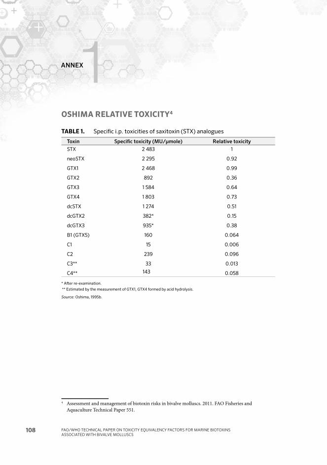
FAO/WHO TECHNICAL PAPER ON TOXICITY EQUIVALENCY FACTORS FOR MARINE BIOTOXINS ASSOCIATED WITH BIVALVE MOLLUSCS
108
ANNEX1OSHIMA RELATIVE TOXICITY4
TABLE 1. Specifi c i.p. toxicities of saxitoxin (STX) analogues
Toxin Specifi c toxicity (MU/μmole) Relative toxicity
STX
neoSTX
GTX1
GTX2
GTX3
GTX4
dcSTX
dcGTX2
dcGTX3
B1 (GTX5)
C1
C2
C3**
C4**
2 483
2 295
2 468
892
1 584
1 803
1 274
382*
935*
160
15
239
33
143
1
0.92
0.99
0.36
0.64
0.73
0.51
0.15
0.38
0.064
0.006
0.096
0.013
0.058
* Aft er re-examination.
** Estimated by the measurement of GTX1, GTX4 formed by acid hydrolysis.
Source: Oshima, 1995b.
4 Assessment and management of biotoxin risks in bivalve molluscs. 2011. FAO Fisheries and Aquaculture Technical Paper 551.

FOR MORE INFORMATION CONTACT:
www.fao.org/food/food-safety-quality/home-page/en/
Shellfish can be contaminated with toxic substances derived from marine
microalgae. In order to protect consumers from poisoning by these toxins,
regulatory authorities have defined limits as to the maximum amount of such
toxins in shellfish intended for human consumption, and analyses must be
conducted in order to ensure that such limits are not exceeded.
Several groups of shellfish toxins have been described, such as the paralytic,
diarrhetic and amnesic toxins. Within each group, several structurally-related
toxins (cogeners) may be present that contribute to the overall toxic potential.
Based on their chemical structure, the toxins included in the Codex Standard
for Live and Raw Bivalve Molluscs (Codex Standard 292–2008) are classified
into five groups, namely, the saxitoxin (STX), okadaic acid (OA), domoicacid
(DA), brevetoxin (BTX) and azaspiracid (AZA) groups.
For risk assessment and management, knowledge of the amount of toxin
congeners in the shellfish is not sufficient. There is also the need to know
the relative toxicity of each of the congeners, so that the total toxicity of the
material in the extract can be estimated. This requires the determination of
Toxicity Equivalency Factors (TEFs).
The 34th Session of the Codex Committee on Fish and Fishery Products
(CCFFP)requested FAO/WHO to provide scientific advice on TEFs for biotoxins
included in the Codex Standard. FAO/WHO set up an Expert Group to provide
scientific advice and recommendations on TEFs for biotoxins. The Expert
Group developed an approach to be used for the development of TEFs for each
group of biotoxins included in the Codex Standard, and in addition considered
tetrodotoxin (TTX), due to its emergence in shellfish.
The Joint FAO/WHO Expert Meeting on Toxicity Equivalency Factors for
Marine Biotoxins was convened in Rome on 22–24 February 2016. This report
summarizes the outcomes of the meeting and identifies the TEFs for each of
the six groups of toxins.
I5970E/1/07.16
ISBN 978-92-5-109345-0
9 7 8 9 2 5 1 0 9 3 4 5 0