tracheostomy laryngectomy managing the patient16... · the procedure requires the trachea ... and...
TRANSCRIPT
Source: Tracheostomy/Laryngectomy Working Group Issue date: 1st August 2017 Page 1 of 16
Status: Approved Review date: 1st August 2018 Document Reference: PP(17) 315
Trust Policy and Procedure PP(17) 315
Tracheostomy / Laryngectomy: Managing the Patient
For use in (clinical areas): F10 – Respiratory Ward
For use by (staff groups): All groups of staff caring for patients with Tracheostomy /
Laryngectomy
For use for (patients/treatments): Patients with Tracheostomy / Laryngectomy
Document owner: Sandy Lewis, Education and Outreach Sisters, Gary Ingalla
Status: Approved
Purpose of the Guideline Tracheostomy is a common procedure for patients who require emergency airway support. As with all procedures the benefits are associated with risk, both during and after insertion .Temporary tracheostomy has become more common place and is frequently an early intervention in critical care units. To ensure patient safety remains a priority, patients who require a tracheostomy should be cared for within an intensive care setting during the acute phase and only discharged to the respiratory ward when the tracheostomy is established (and their medical condition no longer requires critical care). A laryngectomy is the permanent removal of the Larynx. This procedure is often carried out to treat laryngeal cancer or swallowing defects. The procedure requires the trachea to be brought forward to form the stoma; this permanently disconnects the lungs connection to the mouth. This policy describes the procedures which should be followed in managing patients with a tracheostomy tube insitu or a laryngectomy, within the respiratory ward. These procedures aim to maximize patient safety and clinical effectiveness. There is also clinical skills advice available through the intranet, at clinical skills.net – Tracheostomy care.
Contents
1. Indications for tracheostomy 2. Initial arrangements for patients with tracheostomy insitu/laryngectomy insitu 3. Local arrangements for patients with a tracheostomy insitu 4. Criteria of patients suitable for discharge to F10 5. Complications of tracheostomy 6. Humidification 7. Care of inner cannula 8. Tracheal suctioning 9. Changing a tracheostomy dressing 10. Tube occlusion 11. Care of a patient with a total laryngectomy 12. References Appendix 1 - Tracheostomy Discharge Process from CCS to F10 Appendix 2 – Emergency Tracheostomy Management – patient upper airway algorithm Appendix 3 – Tracheostomy bed head sign Appendix 4 – The Tracheostomy Box contents list: Appendix 5 – Emergency Laryngectomy management Appendix 6 – Laryngectomy bed head sign Appendix 7 – Critical Care Education/Outreach - Tracheostomy/Laryngectomy Care Record
Source: Tracheostomy/Laryngectomy working group Issue date: 1st August 2017 Page 2 of 16
Status: Approved Review date: 1st August 2018 Document Reference: PP(17) 315
1. Indications for tracheostomy To relieve upper airway obstruction
To facilitate mechanical ventilation and weaning from respiratory support
To facilitate removal of bronchial secretions
To protect / minimise aspiration risk in the absence of laryngeal reflexes.
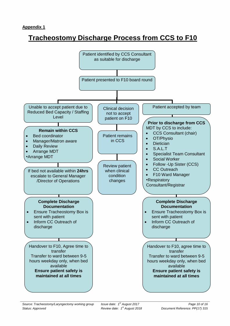
2. Initial arrangements for patients with tracheostomy / laryngectomy Patient admitted to WSH with a Tracheotomy or Laryngectomy should be discussed with both the Critical care and Respiratory consultant to identify the most appropriate place for admission. The purpose of admitting patients to Critical Care or the Respiratory Ward is to ensure they receive the specialised nursing care they require. The responsibility of these patients remains with the admitting consultant, throughout the patients stay. Patient identified as suitable for discharge from Critical Care will need to be discussed with the Respiratory Consultants to identify suitability for discharge to F10 as per agreed tracheostomy/laryngectomy discharge process (see Appendix1) 3. Local arrangements for patients with a tracheostomy insitu
/Laryngectomy in situ All patients with a tracheostomy, outside of Critical Care, should be placed onto F10 the Respiratory ward. This helps to concentrate staff expertise and training. Critical care will hold a centralised stock of tracheostomy tubes, accessories and the tracheostomy emergency boxes for the Trust. To obtain a tracheostomy emergency box contact the Critical Care coordinator or contact the Critical Care Outreach Team (CCOT) on Bleep 666, the ward will be cross-charged for equipment and accessories used from this stock. All patients admitted to the respiratory ward should have the following documentation and signage at their bedside:
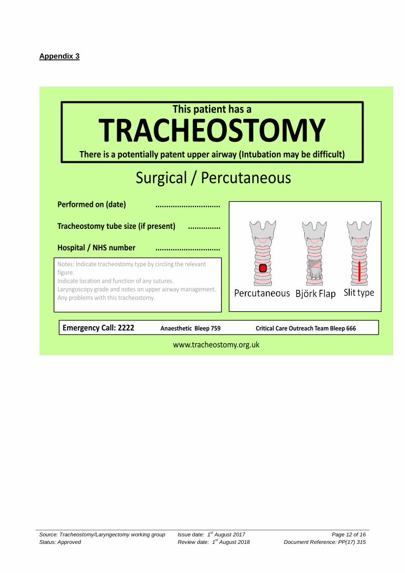
Laminated above bed head Tracheostomy /laryngectomy signage (see Appendix 3)
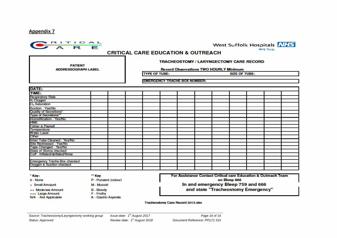
Tracheostomy/laryngectomy Care Record
Tracheostomy Emergency - Tube Occlusion Guide
Laryngectomy Emergency Management Guide
WARNING
Do not leave a patient without a Tracheostomy Emergency Box - check it daily as per checklist. Suction equipment and oxygen delivery systems must always be available, functioning and checked every shift.
Staff looking after a patient with a tracheostomy/ laryngectomy should seek advice from the CCOT if they require any support/education to assist with caring for the patient. Staff must make themselves familiar with the box contents, its use, and sign the tracheostomy care sheet to this effect.
4. Criteria of patients suitable for discharge to F10- Respiratory Ward Patients ready for discharge from Critical Care following a tracheostomy, should have an established tracheostomy tube in place enabling the patient to breathe spontaneously. This group
Source: Tracheostomy Working Group Issue date: 1st August 2017 Page 3 of 16
Status: Approved Review date: 1st August 2018 Document Reference: PP(17) 315
of patients are likely to have the tracheostomy as a result of the following conditions, however this list is not exhaustive and individual assessment will be required.
Past pneumonia or other chronic illness resulting in a residual weak cough or swallowing.
Past traumatic brain injury or post op neurological surgery prior to transfer for rehabilitation centre.
Recovering stroke patients.
Patient with an established laryngectomy admitted with an additional condition that has been stabilised
Palliative Tracheostomy for inoperable Laryngeal tumour
Long term Tracheostomy patient requiring ongoing care following acute phase Prior to discharge and following discussion and acceptance from the respiratory team, these patients require a definitive treatment plan in place, including escalation. They should also have an established physiotherapy and occupational therapy care plan in place.
5. Complications of tracheostomy Immediate complications:
- Haemorrhage - Misplacement - Pneumothorax - Occlusion of tube by cuff herniation (over inflated) Delayed complications:
- Tube occlusion e.g. with secretions - Infection of bronchial tree or stoma
- Mucosal ulceration (caused by asymmetrical or excessive cuff inflation or tube movement)
- Tracheal distension (caused by cuff over inflation) - Tracheo-oesophageal fistula - Impaired swallow Late complications:
- Tracheal dilation, stenosis or granulation - Excessive scar tissue formation causing tracheal tethering.
6. Humidification The delivery of dry gases will slow the mucociliary elevator allowing secretion accumulation, mucosal crusting, increased airway resistance and a susceptibility to bronchial infection. These secretions may thicken and prove difficult to clear and in turn block the tracheostomy/airway. Inadequate humidification and poor hydration may lead to thick secretions which can lead to a potential fatal airway obstruction. Humidification will be required and should be provided for all patients with a tracheostomy.
Patient assessment for humidification Regular assessment will identify effectiveness and adequacy of the humidification of the patient and alongside regular inner cannula care and suctioning can identify warning signs which will allow for appropriate management to prevent tube blockage. Patient assessment should include:
Frequency of suctioning and/or cleaning of inner cannula
Source: Tracheostomy/Laryngectomy working group Issue date: 1st August 2017 Page 4 of 16
Status: Approved Review date: 1st August 2018 Document Reference: PP(17) 315
Tenacity of secretions
Evidence of airflow via tracheostomy
Respiratory rate
Use of accessory muscles
Patient coughing ( ineffective or excessive )
Requirement for supplementary oxygen The practitioner should be aware of the effect that systemic dehydration and poor oral care can have on patient comfort and respiratory tract secretions.
WARNING Failure to humidify adequately can result in tube occlusion, as secretions become thick, sticky and difficult to clear, potentially resulting in an obstructed airway.
Methods of humidification
Hot water humidification – actively humidifies inspired air / oxygen for patients with thick
sputum. Fisher & Paykel humidifiers are available and stored in the medical equipment library.
Circuits are available from CCS (wards will be cross-charged) and must be changed weekly or when the humidifier has been allowed to cool down. The Critical Care Education and Outreach Team can support with these changes.
Heat Moisture Exchange Devices, (HME or “Swedish nose”) - these devices help to retain
the patients’ natural moisture content of their expired breath. Suitable for patients whose sputum is not excessive. Check them at least 2 hourly and change if soiled. Change every 24 hours.
Buchanon Laryngectomy Protector or “bib” – a disposable foam filter applied directly over
the stoma often for long-term laryngectomies. Change every 24 hours.
Saline nebulisers - may be used for tenacious or difficult to suction secretions. Attached to a
tracheostomy mask, 5mls every 2 to 4 hours may be prescribed.
7. Care of inner cannula Wherever possible, tracheotomy tubes with an inner cannula should be used for all patients. The inner cannula must be removed, inspected and when necessary cleaned at regular intervals. Secretions can adhere to the internal lumen of the tracheostomy tube causing reduced airflow, increased work of breathing and tube occlusion. For this reason all tracheostomy tubes outside of Critical Care should have inner cannula or liners which can be cleaned or replaced as necessary. These cannula are single patient use.
Guidelines for changing an inner cannula Preoxygenate and suction as necessary with the inner cannula in situ
The inner cannula should be examined to assess the need for cleaning every two to four hours.
To clean the inner cannula, wearing gloves remove and clean under warm running tap water and rinse with sterile normal saline or water, and reinsert promptly (Russell, 2005).
Never leave the inner cannula to soak during cleaning as this increases the risk of bacterial growth
Always have a spare inner cannula available.
Source: Tracheostomy Working Group Issue date: 1st August 2017 Page 5 of 16
Status: Approved Review date: 1st August 2018 Document Reference: PP(17) 315
8. Tracheal suctioning Tracheal suction is an essential component of secretion control and maintenance of tube patency; however it may be both painful and distressing for the patient and can be complicated by hypoxemia and bradycardia (particularly in patients with autonomic dysfunction such as spinal injuries). Each patient’s individual needs for tracheal suctioning should be constantly reassessed to ascertain the frequency of suctioning required, and where possible patients should be encouraged to expectorate their own secretions. Suctioning is indicated for patients with audible secretions, coughing or those having difficulty with breathing. When selecting a suction catheter , the principle is to use a suction catheter no larger than half of the inner diameter of the tube which will prevent hypoxia and trauma (Glass and Grab, 1995) most adult patients will require either 10FG or 12FG ( Woodrow, 2002). Vacuum pressures (suction) should generally not exceed 100 - 120 mmHg (Intensive Care Society 2008). Higher vacuums can cause mucosal damage and atelectasis (Day 2000).
To perform tracheal suctioning Always use protective apron, sterile gloves and / or goggles
Explain the procedure (and need) to the patient and warn them that it will cause them to cough
Turn on the suction apparatus and ensure it works (not exceeding 120mmHg).
Position the patient upright in neutral head alignment
Put a sterile disposable glove on your dominant hand and hold a sterile catheter with it.
Introduce the catheter to the tracheostomy tube and with no vacuum, insert it until the patient coughs.
Pull back 0.5cm, and then apply continuous suction while slowly withdrawing the catheter.
Key points Do not apply any vacuum while introducing the suction catheter, only on withdrawal.
Tracheostomy tube inner cannula must be in-place during suctioning. If the tracheostomy tube is fenestrated (with a window in the tube) a non-fenestrated inner cannula should be inserted during suctioning (Bilau , 2004).
Pulse oximetery should be used for patients at risk of desaturating during suctioning.
Patients requiring oxygen therapy of 40% or more, or patients who desaturate should be considered for pre-oxygenation. To do this; double the oxygen concentration for three minutes prior to suctioning. For COPD patients requiring pre-oxygenation, increase inspired oxygen by 20 %( Rogge et al, 1989).
A new sterile catheter and glove should be used for each suction attempt
Do not suction for more than 10 seconds.
Instillation of saline to ‘aid’ suctioning is not recommended
Allow the patient time to recover and repeat if necessary.
After suctioning Re-apply the patient’s oxygen and/or humidification apparatus
Dispose of the catheter and glove and rinse the suction tube through with water
Check the patient’s respiratory status is within normal parameter for that patient
Wash your hands
Source: Tracheostomy/Laryngectomy working group Issue date: 1st August 2017 Page 6 of 16
Status: Approved Review date: 1st August 2018 Document Reference: PP(17) 315
Note the nature and amount of tracheal aspirate and record on the Tracheostomy Care Record (Appendix 7)
Change suction tubing and suction container every 24hrs or when heavily soiled / full and between each patient.
Closed Suction (multiple use units) are useful for oxygen dependent patients and patients with copious or infected sputum - particularly MRSA. These units may be used repeatedly as the catheter is enclosed and asepsis is maintained. The units should be changed every 24 hours. Contact the Critical Care Education and Outreach Team via bleep 666.
9. Tracheostomy dressing change Secretions that collect above the cuff ooze out of the stoma site producing a moist environment leading to excoriation and infection. Tracheostomy stoma care aims to reduce the risk of skin irritation and infection by keeping the area clean and dry. Assess the need for dressing changes frequently – at least every shift –change is indicated by the amount of oozing from the stoma
Key point This is a two-person procedure, one person to change the dressing, and another to gently but securely hold the tube in place.
You will need:
Gloves and apron
Dressing pack
Normal saline 0.9%
Pre-cut keyhole dressing
Fresh tracheostomy tube ties.
Changing the tracheostomy dressing:
Wash hands and prepare dressing equipment.
Explain procedure to the patient and position them with neck slightly extended.
One nurse must hold the tracheostomy tube in position while the second nurse removes the soiled dressing and tracheostomy ties - discard.
Assess stoma for signs of infection or inflammation.
Clean around the stoma with normal saline if required and dry thoroughly.
Apply keyhole dressing and secure tracheostomy tube with tracheostomy ties tightly enough to prevent movement during coughing but maintaining patient comfort.
10. Tube occlusion A blocked or displaced tracheostomy tube generally presents with respiratory difficulty. The nature of the problem will often be obvious, but if not it is important to adopt a systematic approach and be aware that acutely ill patients may have other cardio-respiratory reasons for difficulty in breathing. Tracheostomy tubes may become dislodged or displaced for a number of reasons .This can be a stressful event for both patients and staff, and prevention is better than cure.
Source: Tracheostomy Working Group Issue date: 1st August 2017 Page 7 of 16
Status: Approved Review date: 1st August 2018 Document Reference: PP(17) 315
Tube occlusion may occur if sputum is thick and the inner tube becomes blocked. It will also result if the tube is misplaced and can happen following replacement, or after a strong cough with loose ties.
Tube occlusion should be suspected if: Breathing is difficult or noisy
Respirations and pulse rates are increased
The patient is anxious
Air flow via tracheostomy is limited or absent
Accessory muscles are being used
The patient is pale, cyanosed or clammy
If tube occlusion is suspected: Call for help , Bleep – 759- Anaesthetic SPR and 666 Critical Care Outreach team
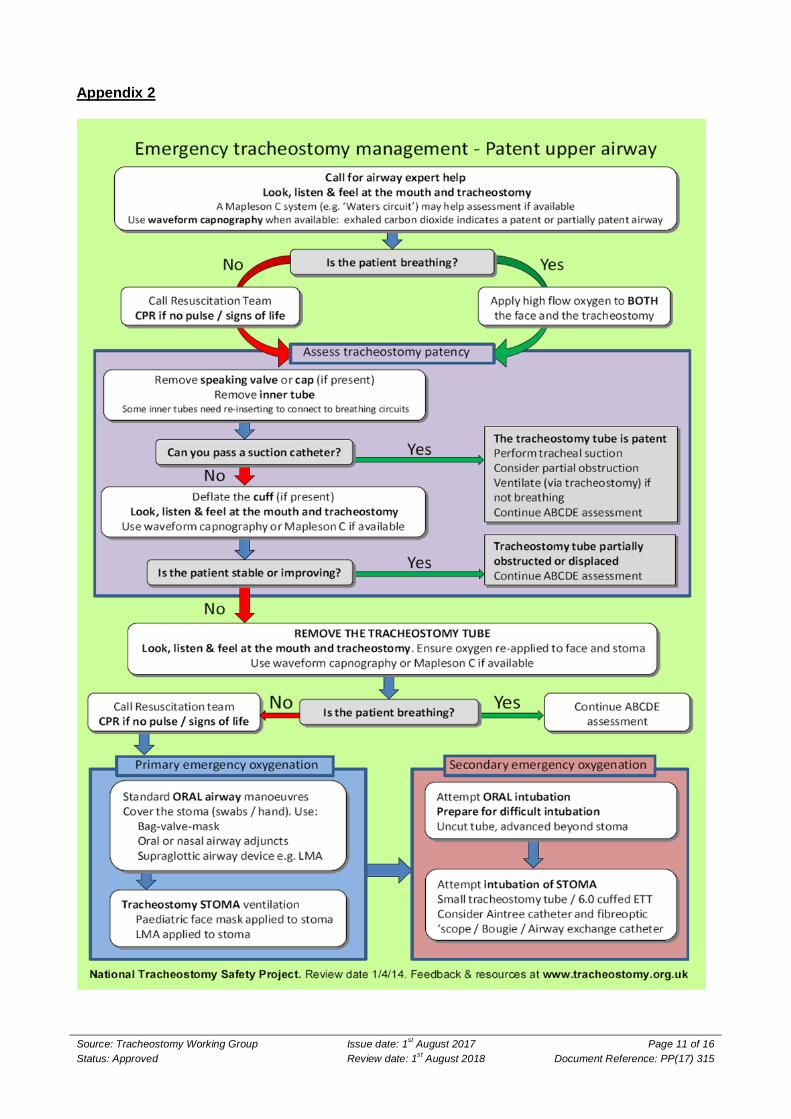
Follow the Emergency Tracheostomy Management Algorithm ( see Appendix 2)
Do not leave the patient – keep calm
Reassure the patient, check oxygen saturation with a pulse oximeter
Sit the patient up, if they are cooperative encourage the patient to give a vigorous cough, this alone may be enough to remove a plug of thick secretions
If the tracheostomy has an inner tube, remove it
Attempt tracheal suction with a suction catheter
If the obstruction is not relieved , deflate the tube cuff , administer oxygen via a face mask if the patient is breathing spontaneously
If the patient is not breathing spontaneously it may be necessary to ventilate the patient with a bag and mask – and an oral airway (Geudal) if tolerated.
If it is not possible to achieve adequate oxygenation , then remove the tracheostomy tube ensuring cuff is fully deflated
Consider passing yankeur or large bore suction catheters directly into the trachea via stoma to remove thick secretions or blood clot
Ensuring the patient is adequately oxygenated , consider replacing the tracheostomy tube
IF BREATHING STOPS AT ANY TIME – CALL THE
CARDIAC ARREST TEAM (2222) and Attempt manual ventilation via the tracheostomy tube with the cuff
up OR
Attempt manual ventilation via facemask with the tracheostomy tube out and the stoma occluded.
11. Care of a patient with a total Laryngectomy Patients who have undergone a total laryngectomy have had a permanent stoma created often as the result of carcinoma of the larynx. The surgical procedure removes the voice box (larynx) and in doing so a new opening for breathing is created in the neck. The oesophagus is detached from its position at the back of the voice box and stitched to create a separate tube in the neck. The end result is the oesophagus is accessed via the mouth where the patient can eat and drink, and an opening in the neck for breathing.
Source: Tracheostomy/Laryngectomy working group Issue date: 1st August 2017 Page 8 of 16
Status: Approved Review date: 1st August 2018 Document Reference: PP(17) 315
Patients who have had a total laryngectomy are unable to speak and have minimal sense of smell. There are a number of options available to patients to assist with speech. These include a trachea oesphageal prosthesis (TEP) which is a small valved device inserted through the back of the trachea into the oesophagus and enables a patient to speak by covering the stoma. The patient can be taught how to do oesophageal speech, or by using an electrolarynx, which is a battery generated device held against the patient’s neck; they then move their mouth to speak as they would have done prior to surgery.
Nursing care required: Daily care requires the skin around the stoma to be kept clean using warm water.
Remove any dried on secretions from the surrounding area.
If suction is required to manage secretions – follow point: 6 tracheal suctioning.
Depending on the patient’s preferences, the site should remain covered to protect from dust, insects, water etc. by using a bib or similar covering.
If a patient with a laryngectomy deteriorates requiring oxygen, ensure it is delivered via the stoma site. Always administer oxygen via the laryngectomy site.
Discuss the patient’s care requirements with the patient or their carer. They will be very familiar with their care requirement.
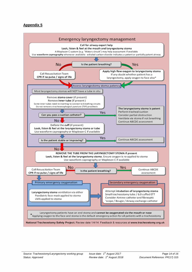
Suspected Stoma occlusion – follow emergency laryngectomy algorithm ( Appendix 5)
IF BREATHING STOPS AT ANY TIME – CALL THE CARDIAC ARREST TEAM (2222) and
Apply high flow oxygen to the laryngectomy stoma
Due to the change to the patient’s anatomy they are unable to receive oxygen via the face or nasal cannula.
Statement of clinical evidence
The Association of Anaesthetists of Great Britain & Ireland grants readers the right to reproduce the algorithms included in this article (Figs 1 and 2) for non-commercial purposes (including in scholarly journals, books and non-commercial websites), without the need to request permission. Each reproduction of any algorithm must be accompanied by the following text: Reproduced from McGrath BA, Bates L, Atkinson D, Moore JA. Multidisciplinary guidelines for the management of tracheostomy and laryngectomy airway emergencies. Anaesthesia. 2012 Jun 26. doi: 10.1111/j.1365-2044.2012.07217, with permission from the Association of Anaesthetists of Great Britain & Ireland/Blackwell Publishing Ltd."
These guidelines are based on St. George’s Healthcare NHS Trust “ Guideline for the care of patients with tracheostomy tubes”. Relevant references are detailed below.
13. References Akgul.S, Akycolcu.N, (2002) Effects of normal saline on endotracheal suctioning , Journal of clinical Nursing, 11:826-30 Billau.C (2004, cited Russell.C, Matta.B ) Tracheostomy a multi professional handbook.Cambridge, Cambridge university press
Source: Tracheostomy Working Group Issue date: 1st August 2017 Page 9 of 16
Status: Approved Review date: 1st August 2018 Document Reference: PP(17) 315
Buglass.E, (1999) Tracheostomy care :tracheal suctioning and humidification.British Journal of Nursing, 8(8) ;500-4 Day.T(2000) Tracheal suctioning:when, why and how .Nursing times,96 (20) 13-15 Day T, Farnell .S, Haynes.S, Wainwright.S, Wilson-Barnett.J, (2002) Tracheal suctioning: an exploration of nursing knowledge and competencies on acute and high dependency ward areas.Journal of Advanced Nursing39(1) 35-45 Glass.C, Grap.M(1995) Ten tips for safe suctioning, American Journal of Nursing, 10(3) 171-8 Harkin.H, Russell.C, (2001) Tracheostomy patient care , Nursing Times, 97 (25) 34-36 Higgins.D (2009) Tracheostomy care part 1: Using suction to remove respiratory secretions via a tracheostomy tube, Nursing Times, 105 (4) 16 National Tracheostomy Safety Project, www.tracheostomy.org.uk Neill.K(2001) Normal saline instillation prior to endotracheal suction.Nursing in Critical Care, 6:34-39 Nelson.L (1999) Wound care, Points of fiction, Nursing Times 95(34) 72-75 Rogge J.A, Bunde.L, Baun.M.M (1989) Effectiveness of oxygen concentrations of less than 100% before and after tracheal suction in patients with chronic pulmonary disease. Heart and Lung (18) 64-71 Russell .C (2005) providing the nurse with a guide to tracheostomy care and management.British Journal of Nursing 14 (8) 428-433 Intensive Care Society (2008) Standards for the care of adult patients with a temporary tracheostomy Woodrow.P(2002) Managing patients with a trachestomy in acute care , Nursing Standard 16(44) 39-48 Contributors and peer review
These guidelines have been developed and reviewed by a representative, multi-professional group encompassing Critical Care, Respiratory Medicine, AHP, and ENT.
Author(s): Sandy Lewis - Head of Patient Safety, Gary Ingalla - Matron Other contributors: Carin Swanevelder , Tracheostomy Working Group, Jessica
White
Approvals and endorsements:
Respiratory Consultants,
Issue no: 2
File name: Managing the Patient with a Tracheostomy
Supercedes: Previous Tracheostomy Guideline
Additional Information:
Source: Tracheostomy/Laryngectomy working group Issue date: 1st August 2017 Page 10 of 16
Status: Approved Review date: 1st August 2018 Document Reference: PP(17) 315
Appendix 1
Patient identified by CCS Consultant
as suitable for discharge
Patient presented to F10 board round
Unable to accept patient due to Reduced Bed Capacity / Staffing
Level
Patient accepted by team
Prior to discharge from CCS
MDT by CCS to include:
CCS Consultant (chair)
OT/Physio
Dietician
S.A.L.T
Specialist Team Consultant
Social Worker
Follow -Up Sister (CCS)
CC Outreach
F10 Ward Manager
•Respiratory
Consultant/Registrar
Complete Discharge Documentation
Ensure Tracheostomy Box is sent with patient
Inform CC Outreach of discharge
Handover to F10, agree time to transfer
Transfer to ward between 9-5 hours weekday only, when bed
available Ensure patient safety is
maintained at all times
Remain within CCS
Bed coordinator
Manager/Matron aware
Daily Review
Arrange MDT
•Arrange MDT
If bed not available within 24hrs
escalate to General Manager /Director of Operations
Complete Discharge Documentation
Ensure Tracheostomy Box is sent with patient
Inform CC Outreach of discharge
Handover to F10, Agree time to transfer
Transfer to ward between 9-5 hours weekday only, when bed
available Ensure patient safety is
maintained at all times
Clinical decision not to accept
patient on F10
Patient remains in CCS
Review patient when clinical
condition changes
Tracheostomy Discharge Process from CCS to F10
Source: Tracheostomy Working Group Issue date: 1st August 2017 Page 11 of 16
Status: Approved Review date: 1st August 2018 Document Reference: PP(17) 315
Appendix 2
Source: Tracheostomy/Laryngectomy working group Issue date: 1st August 2017 Page 12 of 16
Status: Approved Review date: 1st August 2018 Document Reference: PP(17) 315
Appendix 3
This patient has a
TRACHEOSTOMYThere is a potentially patent upper airway (Intubation may be difficult)
www.tracheostomy.org.uk
Surgical / Percutaneous
Performed on (date) ..............................
Tracheostomy tube size (if present) ...............
Hospital / NHS number ..............................
Notes: Indicate tracheostomy type by circling the relevant figure.Indicate location and function of any sutures.Laryngoscopy grade and notes on upper airway management.Any problems with this tracheostomy.
Emergency Call: 2222 Anaesthetic Bleep 759 Critical Care Outreach Team Bleep 666
Source: Tracheostomy Working Group Issue date: 1st August 2017 Page 13 of 16
Status: Approved Review date: 1st August 2018 Document Reference: PP(17) 315
Appendix 4
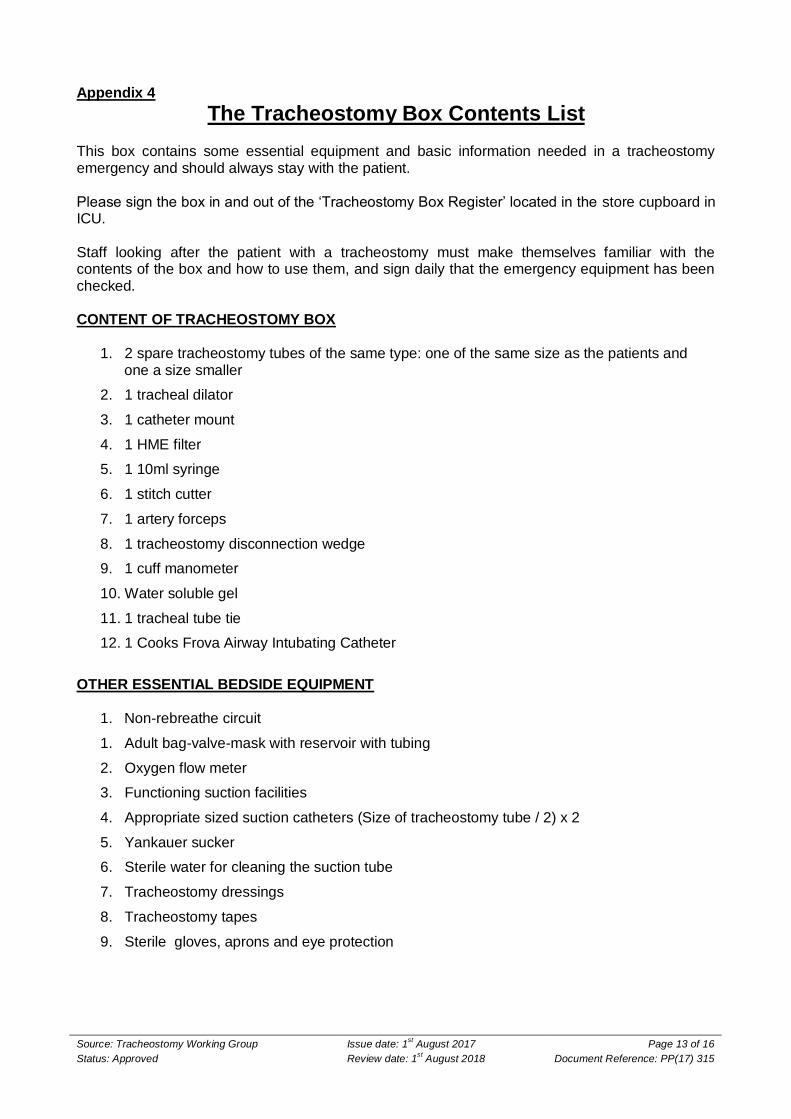
The Tracheostomy Box Contents List This box contains some essential equipment and basic information needed in a tracheostomy emergency and should always stay with the patient. Please sign the box in and out of the ‘Tracheostomy Box Register’ located in the store cupboard in ICU. Staff looking after the patient with a tracheostomy must make themselves familiar with the contents of the box and how to use them, and sign daily that the emergency equipment has been checked. CONTENT OF TRACHEOSTOMY BOX
1. 2 spare tracheostomy tubes of the same type: one of the same size as the patients and one a size smaller
2. 1 tracheal dilator
3. 1 catheter mount
4. 1 HME filter
5. 1 10ml syringe
6. 1 stitch cutter
7. 1 artery forceps
8. 1 tracheostomy disconnection wedge
9. 1 cuff manometer
10. Water soluble gel
11. 1 tracheal tube tie
12. 1 Cooks Frova Airway Intubating Catheter
OTHER ESSENTIAL BEDSIDE EQUIPMENT
1. Non-rebreathe circuit
1. Adult bag-valve-mask with reservoir with tubing
2. Oxygen flow meter
3. Functioning suction facilities
4. Appropriate sized suction catheters (Size of tracheostomy tube / 2) x 2
5. Yankauer sucker
6. Sterile water for cleaning the suction tube
7. Tracheostomy dressings
8. Tracheostomy tapes
9. Sterile gloves, aprons and eye protection
Source: Tracheostomy/Laryngectomy working group Issue date: 1st August 2017 Page 14 of 16
Status: Approved Review date: 1st August 2018 Document Reference: PP(17) 315
Appendix 5
Source: Tracheostomy Working Group Issue date: 1st August 2017 Page 15 of 16
Status: Approved Review date: 1st August 2018 Document Reference: PP(17) 315
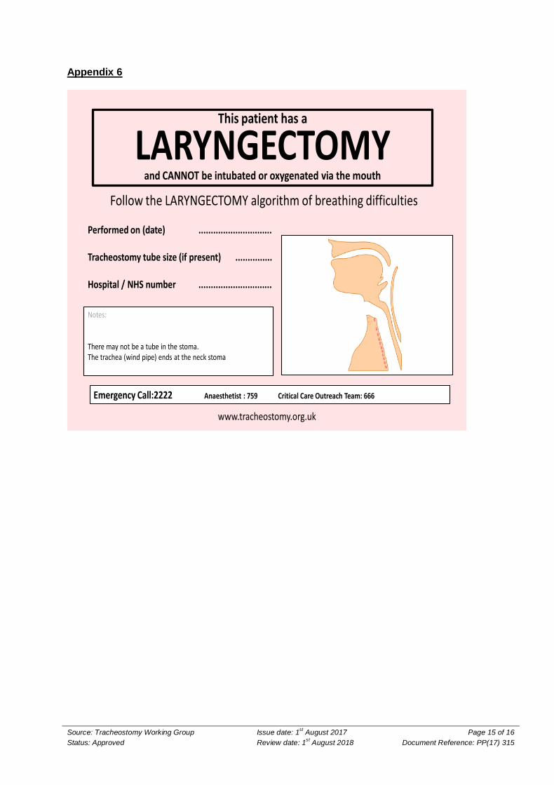
Appendix 6
This patient has a
LARYNGECTOMYand CANNOT be intubated or oxygenated via the mouth
www.tracheostomy.org.uk
Follow the LARYNGECTOMY algorithm of breathing difficulties
Performed on (date) ..............................
Tracheostomy tube size (if present) ...............
Hospital / NHS number ..............................
Notes:
There may not be a tube in the stoma.The trachea (wind pipe) ends at the neck stoma
Emergency Call:2222 Anaesthetist : 759 Critical Care Outreach Team: 666
Source: Tracheostomy/Laryngectomy working group Issue date: 1st August 2017 Page 16 of 16
Status: Approved Review date: 1st August 2018 Document Reference: PP(17) 315
Appendix 7