traj protein of plasmid rp4 binds to a 19-base pair invert
TRANSCRIPT

0 1989 by The American Society for Biochemistry and Molecular Biology, Inc. THE JOURNAL OF BIOLOGICAL CHEMISTRY Vol. 264, No. 20, Issue of July 15, pp. 11989-11994,1989
Printed in U.S.A.
TraJ Protein of Plasmid RP4 Binds to a 19-Base Pair Invert Sequence Repetition within the Transfer Origin*
(Received for publication, November 7, 1988)
Giinter Ziegelin, Jens Peter Fiirste, and Erich LankaS From the Max-Planck-Institut fur Molekulare Genetik, Abteilung Schuster, Ihnestrasse 73, 0-1000 Berlin 33, West Germany
Transfer of plasmid RP4 during bacterial conjuga- tion requires the plasmid-encoded TraJ protein, which binds to the transfer origin (Fiirste, J. P., Pansegrau, W., Ziegelin, G., Kroger, M., and Lanka, E. (1989) Proc. Natl. Acad. Sci. U. S. A. 86, 1771-1775). As indicated by traJ mutants, the TraJ protein is a con- stituent of the relaxosome, the initiation complex of transfer DNA replication. The trdgene maps adjacent to the transfer origin (orin. The structural gene con- sists of a 372-base pair sequence encoding a polypep- tide of 122 amino acids (13,282 Da). TraJ was purified from an Escherichia coli strain overproducing the pro- tein. DNA footprinting experiments involving DNase I demonstrated that the purified protein binds to the right arm of a 19-base pair inverted repeat within oriT. Hydroxyl radical footprints of the DNA-protein complex revealed that TraJ protein is bound to only one side of the DNA helix.
Conjugative transfer of the promiscuous IncPa plasmid RP4 starts at a unique site designated the transfer origin (oriT). Transfer proceeds unidirectionally as concluded from genetic data (1,2). The oriT of the 60-kb’ plasmid RP4 maps within the 17-kb Tral region, which encodes essential func- tions for conjugative transfer. Products from the genes flank- ing oriT are required for the formation of relaxosomes, the initiation complexes of transfer DNA replication (3). The genes for the complex forming proteins comprise about 2.2 kb of the Tral region. These Tral core products serve to direct strand- and site-specific cleavage at the nick location within or iT to generate the unique plasmid strand that is transferred to the recipient cell (T-strand). The TraJ protein of the Tral core components has recently been shown to recognize oriT- DNA specifically (3).
We report here the nucleotide sequence of the t r d gene and the purification of the TraJ protein from an overproduc- ing strain. We demonstrate that the TraJ protein binds spe- cifically to one arm of the 19-bp inverted repeat within oriT. We presume that this nucleoprotein structure is the initial complex in the pathway to assemble a functional relaxosome.
a The costs of publication of this article were defrayed in part by the payment of page charges. This article must therefore be hereby marked “aduertisemenf” in accordance with 18 U.S.C. Section 1734 solely to indicate this fact.
The nucleotide sequence($ reported in this paper has been submitted to the GenBankTM/EMBL Data Bank with accession numberfs) 504942.
4 To whom correspondence should be addressed. The abbreviations used are: kb, kilobase(s); bp, base pair(s);
Mops, 3-(N-morpholino)propanesulfonic acid.
EXPERIMENTAL PROCEDURES
Bacterial Strain, Plasmids, and Medium-Escherichia coli HBlOl (4) was the host for plasmids pJF142n, pJF143n, pJF166u, pJF166uA5, pJF166uA6, pMS226n (3), and pJF118EH (5). YT me- dium (10 g of Tryptone, 5 g of yeast extract, and 5 g of NaCl/liter) was supplemented with Mops, sodium salt (pH 8.0, 25 mM), glucose (0.1%), thiamine HCI (25 pglml), and ampicillin, sodium salt (100
DNA Sequence Analysis-DNA was sequenced by the chain ter- mination method (6) using supercoiled plasmid DNA (7) and single- stranded phage M13mp8/9 subclones as described (8). DNA se- quences were analyzed using the UWGCG programs (University of Wisconsin Genetics Computer Group, Madison, WI (9)).
TraJ-DNA Complex Formation-End-labeled DNA fragments were incubated with various amounts of TraJ protein (fraction V) for 30 min at 37 “C in a total volume of 20 rl of buffer containing 20 mM Tris-HC1 (pH 7.6), 50 mM NaCl, 5 mM MgC12, and 10 pg/ml bovine serum albumin.
DNase I Protection Experiments-DNase I cleavage reactions were performed essentially as described (10, 11). After forming the TraJ- DNA complex the following components were added: CaC12 to 2.5 mM, KC1 to 25 mM, salmon sperm DNA to 2.5 pg/ml, and DNase I to 25 ng/ml (final volume of 40 pl). After a 15-min incubation at 30 “C, the reaction was stopped by adding 10 p1 of a 3 M ammonium acetate, 100 mM EDTA solution, 4 pl of salmon sperm DNA (1 mg/ ml), and 250 p1 of ethanol. The DNA was precipitated at -75 “C for 2 h, centrifuged (20,000 X g), washed with 70% ethanol, dissolved in formamide/dye solution, and electrophoresed on 6% (w/v) polyacryl- amide-urea sequencing gels (12). The cleavage pattern was visualized by autoradiography.
Hydroxyl Radical Protection Experiments-Cleavage reactions were performed as described (13,14) with minor modifications. A solution of iron(1I) EDTA (5 pl), prepared by mixing equal volumes of 20 mM EDTA and 10 mM (NH4)2Fe(S04)2, was added to 25 pl of 3% H202, 5 pl of 100 mM L-ascorbic acid, and 15 pl of H20. 20 pl of this mixture was added to the TraJ-DNA complexes. After a 3-min incubation at 37 “C, cleavage was stopped by adding 10 pl of 100 mM thiourea, 5 pl of 3 M sodium-acetate (pH 5.5), 4 pl of salmon sperm DNA (1 mg/ ml), and 250 pl of ethanol. The DNA was ethanol-precipitated and electrophoresed as described under DNase I protection experiments. Autoradiographs were scanned with a LKB 2222-020 UltroScan XL laser densitometer with a rectangular beam (50 X 800 pm) and a LKB 2220 recording integrator.
d m U .
RESULTS
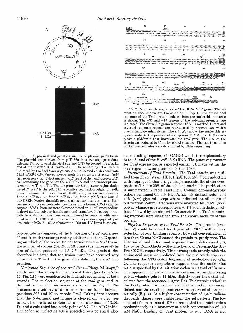
Molecular Cloning of the RP4 traJ Gene-The transfer origin of plasmid RP4 is an intercistronic region centered between two operons of the Tral core region (3). Expression vector cloning of the operon to the left of oriT, encoding genes essential for the relaxation process, resulted in plasmid pJF166uA5 (Fig. lA). This construct contains the complete t r d gene because the size of the inducible traJ product (11 kDa) remained unaltered in contrast to plasmids carrying smaller inserts. The deletion derivative pJF166uA6 lacking the AuaI-BarnHI region (positions 538-852, Fig. lA) specifies a polypeptide slightly larger (12.5 kDa) than the t r d gene product (Fig. 1B). This indicates the formation of a fusion polypeptide since it cross-reacts with anti-TraJ serum. The
11989
11990 IncP oriT Binding Protein
B
12.5 kDa 11 kDa
- BSA
- LYS
120 140 160 180 200
220 240 260 280 300 ma1 I I
320 340 360 380 400
420 440 460 480 500
520 540 560 -10 Accl
580 -35
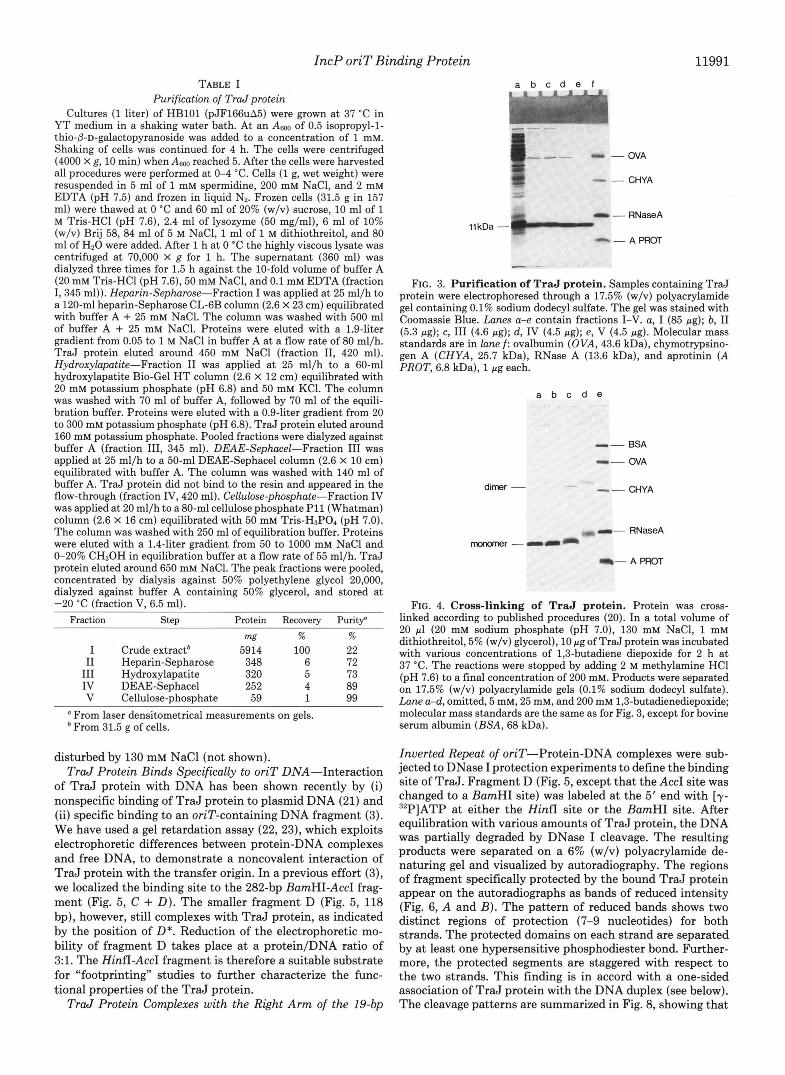
FIG. 2. Nucleotide sequence of the RP4 t r d gene. The re- striction sites shown are the same as in Fig. 1. The amino acid sequence of the TraJ protein deduced from the nucleotide sequence is shown. The -35 and -10 regions of the potential promoter are indicated. The Shine-Dalgarno sequence ( S D ) is marked. Direct and inverted sequence repeats are represented by arrows; dots within arrows indicate mismatches. The triangles above the nucleotide se- quence indicate the position of transposon Tnl725 inserts (17) into plasmid pMS226n that inactivate the traJ gene. The size of the inserts was reduced to 35 bp by EcoRI cleavage. The exact positions of the insertion sites were determined by DNA sequencing.
FIG. 1. A , physical and genetic structure of plasmid pJF166uA5. The plasmid was derived from pJF166u in a two-step procedure, deleting 174 bp toward the AccI site and 1717 bp toward the BssHII end of the inserted RP4 fragment (3). The remaining RP4 DNA is indicated by the bold black segment. AccI is located at kb coordinate 51.16 of RP4 (15). Curued arrows mark the extension of genes: ladQ (lac repressor); bla (p-lactamase); rrnB (part of the rrnB operon of E. coli containing the gene for the 5 S rRNA and the transcriptional terminators TI and TZ). The tac promoter-lac operator region desig- nated P. oriV is the pBR322 vegetative replication origin. B, solid phase immunoblot of extracts of HBlOl carrying various plasmids. Lune a, pJF166uA6; lune b, pJF166uA5; lane c, pMS226n; lane d, pJF118EH (vector plasmid); lane e, molecular mass standards: fluo- rescein isothiocyanate-labeled bovine serum albumin (BSA) and ly- sozyme (LYS). Proteins were electrophoresed on 17.5% (w/v) sodium dodecyl sulfate-polyacrylamide gels and transferred electrophoreti- cally to a nitrocellulose membrane, followed by reaction with anti- TraJ serum (1:400) and fluorescein isothiocyanate-conjugated goat anti-rabbit IgGs (5,16). A photograph (360-nm UV light) is shown.
polypeptide is composed of the 5' portion of traJ and a new 3' end from the vector providing additional codons. Depend- ing on which of the vector frames terminates the traJ frame, the number of codons (14,20, or 23) limits the increase of the size of fusion products to 1.5-2.5 kDa. The product size therefore indicates that the fusion must have occurred very close to the 3' end of the gene, thus defining the traJ map position.
Nucleotide Sequence of the traJ Gene-Phage M13mp8/9 subclones of the 565-bp fragment XmaIII-AccI (positions 575- 10, Fig. lA) were constructed to facilitate sequencing of both strands. The nucleotide sequence of the traJ gene and its deduced amino acid sequences are shown in Fig. 2. The sequence analysis revealed an open reading frame between positions 396 and 27 to encode TraJ. Taking into account that the N-terminal methionine is cleaved off in vivo (see below), the predicted protein has a molecular mass of 13,282 Da and a calculated isoelectric point of 8.7. The ATG initia- tion codon at nucleotide 396 is preceded by a potential ribo-
some-binding sequence (5'-GAGG) which is complementary to the 3'-end of the E. coli 16 S rRNA. The putative promoter for TraJ expression, as reported earlier (3), maps within the oriT region between positions 562 and 589.
Purification of TraJ Protein-The TraJ protein was puri- fied from E. coli strain HBlOl (pJF166uA.5). Upon induction with isopropyl-1-thio-P-D-galactopyranoside, the strain over- produces TraJ to 20% of the soluble protein. The purification is summarized in Table I and Fig. 3. Column chromatography buffers contained 0.1 mM EDTA, 2.5 mM dithiothreitol, and 10% (w/v) glycerol except where indicated. At all stages of purification, column fractions were analyzed by 17.5% (w/v) polyacrylamide gel electrophoresis (0.1% sodium dodecyl sul- fate) followed by staining with Coomassie Blue; TraJ-contain- ing fractions were identified from the known mobility of this protein.
Physical Properties of the TraJ Protein-The protein (frac- tion V) could be stored for 1 year at -20 "C without any reduction of oriT binding capacity. Low salt concentration of less than 50 mM NaCl caused the protein to precipitate. The N-terminal and C-terminal sequences were determined (18, 19) to be NH2-Ala-Asp-Glu-Thr-Lys and Pro-Arg-Ala-Glu- Pro-COOH, respectively. This corresponds exactly with the amino acid sequence predicted from the nucleotide sequence following the ATG codon beginning at nucleotide 396 (Fig. 2). The sequence comparison suggests that the methionine residue specified by the initiation codon is cleaved off in vivo. The apparent molecular mass as determined on denaturing polyacrylamide gels is 11 kDa, slightly lower than that cal- culated from the sequence (13,282 Da). To determine whether the TraJ protein forms oligomers, purified protein was cross- linked, and the resulting products were separated electropho- retically (Fig. 4). At a higher concentration of 1,3-butadiene diepoxide, dimers were visible from the gel pattern. The low amount of dimers (about 10%) suggests that the protein exists predominantly as a monomer at a salt concentration of 130 mM NaC1. Binding of TraJ protein to oriT DNA is not
IncP oriT Binding Protein 11991 TABLE I
Purification of Trdprotein Cultures (1 liter) of HBlOl (pJF166uA5) were grown at 37 "C in
YT medium in a shaking water bath. At an Asm of 0.5 isopropyl-l- thio-0-D-galactopyranoside was added to a concentration of 1 mM. Shaking of cells was continued for 4 h. The cells were centrifuged (4000 X g, 10 min) when Am reached 5. After the cells were harvested all procedures were performed a t 0-4 "C. Cells (1 g, wet weight) were resuspended in 5 ml of 1 mM spermidine, 200 mM NaC1, and 2 mM EDTA (pH 7.5) and frozen in liquid Nz. Frozen cells (31.5 g in 157 ml) were thawed at 0 "C and 60 ml of 20% (w/v) sucrose, 10 ml of 1 M Tris-HC1 (pH 7.6), 2.4 ml of lysozyme (50 mg/ml), 6 ml of 10% (w/v) Brij 58, 84 ml of 5 M NaCl, 1 ml of 1 M dithiothreitol, and 80 ml of H20 were added. After 1 h at 0 "C the highly viscous lysate was centrifuged at 70,000 X g for 1 h. The supernatant (360 ml) was dialyzed three times for 1.5 h against the 10-fold volume of buffer A (20 mM Tris-HC1 (pH 7.6), 50 mM NaC1, and 0.1 mM EDTA (fraction I, 345 ml)). Heparin-Sephrose-Fraction I was applied at 25 ml/h to a 120-ml heparin-Sepharose CL-GB column (2.6 X 23 cm) equilibrated with buffer A + 25 mM NaC1. The column was washed with 500 ml of buffer A + 25 mM NaC1. Proteins were eluted with a 1.9-liter gradient from 0.05 to 1 M NaCl in buffer A at a flow rate of 80 ml/h. TraJ protein eluted around 450 mM NaCl (fraction 11, 420 ml). Hydroxylapatite-Fraction I1 was applied at 25 ml/h to a 60-ml hydroxylapatite Bio-Gel HT column (2.6 X 12 cm) equilibrated with 20 mM potassium phosphate (pH 6.8) and 50 mM KC1. The column was washed with 70 ml of buffer A, followed by 70 ml of the equili- bration buffer. Proteins were eluted with a 0.9-liter gradient from 20 to 300 mM potassium phosphate (pH 6.8). TraJ protein eluted around 160 mM potassium phosphate. Pooled fractions were dialyzed against buffer A (fraction 111, 345 ml). DEAE-Sephcel-Fraction I11 was applied at 25 ml/h to a 50-ml DEAE-Sephacel column (2.6 X 10 cm) equilibrated with buffer A. The column was washed with 140 ml of buffer A. TraJ protein did not bind to the resin and appeared in the flow-through (fraction IV, 420 ml). Cellulose-phosphate-Fraction IV was applied at 20 ml/h to a 80-ml cellulose phosphate P11 (Whatman) column (2.6 X 16 cm) equilibrated with 50 mM Tris-HSPO, (pH 7.0). The column was washed with 250 ml of equilibration buffer. Proteins were eluted with a 1.4-liter gradient from 50 to 1000 mM NaCl and 0-20% CH30H in equilibration buffer a t a flow rate of 55 ml/h. TraJ protein eluted around 650 mM NaC1. The peak fractions were pooled, concentrated by dialysis against 50% polyethylene glycol 20,000, dialyzed against buffer A containing 50% glycerol, and stored at -20 "C (fraction V, 6.5 ml).
Fraction step Protein Recovery Puritf
w % % I Crude extractb 5914 100 22 I1 Heparin-Sepharose 348 6 72 I11 Hydroxylapatite 320 5 73 IV DEAE-Sephacel 252 4 89 V Cellulose-phosphate 59 1 99
From laser densitometrical measurements on gels. From 31.5 g of cells.
disturbed by 130 mM NaCl (not shown). TraJ Protein Binds Specifically to oriT DNA-Interaction
of TraJ protein with DNA has been shown recently by (i) nonspecific binding of TraJ protein to plasmid DNA (21) and (ii) specific binding to an oriT-containing DNA fragment (3). We have used a gel retardation assay (22, 23), which exploits electrophoretic differences between protein-DNA complexes and free DNA, to demonstrate a noncovalent interaction of TraJ protein with the transfer origin. In a previous effort (3), we localized the binding site to the 282-bp BamHI-AccI frag- ment (Fig. 5 , C + D). The smaller fragment D (Fig. 5, 118 bp), however, still complexes with TraJ protein, as indicated by the position of D*. Reduction of the electrophoretic mo- bility of fragment D takes place at a protein/DNA ratio of 3:l. The HinfI -AccI fragment is therefore a suitable substrate for "footprinting" studies to further characterize the func- tional properties of the TraJ protein.
TraJ Protein Complexes with the Right Arm of the 19-bp
11 kDa -
. OVA
. CHYA
- RNaseA
- A PROT
FIG. 3. Purification of TraJ protein. Samples containing TraJ protein were electrophoresed through a 17.5% (w/v) polyacrylamide gel containing 0.1% sodium dodecyl sulfate. The gel was stained with Coomassie Blue. Lanes a-e contain fractions I-V. a, I (85 pg); b, I1 (5.3 pg); c, I11 (4.6 pg); d, IV (4.5 pg); e, V (4.5 pg). Molecular mass standards are in lane f : ovalbumin (OVA, 43.6 kDa), chymotrypsino- gen A (CHYA, 25.7 kDa), RNase A (13.6 kDa), and aprotinin (A PROT, 6.8 kDa), 1 pg each.
a b c d e
dimer -
monomer -a
- BSA
- OVA
- CHYA
- RNaseA
I- A PROT
FIG. 4. Cross-linking of TraJ protein. Protein was cross- linked according to published procedures (20). In a total volume of 20 pl (20 mM sodium phosphate (pH 7.0), 130 mM NaCl, 1 mM dithiothreitol, 5% (w/v) glycerol), 10 pg of TraJ protein was incubated with various concentrations of 1,3-butadiene diepoxide for 2 h a t 37 "C. The reactions were stopped by adding 2 M methylamine HCl (pH 7.6) to a final concentration of 200 mM. Products were separated on 17.5% (w/v) polyacrylamide gels (0.1% sodium dodecyl sulfate). Lane a-d, omitted, 5 mM, 25 mM, and 200 mM 1,3-butadienediepoxide; molecular mass standards are the same as for Fig. 3, except for bovine serum albumin (BSA, 68 kDa).
Inverted Repeat of oriT-Protein-DNA complexes were sub- jected to DNase I protection experiments to define the binding site of TraJ. Fragment D (Fig. 5, except that the AccI site was changed to a BamHI site) was labeled at the 5' end with [y- 32P]ATP at either the HinfI site or the BamHI site. After equilibration with various amounts of TraJ protein, the DNA was partially degraded by DNase I cleavage. The resulting products were separated on a 6% (w/v) polyacrylamide de- naturing gel and visualized by autoradiography. The regions of fragment specifically protected by the bound TraJ protein appear on the autoradiographs as bands of reduced intensity (Fig. 6, A and B). The pattern of reduced bands shows two distinct regions of protection (7-9 nucleotides) for both strands. The protected domains on each strand are separated by at least one hypersensitive phosphodiester bond. Further- more, the protected segments are staggered with respect to the two strands. This finding is in accord with a one-sided association of TraJ protein with the DNA duplex (see below). The cleavage patterns are summarized in Fig. 8, showing that

11992 IncP oriT Binding Protein
bp
369
246
123
abed
- A B
- D* “c
“D
- C / D I B I A
BamHl HinfI Accl HinfI BamHl FIG. 5. Gel electrophoresis of TraJ protein-oriT DNA com-
plexes under nondenaturing conditions. The 790-bp BamHI fragment of pJF142n containing the transfer origin of RP4 was isolated and cleaved by AccI and HinfI. Resulting fragments are A, B, C, and D. The bold black bar marks the oriTfragment (D). Mixtures of TraJ protein and 1.5 pg of DNA fragment were incubated in a total volume of 20 pl for 30 min at 37 “C and loaded onto a 3.5% (w/ v) polyacrylamide gel (8 V/cm). Lane b, DNA fragments with TraJ protein omitted lanes c and d, DNA fragments in the presence of 0.03 and 0.15 pg of TraJ protein, respectively; lane a, DNA fragment size markers (123-bp ladder, Bethesda Research Laboratories). The position of TraJ-oriT complexes is indicated by an asterisk.
the target for TraJ protein is the right arm of the 19-bp inverted repeat within the transfer origin.
TraJ Protein Binds to Only One Side of the DNA Helix- To obtain a higher resolution picture of the protein-DNA complex, we used the hydroxyl radical footprinting technique (13, 14). The cleavage patterns generated on both strands of fragment D were analyzed for protected nucleotide positions by densitometer tracings (Fig. 7). The footprints (summarized in Fig. 8, upper part) show two patches of protected sequence for both strands, separated by a continuous set of unprotected backbone deoxyriboses. The mapping of this pattern onto a scheme of B-DNA (Fig. 8, lowerpart) reveals that the protein DNA contacts lie on one side of the DNA molecule over approximately one helical turn.
DISCUSSION
The isolation of a specific oriT binding protein is likely to be a key step in elucidating the mechanisms involved in initiation of transfer DNA replication. We have purified the plasmid RP4-encoded TraJ protein and have shown that it specifically interacts with a 19-bp inverted repeat within oriT. The perfect sequence symmetry of the repeat is disturbed by three mismatches, allowing TraJ to bind exclusively to the right arm, as indicated by DNase I footprints and hydroxyl radical protection (Fig. 8).
In addition, the right arm of the repeat contains a 10-bp sequence with approximately 2-fold symmetry, suggesting that TraJ protein specifically recognizes this structure. Ap- parently, this sequence motif is structurally conserved be- tween RP4 (IncPa) and the related IncPp plasmid R751. A comparison of their oriT sequences revealed a common 10-bp palindrome within the right arm of a 19-bp inverted repeat (3). However, the palindromic sequences differ in 4 (out of 10) positions. This sequence disparity within the TraJ binding site parallels the oriT specificity of TraJ, i.e. the exclusive interaction of the RP4 TraJ protein with the RP4 transfer
A a b c d e f g h
. A T C
T ‘I C
A .r
B a b c d e f g h
\ C C G G G T G G G C G
\: \\t G
T
FIG. 6. Footprinting analyses of oriT DNA-TraJ protein complexes. Plasmid pJF143n served as a source for the fragment HinfI -BamHI (123 bp) corresponding to the DNA stretch between HinfI and AccI (positions 450-569, Fig. 2, and bund D, Fig. 5). The fragment was 5’-phosphorylated (32P) at the HinfI or the BumHI site by using standard procedures (24). Panel A, DNase I footprint of top strand (Fig. 2) that was labeled at the HinfI -generated end; Panel B, DNase I footprint of bottom strand (Fig. 2) that was labeled at the BamHI-generated end. Lanes e-h, the labeled substrate was incubated with 0,0.15,0.6, and 1.5 pg of purified TraJ protein, respectively, and treated as described under “Experimental Procedures.” The protected nucleotide sequences are indicated with brackets. Lanes a-d, plasmid sequencing reactions of pJF143n containing dideoxynucleoside tri- phosphates of A, C, G, and T, respectively. A 17-mer (26-mer) primer was used which is identical to the first 17 (26) nucleotides of the top (bottom) strand (Fig. 2). Primers were 5’-phosphorylated to provide identical 5’ ends compared to the labeled DNA cleavage products. Slight differences of the electrophoretic mobilities of DNA polymer- ase I- and DNase I-generated products are due to 2’-deoxy-7-deaza- guanosine 5”triphosphate in the sequencing reactions.

IncP oriT Binding Protein 11993
L
FIG. 7. Densitometer scans of hydroxyl radical footprints of oriT DNA-TraJ complexes. Panel A , top strand; panel B, bottom strand (Fig. 2). Upper, degradation of DNA by hydroxyl radical in the absence of TraJ. Lower, degradation of oriT DNA-TraJ complexes. The letters above each peak represent the base whose attached deoxyribose was fragmented by reaction with the hydroxyl radical. Substrates for the cleavage reactions (see "Experimental Procedures") were the same as described in the legend to Fig. 6 as well as the plasmid sequencing reactions for properly aligning the nucleotide sequence to hydroxyl radical-created bands. Protected positions are dotted (lower). The stars mark the 3' end of the se- quence.
-.-.- TGTCACTTCTTCCTTGTGGGCGAGCGFFCACCCGG~?~~GTGGATAGGACGGG
__.-..- I ~. $1 . . ~ ., . ..
~ "..... ' ' .~ :. y.,.i,&;; j ""." ~."
3'
5'
FIG. 8. Upper part, TraJ-protected site within the oriT sequence. The sequence shown corresponds to positions 96-43 of Fig. lA and to positions 485-538 of Fig. 2, respectively. The wedge on the upper strand marks the 3' position of the nick site determined on relaxo- somes in uitro (W. Pansegrau and E. Lanka, manuscript in prepara- tion). Symmetric sequences are indicated by arrows between the strands; mismatches are dotted. Brackets mark the regions protected from DNase I cleavage; nucleotide positions of phosphodiester bonds hypersensitive to DNase I are stippled. Bases whose deoxyriboses are protected by bound TraJ protein from attack by hydroxyl radical are indicated by filled circles above and below the sequence; the size of these circles represents the relative strength of protection. Lower part, pattern of hydroxyl radical protection mapped onto a scheme of B-DNA. Filled circles along the DNA backbone represent deoxyri- boses protected by bound TraJ protein from hydroxyl radical attack.
origin, as concluded from genetic data and binding studies (3).
The hydroxyl radical footprints of bound TraJ protein revealed two patches of protection, approximately 10 bp apart, separated by a region that is still cut by the hydroxyl radical (Fig. 8). These data are consistent with a model that depicts the TraJ protein(s) bound to only one side of the DNA helix, with the back side of the molecule unencumbered by pro- tein(s). Interestingly, the oriT-specific nick was mapped 8 bp to the right of the 19-bp inverted repeat (Fig. 8). This finding might provide a clue of the TraJ function, since the nick site and the TraJ contact points lie on the same side of the DNA duplex, separated by one helical turn. This arrangement could
allow TraJ and the protein(s) which introduce the strand- and site-specific nick to be juxtaposed in a manner suitable for intimate protein-protein interactions. Thus, binding of TraJ to the transfer origin of RP4 might be the initial event for the assembly of the multiprotein complex that directs the oriT-specific initiation of transfer replication.
The above model requires TraJ protein to be an essential relaxosome component. This was substantiated by the prop- erties of two traJ mutations in plasmid pMS226n. The inser- tion mutation, 35-bp remnants of an excised transposon, disrupt the traJ reading frame at positions 228 or 276 (Fig. 2). TraJ cross-reacting material has not been detected in mutant extracts, indicating the absence of any of TraJ fusion products. Plasmid mobilization by the helper plasmid R751 is abolished by these traJ mutations, and no specifically relaxed mutant plasmid DNA can be detected (data not shown).
The traJ gene maps promoter proximal in the polycistronic operon which is to the left of the transfer origin (3). Tran- scription of this operon most likely starts within the transfer origin from a potential promoter about 170 nucleotides up- stream of the coding sequence for TraJ (Fig. 2). The TraJ binding site is located in the noncoding region between the promoter and the t r d gene. It is therefore tempting to suggest the TraJ regulates its own expression. However, autoregula- tion seems unlikely, since the presence of the TraJ binding site in plasmid pJF166uA5 (Fig. L4) is not an obstacle for overexpression of the protein.
The sequence 5"GTTTTAGCGGCTAAAA (Fig. 2, posi- tion 6) occurs in the region between the end of traJ and the following gene in this operon, tral. This sequence contains the inverted repeat 5'-TTTAGC that is common to the pro- posed KorB binding sites (25). KorB is involved in the regu- lation of IncP vegetative replication (25). The occurrence of this sequence motif within the Tral core region of plasmid RP4 may have broad implications regarding the coordination of vegetative and transfer DNA synthesis.
The N-terminal and C-terminal amino acid sequences of TraJ protein coincide with those deduced from the DNA sequence, except that the N-terminal methionine is absent in the protein. The sequence analysis did not reveal domains with significant similarity to the consensus sequences of DNA binding proteins such as the helix-turn-helix motif (26, 27), the zinc finger (28), or the leucine zipper (29). The amino acid composition of TraJ is unusual in that 31% of the amino acid residues contain hydrophobic side chains. This is in accord with the observed protein properties. The yields of TraJ protein are rather low when nonionic detergents are omitted during the purification procedure. The hydrophobicity of TraJ might be required for protein-protein or protein-membrane interactions which are likely to be involved in relaxosome assembly and in possible interactions of the relaxosome with the cell membrane(s).
The data gained by cross-linking of TraJ with 1,3-butadiene diepoxide indicates the protein is predominantly in a mono- meric form. However, as with any cross-linking reagent, the absence of multimers does not preclude the possibility of multimer formation via contacts not accessible or not linkable by the reagent used. The 10-bp palindromic sequence within the TraJ binding region suggests a symmetrical interaction with at least two protein molecules. However, DNase I and hydroxyl radical footprints reveal asymmetric binding of the protein(s) over the pseudo-dyad axis of the 10-bp palindrome. This asymmetry might be of functional relevance, since trans- fer DNA replication is a unidirectional process. Possibly, for this reason, the TraJ protein does not form a symmetrical multimer.

11994 IncP oriT Binding Protein
Perhaps the most interesting finding is the biased interac- tion of TraJ protein with the 19-bp inverted repeat. The structural conservation of this repeat among IncP plasmids suggests a functional importance during either initiation or termination of transfer DNA replication. Why then has an inverted sequence motif evolved that binds TraJ to only one of its arms? It is conceivable that the conservation of the repeat may be unrelated to interaction with TraJ. However, we hypothesize that the hairpin configuration of the repeat may prove to be an alternative binding site for the TraJ protein. One could envisage intrastrand base pairing due to cruciform extrusion under superhelical tension. In addition, a hairpin could form during the single-stranded transfer of plasmid DNA. Experiments are under way to test these pos- sibilities.
Acknowledgments-We are grateful to Heinz Schuster for generous support and stimulating discussions. We are indebted to Makoto Kimura for sequencing the N terminus of the TraJ protein. We thank Karl Rak for preparing anti-TraJ serum, and Wolfgang Schilf and Ingeborg Klaus for providing TraJ mutants. The expert technical assistance of Marianne Schlicht is greatly appreciated.
REFERENCES 1. 2.
3.
4.
5.
6.
7.
Grinter, N. J. (1981) Plasmid 5 , 267-276 Al-Doori, Z., Watson, M. & Scaife, J. (1982) Genet. Res. Carnb.
Furste, J . P., Pansegrau, W., Ziegelin, G., Kroger, M. & Lanka,
Boyer, H. W. & Roulland-Dussoix, D. (1969) J. Mol. Bwl. 4 1 ,
Furste, J . P., Pansegrau, W., Frank, R., Blocker, H., Scholz, P., Bagdasarian, M. & Lanka, E. (1986) Gene (Amst.) 48,119-131
Sanger, F., Nicklen, S. & Coulson, A. R. (1977) Proc. Natl. Acad. Sci. U. S. A. 74 , 5463-5467
Chen, E. Y . & Seeburg, P. H. (1985) DNA 4,165-170
39,99-103
E. (1989) Proc. Natl. Acad. Sci. U. S. A. 8 6 , 1771-1775
459-472
8. Pansegrau, W., Miele, L., Lurz, R. & Lanka, E. (1987) Plusmid
9. Devereux, J., Haeberli, P. & Smithies, 0. (1984) Nucleic Acids
10. Galas, D. J. & Schmitz, A. (1978) Nucleic Acids Res. 5 , 3157-
11. Johnson, A. D., Meyer, B. J. & Ptashne, M. (1979) Proc. Natl.
12. Garoff, H. & Ansorge, W. (1981) Anal. Biochern. 115.450-457
18 , 193-204
Res. 12,387-395
3170
Acad. Sci. U. S. A. 76,5061-5065
13. Tullius, T. D. & Dombroski, B.. A. (1986) Proc. Natl: Acad. Sci. U. S. A. 83,5469-5473
14. Hatfull, G. F., Noble, S. M. & Grindley, N. D. F. (1987) Cell 4 9 ,
15. Pansegrau, W. & Lanka, E. (1987) Nucleic Acids Res. 15 , 2385 16. Towbin, H., Staehelin, T. & Gordon, J. (1979) Proc. Natl. Acad.
17. Ubben, D. & Schmitt, R. (1986) Gene (Amst.) 41 , 145-152 18. Wittmann-Liebold, B. & Kimura, M. (1984) in Methods in Mo-
lecular Biology (Walker, J. M., ed) Vol. I, pp. 221-242, Humana
19. Tsugita, A. & van den Broek, R. (1980) in Methods in Peptide Press Inc., Clifton, NJ
and Protein Sequence Analysis (Birr, C., ed) pp. 359-369, El- sevier/North-Holland Biomedical Press, Amsterdam
20. Baumert, H. G., Skold, S.-E. & Kurland, C. G. (1978) Eur. J. Biochem. 89,353-359
21. Furste, J. P., Ziegelin, G., Pansegrau, W. & Lanka, E. (1987) UCLA Symp. Mol. Cell. Biol. New Ser. 47, 553-564
22. Fried, M. & Crothers, D. M. (1981) Nucleic Acids Res. 9, 6505- 6525
23. Garner, M. M. & Revzin, A. (1981) Nucleic Acids Res. 9 , 3047- 3060
24. Maniatis, T., Fritsch, E. F. & Sambrook, J. (1982) Molecular Cloning: A Laboratory Manual, p. 122, Cold Spring Harbor
25. Smith, C. A., Shingler, V. & Thomas, C. M. (1984) Nucleic Acids Laboratory, Cold Spring Harbor, NY
Res. 12,3619-3630 26. Pabo, C. 0. & Sauer, R. T. (1984) Annu. Rev. Biochern. 5 3 , 293-
321 27. Dodd, I. B. & Egan, J. B. (1987) J. Mol. Bwl. 194,557-564 28. Berg, J. M. (1986) Science 232,485-487 29. Landschulz, W. H., Johnson, P. F. & McKnight, S. L. (1988)
103-110
Sci. U. S. A. 76,4350-4354
Science 240, 1759-1763