transcatheter aortic valve replacement/implant
TRANSCRIPT

Transcatheter Aortic Valve Replacement (TAVR): Getting it right with echo
Jin Kyung Kim, MD PhD FACC FASE Associate Professor of Medicine University of California, Irvine Division of Cardiology School of Medicine

2 2
No conflict
Department Name | Month X, 201X

3 3 Department Name | Month X, 201X Courtesy of Dr. Ihab Alomari

4 4 Department Name | Month X, 201X
Courtesy of Dr. Ihab Alomari
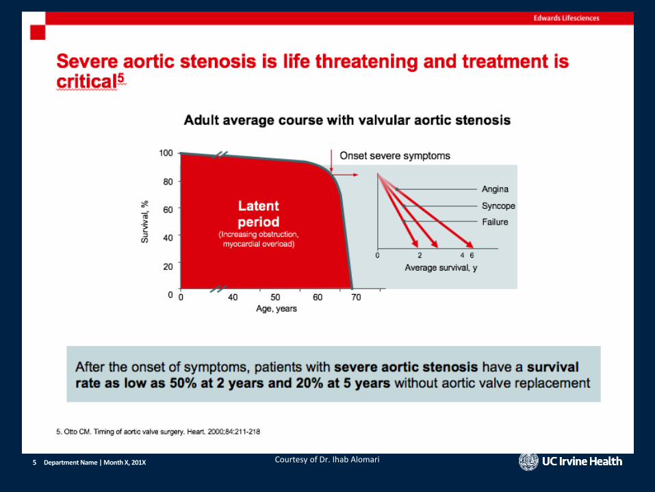
5 5 Department Name | Month X, 201X Courtesy of Dr. Ihab Alomari

6 6 Department Name | Month X, 201X Courtesy of Dr. Ihab Alomari

7 7 Department Name | Month X, 201X Courtesy of Dr. Ihab Alomari

8 8 Department Name | Month X, 201X Courtesy of Dr. Ihab Alomari

9 9 Department Name | Month X, 201X Courtesy of Dr. Ihab Alomari

10 10
TAVR: Transcatheter Aortic Valve Replacement
Department Name | Month X, 201X Courtesy of Dr. Ihab Alomari

11 11 Department Name | Month X, 201X
12 12
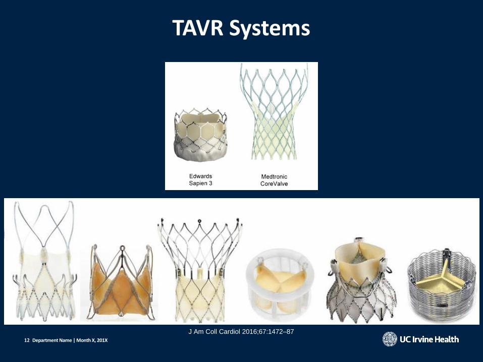
TAVR Systems
Department Name | Month X, 201X
J Am Coll Cardiol 2016;67:1472–87

13 13 Department Name | Month X, 201X Courtesy of Dr. Ihab Alomari

14 14
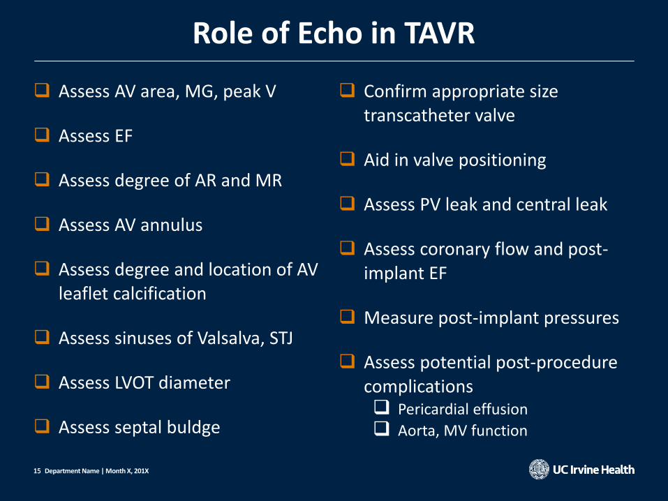
Role of Echo in TAVR
• Patient selection
• Intra-procedural
guidance
• Post-procedural
evaluation
Department Name | Month X, 201X
https://www.wakemed.org/heart-vascular-tavr
15 15
Assess AV area, MG, peak V
Assess EF
Assess degree of AR and MR
Assess AV annulus
Assess degree and location of AV leaflet calcification
Assess sinuses of Valsalva, STJ
Assess LVOT diameter
Assess septal buldge
Confirm appropriate size transcatheter valve
Aid in valve positioning
Assess PV leak and central leak
Assess coronary flow and post-implant EF
Measure post-implant pressures
Assess potential post-procedure complications Pericardial effusion
Aorta, MV function
Department Name | Month X, 201X
Role of Echo in TAVR

16 16
• TTE is the first test to confirm if a patient is eligible for TAVR • Accurate severity of AS:
Patient selection
Department Name | Month X, 201X
LVOT diameter is measured in
• zoomed PLX view
• in mid-systole
• from the white–black interface of the
septal endocardium to the anterior mitral
• Leaflet
• parallel to the AV plane
• within 0.5–1.0 cm of the valve orifice.
Asecho.org

17 17
• Accurate severity of AS:
Patient selection
Department Name | Month X, 201X
LVOT velocity is measured
• from the apical approach
• the sample volume length (or gate) of
3–5 mm
• positioned just proximal to the region
of flow acceleration into the jet.
• The VTI is measured by tracing the
modal velocity (middle of the dense
signal) for use in the continuity
equation or calculation of stroke
volume
Asecho.org

18 18
• Accurate severity of AS:
Patient selection
Department Name | Month X, 201X
• A common source of error for
gradient measurement is
misalignment of the beam
important to use multiple
acoustic windows for the CW
Doppler assessment of AS to
get the best alignment
between Doppler signal and
AV flow. Asecho.org

19 19
• Accurate severity of AS:
Patient selection
Department Name | Month X, 201X
• A dense outer edge of the
smooth velocity curve with a
and clear maximum velocity
should be recorded.
• Vm is measured at the outer
edge of the dark signal; fine
linear signals should not be
included in measurements. Asecho.org
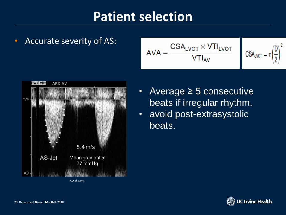
20 20
• Accurate severity of AS:
Patient selection
Department Name | Month X, 201X
• Average ≥ 5 consecutive
beats if irregular rhythm.
• avoid post-extrasystolic
beats.
Asecho.org

21 21
• Importance of EF and dobutamine stress echocardiography in definition of severe AS D2
Patient selection
Department Name | Month X, 201X
AVA ≤ 1.0 cm2 (AVAi ≤0.6 cm2/m2)
Vmax ≥ 4 m/s or mean ΔP ≥ 40 mm Hg AVA ≤ 1.0 cm2 (AVAi ≤0.6 cm2/m2)
Vmax < 4 m/s or mean ΔP < 40 mm Hg
D1 Severe AS
D2 Severe AS
D3 Severe AS
Low LV EF < 50 % Normal LVEF > 50 %
DSE
AVA ≤1.0 cm2 with Vmax ≥4 m/s
Stroke volume index
< 35 mL/m2
Low gradient AS
22 22
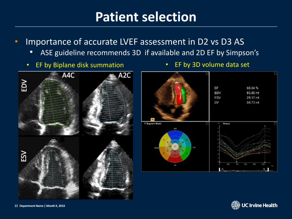
• Importance of accurate LVEF assessment in D2 vs D3 AS • ASE guideline recommends 3D if available and 2D EF by Simpson’s
Patient selection
Department Name | Month X, 201X
• EF by Biplane disk summation • EF by 3D volume data set
23 23
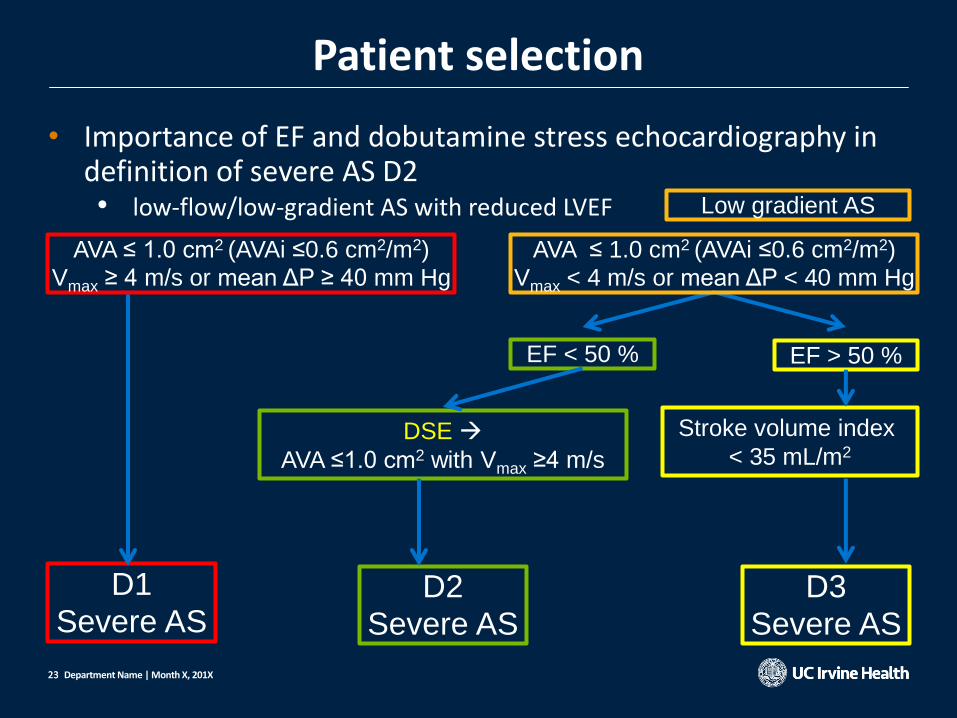
• Importance of EF and dobutamine stress echocardiography in definition of severe AS D2 • low-flow/low-gradient AS with reduced LVEF
Patient selection
Department Name | Month X, 201X
AVA ≤ 1.0 cm2 (AVAi ≤0.6 cm2/m2)
Vmax ≥ 4 m/s or mean ΔP ≥ 40 mm Hg
D1 Severe AS
D2 Severe AS
D3 Severe AS
EF < 50 % EF > 50 %
DSE
AVA ≤1.0 cm2 with Vmax ≥4 m/s
Stroke volume index
< 35 mL/m2
AVA ≤ 1.0 cm2 (AVAi ≤0.6 cm2/m2)
Vmax < 4 m/s or mean ΔP < 40 mm Hg
Low gradient AS

24 24
• Dob stress echo for low flow low gradient AS: • Start dob @ 2.5 – 5 to max 10 – 20 mg/kg/min • Stop infusion when
– Positive result: AVA ≤ 1.0 cm2 with Vmax ≥ 4 m/s at any flow rate – HR 10 – 20 BPM over baseline – > 100 BPM – ↓BP – +Sx – + significant arrhythmia
• Doppler data recorded at each state
• LVOT diameter from baseline used for each stage
Patient selection
Department Name | Month X, 201X

25 25
• Results from Low Dose Dobutamine Echo • An increase in effective AVA > 1.0 cm2 Not severe AS
• AS jet velocity ≥ 4.0 m/s or MG > 30-40 mmHg and AVA < 1.0 cm2 at
any flow rate severe AS
• Absence of contractile reserve (failure to increase SV by 20 %) is a
predictor of high surgical mortality and poor long-term outcome
Department Name | Month X, 201X
Patient selection

26 26
• Importance of stroke volume index in definition of severe AS D3 • low-gradient AS with normal LVEF
• paradoxical low-flow severe AS
Patient selection
Department Name | Month X, 201X
AVA ≤ 1.0 cm2 (AVAi ≤0.6 cm2/m2)
Vmax ≥ 4 m/s or mean ΔP ≥ 40 mm Hg
D1 Severe AS
D2 Severe AS
D3 Severe AS
EF < 50 % EF > 50 %
DSE
AVA ≤1.0 cm2 with Vmax ≥4 m/s
Stroke volume index
< 35 mL/m2
AVA ≤ 1.0 cm2 (AVAi ≤0.6 cm2/m2)
Vmax < 4 m/s or mean ΔP < 40 mm Hg
Low gradient AS

27 27
• Stoke volume by echo • LVOT area x LVOT VIT
• EDV – ESV (by Simpson’s disk summation or by 3D)
• Stroke volume index = SV body surface area
Patient selection
Department Name | Month X, 201X

28 28
• Evolution of Anesthesia & Echo Imaging for TAVR
Intra-procedural Guidance
Department Name | Month X, 201X
29 29
• TEE used • Supplement to pre-procedure CT data • Inadequate CT quality • CT not tolerated or contraindicated
• What to look for during TAVR on echo? • Guide position of transcatheter valve • TAVR Valve Function • Paravalvular Leak • Complications
Intra-procedural Guidance
Department Name | Month X, 201X
30 30
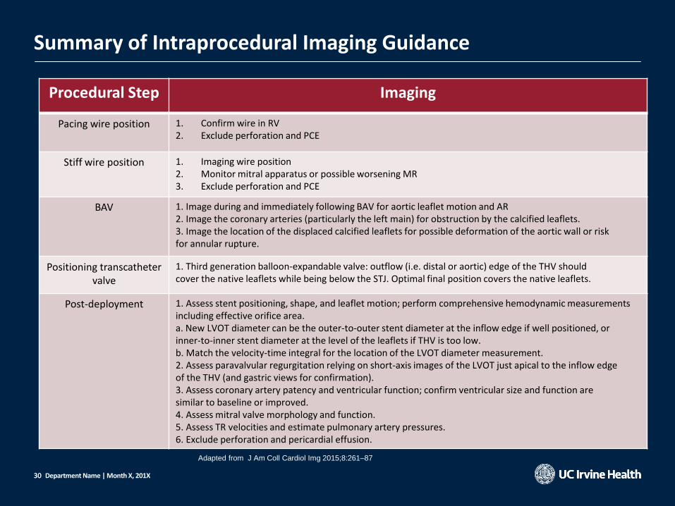
Summary of Intraprocedural Imaging Guidance
Procedural Step Imaging
Pacing wire position 1. Confirm wire in RV 2. Exclude perforation and PCE
Stiff wire position 1. Imaging wire position 2. Monitor mitral apparatus or possible worsening MR 3. Exclude perforation and PCE
BAV 1. Image during and immediately following BAV for aortic leaflet motion and AR 2. Image the coronary arteries (particularly the left main) for obstruction by the calcified leaflets. 3. Image the location of the displaced calcified leaflets for possible deformation of the aortic wall or risk for annular rupture.
Positioning transcatheter valve
1. Third generation balloon-expandable valve: outflow (i.e. distal or aortic) edge of the THV should cover the native leaflets while being below the STJ. Optimal final position covers the native leaflets.
Post-deployment 1. Assess stent positioning, shape, and leaflet motion; perform comprehensive hemodynamic measurements including effective orifice area. a. New LVOT diameter can be the outer-to-outer stent diameter at the inflow edge if well positioned, or inner-to-inner stent diameter at the level of the leaflets if THV is too low. b. Match the velocity-time integral for the location of the LVOT diameter measurement. 2. Assess paravalvular regurgitation relying on short-axis images of the LVOT just apical to the inflow edge of the THV (and gastric views for confirmation). 3. Assess coronary artery patency and ventricular function; confirm ventricular size and function are similar to baseline or improved. 4. Assess mitral valve morphology and function. 5. Assess TR velocities and estimate pulmonary artery pressures. 6. Exclude perforation and pericardial effusion.
Department Name | Month X, 201X
Adapted from J Am Coll Cardiol Img 2015;8:261–87
31 31
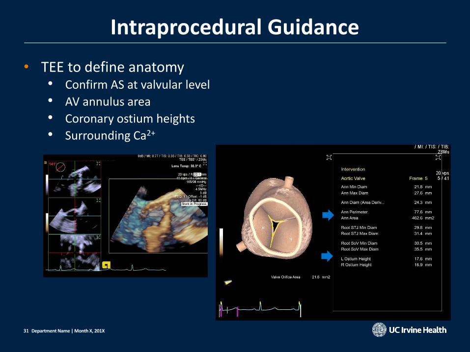
• TEE to define anatomy • Confirm AS at valvular level
• AV annulus area
• Coronary ostium heights
• Surrounding Ca2+
Intraprocedural Guidance
Department Name | Month X, 201X

32 32
• AV annulus area • Important for valve sizing
• Automated 3D or multi-planar tracing
Intraprocedural Guidance
Department Name | Month X, 201X

33 33
• Coronary heights • Automated 3D detection
• SC2000 from Siemens
• Off-axis 2D view
Intraprocedural Guidance
Department Name | Month X, 201X

34 34
• Coronary ostia can be seen by withdrawing the probe above the AV leaflet level and appropriate angulation (yellow arrows)
• Biplane imaging to visualize long axis view
Intraprocedural Guidance
Department Name | Month X, 201X
Echocardiography.. 2018;35:1020–1041. Adapted from J Am Coll Cardiol Img 2015;8:261–87
35 35
Intraprocedural Guidance
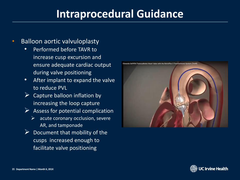
• Balloon aortic valvuloplasty • Performed before TAVR to
increase cusp excursion and
ensure adequate cardiac output
during valve positioning
• After implant to expand the valve
to reduce PVL
Capture balloon inflation by
increasing the loop capture
Assess for potential complication acute coronary occlusion, severe
AR, and tamponade
Document that mobility of the
cusps increased enough to
facilitate valve positioning
Department Name | Month X, 201X

36 36
Intraprocedural Guidance
• TEE helps guide position of the valve before deployment • Optimal position - the distal end of
the crimped valve should cover
the native leaflets but remain 1 to
2 mm below the sinotubular
junction
• Capture deployment of the balloon-expandable valve
Department Name | Month X, 201X

37 37
• Assess • valve position & shape
• leaflet motion
• gradients
• Color Doppler imaging - • presence, location, and severity of AR
• coronary patency
• mitral valve function
• In the setting of hemodynamic compromise – • LV/RV dysfunction
• aortic root catastrophe
Post-implant Assessment
Department Name | Month X, 201X
38 38
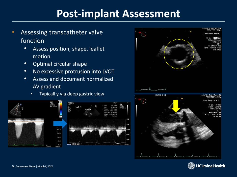
Post-implant Assessment
• Assessing transcatheter valve function • Assess position, shape, leaflet
motion
• Optimal circular shape
• No excessive protrusion into LVOT
• Assess and document normalized
AV gradient • Typicall y via deep gastric view
Department Name | Month X, 201X
39 39
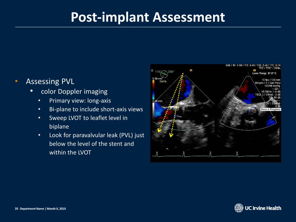
Post-implant Assessment
• Assessing PVL • color Doppler imaging
• Primary view: long-axis
• Bi-plane to include short-axis views
• Sweep LVOT to leaflet level in
biplane
• Look for paravalvular leak (PVL) just
below the level of the stent and
within the LVOT
Department Name | Month X, 201X

40 40
Post-implant Assessment
• Small paravalvular jets following TAVR
may spontaneously regress over 10 to
15 min and require no further
intervention
Department Name | Month X, 201X
41 41
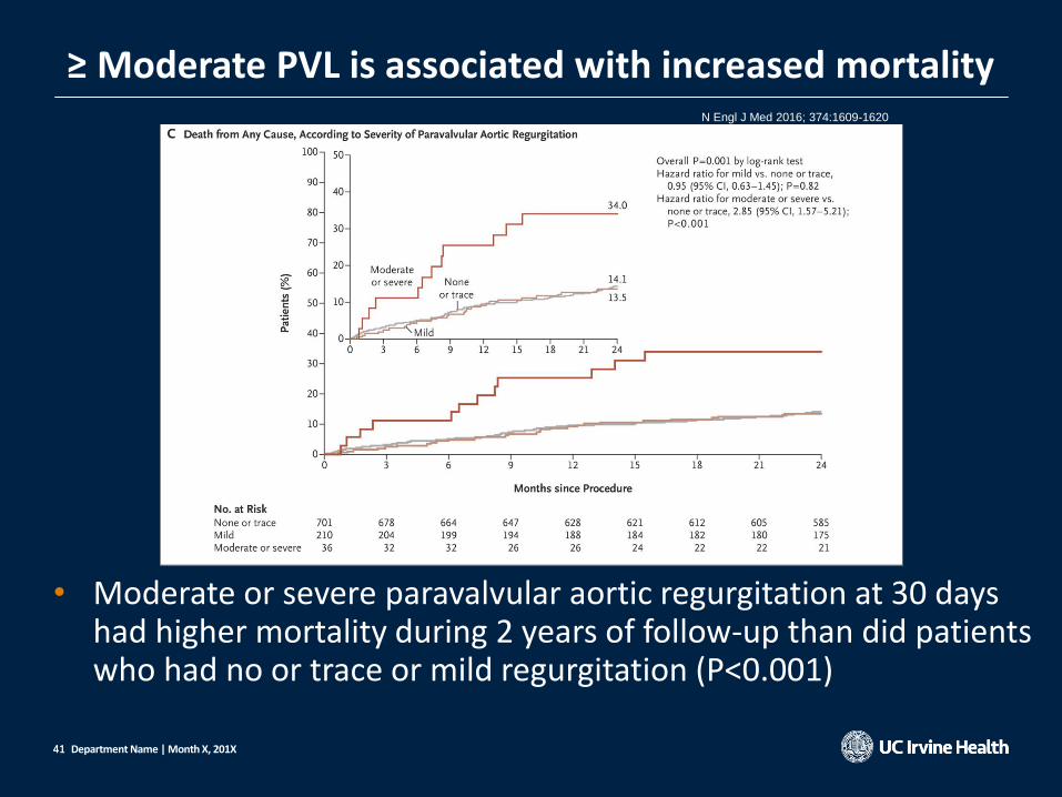
≥ Moderate PVL is associated with increased mortality
Department Name | Month X, 201X
• Moderate or severe paravalvular aortic regurgitation at 30 days had higher mortality during 2 years of follow-up than did patients who had no or trace or mild regurgitation (P<0.001)
N Engl J Med 2016; 374:1609-1620

42 42 Department Name | Month X, 201X
Post-implant Assessment

43 43 Department Name | Month X, 201X
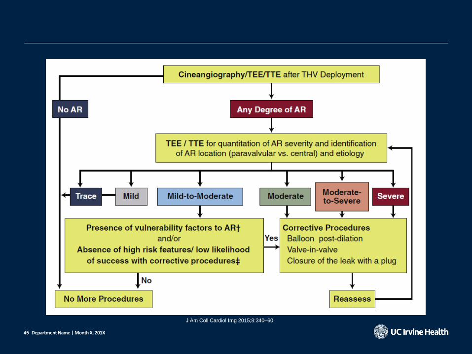
J Am Coll Cardiol Img 2015;8:340–60

44 44
Trace Mild Mild-to-Moderate
Moderate Moderate-to-Severe
Severe
Circumferential extent of PVR (%): color Doppler
<10 <10 10–20 20–30 >30 >30
Department Name | Month X, 201X
TEE Color Doppler Views for the Assessment of PVR
J Am Coll Cardiol Img 2015;8:340–60

45 45
Circumferential Extent of PVR Severity by TTE
Department Name | Month X, 201X
J Am Coll Cardiol Img 2015;8:340–60
Trace Mild Mild to Moderate
Mild to Moderate Moderate Moderate to severe
46 46 Department Name | Month X, 201X
J Am Coll Cardiol Img 2015;8:340–60

47 47
• Complications of TAVR • Acute changes in BP or increase in PAPs
• Check for AR, MR, LV function, PCE
• Major bleeding underfilling of the ventricle • Pericardial effusions
• may indicate localized bleeding, aortic catastrophe or RV perforation by the temporary pacing wire
• Many TAVR patients have small, hypertrophied, and “stiff” ventricles even small effusions tolerated poorly
Department Name | Month X, 201X
Post-implant Assessment

48 48
• Appropriate patient selection • Be accurate about determining AS severity • D2 AS: LFLG AS w reduced EF dob echo • D3 AS: LFLG AS w normal EF SVI ≤ 34 ml/m2
• Pre-procedure planning • Associated unfavorable anatomy • Valve sizing: AV annulus area (TEE)
• Intraprocedural guidance by TEE • Positioning of valve • Assessing for potential complications and/or hemodynamic changes
• MV function • PCE
• PVL assessment - Circumferential extent of PVR less than 20 %
Summary
Department Name | Month X, 201X

Thank you for you attention!
Department Name | Month X, 201X