transcatheter mitral valve-in-valve implantation · figure 1 bioprosthetic mitral valve...
TRANSCRIPT
J A C C : C A S E R E P O R T S V O L . 2 , N O . 1 , 2 0 2 0
ª 2 0 2 0 T H E A U T H O R S . P U B L I S H E D B Y E L S E V I E R O N B E H A L F O F T H E A M E R I C A N
C O L L E G E O F C A R D I O L O G Y F OU N D A T I O N . T H I S I S A N O P E N A C C E S S A R T I C L E U N D E R
T H E C C B Y - N C - N D L I C E N S E ( h t t p : / / c r e a t i v e c o mm o n s . o r g / l i c e n s e s / b y - n c - n d / 4 . 0 / ) .
PERIPARTUM CARDIOVASCULAR DISEASE MINI-FOCUS ISSUE
CASE REPORT: CLINICAL CASE
Transcatheter Mitral Valve-In-ValveImplantation
An Option for Failed BioprostheticMitral Valve Stenosis During PregnancySophie Ribeyrolles, MD, Christelle Diakov, MD, Aurélie Veugeois, MD, Alain Berrebi, MD, Christophe Caussin, MD
JACC: CASE REPORTS CME/MOC/ECME
This article has been selected as this issue’s CME/MOC/ECME activity,
available online at http://www.acc.org/jacc-journals-cme by selecting the
JACC Journals CME/MOC/ECME tab.
Accreditation and Designation Statement
The American College of Cardiology Foundation (ACCF) is accredited by
the Accreditation Council for Continuing Medical Education (ACCME) to
provide continuing medical education for physicians.
The ACCF designates this Journal-based CME/MOC/ECME activity for a
maximum of 1 AMA PRA Category 1 Credit(s)�. Physicians should only
claim credit commensurate with the extent of their participation in the
activity. Successful completion of this CME activity, which includes
participation in the evaluation component, enables the participant to
earn up to 1 Medical Knowledge MOC point in the American Board of
Internal Medicine’s (ABIM) Maintenance of Certification (MOC) program.
Participants will earn MOC points equivalent to the amount of CME
credits claimed for the activity. It is the CME activity provider’s re-
sponsibility to submit participant completion information to ACCME for
the purpose of granting ABIM MOC credit.
Transcatheter Mitral Valve-In-Valve Implantation: An Option for Failed
Bioprosthetic Mitral Valve Stenosis During Pregnancy will be accredited by
the European Board for Accreditation in Cardiology (EBAC) for 1 hour of
External CME credits. Each participant should claim only those hours of
credit that have actually been spent in the educational activity.
The Accreditation Council for Continuing Medical Education (ACCME)
and the European Board for Accreditation in Cardiology (EBAC) have
recognized each other’s accreditation systems as substantially equiva-
lent. Apply for credit through the post-course evaluation. While
offering the credits noted above, this program is not intended to pro-
vide extensive training or certification in the field.
ISSN 2666-0849
From the Department of Cardiovascular Medicine, Institut Mutualiste Mon
Edwards Lifesciences. All other authors have reported that they have no re
disclose.
Informed consent was obtained for this case.
Manuscript received November 1, 2019; revised manuscript received Novem
Method of Participation and Receipt of CME/MOC/ECME Certificate
To obtain credit for this CME/MOC/ECME activity, you must:
1. Be an ACC member or JACC: Case Reports subscriber.
2. Carefully read the CME/MOC/ECME-designated article available
online and in this issue of the journal.
3. Answer the post-test questions. A passing score of at least 70%must be
achieved to obtain credit.
4. Complete a brief evaluation.
5. Claim your CME/MOC/ECME credit and receive your certificate
electronically by following the instructions given at the conclusion
of the activity.
CME/MOC/ECME Objective for This Article: Upon completion of this activ-
ity, the learner should be able to: 1) identify the risks of carrying a preg-
nancy in women with valvular heart disease, and especially with mitral
valve stenosis; 2) discuss the recommended management of women with
valvular heart disease, especially with mitral valve stenosis, from
conception to delivery; and 3) select the recommended regimen of anti-
coagulation therapy during pregnancy.
Author Disclosures: Dr. Caussin is a proctor for Edwards Lifesciences. All
other authors have reported that they have no relationships relevant to
the contents of this paper to disclose.
Medium of Participation: Online (article and quiz).
CME/MOC/ECME Term of Approval
Issue Date: January 2020
Expiration Date: December 31, 2020
https://doi.org/10.1016/j.jaccas.2019.11.050
tsouris, Paris, France. Dr. Caussin is a proctor for
lationships relevant to the contents of this paper to
ber 23, 2019, accepted November 24, 2019.
Ribeyrolles et al. J A C C : C A S E R E P O R T S , V O L . 2 , N O . 1 , 2 0 2 0
Transcatheter Mitral Valve-In-Valve Implantation During Pregnancy J A N U A R Y 2 0 2 0 : 1 4 5 – 9
146
Transcatheter Mitral Valve-In-Valve Implantation
An Option for Failed BioprostheticMitral Valve Stenosis During Pregnancy
Sophie Ribeyrolles, MD, Christelle Diakov, MD, Aurélie Veugeois, MD, Alain Berrebi, MD, Christophe Caussin, MD
ABSTRACT
L
�
�
A pregnant woman presented with symptomatic bioprosthetic mitral valve stenosis. We discuss the difficulties of decision
making in this particular situation where two lives are at stake, the fetus’s and the mother’s, questioning whether
transcatheter mitral valve-in-valve implantation can be an effective and safe option for this challenging condition.
(Level of Difficulty: Advanced.) (J Am Coll Cardiol Case Rep 2020;2:145–9) © 2020 The Authors. Published by Elsevier
on behalf of the American College of Cardiology Foundation. This is an open access article under the CC BY-NC-ND license
(http://creativecommons.org/licenses/by-nc-nd/4.0/).
HISTORY OF PRESENTATION
A 28-year-old woman from Chad was admitted to ourcardiology department for acute dyspnea; she was18 weeks pregnant and had a history of bioprostheticmitral valve (BPMV) replacement 5 years earlier dueto rheumatic fever.
MEDICAL HISTORY
This young woman was born and lived in Chad, whereshe had already given birth to 2 children. At the endof her second pregnancy, 5 years earlier, she experi-enced acute pulmonary edema, which revealed a se-vere rheumatic mitral stenosis. She was referred toour cardiovascular department for surgery 1 monthlater, and mitral valve replacement with BPMV Peri-carbon no. 29 (SORIN, Milan, Italy) was performed.The choice of a biological valve in this young patientwas justified by the absence of birth control (with arisk of unplanned pregnancies), language barrier,difficult access to medical follow-up, and, conse-quently, the risk of noncompliance with oral anti-coagulation. She also had a history of paroxysmalatrial fibrillation. She was treated with vitamin Kantagonists (VKA) and beta-blockers.
EARNING OBJECTIVES
To determine the current role of trans-catheter mitral valve implantation as analternative to surgery in the particular caseof pregnant women.To discuss the management of valvular heartdisease in pregnant women.
DIFFERENTIAL DIAGNOSIS
Because of her past medical history, acute dyspnea inthe setting of history of BPMV replacement mayreveal pulmonary edema due to valvular dysfunctionor recurrence of atrial fibrillation. Premature bio-prosthetic valve deterioration must be suspected.
INVESTIGATIONS
On arrival, our patient was 18 weeks pregnant, andtreatment for acute pulmonary edema compoundedby atrial fibrillation had already been initiated inN’Djamena, Chad, before transfer. She was hemody-namically stable on arrival, in sinus rhythm, andreceiving oral diuretics; VKA had been switched forunfractionated heparin for atrial fibrillation. Bloodtests showed no signs of infection; she had normalblood gas and average levels of creatinine and he-moglobin. Fetal echography showed a healthy fetus.
Transthoracic echocardiography (TTE) (Figure 1,Video 1) showed a significant reduction in BPMVopening, with 3 cusps thickened and calcified. Themitral surface was measured as 0.60 cm2 in planim-etry, effective orifice area was calculated as 0.67 cm2,and mean transvalvular gradient was 26 mm Hg. Shedid not have any other significant valve disease, andleft ventricular ejection fraction was normal. Severestenotic BPMV degeneration was diagnosed. Trans-esophageal echocardiography was performed andconfirmed the diagnosis (Figure 2, Video 2).
MANAGEMENT
After discussion with the obstetric team, percuta-neous intervention was chosen over surgical valve

AB BR E V I A T I O N S
AND ACRONYM S
BPMV = bioprosthetic mitral
valve
MVS = mitral valve stenosis
TMVI = transcatheter mitral
valve implantation
TTE = transthoracic
echocardiography
VKA = vitamin K antagonists
J A C C : C A S E R E P O R T S , V O L . 2 , N O . 1 , 2 0 2 0 Ribeyrolles et al.J A N U A R Y 2 0 2 0 : 1 4 5 – 9 Transcatheter Mitral Valve-In-Valve Implantation During Pregnancy
147
replacement. The patient was therefore scheduled fortranscatheter mitral valve implantation (TMVI). Dur-ing pre-operative assessment, we performed onlycomputed tomography scanning centered solely onthe thorax to avoid fetal radiation exposure. TheTMVI procedure was done without a lead apron onthe patient to decrease fetal exposure and minimizeskin radiation. TMVI was performed when the patientwas 20 weeks pregnant (Figure 3, Videos 3A and 3B)with a transfemoral vein approach. The procedurewas a success, with a mean gradient at the end of theprocedure of 6 mm Hg (Figure 4, Video 4) and noparavalvular leak or pericardial effusion. The totaldose area product for this procedure was of 5,954.6cGy/cm2. The total radiation exposure for the patientwas 624 mGy. The fetus was not in the primary beamand was exposed only to scattered radiation; totalfetal exposure was calculated to be 20 mGy.
DISCUSSION
According to the modified World Health Organizationclassification (1), severe mitral valve stenosis (MVS)during pregnancy is classified as class IV, for whichpregnancy termination should be considered. Luckily,in most cases, these young women benefit from closefollow-up, and pregnancies are anticipated. Theseyoung patients are then referred to a pregnancy heartteam to plan the course of pregnancy. In the case ofsevere MVS, intervention is recommended, with ahigh level of proof when mitral valve area is inferior to1 cm2, regardless of symptoms.
In the case of unanticipated pregnancy for awoman with severe MVS, such as our patient, inter-ventional therapy should be considered instead of
FIGURE 1 Bioprosthetic Mitral Valve Stenosis–Transthoracic Echoca
(A) Parasternal long-axis view showing a calcified thickened mitral biopr
gradient of 26 mm Hg, indicating severe bioprosthesis stenosis.
surgery, considering the fact that risk of fetalmortality during surgery varies from 16% to33% (2). Percutaneous mitral commissur-otomy should be considered first if the pa-tient is eligible (in case of native valve) sothat she can continue her pregnancy. If thepatient does not fit the criteria for this pro-cedure, TMVI is a promising alternative. Itmust nevertheless be considered as a rescueprocedure because published reports lackevidence for the particular management of
pregnant women with severe and unanticipatedvalvular heart disease. Only 1 study (3) focuses spe-cifically on a pregnant woman who underwent asuccessful procedure of concomitant TMVI andtranscatheter aortic valve replacement. Two largercohort studies (4,5) dedicated to TMVI, with 70% and54% women, respectively, reported good outcomesfor TMVI in patients with degenerated BPMV (13.2%and 14% mortality at 12 months for valve-in-valvepatients, respectively), which are encouraging re-sults for the women who underwent these proced-ures. Furthermore, these particular patients were noteligible for surgery because of high comorbidities.Pregnant women, being young, mostly do not sharethis burden, leading to hope that their prognosis willbe even better after TMVI, with a possible chance todefer or even never have surgery in the future.Regarding an antithrombotic regimen, the optimaltherapy after TMVI is still under debate and, becauseof the lack of studies, depends mostly on the team’sexperience. In our center, patients are given doubleantiplatelet therapy for 1 to 3 months and then onlyaspirin. In the case of long-term treatment with an-ticoagulants without bleeding risk, they are givenrdiography
osthesis. (B) Continuous-wave Doppler showing a transmitral mean

FIGURE 2 Bioprosthetic Mitral Valve Stenosis–
Transesophageal Echocardiography
The 3D en face view, showing a limited opening of the mitral
bioprosthesis in systole and thickened leaflets.
Ribeyrolles et al. J A C C : C A S E R E P O R T S , V O L . 2 , N O . 1 , 2 0 2 0
Transcatheter Mitral Valve-In-Valve Implantation During Pregnancy J A N U A R Y 2 0 2 0 : 1 4 5 – 9
148
aspirin for 1 month and then anticoagulant treatmentalone. For our pregnant patient, we considered thehemorrhagic risk and chose to give her only low-molecular-weight heparin.
FOLLOW-UP
The patient was discharged 1 week after the proced-ure, taking low-molecular-weight heparin and beta-blockers. The transmitral mean gradient was8 mm Hg on discharge. TTE was scheduled every
FIGURE 3 Transcatheter Mitral Valve-In-Valve Implantation
Transesophageal echocardiography. (A) Positioning the catheter throug
prosthetic mitral valve: inflating the balloon.
month for the rest of the pregnancy, and the meangradient rose to 11 mm Hg at the end of the term, withhigher cardiac output. She had a planned birth bycesarean and gave birth to a healthy baby boy. Shewas switched back to long-term VKA (warfarin) afterthe birth and then returned to Chad, where she iscurrently followed up, and so far she has not beenreferred again to our hospital.
CONCLUSIONS
Severe BPMV stenosis during pregnancy is chal-lenging to manage. Indeed, 2 patients must beconsidered: the mother and the fetus. TMVI forfailed bioprosthesis seems to be an acceptable op-tion because it ensures protection of the fetus andshows promising results for the mother. It mustnevertheless remain an exceptional situation; thisclinical case stresses the importance of close follow-up for pregnant women with valvular heart diseasesto anticipate pregnancies and avoid difficult situa-tions where the mother’s and fetus’s lives are atrisk.
ADDRESS FOR CORRESPONDENCE: Dr. SophieRibeyrolles, Département de cardiologie, InstitutMutualiste Montsouris, 42 boulevard Jourdan, 75014Paris, France. E-mail: [email protected]: @RbsSophie.
h the bioprosthetic mitral valve. (B) Implantation of the new bio-
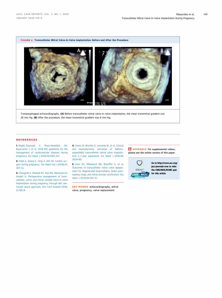
FIGURE 4 Transcatheter Mitral Valve-In-Valve Implantation: Before and After the Procedure
Transesophageal echocardiography. (A) Before transcatheter mitral valve-in-valve implantation, the mean transmitral gradient was
26 mm Hg. (B) After the procedure, the mean transmitral gradient was 6 mm Hg.
J A C C : C A S E R E P O R T S , V O L . 2 , N O . 1 , 2 0 2 0 Ribeyrolles et al.J A N U A R Y 2 0 2 0 : 1 4 5 – 9 Transcatheter Mitral Valve-In-Valve Implantation During Pregnancy
149
RE F E RENCE S
1. Regitz-Zagrosek V, Roos-Hesselink JW,Bauersachs J, et al. 2018 ESC guidelines for themanagement of cardiovascular diseases duringpregnancy. Eur Heart J 2018;39:3165–241.
2. Patel A, Asopa S, Tang A, Otri SK. Cardiac sur-gery during pregnancy. Tex Heart Inst J 2008;35:307–12.
3. Chengode S, Shabadi RV, Rao RN, Alkemyani N,Alsabti H. Perioperative management of trans-catheter, aortic and mitral, double valve-in-valveimplantation during pregnancy through left ven-tricular apical approach. Ann Card Anaesth 2018;21:185–8.
4. Urena M, Brochet E, Lecomte M, et al. Clinicaland haemodynamic outcomes of balloon-expandable transcatheter mitral valve implanta-tion: a 7-year experience. Eur Heart J 2018;39:2679–89.
5. Yoon SH, Whisenant BK, Bleiziffer S, et al.Outcomes of transcatheter mitral valve replace-ment for degenerated bioprosthesis, failed annu-loplasty rings, and mitral annular calcification. EurHeart J 2019;40:441–51.
KEY WORDS echocardiography, mitralvalve, pregnancy, valve replacement
APPENDIX For supplemental videos,please see the online version of this paper.
Go to http://www.acc.org/jacc-journals-cme to takethe CME/MOC/ECME quizfor this article.