transcatheteraorticvalvereplacementincidentalomas...
TRANSCRIPT
Case ReportTranscatheter Aortic Valve Replacement Incidentalomas:A Multimodality Imaging Case of Giant RightCoronary Aneurysm
Jamal Janjua ,1 Esosa G. Odigie-Okon,2 Premnauth Rabindranauth,3
Richard J. Wittchow,4 and Aiman Riaz1
1Department of Internal Medicine, Gundersen Health System, La Crosse, WI 54601, USA2Department of Cardiology, Gundersen Health System, La Crosse, WI 54601, USA3Department of Cardiothoracic Surgery, Gundersen Health System, La Crosse, WI 54601, USA4Department of Pathology, Gundersen Health System, La Crosse, WI 54601, USA
Correspondence should be addressed to Jamal Janjua; [email protected]
Received 23 November 2017; Revised 15 February 2018; Accepted 3 March 2018; Published 26 March 2018
Academic Editor: Ertugrul Ercan
Copyright © 2018 Jamal Janjua et al. )is is an open access article distributed under the Creative Commons Attribution License,which permits unrestricted use, distribution, and reproduction in any medium, provided the original work is properly cited.
Giant coronary artery aneurysms (CAAs) are defined as having a diameter of greater than 2 cm.We report a case of an 82-year-oldmale with severe aortic stenosis incidentally diagnosed with giant right coronary artery aneurysm (gRCAA) while undergoingevaluation for transcather aortic valve replacement (TAVR). It was causing a mass effect on the right cardiac chambers but wasotherwise asymptomatic. Our patient was successfully treated with surgical excision of aneurysm with concomitant coronaryartery bypass grafting (CABG) and surgical aortic valve replacement (SAVR). )e patient remained stable at discharge and onserial follow-ups for two years. In conclusion, due to the associated complication and increased risk of mortality with giantcoronary aneurysms, we recommend surgical approach instead of medical management alone. We also call for evidence-basedrecommendations and guidelines for management of TAVR incidentalomas.
1. Introduction
Transcatheter aortic valve replacement (TAVR) has beenproven to be a safe and effective minimally invasive pro-cedure for aortic stenosis and has been shown to reducemortality among high-risk patients with aortic valve stenosisirrespective of their candidacy for surgical replacement,typically octogenarians and nonagenarians [1]. As a result ofpatients’ advanced age, incidentalomas ranging from in-tracardiac thrombi, extracardiac malignancies and con-genital heart diseases are frequently encountered and alterthe surgical plan. Coronary artery aneurysm (CAA) is a rareentity typically diagnosed incidentally through imagingstudies or at postmortem. Giant coronary artery aneurysms(gCAAs) are even rarer and are defined as having a diametermore than 2 cm. It can be broadly classified into congenitalor acquired (e.g., iatrogenic, atherosclerosis, and infectious).
We present a multimodality imaging case of a giant rightcoronary artery aneurysm (gRCAA) found incidentallyduring workup for aortic valve replacement and outline theunique challenge it presented in this setting.
2. Case Report
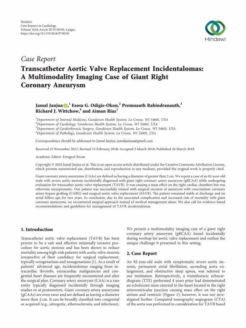
An 82-year-old male with symptomatic severe aortic ste-nosis, permanent atrial fibrillation, ascending aorta en-largement, and obstructive sleep apnea, was referred toour institution. Retrospectively, a transthoracic echocar-diogram (TTE) performed 4 years prior had demonstratedan echolucent mass external to the heart located in the rightatrioventricular junction causing mass effect on the rightatrium and ventricle (Figure 1); however, it was not inve-stigated further. Computed tomography angiogram (CTA)of the aorta was performed in consideration for TAVR based
HindawiCase Reports in CardiologyVolume 2018, Article ID 9738530, 4 pageshttps://doi.org/10.1155/2018/9738530
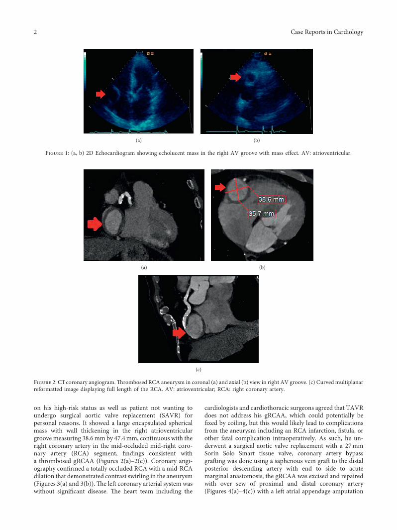
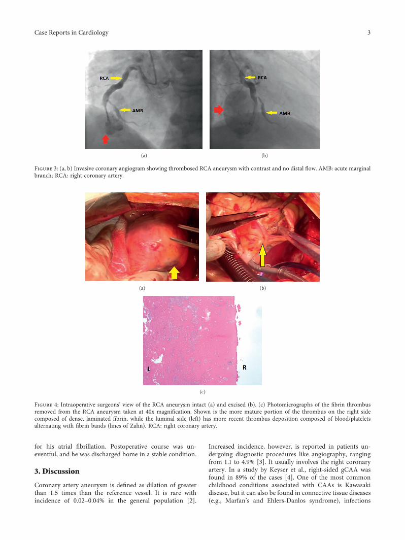
on his high-risk status as well as patient not wanting toundergo surgical aortic valve replacement (SAVR) forpersonal reasons. It showed a large encapsulated sphericalmass with wall thickening in the right atrioventriculargroove measuring 38.6mm by 47.4mm, continuous with theright coronary artery in the mid-occluded mid-right coro-nary artery (RCA) segment, findings consistent witha thrombosed gRCAA (Figures 2(a)–2(c)). Coronary angi-ography confirmed a totally occluded RCA with a mid-RCAdilation that demonstrated contrast swirling in the aneurysm(Figures 3(a) and 3(b)). )e left coronary arterial system waswithout significant disease. )e heart team including the
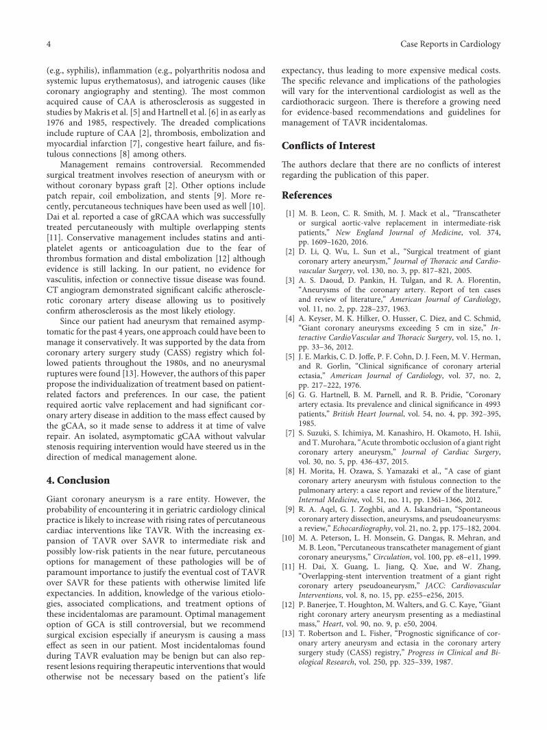
cardiologists and cardiothoracic surgeons agreed that TAVRdoes not address his gRCAA, which could potentially befixed by coiling, but this would likely lead to complicationsfrom the aneurysm including an RCA infarction, fistula, orother fatal complication intraoperatively. As such, he un-derwent a surgical aortic valve replacement with a 27mmSorin Solo Smart tissue valve, coronary artery bypassgrafting was done using a saphenous vein graft to the distalposterior descending artery with end to side to acutemarginal anastomosis, the gRCAA was excised and repairedwith over sew of proximal and distal coronary artery(Figures 4(a)–4(c)) with a left atrial appendage amputation
(a) (b)
Figure 1: (a, b) 2D Echocardiogram showing echolucent mass in the right AV groove with mass effect. AV: atrioventricular.
(a) (b)
(c)
Figure 2: CTcoronary angiogram.)rombosed RCA aneurysm in coronal (a) and axial (b) view in right AV groove. (c) Curvedmultiplanarreformatted image displaying full length of the RCA. AV: atrioventricular; RCA: right coronary artery.
2 Case Reports in Cardiology
for his atrial fibrillation. Postoperative course was un-eventful, and he was discharged home in a stable condition.
3. Discussion
Coronary artery aneurysm is defined as dilation of greaterthan 1.5 times than the reference vessel. It is rare withincidence of 0.02–0.04% in the general population [2].
Increased incidence, however, is reported in patients un-dergoing diagnostic procedures like angiography, rangingfrom 1.1 to 4.9% [3]. It usually involves the right coronaryartery. In a study by Keyser et al., right-sided gCAA wasfound in 89% of the cases [4]. One of the most commonchildhood conditions associated with CAAs is Kawasakidisease, but it can also be found in connective tissue diseases(e.g., Marfan’s and Ehlers-Danlos syndrome), infections
(a) (b)
Figure 3: (a, b) Invasive coronary angiogram showing thrombosed RCA aneurysm with contrast and no distal flow. AMB: acute marginalbranch; RCA: right coronary artery.
(a) (b)
(c)
Figure 4: Intraoperative surgeons’ view of the RCA aneurysm intact (a) and excised (b). (c) Photomicrographs of the fibrin thrombusremoved from the RCA aneurysm taken at 40x magnification. Shown is the more mature portion of the thrombus on the right sidecomposed of dense, laminated fibrin, while the luminal side (left) has more recent thrombus deposition composed of blood/plateletsalternating with fibrin bands (lines of Zahn). RCA: right coronary artery.
Case Reports in Cardiology 3
(e.g., syphilis), inflammation (e.g., polyarthritis nodosa andsystemic lupus erythematosus), and iatrogenic causes (likecoronary angiography and stenting). )e most commonacquired cause of CAA is atherosclerosis as suggested instudies byMakris et al. [5] andHartnell et al. [6] in as early as1976 and 1985, respectively. )e dreaded complicationsinclude rupture of CAA [2], thrombosis, embolization andmyocardial infarction [7], congestive heart failure, and fis-tulous connections [8] among others.
Management remains controversial. Recommendedsurgical treatment involves resection of aneurysm with orwithout coronary bypass graft [2]. Other options includepatch repair, coil embolization, and stents [9]. More re-cently, percutaneous techniques have been used as well [10].Dai et al. reported a case of gRCAA which was successfullytreated percutaneously with multiple overlapping stents[11]. Conservative management includes statins and anti-platelet agents or anticoagulation due to the fear ofthrombus formation and distal embolization [12] althoughevidence is still lacking. In our patient, no evidence forvasculitis, infection or connective tissue disease was found.CT angiogram demonstrated significant calcific atheroscle-rotic coronary artery disease allowing us to positivelyconfirm atherosclerosis as the most likely etiology.
Since our patient had aneurysm that remained asymp-tomatic for the past 4 years, one approach could have been tomanage it conservatively. It was supported by the data fromcoronary artery surgery study (CASS) registry which fol-lowed patients throughout the 1980s, and no aneurysmalruptures were found [13]. However, the authors of this paperpropose the individualization of treatment based on patient-related factors and preferences. In our case, the patientrequired aortic valve replacement and had significant cor-onary artery disease in addition to the mass effect caused bythe gCAA, so it made sense to address it at time of valverepair. An isolated, asymptomatic gCAA without valvularstenosis requiring intervention would have steered us in thedirection of medical management alone.
4. Conclusion
Giant coronary aneurysm is a rare entity. However, theprobability of encountering it in geriatric cardiology clinicalpractice is likely to increase with rising rates of percutaneouscardiac interventions like TAVR. With the increasing ex-pansion of TAVR over SAVR to intermediate risk andpossibly low-risk patients in the near future, percutaneousoptions for management of these pathologies will be ofparamount importance to justify the eventual cost of TAVRover SAVR for these patients with otherwise limited lifeexpectancies. In addition, knowledge of the various etiolo-gies, associated complications, and treatment options ofthese incidentalomas are paramount. Optimal managementoption of GCA is still controversial, but we recommendsurgical excision especially if aneurysm is causing a masseffect as seen in our patient. Most incidentalomas foundduring TAVR evaluation may be benign but can also rep-resent lesions requiring therapeutic interventions that wouldotherwise not be necessary based on the patient’s life
expectancy, thus leading to more expensive medical costs.)e specific relevance and implications of the pathologieswill vary for the interventional cardiologist as well as thecardiothoracic surgeon. )ere is therefore a growing needfor evidence-based recommendations and guidelines formanagement of TAVR incidentalomas.
Conflicts of Interest
)e authors declare that there are no conflicts of interestregarding the publication of this paper.
References
[1] M. B. Leon, C. R. Smith, M. J. Mack et al., “Transcatheteror surgical aortic-valve replacement in intermediate-riskpatients,” New England Journal of Medicine, vol. 374,pp. 1609–1620, 2016.
[2] D. Li, Q. Wu, L. Sun et al., “Surgical treatment of giantcoronary artery aneurysm,” Journal of ,oracic and Cardio-vascular Surgery, vol. 130, no. 3, pp. 817–821, 2005.
[3] A. S. Daoud, D. Pankin, H. Tulgan, and R. A. Florentin,“Aneurysms of the coronary artery. Report of ten casesand review of literature,” American Journal of Cardiology,vol. 11, no. 2, pp. 228–237, 1963.
[4] A. Keyser, M. K. Hilker, O. Husser, C. Diez, and C. Schmid,“Giant coronary aneurysms exceeding 5 cm in size,” In-teractive CardioVascular and ,oracic Surgery, vol. 15, no. 1,pp. 33–36, 2012.
[5] J. E. Markis, C. D. Joffe, P. F. Cohn, D. J. Feen, M. V. Herman,and R. Gorlin, “Clinical significance of coronary arterialectasia,” American Journal of Cardiology, vol. 37, no. 2,pp. 217–222, 1976.
[6] G. G. Hartnell, B. M. Parnell, and R. B. Pridie, “Coronaryartery ectasia. Its prevalence and clinical significance in 4993patients,” British Heart Journal, vol. 54, no. 4, pp. 392–395,1985.
[7] S. Suzuki, S. Ichimiya, M. Kanashiro, H. Okamoto, H. Ishii,and T.Murohara, “Acute thrombotic occlusion of a giant rightcoronary artery aneurysm,” Journal of Cardiac Surgery,vol. 30, no. 5, pp. 436-437, 2015.
[8] H. Morita, H. Ozawa, S. Yamazaki et al., “A case of giantcoronary artery aneurysm with fistulous connection to thepulmonary artery: a case report and review of the literature,”Internal Medicine, vol. 51, no. 11, pp. 1361–1366, 2012.
[9] R. A. Aqel, G. J. Zoghbi, and A. Iskandrian, “Spontaneouscoronary artery dissection, aneurysms, and pseudoaneurysms:a review,” Echocardiography, vol. 21, no. 2, pp. 175–182, 2004.
[10] M. A. Peterson, L. H. Monsein, G. Dangas, R. Mehran, andM. B. Leon, “Percutaneous transcatheter management of giantcoronary aneurysms,” Circulation, vol. 100, pp. e8–e11, 1999.
[11] H. Dai, X. Guang, L. Jiang, Q. Xue, and W. Zhang,“Overlapping-stent intervention treatment of a giant rightcoronary artery pseudoaneurysm,” JACC: CardiovascularInterventions, vol. 8, no. 15, pp. e255–e256, 2015.
[12] P. Banerjee, T. Houghton, M. Walters, and G. C. Kaye, “Giantright coronary artery aneurysm presenting as a mediastinalmass,” Heart, vol. 90, no. 9, p. e50, 2004.
[13] T. Robertson and L. Fisher, “Prognostic significance of cor-onary artery aneurysm and ectasia in the coronary arterysurgery study (CASS) registry,” Progress in Clinical and Bi-ological Research, vol. 250, pp. 325–339, 1987.
4 Case Reports in Cardiology

Stem Cells International
Hindawiwww.hindawi.com Volume 2018
Hindawiwww.hindawi.com Volume 2018
MEDIATORSINFLAMMATION
of
EndocrinologyInternational Journal of
Hindawiwww.hindawi.com Volume 2018
Hindawiwww.hindawi.com Volume 2018
Disease Markers
Hindawiwww.hindawi.com Volume 2018
BioMed Research International
OncologyJournal of
Hindawiwww.hindawi.com Volume 2013
Hindawiwww.hindawi.com Volume 2018
Oxidative Medicine and Cellular Longevity
Hindawiwww.hindawi.com Volume 2018
PPAR Research
Hindawi Publishing Corporation http://www.hindawi.com Volume 2013Hindawiwww.hindawi.com
The Scientific World Journal
Volume 2018
Immunology ResearchHindawiwww.hindawi.com Volume 2018
Journal of
ObesityJournal of
Hindawiwww.hindawi.com Volume 2018
Hindawiwww.hindawi.com Volume 2018
Computational and Mathematical Methods in Medicine
Hindawiwww.hindawi.com Volume 2018
Behavioural Neurology
OphthalmologyJournal of
Hindawiwww.hindawi.com Volume 2018
Diabetes ResearchJournal of
Hindawiwww.hindawi.com Volume 2018
Hindawiwww.hindawi.com Volume 2018
Research and TreatmentAIDS
Hindawiwww.hindawi.com Volume 2018
Gastroenterology Research and Practice
Hindawiwww.hindawi.com Volume 2018
Parkinson’s Disease
Evidence-Based Complementary andAlternative Medicine
Volume 2018Hindawiwww.hindawi.com
Submit your manuscripts atwww.hindawi.com