transcranial doppler up stroke time fraction (ustf) and severe carotid stenosis
TRANSCRIPT

Combination Of Carotid Duplex And Transcranial Doppler For The
Assessment Of Carotid Stenosis
Roberto Hirsch*, Donald H. Lee**, Milberto Scaff*
*Department of Neurology of the Hospital das Clínicas da Faculdade de Medicina da Universidade de São Paulo,
Brazil
**Department of Neuroradiology of the University Hospital of the University of Western Ontario, London, Canada

Background
� NASCET published data from the severe stenosis randomization phase
� 70% ICA stenosis arbitrary cutoff
� “Poor results” achieved by ultrasound in correlating with angio measurements
� Why should this threshold represent a greater risk for stroke if not corrected by endarterectomy
� Can TCD and DCU address this question?

Background
� Is there a physiological phenomenon at this boundary that might be detected by physiological approach?
� Is there room for non invasive approach for the assessment of ICA stenosis?

Background
� Can TCD provide additional information in order to optimize DCU results in determining ICA degree of stenosis?
� Is the combination TCD/DCU more reliable than carotid ultrasound alone in determining severe stenosis?

NASCET angiographic criteria
Bulbo de ACI
Diâmetro da luz
Diâmetro distal
Método NASCET angiográfico % = 1- luz/luz distal
� The NASCET angio measurement technique brought clear standards reducing interobserver variation, unlike ultrasound at the beggining
Methods
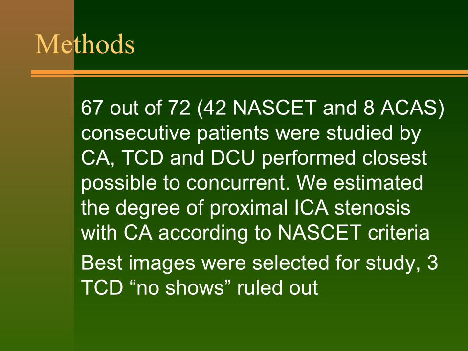
� 67 out of 72 (42 NASCET and 8 ACAS) consecutive patients were studied by CA, TCD and DCU performed closest possible to concurrent. We estimated the degree of proximal ICA stenosis with CA according to NASCET criteria
� Best images were selected for study, 3 TCD “no shows” ruled out

Methods
� 134 arteries were randomized according to angio degree of ICA stenosis into below and over 70% stenosis and ICA occlusion, and had their TCD and DCU readings compared
� Multivariate and univariate analysis with logistic regression and chi-square and T-student test performed for each and total of TCD and DCU parameters having angiogram as gold standard
Methods
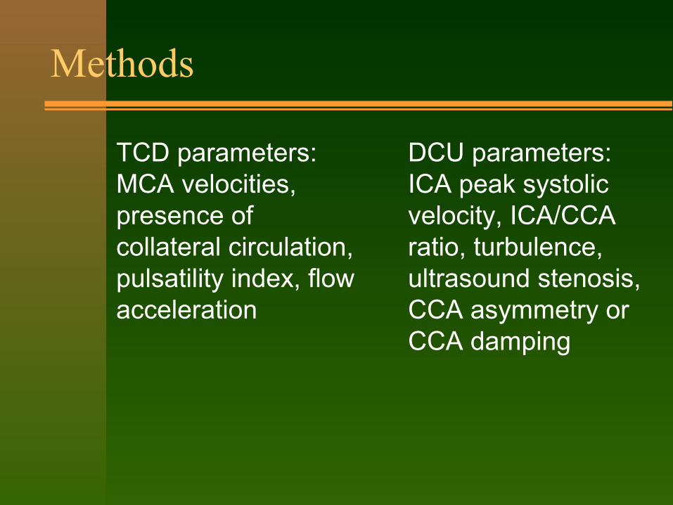
� TCD parameters: MCA velocities, presence of collateral circulation, pulsatility index, flow acceleration
� DCU parameters: ICA peak systolic velocity, ICA/CCA ratio, turbulence, ultrasound stenosis, CCA asymmetry or CCA damping

Upstroke time fraction
x
y
UST-F = x/y
ACINormal
B
� MCA reading distal and ipsilateral to normal ICA. Note the flow acceleration showing steep elevation of systolic flow velocity reaching its peak early in the cardiac cycle
Upstroke time fraction
x
y
UST-F = x/y
cas o de s ub-oclus ão
A
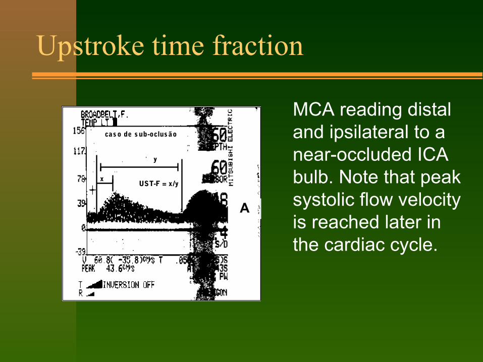
� MCA reading distal and ipsilateral to a near-occluded ICA bulb. Note that peak systolic flow velocity is reached later in the cardiac cycle.

Upstroke time fraction
x
y
UST-F = x/y
cas o de s ub-oclus ão
A
� UST-f was taken measuring the linear distance (time) for systolic velocity to be first achieved (x) and dividing it by the linear distance that represents one cardiac cycle.

Reproducibility of UST-f
� Patient presenting ICA near-occlusion, being the only conflictive result among the two readings (D.Lee = 7,82 ; RH = 15,92)

Reproducibility of UST-f
� By shifting background colour one can identify 2 spectra

Reproducibility of UST-f
� Superimposed retrograde ACA showing quicker flow acceleration time and underlying MCA affected by proximal ICA near-occlusion

Univariate analysis of TCD and DCU parameters - under 70% angio
Mean SD p
MCA mfv 48.5 10.62 0.0014
MCA UST-f 8.7 1.37 0.0001
ECICA psv 158.2 97.68 0.0000
ICA/CCAratio
2.05 1.38 0.0001
CCAdamp/asym
79.81 19.03 0.0000

Univariate analysis of TCD and DCU parameters - over 70% angio, excluding ICA occlusion
Mean SD p
MCA mfv 41.32 11.1 0.0014
MCA UST-f 19.8 3.42 0.0001
ECICA psv 338.89 125.43 0.0000
ICA/CCAratio
6.63 4.16 0.0001
CCAdamp/asym
57.52 19.92 0.0000

Most important TCD parameters - UST-f
� Upstroke time fraction could not be predictive in multivariate analysis because of the wide SD, but in univariate analysis was predictive with 100% of both specificity and sensitivity, provided there were no proximal ICA occlusion
Ust-f<70%
Ust-f>70%
Occl
< 70% 103 0 0
>70% 0 31 0
Occl 0 0 10

Most important TCD parameters - presence of collateral circulation
� Presence of collaterals in all patients with severe stenosis except when more severe stenosis or occlusion was present in contralateral side (p=0.000)
Yes no total
< 70% 9895.15%
54.85%
103
> 70% 412.90%
2787.10%
31
total 102 32 134

Multivariate analysis
� Logistic regression, with analysis of maximum likelihood estimates, shows that CCA damp/asymmetry has 101.686 odds ratio and 95% confidence limits. This parameter was arbitrarily defined as a more than 14 cm/s velocity reduction below the stenotic site or spectral damping. It may be jeopardized when contralateral to an ICA severe stenosis or occlusion

Probability of peak systolic extra-cranial ICA velocity to determine severe ICA stenosis
0
0,1
0,2
0,3
0,4
0,5
0,6
0,7
0,8
0,9
1
0 100 200 300 400 500
EC_ICA
Pro
bab
ilit
y
CCADAMP=N
CCADAMP=S

Sensitivity and specificity for CCA asymmetry to determine severe ICA stenosis
0
20
40
60
80
100
0 0,1 0,2 0,3 0,4 0,5 0,6 0,7 0,8 0,9 1
PROBABILITY
SENSIBILIDADE
ESPECIFICIDADE
� This model gives 90% of both sensitivity and specificity for CCA asymmetry presence to indicate severe stenosis

Conclusion
� Combined TCD and DCU parameters can reliably predict ICA proximal stenosis greater than 70% according to NASCET angiographic criteria.
� If indication of endarterectomy in a symptomatic patient is to be based only upon degree of stenosis, it can safely be done solely on non-invasive combined transcranial and cervical ultrasound approach

Conclusion
� These findings may bring us to the point that hemodynamic features, detectable by physiological methods, may play an important additional role in the genesys of ischemic events in symptomatic patients suffering from ICA stenosis along with embolic risk of a given plaque.