transcription regulation and gene expression in … regulation and gene expression in eukaryotes fs...
TRANSCRIPT
Transcription Regulation And Gene Expression in Eukaryotes
FS 2016 Graduate Course G2P Matthias and RG Clerc
Pharmazentrum Hörsaal 2 16h15-18h00
MODULAR STRUCTURE OF UPSTREAM TRANSCRIPTION FACTORS
•Experimental approaches
•DNA binding domains (DBD)
•Transcription regulation domains (TAD, TRD)
•Ligand binding domains
RG Clerc. March 16, 2016

TFIIB
RNAPII
TFIIATFIID
TFIIH TFIIFTFIIE
The Players in Transcription Regulation• DNA-binding transcription factors (upstreamfactors)
• Chromatin regulators• Coactivators and corepressors: Mediator, etc..• Basal Machinery: RNA PolII, GTFs
Gene
BasalMachinery
DNA-binding TFs
Chromatin regulatorsMediator
Coactivator

MODULAR STRUCTURE OF UPSTREAM TRANSCRIPTION FACTORS
•Experimental approaches “gel shift assay” (EMSA)
•DNA binding domains (DBD)
(modular highly ordered structure, classification of upstream TF)
•Transcription regulation domains (TAD, TRD):
(induced structure, not ordered on their own)
•Hormone binding domains
(structured modules, pharmacological tractable)

Typical gene and components involved in gene activation and inactivation: the players in transcription regulation
Assembly of the RNA Pol II preinitiation complex (PIC) and the major players in transcription regulation
DBDs are responsible for localizing a TAD domain in the proximity of the basal apparatus, recruiting co-factors specificity
S. Buratowski. 2012. Nature 483:286-287

Modular structure of upstream transcription factors
C
DBD
Structural domain
Transcription regulation (TAD, TRD)
GENEDNA binding site (RE)
TADStructural domain DBDN
N Structural domain DBD CTRD

Modular structure of upstream transcription factors
C
CStructural domain DBD 2 TAD 1
“domain swapping experiment”
N
TAD 1Structural domain DBD 1N
Structural domain DBD 2 CN TAD 2X X

Electrophoresis mobility shift assay (EMSA): a corner stone in deciphering DNA binding specificities

EMSA competition experiment
DNA methylation interference assay

DNase I footprinting assay

Affinity chromatography to purify DNA:protein and protein:protein interactions

Southwestern: Expression cloning of DNA binding protein eg. cloning of POU domain Oct-2
Staudt L. Clerc RG and Baltimore D. Science 241:577-581

TRX Factor Binding Assay on Random DNA oligomers: PCR Binding Site Selection
Bertolino E. Clerc RG. J Biol Chem 270:31178-31188

TRX Factor Binding Assay on Random DNA oligomers: PCR Binding Site Selection
Bertolino E. Clerc RG. J. Biol Chem.270:31178-31188

•DNA binding domain: the “motif mindset” concept: no one had anticipated that basic stuctural scaffolds would be used over and over again to recognize DNA and other proteins.
•Surprisingly large variety of protein familiy motifs involved in DNA sequence -specific recognition (promoter/enhancer contain matrices of short consensus sequences (RE)
•The role of DNA binding domains is to specifically deliver the transcription activation domain into the vincinity of the PIC
DNA Binding Domains (DBD)

•When a new protein was discovered, its gene cloned, the first questions were and to some extends still is: does it has any motifs we can understand? A helix-turn-helix? A zinc finger? An HMG domain? A β sheet DNA Motif?
•Uptream Trx factors are grouped with respect to their respective highly conserved DNA binding domains (DBD)
•In the case of DNA binding transcription factors the initial concept came from major stuctural determinations of bacterial regulators (lambda phage repressor, lambda Cro, Trp, 434 repressor, and CAP (catabolite activator protein from E. coli)
DNA Binding Domains (DBD)

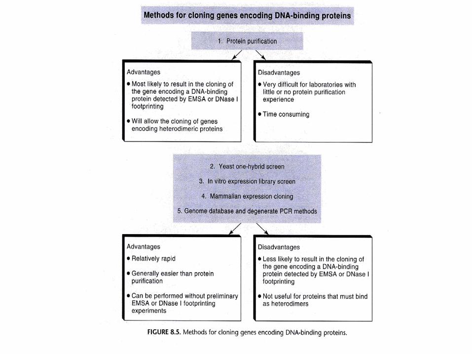
A proteome-wide classification of DNA-binding protein DBD families
helix-turn-helix
zinc finger
helix-loop-helix
leucine zipper
Number of Swissprot (SW) entries for the respective DNA binding folds

Helix-Turn-Helix Motif (HTH) Domain: high degree of homology between bacterial and eukaryotes DBD

Typically relatively short motifs are responsible for specific binding to DNA

Homeodomain-DNA Complex (PDB 1hdd)
Gln50
Asn51Ile47
Arg3
Arg5
5’
T
A
A
T
T
3’
Computer drawing of the tridimensional structure as determined by X-Ray crystallography of Engrailed homeodomain
Vollmer JY and Clerc RG J. Neurochem. 71:1-20


Homeodomain protein classes implicated in the mouse brain development
Vollmer JY and Clerc RG J. Neurochem. 71:1-20

posterior late anterior early
low RA response high RA response
trunk hindbrain
Body plan is constructed through interaction of the developmentally regulated homeotic genes
Duboule D. editor Guidebook Homeoboxgenes. Oxford Press 1994

The POU domain: a large conserved region in the mammalian pit-1, oct1,2 and C. elegans unc96 gene products
Herr W. Clerc RG and Baltimore D. Genes & Dev 12:1513-1516

POU domain DNA interaction
5’ATGCAAAT 3’ (Octamer)Müller-Immerglück M, Matthias P. EMBO J. 9:1625-1631.

A zinc finger domain is a DBD
Originally identified in TFIIIA (5S rRNA), Zinc fingers form alpha helices that insert into the major groove associated with stabilizing beta sheets

A

TFIIIA contains 9 sequential CysHis zinc fingers with distinct DNA binding and TRX functions
DNA binding
in vitro transcription
recruitment of TFIIIC
Rothfels K et al. (2007) Nucl Acid Res 35:4869-81
TFIIIA-DNA
TFIIIA-TFIIIC-DNA
DNA

Originally identified in TFIIIA (5S rRNA), Zinc fingers form alpha helices that insert into the major groove associated with stabilizing beta sheets

Individual zinc finger domains that recognize DNA triplets with high specificity and affinity are fused to nonspecific nuclease domain to create designer TRX F for gene targeting
Targeted genome editing using zinc finger nucleases: custom DBD for transcription factors and nucleases (http://www.zincfingertools.org)
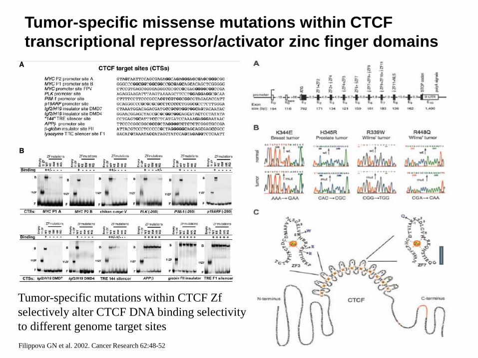
Tumor-specific missense mutations within CTCF transcriptional repressor/activator zinc finger domains
Filippova GN et al. 2002. Cancer Research 62:48-52
Tumor-specific mutations within CTCF Zf selectively alter CTCF DNA binding selectivity to different genome target sites

A distinct form of Zn finger motifs in Nuclear Receptors

Glucocorticoid Receptor-DNA Complex (PDB 1glu)
Zn finger
K461 V462 R466
5’A G A A C A NNN T G T T C T3’
T C T T G T NNN A C AA G A

Generation of DNA binding specificity mutants: glucocorticoid receptor ZnF DBD acquires estradiol specificity (ERE)

bZIP domain: basic “leucine zipper” (leucine residues spaced by 7 AA residues)
bZIP domain: prototypical amphipathic coiled-coil alpha helix

c-Fos-c-Jun-DNA Complex (PDB 1fos)
5’ T G A G T C A 3’
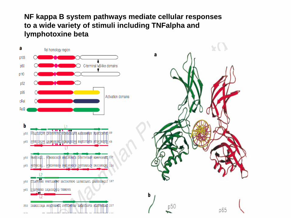
NF kappa B system pathways mediate cellular responses to a wide variety of stimuli including TNFalpha and lymphotoxine beta

Only one of the two DNA-bound orientations of AP-1 (fos-jun) found in solution cooperates with NFATp
Oakley M. and Verdine GL. Curr Biol 5:882 (1995)

Oakley M. and Verdine GL. Curr Biol 5:882 (1995)
Only one of the two DNA-bound orientations of AP-1 found in solution cooperates with NFATp

Helix-Loop-Helix Domains: Prototypical TRXF dimerizers
Each amphipathic helix presents a face of hydrophobic residues on one side and charged residues on the other side
bHLH proteins typically bind to a consensus sequence called an E-box, CANNTG

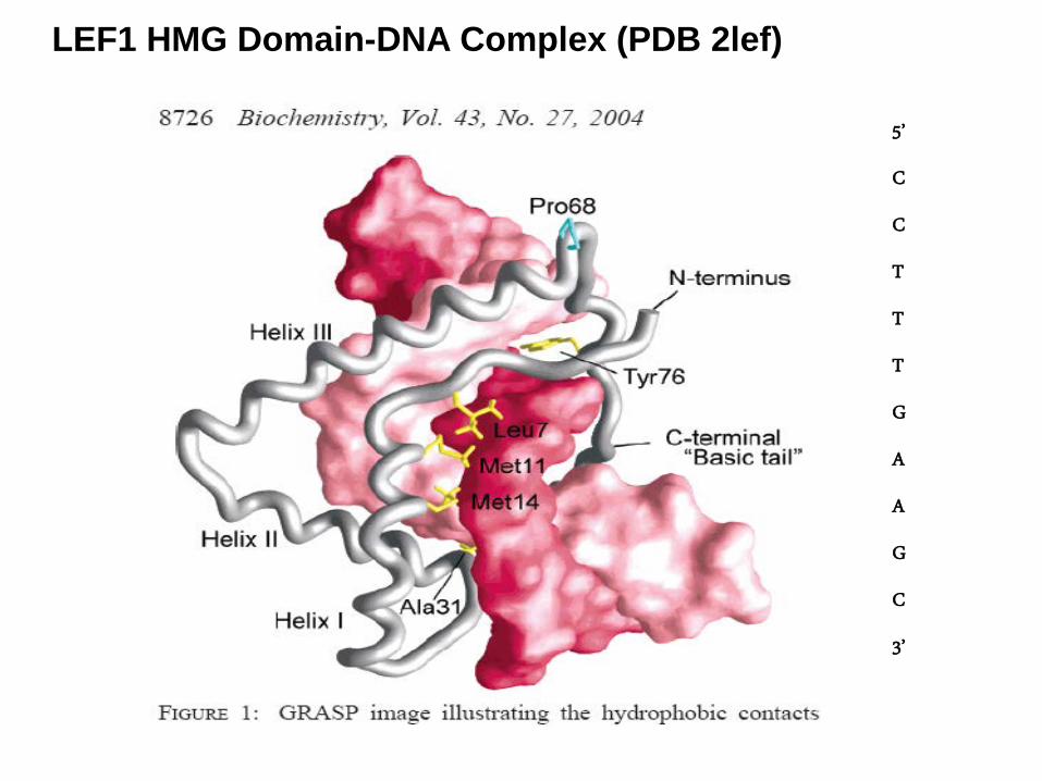
HMG “High Mobility Group” Motif (eg.lymphoid (T-cell) enhancer binding factor LEF-1)

DNA bending analysis by EMSA

LEF1 HMG Domain-DNA Complex (PDB 2lef)
L6
M13M10
E28
HELIX 2
HELIX 1
HELIX 3BASIC C-TERM
5’
C
C
T
T
T
G
A
A
G
C
3’
Y76
LEF1 HMG Domain-DNA Complex (PDB 2lef)
5’
C
C
T
T
T
G
A
A
G
C
3’

TBP : TATAA – Binding protein

• Acidic domains (DDD, EEE), VP16, GAL4, GCN4, GR
• Glutamine-rich domain (QQQXXXQQQ), SP1
• Proline-rich domains (PPPXXXPPP), CTCF, AP2
• Isoleucine-rich domain IIXXII), NTF
Several motifs involved in transactivation
Transcription Activation Domains -TADs
protein motives commonly referred to as “negative noodles, acid blobs” ! (Sigler P. Nature 333:210-212)

Transcription Activation Domains -TADs
• in contrary to DBDs, few structural data on TADs available
• TADs are intrinsically disordered unfolded proteins (resistant to crystal packing)
• TADs indirectly recruit Pol II by recruiting co-activators that serve as physical bridge between TADs and PIC/RNA Pol II holoenzyme
• TADs were reported to become structured (eg p53 helical TAD) upon binding to co-activators
Wells et al. PNAS 105:5762-5767

Ser/Thr and Gln Transcription activation domains -SP1, a prototypical constitutive TAD
5'-(G/T)GGGCGG(G/A)(G/A)(C/T)-
SP1 contains two glutamine-serine/threonine-rich TADs which interact with TAF110 (Gill g., Tjian R. PNAS 91:192-196)
(Gill G., Tjian R. PNAS 91:192-196)

Prototypical ligand mediated transcription activation domain (eg acidic negatively charged GR TAD)
unliganded liganded
co-repressor binding co-activator binding
“mouse trap”

HPV transactivation domain TAD forms a dimer
Harris SF, Botchan M. Science 284: 1673-1677
Antson AA et al. Nature 403: 805-809

HPV transactivation domain
Antson AA et al. Nature 403: 805-809Knight JD, Botchan M. PNAS 88:3204-3208
E2 binding site
E2 binding site
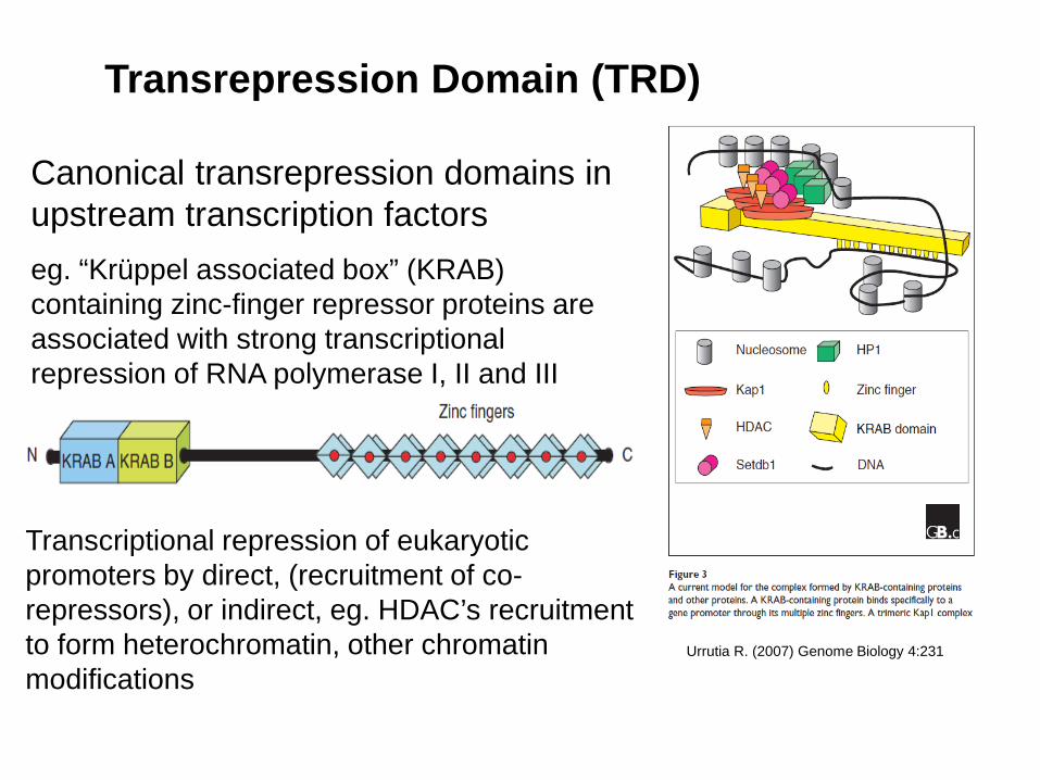
Canonical transrepression domains in upstream transcription factorseg. “Krüppel associated box” (KRAB) containing zinc-finger repressor proteins are associated with strong transcriptional repression of RNA polymerase I, II and III
Transrepression Domain (TRD)
Transcriptional repression of eukaryotic promoters by direct, (recruitment of co-repressors), or indirect, eg. HDAC’s recruitment to form heterochromatin, other chromatin modifications
Urrutia R. (2007) Genome Biology 4:231
