transferofbone-marrow-derivedmesenchymal
TRANSCRIPT
Hindawi Publishing CorporationJournal of Biomedicine and BiotechnologyVolume 2012, Article ID 165296, 7 pagesdoi:10.1155/2012/165296
Research Article
Transfer of Bone-Marrow-Derived MesenchymalStem Cells Influences Vascular Remodeling and Calcification afterBalloon Injury in Hyperlipidemic Rats
Jianquan Liao,1 Xiaochun Chen,2 Yongdong Li,2 Zhiping Ge,2 Hongyu Duan,2
Yunzeng Zou,1 and Junbo Ge1
1 Shanghai Institute of Cardiovascular Diseases, Zhongshan Hospital, Fudan University, Shanghai 200032, China2 Department of Cardiology, The Third Affiliated Hospital, Inner Mongolia Medical College, BaoTou, Inner Mongolia 014010, China
Correspondence should be addressed to Xiaochun Chen, [email protected] Junbo Ge, [email protected]
Received 19 December 2011; Revised 27 February 2012; Accepted 29 February 2012
Academic Editor: Monica Fedele
Copyright © 2012 Jianquan Liao et al. This is an open access article distributed under the Creative Commons Attribution License,which permits unrestricted use, distribution, and reproduction in any medium, provided the original work is properly cited.
Bone-marrow-derived mesenchymal stem cells (BM-MSCs) were found to markedly increase atherosclerotic lesion size. The aimof this paper was to investigate whether BM-MSCs contribute to vascular remodeling and calcification after balloon injury inhyperlipidemic rats. Labeled BM-MSCs were found in the lesion of hyperlipidemic rats after balloon injury. Comparing injurygroup, transferred BM-MSCs significantly triggered vascular negative remodeling, characterized by the changes of remodelingindex (0.628±0.0293 versus 0.544±0.0217), neointimal area (0.078±0.015 mm2 versus 0.098±0.019 mm2), PCNA index (23.91±6.59% versus 43.11±5.31%), and percentage of stenosis (18.20±1.09% versus 30.58±1.21%). Apparent vascular calcification wasdetected in medial layers at 6 weeks after balloon angioplasty, which may be associated with upregulation of bone morphogeneticprotein-2 (BMP-2). Our data indicated that unselected BM-MSCs transfer may induce vascular remodeling and calcification afterballoon injury in hyperlipidemic rats.
1. Introduction
Cellular therapy has been an area of intense research over fewdecades, which is widely used for treatment of cardiovasculardisease. Bone-marrow-derived mesenchymal stem cells (BM-MSCs) were isolated from adult bone marrow, along withthe ability for self-renewal and multilineage differentiationinto cardiomyocytes, smooth muscle cells, endothelium,osteoblasts, and chondroblasts [1, 2]. Accumulating evidencefrom a growing body of animal studies demonstrated theextensive capacity of MSCs to engraft, differentiate andproduce substantial functional recovery [3]. Transplantationhas been considered as a potential clinical strategy for thetreatment of ischemic heart diseases and heart failure.
Although a growing body of evidence from animal exper-iments and clinical trials indicated the beneficial therapeuticpotential of MSCs in cardiovascular diseases, the safety ofMSCs transplantation is a critical problem that should be
kept in mind. The findings that MSCs transplantation couldresult in tumor formation and alteration of electrophysio-logic properties caused wide concerns [4, 5]. Furthermore,unexpected severe intramyocardial calcification was elicitedby direct transplantation of unselected bone marrow cellsinto acutely infarcted myocardium [6]. It has been demon-strated that transfer of bone marrow and endothelial pro-genitor cells accelerated atherosclerosis and influenced theplaque stability [7]. Increasing evidence indicates a linkbetween MSCs therapy and pathogenesis of atherosclerosis[8, 9].
Our previous studies demonstrated that oxidized LDLsynergistically promoted osteodifferentiation of bone-mar-row-derived MSCs in response to osteogenic inductor. In thisstudy, we investigated the effects of transfer of unselectedBM-MSCs on vascular remodeling and calcification in thehyperlipidemic rat.

2 Journal of Biomedicine and Biotechnology
2. Method
2.1. Preparation of BM-MSCs. Unselected BM-MSCs wereextracted from male Sprague-Dawley (SD) rats (150∼200 g)femurs by flushing the bone marrow cavities with Dulbecco’smodified Eagle’s medium-low glucose (DMEM) and cul-tured in DMEM supplemented with 10% fetal bovine serum(FBS). After 24 h, the medium was discarded to removehematopoietic stem cells and nonadherent cells. Medium waschanged every 1∼2 days. Cells became almost confluent after8 days and were trypsinized with 0.25% trypsin containing1 mM EDTA for 3 min at 37◦C. All experiments were per-formed on cells cultured for up to 3∼5 passages. BM-MSCswere identified as CD90-positive and CD34-negative by flowcytometry.
2.2. Animals. All animal experiments were approved by theAnimal Care and Use Committee of Fudan University ac-cording to the Guide for the Care and Use of LaboratoryAnimals, published by the US National Institutes of Health(NIH publication no. 85-23, revised 1996). Animals were fedwith high cholesterol diet containing 10% fat, 5% choles-terol, 0.5% sodium cholate, and 0.2% propylthiouracil andreceived vitamin D3 (600,000 U/kg body weight) for 1 weekbefore balloon denudation. Female SD rats were randomlyallocated to three groups: (1) rats without denudation werefed with high cholesterol diet (Control, n = 8); (2) rats weresubjected to balloon denudation and fed with high choles-terol diet (Injury group, n = 8); (3) rats were subjectedto balloon denudation and BM-MSCs transplantation pluswith high cholesterol diet (MSC group, n = 8). Ratswere anesthetized with ketamine (150 mg/kg body weightIP) and heparinized with 100 U/kg heparin sodium beforeprocedures. A well-established rat model of aortic arteryballoon injury was used in this study. The abdominal aortawas isolated and stripped of adventitia. A 6 F balloon catheterwas introduced through incision under the renal arterybifurcation about 1 cm and advanced into the thoracic aorta.The balloon was inflated with saline and pulled back to theabdominal aorta. After balloon denudation was performedfor 3 times, saline or BMSCs labeled by DAPI were injectedbefore removal of the catheter. After balloon denudation, ratswere fed with high cholesterol diet containing 10% fat, 5%cholesterol, 0.5% sodium cholate, and 0.2% propylthiouracilfor 6 weeks after balloon denudation. In addition, ratswithout denudation were fed with a normal diet to comparewith those with a high cholesterol diet.
2.3. Analyses of Plasma Lipid. After feeding, the rats werefasted overnight and the samples were collected and cen-trifuged at 3000 rpm for 15 min. The plasma lipid profile wasdetermined by measuring the contents of total cholesterol(TC) and low-density lipoprotein (LDL) by standard enzy-matic procedures using reagent kits.
2.4. Histology. Paraffin sections of vascular tissue werestained with hematoxylin-eosin for morphometric analysis
as previously described [10]. Histomorphometric parame-ters, including luminal area (LA), internal elastic lamina area(IELA), external elastic lamina area (EELA), and neointimalarea were defined and determined using the software of LeicaQWin Plus as previously described [11]. The chronic varia-tion of the area surrounded by the external elastic lamina wasevaluated by remodeling index compared with a referencenormal segment upstream from the lesion site, which wascalculated as a percentage as follows: EELA(lesion)/EELA(reference). The percentage of stenosis was calculated as(IELA−LA/IELA) × 100.
2.5. Von Kossa Stain. Von Kossa stain (Sigma-Aldrich, USA)was performed to evaluate abnormal deposits of calciumwithin vascular wall. Paraffin-fixed tissue sections were incu-bated with silver nitrate solution under a strong light. Thecalcium was replaced with silver deposit, visualized as metal-lic silver. Procedural details are described elsewhere [6].
2.6. Immunohistochemistry. Proliferative activity was deter-mined in vascular tissue using analysis of proliferating cellnuclear antigen (PCNA) expression defined immunohisto-chemically. In brief, tissue sections were quenched for endo-genous peroxidase and antigen-retrieved in 0.01 M citratebuffer (pH 6.0) for 15 min in a microwave oven at 100◦C. Thesections were blocked with normal goat serum and thenincubated with mouse anti-PCNA antibody (Boster Com-pany, China) overnight in a humid chamber at 4◦C. Afterincubation with biotinylated goat anti-mouse IgG, sectionswere visualized by using avidin-biotin horseradish peroxi-dase visualization system. Sections were then counterstainedwith haematoxylin. Images were recorded using Leica QwinPlus.
2.7. Immunofluorescence Staining. Immunofluorescencestaining was performed according to standard procedures.Briefly, vascular tissue sections were deparaffinized andhydrated. After preincubation with normal goat serum,sections were incubated with rabbit anti-BMP-2 antibody(Boster Company, China) overnight in humid chamber at4◦C followed by incubation with appropriate FITC-linkedsecondary antibody. Images were captured by using LeicaQwin Plus.
2.8. Statistical Analysis. Data were presented as means ±SEM. Comparison was performed using one-way analysis ofvariance (ANOVA) with Tukey test for post hoc analysis todetermine the difference among groups. P < 0.05 was con-sidered statistically significant.
3. Result
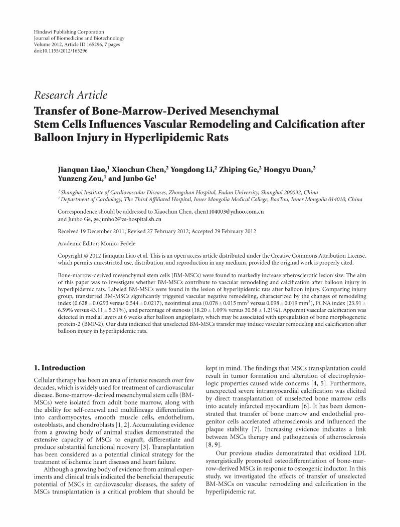
3.1. Identification of BM-MSCs Prepared for Transplantation.For transplantation, unselected BM-MSCs were isolatedfrom bone marrow according to standard technique. Afew small, bright, round cells were present in the primaryculture, which gradually disappeared by the second genera-tion or later (Figure 1(a)). BM-MSCs were characterized as
Journal of Biomedicine and Biotechnology 3
(a) (b)
30
25
20
15
10
5
0Normal
diet
TC
(m
mol
/L)
Injury group
MSCs group
High cholesterol diet
∗ ∗ ∗
(c)
20
15
10
5
0
LDL-
C (
mm
ol/L
)
Normal diet
Injury group
MSCs group
High cholesterol diet
∗ ∗ ∗
(d)
Figure 1: Identification of BM-MSCs and the profile of lipid level in cholesterol-rich fed rats. Phase contrast microscopy depicted that a fewsmall, bright, round cells were present in the primary culture (a). Phase contrast microscopy revealed that BM-MSCs were characterized asa fibroblast-like spindle-shaped morphology (b). Baseline of serum lipid content before received cholesterol-rich diet did not significantlydiffer among three groups. Cholesterol-rich diet significantly increased total serum cholesterol (c), and LDL (d) levels after angioplasty inrats. The data were expressed as means ± SEM. n = 8 per group.∗P < 0.05 versus normal diet group.
a fibroblast-like spindle-shaped morphology (Figure 1(b)).More than 95.69% of cells isolated from bone marrow werepositive for CD90, and negative for the early hematopoieticmarker CD34, indicating specific phenotype of BM-MSCsmarkers.
3.2. The Profile of Lipid Level in Rats with Cholesterol-RichDiet. Baseline of serum lipid content before received cho-lesterol-rich diet did not significantly differ among threegroups. The addition of cholesterol to the diet significantlyincreased total serum cholesterol, and LDL levels. Proceduresof balloon angioplasty and BM-MSCs transplantation didnot affect plasma lipid levels in this study (Figures 1(c) and1(d)).
3.3. Effect of BM-MSCs on Vascular Remodeling after BalloonAngioplasty. To address vascular remodeling in vivo, histo-logical analysis of vascular wall was assessed at 6 weeks afterballoon angioplasty. Ruptured internal elastic lamina waspresent at the site of lesion, whereas splitting of the intima
was replaced with significant neointimal formation afterballoon angioplasty in injury group and MSC group. Datafrom histomorphometric parameters depicted that LA, IELA,EELA, and remodeling index significantly decreased afterballoon angioplasty (LA: 1.632 ± 0.043 mm2 versus 1.335 ±0.056 mm2 versus 1.133 ± 0.074 mm2; IELA: 1.632 mm2 ±0.043 mm2 versus 1.414 mm2 ± 0.073 mm2 versus1.185 mm2 ± 0.086 mm2; EELA: 2.403 mm2 ± 0.067 mm2
versus 1.509 ± 0.058 mm2 versus1.308 ± 0.044; remodelingindex: 1 versus 0.628±0.0293 versus 0.544 ± 0.0217; controlversus injury group versus MSC group, resp., P < 0.05),whereas significant neointimal area and percentage ofstenosis were increased, compared with control (neointimalarea: 0 versus 0.078 ± 0.015 mm2 versus 0.098 ± 0.019 mm2,P < 0.05; percentage of stenosis: 0 versus 18.20 ± 1.09%versus 30.58 ± 1.21%; control versus injury group versusMSC group, resp., P < 0.05). BM-MSCs transplantationsignificantly aggravated vascular negative remodeling whencompared with injury group, characterized by the decreaseof LA, IELA, EELA, and remodeling index and increasedneointimal area and percentage of stenosis (Figure 2).

4 Journal of Biomedicine and Biotechnology
Control Injury group MSCs group
(a)
1.81.61.41.2
10.80.60.40.2
0Control Injury
groupMSCs group
∗#
1.81.61.41.2
10.80.60.40.2
0Control Injury
groupMSCs group
∗#
1
0.5
1.5
Control Injury group
MSCs group
∗#
2
2.5
3
0
0.12
Control Injury group
MSCs group
0.1
0.08
0.06
0.04
0.02
0
∗
#
1.2
1
0.8
0.6
0.4
0.2
0Control Injury
groupMSCs group
∗#
35
Sten
osis
(%
)
30
25
20
15
10
5
0Control Injury
groupMSCs group
∗
#
LA (
mm
2)
Neo
inti
mal
are
a (m
m2)
Rem
odel
ing
inde
xIE
LA (
mm
2)
EE
LA (
mm
2)
(b)
Figure 2: Effect of BM-MSCs on vascular remodeling after balloon angioplasty. Vascular tissues were stained with hematoxylin-eosin formorphometric analysis. (a) Ruptured internal elastic lamina was present at the site of lesion, whereas splitting of the intima was replacedwith significant neointimal formation after balloon angioplasty. (b) Histomorphometric parameters including LA, IELA, EELA, remodelingindex, neointimal area, and percentage of stenosis indicated vascular negative remodeling. The scale bar indicated 500 µm (top row, 40x) and100 µm (bottom row, 400x), respectively. The data were expressed as means ± SEM. n = 8 per group. ∗P < 0.05 versus control, #P < 0.05versus injury group.
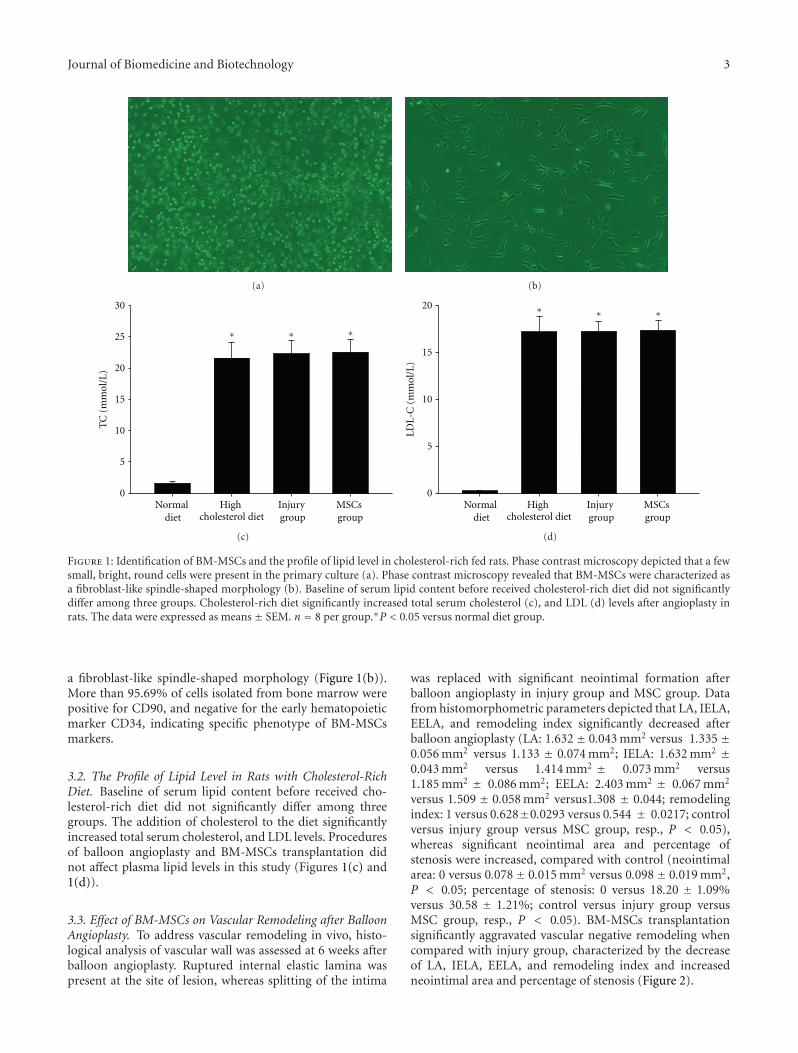
3.4. Effect of BM-MSCs on Neointimal Formation afterBalloon Angioplasty. To investigate the neointimal formationin vivo, the PCNA index was evaluated. PCNA-stainednuclei were detected in the neointimal and medial layers ofvascular wall at 6 weeks after vascular denudation (Figure 3).Data demonstrated that BM-MSCs resulted in a significantincrease in the neointimal area (0.078 ± 0.015 mm2 versus
0.098 ± 0.019 mm2, P < 0.05), PCNA index (23.91 ± 6.59%versus 43.11 ± 5.31%, P < 0.05), and percentage of stenosis(18.20 ± 1.09% versus 30.58 ± 1.21%, P < 0.05) comparedwith injury group.
3.5. Effect of BM-MSCs on Vascular Calcification after BalloonAngioplasty. To further characterize vascular calcification in
Journal of Biomedicine and Biotechnology 5
Control Injury group MSCs group
400x 400x 400x
(a)
60
50
40
30
20
10
0Control Injury group MSCs group
PC
NA
inde
x (%
)
#
∗
(b)
Figure 3: Effect of BM-MSCs on neointimal formation after balloon angioplasty. (a) The PCNA expression was evaluated immunohisto-chemically to investigate proliferative activity in vascular tissue. PCNA-stained nuclei were detected in the neointimal and medial layers ofvascular wall at 6 weeks after vascular denudation. (b) PCNA index was significantly increased after BM-MSCs transplantation comparedwith injury group. The data were expressed as means ± SEM. n = 8 per group. ∗P < 0.05 versus control, #P < 0.05 versus injury group.
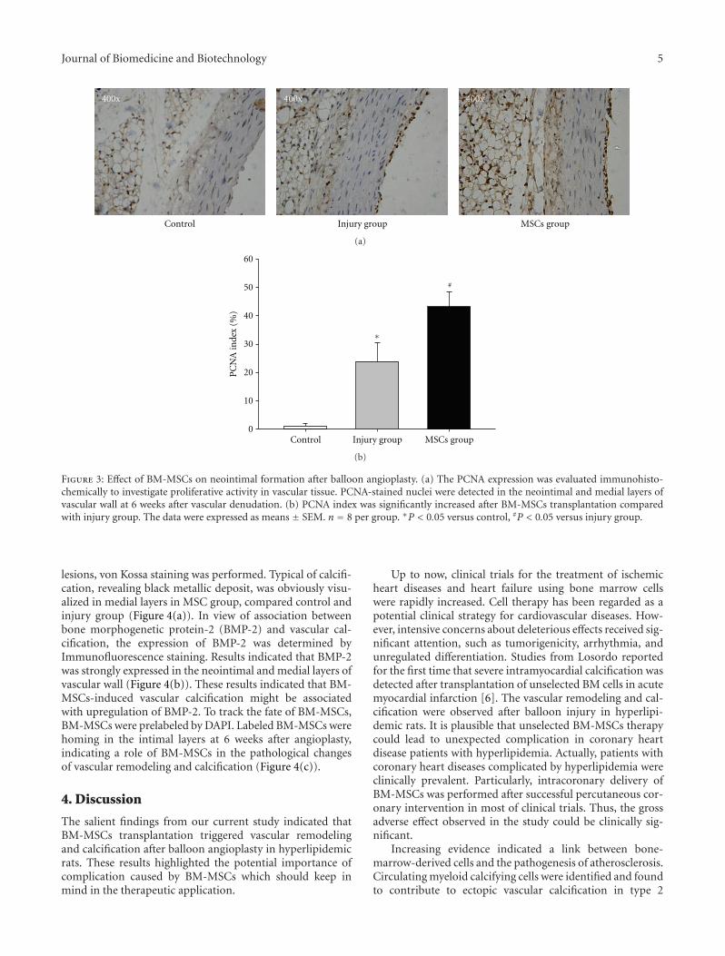
lesions, von Kossa staining was performed. Typical of calcifi-cation, revealing black metallic deposit, was obviously visu-alized in medial layers in MSC group, compared control andinjury group (Figure 4(a)). In view of association betweenbone morphogenetic protein-2 (BMP-2) and vascular cal-cification, the expression of BMP-2 was determined byImmunofluorescence staining. Results indicated that BMP-2was strongly expressed in the neointimal and medial layers ofvascular wall (Figure 4(b)). These results indicated that BM-MSCs-induced vascular calcification might be associatedwith upregulation of BMP-2. To track the fate of BM-MSCs,BM-MSCs were prelabeled by DAPI. Labeled BM-MSCs werehoming in the intimal layers at 6 weeks after angioplasty,indicating a role of BM-MSCs in the pathological changesof vascular remodeling and calcification (Figure 4(c)).
4. Discussion
The salient findings from our current study indicated thatBM-MSCs transplantation triggered vascular remodelingand calcification after balloon angioplasty in hyperlipidemicrats. These results highlighted the potential importance ofcomplication caused by BM-MSCs which should keep inmind in the therapeutic application.
Up to now, clinical trials for the treatment of ischemicheart diseases and heart failure using bone marrow cellswere rapidly increased. Cell therapy has been regarded as apotential clinical strategy for cardiovascular diseases. How-ever, intensive concerns about deleterious effects received sig-nificant attention, such as tumorigenicity, arrhythmia, andunregulated differentiation. Studies from Losordo reportedfor the first time that severe intramyocardial calcification wasdetected after transplantation of unselected BM cells in acutemyocardial infarction [6]. The vascular remodeling and cal-cification were observed after balloon injury in hyperlipi-demic rats. It is plausible that unselected BM-MSCs therapycould lead to unexpected complication in coronary heartdisease patients with hyperlipidemia. Actually, patients withcoronary heart diseases complicated by hyperlipidemia wereclinically prevalent. Particularly, intracoronary delivery ofBM-MSCs was performed after successful percutaneous cor-onary intervention in most of clinical trials. Thus, the grossadverse effect observed in the study could be clinically sig-nificant.
Increasing evidence indicated a link between bone-marrow-derived cells and the pathogenesis of atherosclerosis.Circulating myeloid calcifying cells were identified and foundto contribute to ectopic vascular calcification in type 2

6 Journal of Biomedicine and Biotechnology
Control Injury group MSCs group
400x 400x 400x
(a)
400x 400x 400x
Control Injury group MSCs group
(b)
400x
(c)
Figure 4: Effect of BM-MSCs on vascular calcification after balloon angioplasty and the fate of transferred BM-MSCs. (a) Von Kossastaining was performed to detect vascular calcification in lesions black metallic deposit was obviously visualized in medial layers in MSCgroup, compared control and injury group. (b) Immunofluorescence staining was performed to determine the expression of BMP-2. Resultsindicated that upregulated BMP-2 was strongly expressed in the neointimal and medial layers of vascular wall in MSC group, comparedControl and Injury groups. (c) Labeled BM-MSCs were homing in the intimal layers at 6 weeks after angioplasty. n = 8 per group.
diabetes [12]. Previous studies demonstrated that transferredBM-MSCs were homing to the vascular lesion and markedlyincreased atherosclerotic lesion size [7]. In this study, thefate of transferred BM-MSCs were traced and found topresent at a low density in the vascular sections. However, thehoming BM-MSCs may be underestimated, as quenching ofthe DAPI cannot be completely excluded at 6 weeks after bal-loon injury. It was indicated that BM-MSCs were potentiallyinvolved in atherosclerotic calcification.
The mechanisms by which transferred BM-MSCs mod-ulate vascular calcification remain unclear. Compelling evi-dence suggested that vascular calcification was regulatedby bone calcification regulatory factors locally expressed inblood vessels [13–15]. Paracrine BMP-2 caused the induction
of osteoblast-like cells in intima and accelerated the ath-erosclerotic calcification in vivo [16]. BMP-2 was foundto promote vascular calcification via increased phosphateuptake and induction of osteogenic phenotype modulationin smooth muscle cells [17]. Our observation that stronglyexpression of BMP-2 was detected in the neointimal andmedial layers indicated a potential role of BMP-2 in the BM-MSCs-induced calcification. Our data demonstrated thatox-LDL was synergistically promoted osteodifferentiation ofBM-MSCs co-cultured with vascular smooth muscle cells inthe presence of osteogenic inductor (unpublished data). Itwas suggested that BM-MSCs together with balloon injuryand hyperlipidemia contribute to the formation of vascularcalcification.

Journal of Biomedicine and Biotechnology 7
Taken together, we demonstrated an important role oftransferred BM-MSCs in vascular remodeling and calcifi-cation. These findings call for caution in potential adverseeffects with respect to BM-MSCs transfer in the future clini-cal trials.
Acknowledgments
This study was supported by the Key Scientific ResearchFund of Inner Mongolia Medical College (NY2004ZD007)and the Natural Scientific Research Fund of Inner Mongolia(2010MS1113).
Authors’ Contribution
J. Liao and X. Chen contributed equally to this work.
References
[1] H. C. Quevedo, K. E. Hatzistergos, B. N. Oskouei et al., “Al-logeneic mesenchymal stem cells restore cardiac function inchronic ischemic cardiomyopathy via trilineage differentiatingcapacity,” Proceedings of the National Academy of Sciences of theUnited States of America, vol. 106, no. 33, pp. 14022–14027,2009.
[2] E. A. Jones, S. E. Kinsey, A. English et al., “Isolation and char-acterization of bone marrow multipotential mesenchyma pro-genitor cells,” Arthritis and Rheumatism, vol. 46, no. 12, pp.3349–3360, 2002.
[3] A. R. Williams and J. M. Hare, “Mesenchymal stem cells: biol-ogy, pathophysiology, translational findings, and therapeuticimplications for cardiac disease,” Circulation Research, vol.109, no. 8, pp. 923–940, 2011.
[4] J. O. Jeong, J. W. Han, J. M. Kim et al., “Malignant tumorformation after transplantation of short-term cultured bonemarrow mesenchymal stem cells in experimental myocardialinfarction and diabetic neuropathy,” Circulation Research, vol.108, no. 11, pp. 1340–1347, 2011.
[5] C. S. Kuo, K. Munakata, C. P. Reddy, and B. Surawicz, “Char-acteristics and possible mechanism of ventricular arrhythmiadependent on the dispersion of action potential durations,”Circulation, vol. 67, no. 6 I, pp. 1356–1367, 1983.
[6] Y. S. Yoon, J. S. Park, T. Tkebuchava, C. Luedeman, and D.W. Losordo, “Unexpected severe calcification after transplan-tation of bone marrow cells in acute myocardial infarction.,”Circulation, vol. 109, no. 25, pp. 3154–3157, 2004.
[7] J. George, A. Afek, A. Abashidze et al., “Transfer of endothelialprogenitor and bone marrow cells influences atheroscleroticplaque size and composition in apolipoprotein E knockoutmice,” Arteriosclerosis, Thrombosis, and Vascular Biology, vol.25, no. 12, pp. 2636–2641, 2005.
[8] A. Saiura, M. Sata, Y. Hirata, R. Nagai, and M. Makuuchi,“Circulating smooth muscle progenitor cells contribute toatherosclerosis,” Nature Medicine, vol. 7, no. 4, pp. 382–383,2001.
[9] M. Sata, A. Saiura, A. Kunisato et al., “Hematopoietic stemcells differentiate into vascular cells that participate in thepathogenesis of atherosclerosis,” Nature Medicine, vol. 8, no.4, pp. 403–409, 2002.
[10] L. Lin, H. Gong, J. Ge et al., “High density lipoproteindownregulates angiotensin II type 1 receptor and inhibitsangiotensin II-induced cardiac hypertrophy,” Biochemical and
Biophysical Research Communications, vol. 404, no. 1, pp. 28–33, 2011.
[11] C. Brasselet, E. Durand, F. Addad et al., “Collagen and elastincross-linking: a mechanism of constrictive remodeling afterarterial injury,” American Journal of Physiology, vol. 289, no.5, pp. H2228–H2233, 2005.
[12] G. P. Fadini, M. Albiero, L. Menegazzo et al., “Widespreadincrease in myeloid calcifying cells contributes to ectopicvascular calcification in type 2 diabetes,” Circulation Research,vol. 108, no. 9, pp. 1112–1121, 2011.
[13] K. A. Hruska, S. Mathew, and G. Saab, “Bone morphogeneticproteins in vascular calcification,” Circulation Research, vol. 97,no. 2, pp. 105–114, 2005.
[14] R. C. Johnson, J. A. Leopold, and J. Loscalzo, “Vascularcalcification: pathobiological mechanisms and clinical impli-cations,” Circulation Research, vol. 99, no. 10, pp. 1044–1059,2006.
[15] M. Ketteler, G. Schlieper, and J. Floege, “Calcification andcardiovascular health: new insights into an old phenomenon,”Hypertension, vol. 47, no. 6, pp. 1027–1034, 2006.
[16] Y. Nakagawa, K. Ikeda, Y. Akakabe et al., “Paracrine osteogenicsignals via bone morphogenetic protein-2 accelerate theatherosclerotic intimal calcification in vivo,” Arteriosclerosis,Thrombosis, and Vascular Biology, vol. 30, no. 10, pp. 1908–1915, 2010.
[17] X. Li, H. Y. Yang, and C. M. Giachelli, “BMP-2 promotesphosphate uptake, phenotypic modulation, and calcificationof human vascular smooth muscle cells,” Atherosclerosis, vol.199, no. 2, pp. 271–277, 2008.

Submit your manuscripts athttp://www.hindawi.com
Stem CellsInternational
Hindawi Publishing Corporationhttp://www.hindawi.com Volume 2014
Hindawi Publishing Corporationhttp://www.hindawi.com Volume 2014
MEDIATORSINFLAMMATION
of
Hindawi Publishing Corporationhttp://www.hindawi.com Volume 2014
Behavioural Neurology
EndocrinologyInternational Journal of
Hindawi Publishing Corporationhttp://www.hindawi.com Volume 2014
Hindawi Publishing Corporationhttp://www.hindawi.com Volume 2014
Disease Markers
Hindawi Publishing Corporationhttp://www.hindawi.com Volume 2014
BioMed Research International
OncologyJournal of
Hindawi Publishing Corporationhttp://www.hindawi.com Volume 2014
Hindawi Publishing Corporationhttp://www.hindawi.com Volume 2014
Oxidative Medicine and Cellular Longevity
Hindawi Publishing Corporationhttp://www.hindawi.com Volume 2014
PPAR Research
The Scientific World JournalHindawi Publishing Corporation http://www.hindawi.com Volume 2014
Immunology ResearchHindawi Publishing Corporationhttp://www.hindawi.com Volume 2014
Journal of
ObesityJournal of
Hindawi Publishing Corporationhttp://www.hindawi.com Volume 2014
Hindawi Publishing Corporationhttp://www.hindawi.com Volume 2014
Computational and Mathematical Methods in Medicine
OphthalmologyJournal of
Hindawi Publishing Corporationhttp://www.hindawi.com Volume 2014
Diabetes ResearchJournal of
Hindawi Publishing Corporationhttp://www.hindawi.com Volume 2014
Hindawi Publishing Corporationhttp://www.hindawi.com Volume 2014
Research and TreatmentAIDS
Hindawi Publishing Corporationhttp://www.hindawi.com Volume 2014
Gastroenterology Research and Practice
Hindawi Publishing Corporationhttp://www.hindawi.com Volume 2014
Parkinson’s Disease
Evidence-Based Complementary and Alternative Medicine
Volume 2014Hindawi Publishing Corporationhttp://www.hindawi.com