transformation of aspergillus sojae with vitreoscilla
TRANSCRIPT

TRANSFORMATION OF Aspergillus sojae WITH Vitreoscilla HEMOGLOBIN GENE
A Thesis Submitted to the Graduate School of Engineering and Sciences of
İzmir Institute of Technology in Partial Fulfillment of the Requirements for the Degree of
MASTER OF SCIENCE
in Biotechnology
by Betül BARDAKCI
December 2010 İZMİR

iv
We approve the thesis of Betül BARDAKCI Assoc. Prof. Dr. Canan TARI Supervisor Assist. Prof. Dr. Alper ARSLANOĞLU Co-Supervisor Assist. Prof. Dr. Sevcan ÜNLÜTÜRK Committee Member Assoc. Prof. Dr. Banu ÖZEN Committee Member Dr. İlhan DOĞAN Committee Member 27 December 2011 Assoc. Prof. Dr. Ahmet KOÇ Prof. Dr. Durmuş Ali DEMİR Head of the Department of Dean of the Graduate School of Biotechnology and Bioengineering Engineering and Sciences

ACKNOWLEDGEMENTS
I would like to thank my supervisor Assoc. Prof. Dr. Canan TARI, providing me
an oppurtunity to work with her. This thesis would not have been possible without her
knowledge in many areas, kindness, understanding, and great guidance.
I also want to thank my co-supervisor Assist. Prof. Dr. Alper ARSLANOĞLU
for his advices, experiences and contributions in this thesis. In addition, I would like to
thank the Thesis Committee: Dr. İlhan DOĞAN, Assist. Prof. Dr. Sevcan ÜNLÜTÜRK
and Assoc. Prof. Dr. Banu ÖZEN for their suggestions improving this project for future
studies, and providing me a perspective.
Additionally, I want to express my appreciation to my laboratory colleagues
especially Nihan GÖĞÜS, Hande DEMİR and Burcu OKUKLU for their time, kindness
and frienships, and also I want to thank all my friends for the great times I had in our
group and their great supports.
Finally, I want to show my appreciation to my mother Nevin BARDAKCI, my
father Hüsnü BARDAKCI and my sister Berna YÖRÜK BARDAKCI for their
encouragement, understanding, and love.

iv
ABSTRACT
TRANSFORMATION OF Aspergillus sojae WITH Vitreoscilla HEMOGLOBIN GENE
Aspergillus sojae known as koji mold is a non-aflatoxigenic fungus, designated
with a GRAS status by FDA. This study considers the transformation of A. sojae with
Vitreoscilla hemoglobin gene. Vitreoscilla hemoglobin is the bacterial hemoglobin,
which enhances the oxygen transfer under microaerophilic conditions. Since industrial
fungal fermentation with the high demand for oxygen are accounted to face oxygen
limitations during the production of value added products like enzymes, antibiotics and
organic acids; oxygen supply becomes an enormous problem to be solved by the
manufacturers. To overcome this problem, an alternative solution using the vgb gene of
Vitreoscilla and recombinant DNA technology and taking the A. sojae organism as
model organism was proposed. The product of interest in this study was the exo-
polygalacturonase enzyme known to have wide applications in food, pharmaceutical,
textile and paper industries. Here vgb gene was tried to be introduced to A .sojae via
both protoplasting and electroporation methods. For transformation process vgb gene
was cloned initially into plasmid ANIp4. For the selection of transformed A. sojae cells,
uridine auxotrophic mutants were tried to be selected after UV mutagenesis. However,
using a procedure based on selection of uridine supported growth did not result in A.
sojae pyrG mutants. The success of transformation was initially observed by means of
PCR analysis and agarose gel electrophoresis, later this was try to be confirmed by
sequence analysis and agarose gel electrophoresis. However, due to the contamination
problems accounted in the procedures the transformation with both methods could not
be assured.

v
ÖZET
Vitreoscilla HEMOGLOBIN GENİYLE Aspergillus sojae’nin
TRANSFORMASYONU
Koji küfü olarak bilinen Aspergillus sojae, FDA tarafından genel olarak güvenli
kabul edilmiştir. Bu organizmayla ilgili sınırlı sayıda çalışma rapor edilmiş olup, bu
çalışma, A. sojae’nin Vitreoscilla hemoglobin geni kullanılarak transformasyonuyla
ilgili yapılan tek çalışmadır. Mikroaerofilik koşullarda oxygen transferini artıran
Vitreoscilla hemoglobini bir bakteriyel hemoglobindir. Yüksek oksijen miktarına
ihtiyaç duyan endüstriyel fungal fermentasyonları, enzim, antibiyotik ve organik asit
gibi değerli ürünlerin üretimi sırasında oksijen sınırlamasıyla karşılaştığından, bu
sistemlere oksijen sağlamak üreticiler tarafından çözülmesi gereken önemli bir
sorundur. Bu problemin çözümü için Vitreoscilla’dan elde edilen vgb geni ve
rekombinant DNA teknolojisi kullanılması ile ilk etapta örnek olarak, A. sojae
organizması model olarak önerilmiştir. Bu çalışmada üretilen ürün, gıda, ilaç, tekstil ve
kağıt endüstrisinde geniş kullanım alanına sahip ekzo-polygalakturonaz enzimidir.
Transformasyon aşaması için vgb geni ilk olarak ANIp4 plasmidine klonlanmıştır.
Transforme edilmiş A. sojae hücrelerinin seçimi için UV mutasyonundan sonra üridin
okzotrofik mutantlar seçilmeye çalışılmış, ancak, üridin destekli büyümeye dayalı seçim
metodu ile A. sojae pyrG mutantlarının seçimi sonuç vermemiştir. Transformasyonun
başarılı oluşu ilk olarak PCR analizleri ve gel electroforezi yöntemiyle daha sonra ise
sekans analizi yöntemleriyle doğrulanmaya çalışılmıştır. Ancak kontaminasyondan ileri
gelen problemlerden dolayı transformasyon işleminin tam olark gerçekleşip
geçekleşmediğinde emin olunamamıştır.

vi
TABLE OF CONTENTS
LIST OF FIGURES ......................................................................................................... iv
LIST OF TABLES............................................................................................................ x
CHAPTER 1. INTRODUCTION ..................................................................................... 1
CHAPTER 2. ASPERGILLUS .......................................................................................... 4
2.1. Aspergillus.............................................................................................. 4
2.2. The Taxonomy of Aspergillus ................................................................ 4
2.3. Aspergillus sojae .................................................................................... 7
CHAPTER 3. PECTINOLYTIC ENZYMES................................................................... 9
3.1. Application of Pectinases ....................................................................... 9
CHAPTER 4. GENETIC MANIPULATIONS .............................................................. 12
4.1. Plasmids................................................................................................ 12
4.2. Transformation ..................................................................................... 13
4.3. Shuttle Vectors ..................................................................................... 14
4.4. Selection Markers for the Aspergilli..................................................... 15
4.4.1. Auxotrophic Markers...................................................................... 15
4.4.2. Dominant Selectable Markers ........................................................ 16
4.5. Protoplast Transformation .................................................................... 17
4.6. Uptake of DNA .................................................................................... 18
4.7. Transformation with Electroporation ................................................... 19
CHAPTER 5. VITREOSCILLA HEMOGLOBIN........................................................... 21
CHAPTER 6. MATERIALS AND METHODS ............................................................ 24
6.1. Materials ............................................................................................... 24
6.1.1. Chemicals ....................................................................................... 24
6.1.2. Media.............................................................................................. 24
6.1.3. Solutions and Buffers ..................................................................... 24

vii
6.2. Methods ................................................................................................ 24
6.2.1. Strains, and Growth Conditions ..................................................... 24
6.2.2. Plasmid Construction ..................................................................... 25
6.2.3. Preparation of Competent E. coli using Calcium Chloride ............ 29
6.2.4. Plasmid Isolation from Transformed E. coli Cells with Alkaline
Lysis (Mini Prep) ........................................................................... 30
6.2.5. Plasmid Isolation from Transformed E. coli Cells with Boiling
Method ........................................................................................... 31
6.2.6. Digestion of the Plasmid DNA with RNAase ................................ 31
6.2.7. Digestion with EcoRI Restriction Enzyme .................................... 32
6.2.8. Agarose Gel Electrophoresis .......................................................... 32
6.2.8.1. Gel Preparation...................................................................... 32
6.2.8.2. Preparation of the Marker...................................................... 32
6.2.8.3 Running the Gel...................................................................... 33
6.2.9. Designing and Ordering Primers..................................................... 33
6.2.10. PCR (Polymerase Chain Reaction) ............................................... 34
6.2.11. Isolation of A. sojae pyrG Negative Mutants by UV Mutation .... 35
6.2.12. Agar Diffusion Method for Detection of Pectinase Production
using Polygalacturonic Acid......................................................... 36
6.2.13. Protoplast Formation and Transformation with Glucanex
Enzyme ......................................................................................... 37
6.2.14. Transformation of Aspergillus sojae with Electroporation
Method ........................................................................................... 38
6.2.15. Plasmid Isolation from Transformed A. sojae............................... 39
CHAPTER 7. RESULTS AND DISCUSSION.............................................................. 40
7.1. Isolation of A. sojae pyrG Negative Mutants by UV Mutation............ 41
7.2. Plasmid Isolation from Transformed E. coli cells with Alkaline
Lysis (Mini Prep) ................................................................................. 44
7.3. Results of Sequence Analysis............................................................... 48
7.4. Sample 1: PCR product vgb gene stock coming from Illinois
Institute of Technology Department of Biological Sciences, USA ..... 49
7.5. Sample 2: Transformed M7/2 A. sojae strain with glucanex
(TraGlux) ............................................................................................. 51

viii
7.6. Sample3: Transformed M7/2 A. sojae strain with electroporation
(TraElectro).......................................................................................... 52
7.7. Sample 4: Mutant 7/2 Aspergillus sojae............................................... 54
7.8. Sample 5: Negative Control with Water .............................................. 55
7.9. Agar Diffusion Method for Detection of Pectinase Production using
Polygalacturonic Acid.......................................................................... 56
CHAPTER 8. CONCLUSION ....................................................................................... 63
REFERENCES ............................................................................................................... 65
APPENDICES
APPENDIX A CHEMICALS USED IN EXPERIMENTS ........................................... 76
APPENDIX B MEDIA................................................................................................... 79
APPENDIX C STOCK SOLUTIONS............................................................................ 87

ix
LIST OF FIGURES
Figure 2.1. Asexual fruiting structure of Aspergillus species illustraiting septate
hyphae, conidiophore, vesicle, phialides and conidiospores. ......................... 5
Figure 2.2. Molecular phylogeny of selected Aspergillus species, including species
belonging to the section Flavi, based on ITS sequence data. ......................... 8
Figure 4.1. Chemical transformation of Aspergillus. Conidiospores subjected to an
enzymatic treatment to lyse the cell walls, in order to generate protoplasts,
which are incubated with transforming DNA in a medium containing CaCl2
and other additives ...................................................................................... 19
Figure 6.1. Plasmid vector Bluescript............................................................................ 28
Figure 6.2. pBluescript SK (+/–) Multiple Cloning Site Region (sequence shown 601-
826). .............................................................................................................. 28
Figure 6.3. Shuttle vector (ANIp4)................................................................................. 28
Figure 6.4. V.stercoraria vgb gene encoding hemoglobin, complete cds from NCBI web
page (The accession number is M27061). .................................................... 34
Figure 6.5. PCR run conditions ...................................................................................... 38
Figure 6.6. Picture of master plate of TraGlux .............................................................. 38
Figure 7.1. A selected plate of UV mutated A. sojae M 7/2 strain. ........................ 42
Figure 7.2. Colony number 2 could not grow without uridine ....................................... 43
Figure 7.3. Colony number 3 could not grow without uridine ....................................... 43
Figure 7.4. Colony number 1 could not grow without uridine ....................................... 43
Figure 7.5. Agarose gel electrophrosis of transformant E.coli ....................................... 45
Figure 7.6. Agarose gel electrophrosis of PCR product from transformant E.coli......... 45
Figure 7.7. Agarose gel electrophrosis of PCR product from transformant A.sojae. ..... 47
Figure 7.8. Agarose gel electrophrosis of transformant A.sojae and M7/2 .................... 47
Figure 7.9. Coding sequence for PCR product of vgb gene stock coming from Illinois
Institute of Technology Department of Biological Sciences, USA .............. 49
Figure 7.10. Alignments of vgb gene sequence analysis result. ..................................... 53
Figure 7.11. Coding sequence for strain TraGlux .......................................................... 51
Figure 7.12. Out put of blast results from Finch TV program by using the sample of
strain TraGlux.. ............................................................................................. 53

x
Figure 7.13. Alignments of strain TraElectro extracted plasmid sequence analysis result.............................................................................................. 53
Figure 7.14. Out put of blast results from Finch TV program by using TraElectro ....... 54
Figure 7.15. Coding sequence for Mutant 7/2 Aspergillus sojae.................................... 54
Figure 7.16. Out put of blast results from Finch TV program by using the sample
of Mutant 7/2 Aspergillus sojae.................................................................... 55
Figure 7.17. Coding sequence for negative control with water. ..................................... 55
Figure 7.18. Output of blast results from Finch TV program by using the sample of
negative control with water........................................................................... 53
Figure 7.19. Solid media containing polygalacturonic acid (PGA) p for detection of enzyme production of the control strain M5/6............................................ 53
Figure 7.20. Solid media containing polygalacturonic acid (PGA) p for detection of
enzyme production of the wild type strain M7 ............................................. 53
Figure 7.21 Solid media containing polygalacturonic acid (PGA) p for detection of
enzyme production of the mutated strain M7/2. ........................................... 60
Figure 7.22. Solid media containing polygalacturonic acid (PGA) p for detection of
enzyme production of transformed strain TraGlux....................................... 61
Figure 7.23. Solid media containing polygalacturonic acid (PGA) p for detection of enzyme production from spore solution of strain TraElectro…....………....53

xi
LIST OF TABLES
Table 2.1. Characteristics of Aspergillus species ............................................................. 6
Table 3.1. Commercial pectinases .................................................................................. 10
Table 3.2. Characterization of microbial pectinases ....................................................... 11
Table 4.1. True mitochondrial plasmids of filamentous fungi ....................................... 13
Table 4.2. List of pyr genes and corresponding enzymes involved in de novo pyrimidine
nucleotide biosynthesis in prokaryotes and Trypanosoma cruzi .................. 53
Table 4.3. Dominant selectable markers used for different organisms .......................... 17
Table 4.4. Enzymes used in Protoplasting ...................................................................... 18
Table 5.1. Kinetic constants for reactions of various hemoglobins with oxygen. .......... 22
Table 5.2. Effects of hemoglobin expression in various microorganisms. ..................... 43
Table 7.1. The survival rate of spores after 10 minutes of UV exposure. ...................... 42
Table 7.2. Output of blast results from Finch TV program by using the sample of vgb
gene stock.. ................................................................................................... 50
Table 7.3. Alignments of strain TraGlux extracted plasmid sequence analysis result. .. 51
Table 7.4. Coding sequence for the sample of transformed A. sojae with electroporation
(TraElectro) ................................................................................................... 53
Table 7.5 Alignments of Mutant M7/2 Aspergillus sojae ............................................. 43
Table 7.6. Alignments of negative control with water. .................................................. 56
Table 7.7. Ratio of the diameters of clearance zones of the control strain M 5/6. ......... 58
Table 7.8. Ratio of the diameters of clearance zones of the wild type strain M7 ......... 59
Table 7.9. Ratio of the diameters of clearance zones of the mutated strain M7/2 .......... 60
Table 7.10. Ratio of the diameters of clearance zones to TraGlux colonies and theirs
average ratios. ............................................................................................... 61
Table 7.11. Ratio of the diameters of clearance zones to TraElectro colonies and their
average ratios ................................................................................................ 62
Table 7.12. Average ratio of the diameters of clearance zones to colonies of strains
used. .............................................................................................................. 62

1
CHAPTER 1
INTRODUCTION
Vitreoscilla is a filamentous bacterium belonging to the Beggiatoa family. It is a
gram-negative aerobic or microaerophilic bacteria. The species V. strecoraria is strictly
aerobic and is able to cope with the hypoxic conditions by synthesizing a soluble
hemoglobin (VHb) (Zhang, et al., 2007). VHb is the first bacterial hemoglobin, which
has 25% sequence homology with leghemoglobin (Kroneck, et al., 1991; Dikshit, et al.,
1992). The synthesis of this hemoglobin is promoted under the hypoxic conditions. The
rate constant for oxygen association (kon) is relatively normal; however the rate constant
for oxygen dissociation (koff) is unusually large. The beneficial effect is based on the
binding characteristics of the hemoglobin to oxygen and inducing the facillated
diffusion into the cell under very low oxygen concentrations where normal diffusion
does not take place with the concentration gradient. Overall the possible mechanism for
vgb action can be summarized such as increasing the flux of oxygen to the respiratory
apparatus, providing higher internal oxygen concentration, altering the internal redox
state, functioning as an efficient terminal oxidase, or improving overall efficiency of
oxygen limited ATP production (Sun, et al., 2002; Yubin, et al., 2009). Substantial
improvements in the production of recombinant proteins such as chloramphenicol acetly
transferase, beta galactosidase, catechol 2,3 dioxygenase, alpha amylase, protease and
human tissue plasminogen activator, antibiotics such as actinorhodin, cephalosporin C,
itaconic acid, erythromycin, ethanol and nicotine production (in Nicotiana tabaccum
plant) using this gene have been reported (Dikshit, et al., 1990; Wu, et al., 2003)
Based on the given literature, introduction of this gene into corresponding
organisms for the production of many other products would provide benefits to those
fermentations which require high aeration and where the production of the products are
very much oxygen dependent. It will enable such fermentations to work under much
lower oxygen concentrations and therefore reduce the air supply and the operational
cost. In large scales, such a strain improvement technique will not only save on air
supply but also increase the product yields and promote cell growth. Fungal submerged
fermentations are widely used in the production of enzymes, antibiotics and organic
2
acids which have lots of applications in the food, medicine, pharmaceutical, chemical
and textile industry. Therefore any problem faced during their production will affect the
final product significantly. One of the major accounted problems is the morphological
change of the culture during the fermentation process. It is well observed that the
fungal culture exhibits two major morphology which are very much determined by
several environmental and genetic factors. These factors can be stated as pH,
composition of the media, inoculation ratio, type of the inoculum, agitation speed,
aeration rate, feeding rate and genetic factors of the culture.
The pellet morphology is desired in downstream processing due to the non-
viscous (Newtonian) rheology of the broth. Since agitation and aeration is also much
easier in such systems, the power input therefore the operating cost will also be reduced.
Also, such morphology makes the downstream processing easier. However, the pellet
formed, is subject to internal mass transfer resistances (oxygen and nutrient) which can
result into low growth rate and productivity due to cell autolysis at the center of the
pellets. Therefore, the presence of such a strain improvement technique will help to
overcome these resistances.
Until 1974, strain improvement techniques were only based on mutation,
screening and selection. These methods were time consuming, laborious and expensive.
The first gene cloning of Aspergillus was reported in 1983 by Tilburn et al. (Lubertozzi
and Keasling, 2009). The transformation techniques for Aspergillus and other
filamentous fungi were more problematic with their multicellular morphology, thick
chitinous cell walls, and lack of plasmids compared to E. coli and S. cerevisia. Methods
for the genetic manipulation of S. cerevisia and E. coli were succeeded by the discovery
of native plasmids; nevertheless, like most fungi, the Aspergilli lack natural
extrachromosomaly replicating DNA elements. Consequently, researchers began to
construct artificial plasmids for Aspergillus (Lubertozzi and Keasling, 2009).
In the transformation process of fungi, selection markers bearing vectors are
essential for determination of transformed cells. The selection markers either depend on
the complementation of a specific auxotrophy or resistance to a specific antibiotic.
Antibiotic resistance markers are more easy to use because they do not require the
availability of specific auxotrophic strains.
Transformation can be mediated either by the electroporation method or
protoplast formation. Protoplast membrane is sufficiently competent for the uptake of
the foreign DNA or the fusion process. To obtain protoplasts from fungal cells, cell wall

3
degradation is an important process. In protoplasting process rather than the non
enzymatic methods, lytic enzymes are generally preferred (Peberdy, 1979).
Protoplasts need osmotic stabilizers in the suspending medium. Removing of the
cell wall makes protoplasts undefended to the osmotic differences. Sorbitol is the most
commonly used stabilizer and is sufficient for all fungi at the concentrations ranging
from 0.8 to 1.2 M. Calcium ions are crucial for nearly all transformation procedures.
Besides, up to 10 volumes of 40% PEG 4000 is used in the transformation mixture
because in the presence of PEG, the treated cells do not clump and facilitate the trapping
of DNA (Fincham, 1989).
As a result, in this study using the transformation techniques described above, an
alternative solution to the important problem of the industrial fungal fermentations will
be proposed.

4
CHAPTER 2
ASPERGILLUS
2.1. Aspergillus
Aspergillus is a genus of filamentous ascomycetes fungi which has medical,
agricultural, industrial, and high pathological importance. Aspergillus species have an
amazing nutritional flexibility since they grow and reproduce on many different carbon
sources (Goldman and Osmani, 2008). There are some 250 species of Aspergillus,
ubiquitous members of the air flora. Many Aspergilli contaminate food, produce toxic
products like aflatoxin. Aflatoxin is synthesized by only a few Aspergillus species
including A. flavus and A. parasiticus. Aflatoxin exposure is difficult to avoid because
A. flavus and A. parasiticus grow in many food products particularly, under high
moisture conditions. The main target organ in mammals is the liver that is why
aflatoxicosis is primarily a hepatic disease.
About 40 to 250 species of Aspergillus, capable of growing at 37oC are
opportunistic human pathogens. A. fumigatus is the major organism associated with
aspergillosis, although A. terreus, A. flavus and A. niger have also been implicated
(Machida and Gomi, 2010; Wainwright, 1992).
Aspergillus species produce various primary and secondary metabolites and are
important in the beverage, pharmaceutical, and enzyme industries. Commonly products
produced by Aspergillus species include citric, gluconic, itaconic and kojic acids which
are commodity products. Industrial enzymes, α-amylase, pectinases, acid proteases, and
glucoamylases are examples produced from Aspergillus species (Wainwright, 1992).
2.2. The Taxonomy of Aspergillus
Fungal taxonomy is a complex issue because it continues to be a subject for
change (Gellisen, 2005). The old taxonomy of Aspergillus focused on the characteristics
of the whole colony (color, size, presence or absence of sclerotia and pigments) and also

5
relied on the morphology including spore bearing structure or conidiophore. It has a
long stipe, in a swollen apex. Expanded apical region bears a series of spore bearing
cells called phialides. In the phialide, repeated mitotic divisions take place and yield a
chain of asexual spores called conidiaspores or conidia. Conidiaspores vary in shape
ranging from spherical to elongate forms, which can be smooth or echinulade (Figure
2.1). Conidia are extremely hydrophobic and are easily dispersed by the air. The new
taxonomies are based on both phenotypical characters and multigene DNA sequences
(Goldman and Osmani, 2008; Samson, et al., 2006).
Aspergillus can be subdivided into eight subgenera. Subgenus Circumdati is
further subdivided into the sections Circumdati, Nigri, Flavi, and Cremei. The section
Flavi currently contains 18 accepted species of which two are domesticated forms, these
are A. sojae and A. oryzae (Table 2.1).
Figure 2.1. Asexual fruiting structure of Aspergillus species, illustrating septate hyphae, conidiophore, vesicle, phialides and conidiospores.
(Source: A. T. Still University, 2010)
Vesicles
Conidiospores
Phialides
Conidiophore
Septate hyphae

Table 2.1.Characteristics of Aspergillus species
(Source: Samson, et al., 2006)
Kojic Acid
Alfatoxin B type
Alfatoxin G type
Cyclopiazonic acid
Aspergillic acid
Ochratoxin A
Chrysogine Parasi-ticolide
Aspergillus arachidicola + + + - + - + + Aspergillus avenaceus - - - - - - - - Aspergillus bombycis + + + - +/- - -/+ - Aspergillus caelatus + - - + - - - - Aspergillus flavus + +/- - + + - - - Aspergillus lanosus + - - - - - - - Aspergillus leporis + - - - - - - - Aspergillus minisclerotium + + + + + - - - Aspergillus nomius + + + - + - -/+ - Aspergillus oryzae (domesticated from A. flavus)
+ - - + - - - -
Aspergillus parasiticus + + + - + - - + Aspergillus parviselerotigenus + + + + + - - - Aspergillus pseudotamarii + + - + - - - - Aspergillus sojae (domesticated from A. parasiticus)
+ - - - + - - -
Aspergillus tamari + - - +/- - - - - Aspergillus toxicarius + + + - + - - + Petromyces albertensis + - - - - + - - Petromyces aliaceus + - - - - + - -
6

7
2.3. Aspergillus sojae
Section Flavi includes both aflatoxigenic species such as A. flavus and A.
parasiticus, and non-aflatoxigenic species such as A. sojae and A. oryzae (Figure 2.2). It
was proposed that A. sojae is a domesticated variant of A. parasiticus and also A. sojae
is virtually never found outside of food fermentation processes. However, these two
species can be distinguished from each other in some ways such as A. parasiticus grows
readily on media containing the antibiotic bleomycin while A. sojae does not; in
addition, A. sojae never produces aflatoxins whereas A. parasiticus produces (Klich, et
al., 1997). The reason why A. sojae does not produce aflatoxins was explained by the
presence of a defect in the aflatoxin pathway regulatory gene homologue aflR, where
the genes are not transcripted (Takahashi, et al., 2002). A. sojae is designated as GRAS
(generally recognized as safe) by FDA (Takahashi, et al., 2008). These features make
this organism a valuable tool for food fermentation and for the production of various
enzymes. Besides, for over 1500 years, A. sojae with A. oryzae and other closely related
species have been used for koji food and beverage processes (Goldman and Osmani,
2008).

8
Figure 2.2. Molecular phylogeny of selected Aspergillus species, including species belonging to the section Flavi, based on ITS sequence data.
(Source: Gellisen, 2005)
A. terreus
A. phoenicis A. foetidus A.tubigensis A. awamori A. niger
A. parasiticus
A. sojae
A. oryzae
A. flavusA. flavofurcatus
A.thornii
A. tamarii
A. alliaceus
A. nomius
A. avenaceus
A. ochraceus A. pertrakii
A. melleus
A. fumigatus
A. rhizopodus A. candidus A. wentii
A. japonicus

9
CHAPTER 3
PECTINOLYTIC ENZYMES
In the industrial process of fruits and vegetables, pectinases are widely used
enzymes. Among, Aspergillus species, especially A. niger are used commercially for
pectinase production, since they produce several enzymes including endo- and exo-
polygalacturonase (PG), pectin lyase (PL), pectin esterase (PE), and oligogalacturonase
(OG) (Solís and Flores, 1997). These enzymes are primarly responsible for the
degradation of the long and complex molecules called pectin which occurs as structural
polysaccharides in the middle lamella and the primary cell walls of young plant cells.
They break down pectin into simpler molecules of galacturonic acid units.
Pectinases are classified under three headings according to whether pectin,
pectic acid or oligo-D-galacturonate is the preferred substrate, whether pectinases act by
trans-elimination or hydrolysis and whether the cleavage is random (endo-, liquefying
of depolymerizing enzymes) or endwise (exo- or saccharifying enzymes). The three
major types of pectinases are as followings: Pectinesterases (PE), depolymerizing
enzymes, and protopectinase. Depolymerizing enzymes contain polygalacturonases
(PG) that catalyze hydrolysis of α-1,4-glycosidic linkages in pectic acid
(polygalacturonic acid). These are also of two types: endo- polygalacturonase and exo-
polygalacturonase. Endo-PG also known as poly (1,4-α-D-galacturonide)
glycanohydrolase, catalyzes the random hydrolysis of α-1,4-glycosidic linkages in
pectic acid. Exo-PG: also known as poly (1,4-α-D-galacturonide) galacturonohydrolase,
catalyzes the hydrolysis in a sequential fashion of α-1,4-glycosidic linkages on pectic
acid in sequencial fashion (Runco, et al., 2001).
3.1. Application of Pectinases
Pectinases mainly serve to prepare fruit and vegetable juices, such as: sparkling
clear juices (apple, pear, grape), juices with clouds (citrus, prune, tomato juice and
nectars), and juices, in which the intent is to preserve the integrity of the plant cells by
selectively hydrolyzing the polysaccharides of the middle lamella. While in the case of

10
the production of clear juices the pectinolytic enzymes are added to increase the yield
during pressing and for clarification. The stabilisation of clouds in orange juices is
achieved by the use of pectic enzymes with high levels of polygalacturonase activity.
Other applications of pectinases can be listed as, coffee and tea fermentation, paper
making, oil extraction, isolation of protoplasts from plants for genetic manipulations
and in clarification of grape juice and wine. The main micro-organism exploited for the
production of commercial pectinase preparations is A. niger. A list of commercially
produced pectinolytic enzymes are presented in Table 3.1. (Runco, et al., 2001)
Furthermore, some of the characteristics of pectinases are given in Table 3.2.
Table 3.1. Commercial pectinases
(Source: Runco, et al., 2001)
Supplier Location Brand name C.H. Boehringer Sohn Ingelheim, West Germany Panzym Ciba-Geigy, A.G. Basel, Switzerland Ultrazyme Grinsteelvaeket Aarthus, Denmark Pectolase Kikkoman Shoyu, Co. Tokyo, Japan Sclase Schweizarische Ferment, A.G. Basel, Switzerland Pectinex Societe Rapidase, S.A. Seclin, France Rapidase, Clarizyme Wallerstein, Co. Des Plaines, USA Klerzyme Rohm, GmbH Darmstadt, West Germany Pectinol, Rohament

11
Table 3.2. Characterization of microbial pectinases
(Source: Kashyap, et al., 2001)
Producer Type of pectinase Opti. pH for activity
Opti. Temp. for activity (0C)
Reference
Acidic pectinases Aspergillus niger CH4
Endo-pectinase, Exo-pectinase
4.5-6.0 3.5-5.0
Below 50
Acuna-Arguelles et al., 1995
Penicillium frequentans
Endopolygalacturonase (Endo-PG)
4.5-4.7 50 Borin et al., 1996
Selerotium rolfsii Endo-PG 3.5 55 Channe and Shewal, 1995 Rhizoctonia solani Endo-PG 4.8 50 Marcus et al., 1986 Mucor pusilus PG 5.0 40 Al-Obaidi et al., 1987 Cloctridium thermosac-charolyticum
Polygalacturonate hydrolase
5.5-7.0 30-40 Rijssel et al., 1993
Alkaline pectinases Bacillus sp. RK9 PGL 10.0 - Fogarty and Kelly, 1983 Bacillus sp. NT-33 PG 10.5 75 Cao et al., 1992 Bacillus polymyxa PG 8.4-9.4 45 Nagel and Vaughn, 1961 Bacillus pumilis PATE 8.0-8.5 60 Dave and Vaughn, 1971 Amucola sp. Pectate lyase (PAL) 10.25 70 Bruhlmann et al., 1994 Xanthomonas compestris
PATE 9.5 25-30 Nasumo and Starr, 1967
Bacillus No. P-4-N PG 10-10.5 65 Horikoshi, 1990 Bacillus stearothermophilus
PATE 9.0 70 Karbassi and Vaughn, 1980
Penicillium italicum CECT 22941
Pectin lyase 8.0 50 Alana et al., 1990
Basillus sp. DT 7 Pectin lyase 8.0 60 Kashyap et al., 2000 Basillus suptilis PAL 8.5 60-65 Chesson and Codner, 1978 Pseudomonas syringae pv. Glycinea
PAL 8.0 30-40 Magro et al., 1994

12
CHAPTER 4
GENETIC MANIPULATIONS
4.1. Plasmids
Plasmids are DNA molecules that can replicate independently of the
chromosome. Plasmids are associated with several traits that can pass down to its
daughter cells like antibiotic resistance, toxin and bacteriocin production, modification
of bacterial virulence and an over-increasing number of metabolic capabilities. These
extra chromosomally determined properties can be taken up from an organism by
natural transfer mechanisms, such as transformation, transduction and conjugation
(Sgorbati, et al., 1982). Plasmids are found in prokaryotes like bacteria, but are
sometimes found in eukaryotic organisms and conduced to the genetic and evolutionary
potential of these microorganisms. Plasmids were first discovered in bacteria (1950s)
(Molbak, et al., 2003), and later analogous molecules were found in eukaryotes
(Griffiths, 1995). In 1970, a major discovery was reported that the yeast Saccharomyces
cerevisiae bears a plasmid. The importance of this discovery relies on the possibility
that plasmids could be transferred by the transformation process. In transformation
process, the plasmid is used as a vector which bears desired gene and can be transmitted
from one cell to another. By using the yeast protoplasts, plasmids can be taken up
through the cell membrane. For this purpose, usually Escherichia coli is used as host
organism to enable sufficient plasmid amount before transformation process.
Transformation in yeast was facilitated by the discovery of yeast plasmids and after the
discovery of the plasmids of filamentous fungi the genetic manipulation of these
organisms could be performed. Today, many plasmids of filamentous fungi have been
recognized (Wainwright, 1992). Double stranded DNA plasmids have been reported in
a number of true fungi; including plant pathogens (Table 4.1). Most fungal plasmids are
found within mitochondria, however, in some yeast species, plasmids are found within
nuclei or in the cytosol. Plasmids are either circular or linear, but the majority of fungal
plasmids are linear. Circular plasmids have been described in only limited number of

13
fungal genera, for example, in the genera Saccharomyces, Cochliobolus pythium and
Neurospora (Katsuya, et al., 1997).
Table 4.1.True mitochondrial plasmids of filamentous fungi
(Source: Samac and Leong, 1989)
Species Plasmid Size (kbp)
Structure Terminal proteins
Terminal repeats
Reference
Agaricus bitorquis pEM pMPJ
7.4 3.7
Linear Linear
Unknown Unknown
Yes Unknown
Mohan et al., 1984
Ceratocystis fimbriata pCF637 pFQ501
8.2 6.0
Linear Linear
Yes Unknown
Unknown Yes
Gaisson and Lalonde, 1987 Normand et al., 1987
Claviceps purpurea pClK1 pClK2 pClK3
6.7 5.5 1.1
Linear Linear Linear
Unknown Unknown Unknown
Yes Unknown Unknown
Tudzynski and Esser, 1986
Fusarium merismoides 2.1 1.8
Unknown Unknown
Unknown Unknown
Unknown Unknown
Rubidge, 1986
F. oxysporum f. sp. conglutinans f. sp. raphani f. sp. matthioli
pFOXC1 pFOXC2 pFOXC3
1.9 1.9 1.9
Linear Linear Linear
Yes Unknown Unknown
Unknown Unknown Unknown
Kistler et al., 1987
F. solani f. sp. cucurbitae pFSC1 pFSC2
9.2 8.3
Linear Linear
Yes Yes
Yes Yes
Samac and Leong, 1998
Gaeumannomyces graminis f. sp. tritici
E1 E2
8.4 7.2
Linear Linear
Unknown Unknown
Unknown Unknown
Honeyman and Currier, 1986
Neurospora crassa Mauriceville 3.6 Circular NA* NA Colins et al., 1981 N. intermedia Fiji
LaBelle Varkud
5.2 4.1 3.8
Circular Circular Circular
NA NA NA
NA NA NA
Stohl et al., 1982 Akins et al.,1988
N. tetrasperma Hawaiian Surinam
5.0 5.0
Circular Circular
NA NA
NA NA
Taylor et al., 1985
*Not applicable
4.2. Transformation
The first transformation of fungal species Neurospora crassa was reported in
1973. The method used for transformation process depeded on the inositol-requiring
mutant (inl). (The inl mutant was thought to crave for inositol, opening the pores of the
cell membranes.) However; this method was not practical since transformants were
revertible to the wild type with a remarkable frequency. Saccharomyces cerevisia was
the first organism whose protoplast was prepared. These protoplasts were generated by
using an enzyme called glucanase to remove the cell wall. However, these protoplasts
were used to study on macromolecular synthesis, only (Fincham, 1989). The first gene
cloning Aspergilllus was reported in 1983 by Tilburn et al. (Lubertozzi and Keasling,
2009). Several laboratory methods for Aspergillus transformation are currently in use.
The most popular technique, adapted from that originally developed for yeast, is the

14
chemical treatment of nucleated protoplasts produced by enzymatic digestion of the
fungal cell wall.
4.3. Shuttle Vectors
The transformation techniques for Aspergillus and other filamentous fungi were
more problematic compared to E. coli and S. cerevisia because of their multicellular
morphology, thick chitinous cell walls, and lack of plasmids. Methods for the genetic
manipulation of S. cerevisia and E. coli were succeeded through the discovery of native
plasmids; nevertheless, like most fungi, the Aspergilli lack natural extrachromosomaly
replicating DNA elements. Consequently, researchers began to construct artificial
plasmids for Aspergillus (Lubertozzi and Keasling, 2009).
The fungal molecular genetic was first applied with the development of E. coli
shuttle vectors for S. cerevisia transformation using auxotropic markers and this was
followed by the model filamentous fungi Neurospora crassa and Aspergillus nidulans
transformation (Ruiz-Díez, 2002). In cloning process, a plasmid is constructed
containing both part of recipient organism and bacterial plasmid. Bacterial plasmid and
recipient organism plasmid must contain replication origin. The recipient organism
origin is recognized by its replication system and the bacterial origin by the bacterium.
These kinds of plasmids are called shuttle vectors. The bacterial plasmid also bears an
antibiotic resistance gene that normally bacterial cells lack. Similarly, a marker gene
cassette was inserted from recipient organism to the plasmid in order to detect the
presence of the plasmid in a recipient organism. For yeast or fungi, usually this marker
has the ability to synthesize a specific amino acid (auxotrophic), but in order to use this
marker the recipient organism has to be unabled initially to synthesize this amino acid.
After DNA insertion the strain will be cured and can grow in a media lacking this amino
acid. Thus selection of plasmid bearing recipient organism can be screened
(Wainwright, 1992). Shuttle vectors, which can multiply in two different organisms are
needed to be designed. A host organism like E.coli is very advantageous since it can be
cloned, propagated and maintained in the laboratory very easily.

15
4.4. Selection Markers for the Aspergilli
In the transformation process of fungi, selection markers bearing vectors are
essential for determination of transformed cells. The selection markers can either
depend on the complementation of a specific auxotrophy or resistance to a specific
antibiotic. Antibiotic resistance markers are easier to use, because they do not require
the availability of specific auxotrophic strains. (Gellisen, 2005)
4.4.1. Auxotrophic Markers
A. nidulans was used as model organism for Aspergillus transformation in which
selection for the transforming DNA depended on auxotrophic mutants or some other
related nutritional markers. A media that lack the required nutrient was used to plate the
transformants. Strains complemented with the nutritional marker encoded by the DNA
construct were able to grow. One of the most used selectable markers is orotidine -5-
phosphate decarboxylase (pyrG). The transformation of A. oryzae using A. niger pyrG
gene was reported (Mattern, et al., 1987). The orotidine-5’-phosphate decarboxylase or
orotidine-5’-monophosphate (OMP) decarboxylase coded by pyrG gene plays an
important role in uridine synthesis. The pyrG deficient strain can only grow on medium
containing uridine or uracil. The selection is based on uridine supplementation. Besides,
5-fluoroorotic acid (5-FOA) can be used in addition in the selective media as it will be
converted into a toxic intermediate 5-fluoro-UMP in prototrophic strains making the
selection easier (Long, et al., 2008).
In order to understand how pyrG deficient strains can be used as selection for
transformation experiments, first the pyrimidine synthese pathway should be
considered. The biosynthesis of purine and pyrimidine nucleotides is an essential
pathway in living organisms. The pathway consists of a small number (six) of enzymes.
It is one of the oldest metabolic pathways and the six enzymatic steps are identical in
eubacteria, archaebacteria, and eukaryotes (Gao, et al., 1999; Nara, et al., 2000). All six
enzymes and their corresponding pyrimidine biosynthetic (pyr) genes and abbreviations
are listed in Table 4.2. Some of the other markers are tryptophan utilization- trpC gene,
arginine auxotrophy- argB gene and acetamidase utilization-acetamidase (Wainwright,
1992).

16
Table 4.2. List of pyr genes and corresponding enzymes involved in de novo pyrimidine nucleotide biosynthesis in prokaryotes and Trypanosoma cruzi
Prokaryote T. cruzi Enzyme (abbreviated) Enzyme code
(EC) number
pyrAaa pyr1ae Glutamine amidotransferase (GATase)
6.3.5.5
pyrAbb pyr1be Carbamoyl-phosphate synthetase (CPSase)
6.3.5.5 (same)
pryBc pyr2 Aspartate carbamoyltransferase (ACTase)
2.1.3.2
PyrC pyr3 Dihydroorotase (DHOase) 3.5.2.3
pyrD pyr4 Dihydroorotate dehydrogenase (DHODase)
1.3.3.1
pyrE pyr5f Orotate phosphoribosyltransferase (OPRTase)
2.4.2.10
pyrF pyr6f Orotidine-50-phosphate decarboxylase (OMPDCase)
4.1.1.23
a) Equivalent to carA in E. coli. b) Equivalent to carB in E. coli. c) The E. coli ACTase is an allosteric enzyme consisting of regulatory and catalytic subunits encoded by pyrI and pyrB, respectively, while the Bacillus ACTase is an unregulated enzyme encoded by pyrB. d) In Dictyostelium discoideum, pyr genes were defined as PYR1 for GATase-CPSase, PYR2 for ACTase, and so on (Faure, et al., 1989; Henriksson et al., 1990) slightly modified the original definition and expanded it to eukaryotes in general, including trypanosomatids and mammals. e) The pyr1a-1b gene encodes a single translation product, glutamine-dependent carbamoyl-phosphate synthetase, with GATase and CPSase domains in T. cruzi. f) The pyr6-5 gene encodes a single protein with OMPDCase and OPRTase domains in T. cruzi; the domain order is opposite to that of mammalian OPRTase-OMPDCase encoded by pyr5-6. Source: (Gao, et al., 1999)
4.4.2. Dominant Selectable Markers
Selection against auxotrophic markers sometimes cannot provide a good solution. An
alternative to auxotrophic selection is to use a dominant selectable marker, which
confers mostly resistance to drugs. A list of dominant selectable markers is given in
Table 4.3.

17
Table 4.3. Dominant selectable markers used for different organisms
(Source: Fincham, 1989)
Marker Species of Origin Phenotype(s) Genera in which marker was used HygBr E. coli Hygromycin B resistance Saccharomyces
Cephalosporium Cochliobolus Colletotrichum Fulvia Septoria Ustilago
Neor E. coli Kaynamycin G418 resistance Schizophyllum Ustilago Phycomyces
Benr N. crassa Benomyl resistance Colletotrichum Gaeumannomyces
oliC Aspergillus niger Oligomycin resistance Aspergillusa amdS+ Aspergillus nidulans Acetamide utilization Penicillium
Cochliobolus Colletotrichum
pyr-4+ N. crassa Pyrimidine synthesis Aspergillus Penicillium
argB+ Aspergillus nidulans Arginine synthesis Aspergillusb Magnaporthe
blac E. coli Β-Lactamase Saccharomyces lacZc E. coli Β-Galactosidase Aspergillus a Aspergillus nidulans. b Aspergillus niger. c For visual selection.
4.5. Protoplast Transformation
Protoplast membrane is sufficiently competent to uptake the foreign DNA or to
undergo the fusion process. To obtain protoplasts from fungal cells, cell wall
degradation is an important process. In protoplasting process lytic enzymes are
generally preferred to the non enzymatic methods (Peberdy, 1979). In the preparation
of the protoplasts capable of transformation, it is important to choose a proper enzyme
or enzyme cocktail. A summary of enzymes used in protoplasting is given in Table 4.4.
In filamentous fungi, different cell types are used for protoplast preparation. In
Neurospora species germinating macroconidia which are polynucleated, are commonly
used. The uninucleate microconidia can also be used, but it is difficult to obtain. On the
other hand young mycelium is an alternative to macroconidia, which are easily
separated from hyphal debris (Fincham, 1989). Both conidia and mycelia are used in
different fungi. In addition, the choice of cell type does not make significant difference
(Ruiz-Díez, 2002). On the other hand, the physiological age of the culture and the

18
culture medium are effective on protoplasting rate. Fungal cell walls from cultures in
the early and mid-exponential phase of growth are more suitable than the cell walls
from older cultures (Peberdy, 1979).
Protoplasts need osmotic stabilizers in the suspending medium, because the
removal of the cell wall makes the protoplasts undefended to osmotic differences.
Sorbitol is the most commonly used stabilizer, which is sufficient at concentrations of
0.8 to 1.2 M for most fungi. Alternatives are sodium chloride, magnesium sulphate,
mannitol and sucrose; these can be stored at -70oC for long term storage (Ruiz-Díez,
2002).
Table 4.4. Enzymes used in Protoplasting
Organism Enzyme Reference
Aspergillus nidulans Novozyme 234 Cullen, 1987
A. niger hemicellulase β-glucuronidase (Sigma) chitinase
De Bekker, et al., 2009
A. nidulans Novozyme 234 Oza and Kafer, 1990
A. nidulans Novozyme 234 Osmani, et al., 1987
A. oryzae Yatalase (Takara Shuzo) Lysing Enzymes (Sigma,St. Louis, MO,USA)
Shin Kanamasa, 2001
A.oryzae Oerskovia's enzyme Yuzuru et al., 1986
A. parasiticus Novozyme 234 (Sigma) β-glucuronidase (Sigma)
Skory, et al., 1990
A. nidulans Novozyme 234 (Sigma) β-glucuronidase (Sigma)
Yelton, et al., 1983
Penicillium expansum P. griseoroseum
Trichoderma harzianum lytic enzyme (Glucanex)
Varavallo et al., 2007
A. nidulans Glucanex (Novozymes) Koukaki, et al., 2003
A. nidulans Novozyme 234 Helicase
Tilburn, et al., 1983
A. fumigatus Novozyme 234 (Interspex,USA) De Lucas et al., 2001
A. niger Novozyme 234 (NOVO) β-glucuronidase (Sigma)
van Hartingsveldt, et al., 1987
4.6. Uptake of DNA
Calcium ions are crucial for nearly all transformation procedures. The only
exception is the non-protoplasting transformation procedure in which lithium is used. In
the presence of lithium, there is no need to both calcium ions and mechanical disruption

19
as described above. Calcium chloride in the amount of 10 mM to 50 mM is used in the
protoplast transformation. The concentration of protoplast in the transformation mixture
is about 108 to 109/ml, whereas the DNA concentration is usually 5 μg/ml-1. Incubation
time for 15-30 min at room temperature is generally sufficient for DNA uptake
(Fincham, 1989).Up to 10 volumes of 40% PEG 4000 is used in the transformation
mixture because in the presence of PEG, the treated cells do not clump which facilitates
the trapping of DNA (Fincham, 1989). The chemical transformation of Aspergillus is
presented in Figure 4.1.
Figure 4.1. Chemical transformation of Aspergillus. Conidiospores subjected to an Enzymatic treatment to lyse the cell walls, in order to generate protoplasts, which are incubated with transforming DNA in a medium containing CaCl2 and other additives. Plating on selective medium allows the regrowth of heterokaryotic transformants; since the conidia are uninucleate in most Aspergillus species, a homokaryotic strain is readily obtained by reselection of progeny. (Source: Lubertozzi and Keasling, 2009)
4.7. Transformation with Electroporation
Electroporation technique relies on the reversible permeabilization of the
membranes by short duration of high amplitude electric fields. During the electric pulse,

20
the membrane lets the uptake of the DNA. This technique can be either used after
pretreatment of a cell wall weakening agent in the case of the protoplasts or used
directly to the recipient cell (Ruiz-Díez, 2002).
In literature, this method is widely used. Sanches and Aguirre in 1996 described
a method for electroporation of A. nidulans germinated conidia in order to avoid
protoplast preparation. Some other examples of electroporation mediated transformation
of filamentous fungi are; N. crassa, Penicillium urticae, Leptosphaeria maculans, A.
oryzae, Scedosporium prolificans, and A. niger (Ruiz-Díez, 2002). However,
electroporation and biolistics have been shown to be inefficient in Aspergillus (De
Bekker, et al., 2009).

21
CHAPTER 5
Vitreoscilla HEMOGLOBIN
Vitreoscilla is a filamentous bacterium belonging to the Beggiatoa family. It is a
gram-negative aerobic or microaerophilic genus. The species V. strecoraria is strictly
aerobic and able to cope with the hypoxic conditions by synthesizing soluble
hemoglobin (VHb) (Zhang, et al. 2007). The early studies on Vitreoscilla hemoglobin
indicated that VHb is a soluble cytochrome o until it was identified as the first bacterial
hemoglobin which has a sequence (25%) homology with leghemoglobin (Kroneck, et
al. 1991; Dikshit, et al., 1992).
VHb is a dimeric protein with two identical subunits each with a molecular
weight of 15,775Da. The synthesis of the hemoglobin is promoted under the hypoxic
conditions. The expression of the vgb gene in Vitreoscilla has shown that it is
transcriptionally regulated by oxygen. Vgb specific transcriptional is increased in E. coli
when oxygen level shifted from 20% to 5% (Dikshit, et al., 1990). VHb has an unusual
property of oxygene association/dissociation rate constant when compared with other
hemoglobins. The rate constant for oxygen association (kon) is relatively normal;
however the rate constant for oxygen dissociation (koff) is unusually large (Table 5.1.).
The reason for this large oxygen disassociation rate is probably related to its function.
The possible role of Vitreoscilla hemoglobin is that the presence of VHb binds to
oxygen and transfers it to the terminal oxidases (Liang, et al., 2007) and increases the
activity of one or both terminal oxidases (Kallio, et al. 1994). As a summary the
possible action of VHb is; increasing the flux of oxygen to the respiratory apparatus,
providing higher internal oxygen concentrations, altering the internal redox state, or
functioning as an efficient terminal oxidase (Sun, et al. 2002; Liu, et al., 2009).
Heterologous expression of VHb by genetic engineering hosts containing vgb is used
for various reasons such as for growth improvement, production of proteins and
secondary metabolites (Sun, et al. 2002).
The VHb gene was first cloned and expressed in recombinant E. coli and the
consequences for oxygen-limited growth were reported as enhanced growth properties
and protein synthesis by Khosla and co-workers (Chen, et al., 1994). VHb seems to be

22
beneficial for many organisms for strain improvement. The effect of hemoglobin
expression in microorganisms is presented in Table 5.2. For example, VHb gene was
introduced into recombinant Chinese hamster ovary cell lines engineered to express
human tissue plasminogen activator (tPA). The expression of VHb exhibited improved
tPA production (Pendse and Bailey, 1994). Another example of the expression of VHb
was demonstrated in fungus Aspergillus terreus in the investigation of the effect of a
short break in aeration during cultivation on the itaconic acid production. Also here a
significant improvement was detected (Lin, et al. 2004). Another transformation
example was in the improved of β-galactosidase production in the recombinant Pichia
pastoris yeast (Wu, et al. 2003).
Table 5.1. Kinetic constants for reactions of various hemoglobins with oxygen.
(Source: Eichhorn and Marzill, 1987; Zhang, et al., 2007)
Protein kon
(μM-1 s-1) koff (s-1)
References
Vitreoscilla hemoglobin (VHb) 78 5000 Orii and Webster 1986
Lupin leghaemoglobin (Lba) 120 5,6 Aplleby 1992 Oryza sativa hemoglobin 1 68 0,038 Arredondo-Peter et al. 1997 Arabidopsis hemoglobin 1 (AHB1)
75 0,12 Trevaskis et al. 1997
Arabidopsis hemoglobin 2 (AHB2)
1,07 0,14 Trevaskis et al. 1997
Escherichia coli flavohemoglobins (HMP)
38 0,44 Gardner et al. 2000
Candida Hb(yeast) 850 17 Eichhorn and Marzilli 1987 Horse myoglobin 14 11 Eichhorn and Marzilli 1987 Soybean Lb 116 5,55 Eichhorn and Marzilli 1987 Glycera Hb (bloodworm) 190 2800 Eichhorn and Marzilli 1987

23
Table 5.2. Effects of hemoglobin expression in various microorganisms.
Organism Effects of Vgb References Aspergillus niger Alteration in the metabolism Hofmann, et al. 2009 A. terreus Alletiviate the effect of short breakin
gaeration Lin, et al.,2004
Pichia pastoris Improved β-galactosidase production
Wu, et al., 2003
Serretia morcescens Alteration in fermentation pahways Wei, et al., 1998 Enterobacter aerogenes Tolerance during exposure to high
oxygen tension Khleifat and Abooud, 2003
Pseudomanads Increased cell growth Liu, et al., 1995a Saccharopolyspora erythrae Increased erythromycin production Brünker, et al., 1998 Enterobacter aerogenes Efficient oxygene uptake Geckil and Gencer, 2003 Gordonia amarae Enhanced biosurfactant production Dogan, et al., 2006
Bacillus subtilis Enchanced total protein secretion and improved the production of α-Amylase and neutral protease
Kallio and Bailey, 1996
Bacillus subtilis Improved poly-γ-glutamic acid production
Su, et al., 2010
Acremonium chrysogenum Improved production of Cephalosporin C
J.A. DeModena 1993
Saccharomyces cerevisia Altered the aerobic metabolism Chen, et al., 1998 Xanthomonas maltophilia Enchanced degradation of Benzoic
Acid Liu, et al. 1995b
Escherichia coli Altered energy metabolism Kallio, et al., 1994 E. coli Altered amylase production Khosravi, et al., 1990 Bacillus thringiensis Improved the cell density and
incecticidal crystal proteins yield Liang, et al., 2006
Chinese Hamster Ovary Cells Improved tPA production Pendse and Bailey, 1994 Cephalosporium acremorium Higher internal oxygene
concentration Liu et al., 2009
24
CHAPTER 6
MATERIALS AND METHODS
6.1. Materials
6.1.1. Chemicals
The chemicals used in the study are listed in Appendix A.
6.1.2. Media
A detailed list of used media and their compositions are provided in Appendix B.
6.1.3. Solutions and Buffers
Solutions and buffers were listed in Appendix C.
6.2. Methods
6.2.1. Strains, and Growth Conditions
DH5 α E. coli cells, which were obtained from Chemical Physical and Biology
Department of Illinois Institute of Technology (IIT), USA, were used as host strain for
plasmid DNA.
The propagation of DH5 α E. coli cells were carried out on Luria-Bertani (LB)
medium at 37oC for 12h. Transformed DH5 α E. coli cells were selected on LB-Amp
(50 μl/ml) medium at 37oC for 12h.
Mutant Aspergillus sojae ATCC 20235 (M7) was used for mutation procedure in
order to obtain pyrG negative mutant A. sojae strains (M7/2).

25
The propagation of M7 was performed on YME (Yeast Malt Extract) medium
and was incubated at 30oC until well sporulation (1 week). These stock spores were
transferred on molasses agar slants and incubated at 30oC until well sporulation (1
week). Spores from the slants were either used to prepare stock cultures prepared with
20% glycerol water and stored at -80oC or harvested from the slants by using 5 ml of
sterile Tween-80 water (0.02%). The spore suspension was collected in a sterile 50 ml
falcon tube and was stored at 4oC. After this procedure, spore counts were determined
by using Thoma bright line hemacytometer (Marienfield, Germany). The sterility of
strain M7 was checked on BHI (Brain Heart Infusion) agar. M7/2 strains were selected
using Minimal Media (MM) and Minimal Media with Uridine supplementation (10 ml
of 2,4% stock in 1L).
6.2.2. Plasmid Construction
The following steps in plasmid construction were performed at Illinois Institute
of Technology Department of Chemical Physical and Biology Sciences, USA by
Associate Professor Dr. Canan Tarı.
In order to increase the vgb amount before ligation to ANIp4 plasmid, vgb was
cloned into plasmid pNKD1 and vgb inserted plasmid pNKD1 was transformed into
DH5 α E. coli cells. (pNKD1 was constructed by inserting vgb into the SmalI site of
plasmid vector Bluescript from the reference given below (Liu, et al., 1994) (Figure 6.1
and Figure 6.2). Afterwards, cells were incubated in LB broth, the vgb insert from E.
coli pNKD1 was extracted and amplification of the vgb was performed using PCR.
Vitreoscilla hemoglobin gene (vgb) was supplied by the Chemical Physical and
Biology department of Illinois Institute of Technology (IIT), USA. Plasmid ANIp4 was
supplied from the reference given below (Storms, et al., 2004). Plasmid ANIp4 was
used as a vector that shuttle DNA between E. coli and Aspergillus sojae. ANIp4 was
constructed from plasmid ANIp1 by inserting the pyrG gene cassette, a selectable
marker for transformation of A. sojae (Storms, et al., 2004). Previously, plasmid pUC18
with an ampicillin resistance marker (Bou, et al. 2000) was cloned into plasmid ANIp1
therefore ampicillin resistance marker was inserted while constructing ANIp4.
Ampicillin resistance marker was necessary for selection of transformed DH5 α E. coli
cells.

26
In order to perform the ligation of vgb gene with ANIp4 plasmid, plasmid
ANIp4 was digested at a place where vgb gene can be cloned (Figure 6.3) using NheI
and FseI restriction enzymes. Besides, vgb gene was also double digested with NheI and
FseI restriction enzymes. Double digestion was performed at 37oC for 2 hours and
enzymes were inactivated right after at 65oC for 20 minutes. For the digestion 1 μl of
NheI enzyme, 1 μl FseI enzyme, 2 μl of buffer N4, 0,2 μl of BSA, 2 μl of undigested
vector DNA sample and 13,8 μl of ultrapure water was mixed. (Buffer 4 and BSA were
sent by the company together with the enzymes). After digestion, 2 μl of sample, 18 μl
ultrapure water and 4 μl of loading dye were added for agarose gel electrophoresis run.
The rest of the sample was kept at either -20oC or used for ligation procedure right after.
The ligation step was performed using the double digested vector and double digested
vgb insert at certain ratios such as 8:1 to 15:1 (insert: vector ratio) and incubated at 16oC
for overnight (16 hours). This ratio was based on the initial base pair ratio of vector and
vgb gene. In this study insert was approximately 500 base pair and the vector was 5574
base pair, therefore the ratio 1:10 was used. The concentration of both the vector and the
insert was 115 ng/μl. Based on this, 0,3 μl of vector, 3 μl of insert, 2 μl of T4 ligase
buffer, 1 μl of the T4 ligase enzyme and 13,7 μl of ultrapure water to make up 20 μl of
total volume was mixed in order to perform the ligation.

27
Figure 6.1. Plasmid vector Bluescript
Figure 6.2. pBluescript SK (+/–) Multiple Cloning Site Region (sequence shown 601-826)
Vgb inserted

28
Figure 6.3. Shuttle vector (ANIp4). vgb was ligated from the restriction sites FseI and
NheI of ANIp4. The various modules in the figure are indicating the pUC18 sequences (Ap/pUC18), glaA transcription termination region (GlaTt), glaA promoter region (GlaPr) and the selectable marker for A. niger (pyrG). (Source: Storms, et al., 2004).

29
6.2.3. Preparation of Competent E. coli using Calcium Chloride
Competency procedure is necessary for DH5 α E. coli cells to enable
transformation procedure and it is fulfilled at the beginning of the transformation.
Competency and transformation procedures were described by Sambrook et al.
(Sambrook, et al., 1989). Single colony from a LB agar medium was picked after
incubation for 16-20 hours at 37oC and was used to inoculate 100 ml LB broth in 1 liter
Erlenmeyer flask at 37oC. For efficient transformation, less than 108 cells/ml viable
cells equivalent to an OD600 of ~ 0.4 was used. Culture was harvested when the OD600
reached 0,35. Ice-cold 50 ml falcon tubes were made ready before the bacterial cells
were transferred and the cells were stored in ice for 10 minutes. After centrifugation for
10 minutes at 4100 rpm at 4oC, supernatant was discarded from the cell pellets. 30 ml
of ice-cold MgCl2-CaCl2 solution (80 mM MgCl2, 20Mm CaCl2) was used to resuspend
cell pellets by gentle vortexing and afterwards cells were recovered by centrifugation at
4100 rpm for 10 minutes at 4oC. For each 50 ml original culture, 2 ml of ice-cold 0.1M
CaCl2 was used for resuspension. These cells now competent, could be used for the
transformation procedure, and rest was dispensed into aliquots and frozen at -80oC (750
μl competent cell solution, 250 μl sterile 20% glycerol solution).
Transformation procedure was described previously (Sambrook, et al., 1989).
According to this procedure, 200 μl of CaCl2 treated competent cells were transferred
into sterile ice-cold falcon tubes. DNA (no more than 50 ng/10 μl) was added to each
tube and gently mixed with the cells and stored in ice for 30 minutes. Afterwards, these
were transferred to 42oC preheated water and bathed for 90 seconds before they were
transferred to an ice bath without shaking, for 1-2 minutes. After adding 800 μl of LB-
amp agar medium to each tube, culture was incubated at 37oC for 45 minutes in a water
bath for recovery. Following this, 100 μl of transformed competent cells were plated on
LB-amp agar plates and incubated at 37oC for 12-16 hours.

30
6.2.4. Plasmid Isolation from Transformed E. coli cells with Alkaline
Lysis (Mini Prep)
This method can be used either to obtain high amount of plasmid or to check if
the cells have the plasmid.
Mini prep protocol was performed by using Qiagen miniprep kit as described. A
single colony of transformed and competent cells were scrapped from over night LB-
amp agar plates and transferred to 1 ml of LB-amp medium in a 10 ml tube and
incubated at 37oC for 12-16 hours. In order to retrieve the plasmid DNA, different
buffers were used. Buffer P1 was the resuspension buffer, buffer P2, the buffer used to
break down the cells, and buffer N3 is the neutralization buffer (It precipitates
everything but the DNA.). Buffer EB was used to rehydrate plasmid DNA (Qiagen
miniprep kit included these buffers).
According to the procedure, 250 μl of P1 buffer was added to the pellet and
resuspended gently until a nice suspension was formed. Afterwards 250 μl of P2 buffer
was added to this whole suspension. A sticky layer was observed after inverting the tube
10-15 times. 250 μl of N3 buffer was added to this solution and the tube was inverted
10-15 times until debris precipitation and cloudy suspension was observed. After adding
these 3 buffers, suspension was centrifugated at 12000 rpm for 10-12 minutes. 750 μl of
isopropanol containing 1,5 ml Eppendorf tubes were prepared and supernatant was
transferred into these tubes. Pellet was thrown away due to the fact that supernatant
contained the plasmid DNA. The whole 1.5 ml bearing Eppendorf tubes were
centrifugated at 12000 rpm for 10-12 minutes and supernatant was thrown away, while
the pellet was kept. 200 μl of 70% ethanol was used to wash the pellet and afterwards
centrifugated at 12000 rpm for 2 minutes. This step was repeated twice. After
discarding the supernatant, the pellet was left for air drying where it turned transparent.
60 μl of EB was added to rehydrate the pellet and kept at 37oC for 5 minutes and stored
afterwards at -20oC for long term storage.

31
6.2.5. Plasmid Isolation from Transformed E. coli Cells with Boiling
Method
This method was applied instead of Qiagen miniprep kit. Lysozyme solution (10
mg/ml) with ultra pure water was prepared in 10 μl of aliquots in Eppendorf tubes. A
single colony of transformed and competent cells were scrapped from over night LB-
amp agar plates and transferred to 5 ml of LB-amp medium in a 10 ml tube and
incubated at 37oC for 12-16 hours. Afterwards this whole content was transferred into
falcon tubes and centrifugated at 3620 rpm for 5 min at 4oC and supernatant was
discarded from the pellet. Pellets were transferred to 1,5 ml Eppendorf tubes and 200 μl
of STET solution was added. The whole content was mixed gently and kept on ice and
10μl of lysozyme solution was added. After 5 seconds the mixture was immersed in the
boiling water for another 40 seconds and centrifuged at 13000 rpm for 5 minutes. The
pellet was removed and supernatant was kept since the supernatant contained the
plasmid. 200 μl of isopropanol was added to the supernatant and mixed with shaking
hands. After keeping the mixture on ice a couple of minutes, the mixture was
centrifugated at 13000 rpm for 10 minutes. Then supernatant was drained and pellet was
washed with 100 μl of 70 % ethanol. After mixing vigorously by vortexing, the sample
was spinned at 13000 rpm for 2 minutes. This step was repeated twice. Then ethanol
was discarded and pellet was left for air drying to remove any traces of ethanol. The
pellet was the desired plasmid. After drying, 200 μl of ultrapure water was added in
order to rehydrate the plasmid and stored at -20oC.
6.2.6. Digestion of the Plasmid DNA with RNAase
This procedure was necessary in order to get rid of the RNA in the plasmid
DNA sample. (In the procedure of Qiagen miniprep kit, this step was not necessary
since the kit included RNAase). Initially RNAase enzyme solution (10 mg/ml) was
immersed in boiling water for 15 minutes and cooled to room temperature. The plasmid
and the 10 μl RNAase enzyme solution was mixed and incubated for 1 hour at room
temperature. After incubation, 100 μl of 3 M Na-acetate at pH5.2 and 2.75 ml of 95%
ethanol was added and mixed well in order to precipitate the plasmid. The whole
suspension was centrifugated at 10000 rpm for 15 minutes and supernatant was drained,
32
pellet was kept. Afterwards pellet was left for air drying and 250 μl of ultrapure water
was added and stored at -20oC.
6.2.7. Digestion with EcoRI Restriction Enzyme
In order to digest the plasmid DNA and form it in the linear form, the restriction
enzyme EcoRI was used. This enzyme cleaves the 5’…G↑AATTC…3’ and
3’…CTTAA↑G…5’ ends.
For the digestion procedure, reagents were thawed and placed on ice. 12 μl
water, 1 μl EcoRI enzyme, 2 μl EcoRI buffer and 5 μl plasmid DNA were mixed
together (total volume has to be 20 ul). The whole mixture was incubated at 37oC for 2
hours. The whole procedure was repeated for the undigested DNA sample as negative
control by adding water instead of EcoRI enzyme and was incubated also at 37oC for 2
hours. After incubation at 37oC for 2 hours, 4 μl of loading buffer was added to the
whole mixture making up the total amount 24 μl. This mixture was ready for the run on
the agarose gel electrophoresis.
6.2.8. Agarose Gel Electrophoresis
6.2.8.1. Gel Preparation
Agarose gel preparation for 50 ml of volume was as followings. 0.5 g of agarose
gel (1%) and 50 ml of 1X TAE buffer (the stock solution of 50X TAE was used to
prepare 1X TAE (Appendix C) were added to 250 ml Erlenmeyer flask and boiled while
stirring. After cooling to 50-55oC, 5 μl of gel stain was added and poured onto the gel
cast. By putting appropriate comb, wells were prepared. After solidifying the gel, the
comb was removed slowly and wells were formed to load the DNA samples.
6.2.8.2. Preparation of the Marker
In this study two markers were used: Gene ruler 100 bp DNA Ladder Plus
(Fermentas) (ready to use) and Lambda DNA/HindIII Marker. Lambda DNA has to be

33
digested with HindIII enzyme before using. For this procedure 1 μl of Lambda DNA
(500 ng/1 μl), 1 μl of HindIII enzyme, 2 μl of buffer N#2 was mixed and 16 μl of water
was added to bring the whole content to 20 μl. In order to perform the digestion, the
mixture was incubated at 37oC for 2 hours where afterwards HindIII enzyme was
inactivated by incubating at 65oC for 15 min. This could be stored either for long term
storage at -20oC or used right after. For loading on the agarose gel, 5 μl of loading dye
was added to this content.
6.2.8.3 Running the Gel
In order to run the agarose gel, the prepared gel as described in section 6.2.8.1
was loaded with all the samples (25 μl) and the markers. The tank was filled with 1X
TAE buffer until the gel was completely immersed and then connected to the power
unit. The power unit was adjusted to 80 V for approximately for 1 hour until the blue
lines came to the other side of the gel. After the gel run was completed, the gel was
carefully removed and the bands were observed under the UV light. Picture of the gel
was captured by a camera and loaded onto the computer (Biolab Transilluminator).
6.2.9. Designing and Ordering Primers
Before any PCR process, the primers (reverse and forward) have to be custom
designed. In this study, primers were ordered from Integrated Technologies in USA. In
this study NheI recognition site was used (GCTAGC). After this, the actual
transcriptional start primer region was used, which was made of the 15-19 bp units on
the whole vgb sequence. These were provided from the NCBI nucleotide sequence for
V. stercoraria vgb (GI: 155317, accession number: M27061) encoding hemoglobin with
total of 745 bp. Based on this, the forward primer which was ordered was as 5’-GAT
CAT GCT AGC ATG TTA GAC CAG CAA ACC ATT-3’
For designing reverse primer, once again control region GATCAT was used and
was followed with FseI recognition site GGCCGGCCT. After that the stop
transcriptional primer region obtained from the NCBI nucleotide sequence for vgb
hemoglobin sequence was followed. Based on this the reverse primer, which was
ordered was as 5’-GAT CAT GGC CGG CCT TCA ACC GCT TGA GCG TAC-3’.

34
While ligating the vgb gene into ANIp4 plasmid, the amplified vgb nucleotide
sequence (Figure 6.4.) excluded the promoter. According to this, prokaryotic vgb
promoter was not cloned into ANIp4 plasmid, hence its expression was achieved by
eukaryotic ANIp4 promoter region.
Figure 6.4. V.stercoraria vgb gene encoding hemoglobin, complete cds from NCBI web page (The accession number is M27061).
6.2.10. PCR (Polymerase Chain Reaction)
The Polymerase Chain Reactions (PCR) was used in order to amplify the vgb
gene sequence for confirmation of the transformation. Therefore, the reagents used for
this procedure were as followings.
a) Maxtermix: (Tag enzyme polymerase+nucleotides) 2X concentration diluted to
1X.
b) Template DNA (20-30 ng/μl): Concentration of the template DNA was
measured by Nanodrop analysis.
c) Reverse primers: 10 nmol/ml of working stock.
Reverse Primer Region
Forward Primer Region

35
d) Forward primers: 10 nmol/ml of working stock
For the PCR procedure, 12.5 μl of maxtermix, 2 μl of template DNA, 2 μl of
forward primer, 2 μl of reverse primer and 6.5 μl of ultrapure water making up the total
volume to 25 μl, was mixed in 200 μl in an Eppendorf tube. The operating step was
summarized in Figure 6.5.
Figure 6.5. PCR run conditions
After the PCR run, 3 μl of PCR product, 17 μl of DI water and 4 μl loading
buffer was mixed and run on the agarose gel.
6.2.11. Isolation of A. sojae pyrG Negative Mutants by UV Mutation
The pyrG gene encodes the orotidine 5’-phosphate carboxylase, the terminal
enzyme in uridine 5’-phosphate biosynthesis. The pyrG negative mutants require
uridine supplementation for growth. After transformation of these mutants with plasmid
ANIp4, pyrG negative mutants will be cured and will no longer need uridine
supplementation, since plasmid ANIp4 bears pyrG cassette. Thus to select the
transformed ones, first pyrG deleted mutants have to be obtained by replica plating
method. Selection depends on uridine auxotrophy.
Isolation of A. sojae pyrG deleted mutants by UV irradiation was as described
by Van Hartingsveldt, et al. 1987. with slight modifications A suspension of freshly
harvested strain M7 (initially UV mutated spores) spores in sterile Tween-80 water

36
(0.02%) was diluted to 107spores/ml with sterile PBS buffer. This solution was exposed
to 600 J/m2 UV light. During 10 minutes of exposure time, 100 μl of spore solution was
taken for every one minute to obtain 10% survival rate. The irradiated spore solution
(100 μl) samples (survival 10%) were added to 900 μl of PBS buffer and placed in ice
bath for 5 minutes. Afterwards samples were kept in dark for 30 minutes and diluted to
1x10² spores/ml with PBS buffer. For each minute, 100 μl of appropriate diluted
samples were used to plate on YME agar and incubated at 30°C for 48 hours. The first
sample before treating the spores by UV irradiation and also the last sample was plated
on BHI for sterility check. After formation of colonies, these were tested for uridine
auxotrophy by replica plating method. In this method colonies were replica plated on
both minimal media and minimal media supplemented with uridine. Colonies surviving
on uridine containing plates were selected.
In these experiments colonies acting like pyrG negative mutants were tested for
the second time with replica plating method. Second verifications were unsuccessful;
nevertheless, the colony acting like pyrG negative mutant that could not pass the second
verification but passed the first verification step was chosen. This colony was
propagated and named as Mutant7/2 (M7/2) and was used for the transformation
procedure.
6.2.12. Agar Diffusion Method for Detection of Pectinase Production
using Polygalacturonic Acid (PGA)
The use of agar diffusion method allows the rapid determination of production
of pectinase from Aspergillus sojae. This method is very useful for the detection of
pectinase production potential of wild type and the mutants of A. sojae before and after
mutations and cloning with vgb. It also provides information about the effect of the
mutations on the pectinase production.
The solid medium used for detection of pectinase, described by Hankin and
Anagnostakis (Hankin and Anagnostakis, 1975) was modified. These media were
inoculated with spore solutions of the strains used. Spores from spore solutions of
harvested molasses agar slants were used. They were picked up by using automatic
pipette and transferred into the plates by puncturing the medium. After incubation at
30°C for 2 days, plates were flooded with 5 N of HCl to show the clear zone formation

37
around a colony, which was measured and compared among the strains M5/6, M7,
M7/2 TraGlux and TraElectro.
6.2.13. Protoplast Formation and Transformation with Glucanex
Enzyme
Protoplast formation and transformation procedures are based on the methods
described by the references (Koukaki, et al. 2003; Tilburn, et al. 1983). According to
these, A. sojae pyrG negative mutant strain (M7/2) was grown at 30°C for 7h 30 min at
100 rpm in broth minimal media supplemented with uridine. Under these conditions,
young mycelium was obtained after inoculation of 250 ml cultures, in 1l flasks, with
1010-11conidiospores. Mycelium was resuspended in 20 ml of isosmotic buffer (1.2M
MgSO4, 10Mm orthophosphate, pH 5.8) and protoplasting was carried out using cell
wall lysing enzyme (Glucanex) at a final concentration of 100 mg per 0.5 mg of
mycelium at 30°C for 90 min. Protoplasts were washed twice in 1M Sorbitol, 10mM
Tris-HCI, 10mM CaCI2, pH 7.4 and resuspended with the same buffer at a
concentration of 108 conidiospores/200 μl and distributed in sterile Eppendorf tubes.
These were used for transformations. 0.1-1 μg of plasmid DNA was added to the
osmotic medium. The protoplasts were incubated with DNA for 20 minutes at room
temperature, and then 10 volumes of 25%w/v PEG 4000, 10mM CaCl2, 10mM Tris-
HCI, pH 7.5 solutions was added. The mixture was left at room temperature for 20
minutes, pelleted and washed with 1.2M sorbitol, 10mM CaCl2, 10mM Tris-HCI pH 7.5
solution and plated on the YME medium in a 2 ml soft agar overlay (Tilburn, et al.
1983). Agar overlay technique allows producing a homogeneous lawn of organism
within a thin layer of agar across the surface of a plate. Organisms are added to a soft
top agar (0.75% agar, as opposed to the usual 1.5% for agar plates), which has been
melted at 100°C and cooled to 45°C. This is warm enough to keep the agar liquid, and
also organisms are not killed (for a period of time). The melted agar/organism
suspension is mixed and poured evenly across the top of an agar plate and allowed to
solidify and incubated at 30°C for 48 hours. After visible colonies formed, (Figure 6.6.)
this new strain was named as transformant using glucanex (TraGlux).

38
Figure 6.6. Picture of master plate of TraGlux with agar over lay technique after 48 hours, in minimal media and minimal media containing uridine. These plates were used for future propagation of the strain TraGlux.
6.2.14. Transformation of Aspergillus sojae with Electroporation
Method
The electroporation procedure was described by the references (Sanchez,
Aquirre, 1996; Weidner, et al., 1998). The A. sojae mutant strain M7/2 was harvested
from the YME plates using Tween 80 solution. The total viable count obtained from this
plate was around 1,25x10⁷ spore/ml which was used for the transformation. Total of 3
ml was collected from the plate corresponding to 3,75x10⁷ total spores used as
inoculum size of 107 spore/ml into 125ml of YG media. After inoculation, the strain
was left for growth at 30oC and 250 rpm. Spores were collected by centrifugation and
resuspended in 200 ml ice-cold sterile water and centrigugated again. Afterwards,
spores were resuspended in 12.5 ml YED and incubated for 60 min at 30oC and 100
rpm. After incubation, spores were collected by centrifugation and resuspended in 1 ml
ice-cold EB with 109 conidia/ml and kept on ice. 50 μl of this conidial suspension was
mixed with 1 or 2 μl DNA in eppendorf tubes and kept on ice for 15 min. For
transformation process, 1000 V for 300 μsec with single pulse was used. 1 ml ice-cold
YED was added and the whole suspension was transferred to 15 ml tube and kept on ice
for 15 min. Afterwards, this was incubated at 30oC for 90 min at 100 rpm on rotary
shaker. 100, 200 and 700 μl spore solutions were spread on minimal medium agar plates
and incubated at 30oC, where transformats were observed after 36-48h.

39
6.2.15. Plasmid Isolation from Transformed A. sojae
After transformation of A. sojae with vgb gene bearing ANIp4 plasmid,
verification of the transformation was necessary. In order to verify this, plasmid
ısolation procedure was performed with slight modifications as described in the
reference given below (Storms, et al., 2004). Freshly prepared spore solution
(107spore/ml) was used to inoculate 150 ml of minimal media broth supplemented with
uridine and incubated in 1L Erlenmeyer for 12-16 hours at 30°C and 100 rpm. After
incubation, mycelium were filtered through buhner funnel. 100 μl lysing solution
consist of 2,5M LiCI, 50 mM Tris-HCI pH 8,0, 4% Triton X-100, and 62.5 mM EDTA,
an equal volume of phenol:chloroform and 0,2 g glass beads (0.45-0.5 mm) were added
into microcentrifuge tubes and vortexed for 2 min, then centrifugated at top speed for 1
minute. Two volumes of 95% ethanol was added in order to precipitate the DNA from
the cleared supernatant and pelleted for 15 min. at top speed. Later, this was washed
twice with 70 % ethanol and dissolved in 20-60 μl water. After the PCR run of this
extracted DNA, agarose gel electrophoresis method was used in order to observe the
vgb bands.

40
CHAPTER 7
RESULTS AND DISCUSSION
Prior to the transformation of Aspergillus sojae with the Vitreoscilla hemoglobin
gene (vgb) certain experiments were performed and their results are discussed below:
1) Transformation of the DH5 α E. coli cells with the shuttle vector (ANIp4).
2) Cell growth of the plasmid bearing cells in LB-Amp broth at 37oC and 225 rpm
in order to increase the plasmid amount.
3) Extraction of the plasmid and verification of the plasmid on the agarose gel.
4) Cell growth of E.coli pNKD1 containing the plasmid vgb in LB-Amp broth in
order to increase its amount.
5) Extraction of the vgb insert from E coli pNKD1 and amplification using PCR.
6) Ligation of the insert vgb with ANIp4 shuttle vector.
7) Transformation of DH5 α E. coli with ligated plasmid (ANIp4 containing the
insert vgb).
8) Cell growth of DH5 α E. coli with ligated plasmid in order to increase its
amount for A. sojae transformation.
9) Extraction of the ligated plasmid and amplification using PCR and verification
on the agarose gel.
10) UV mutation of A. sojae in order to obtain pyridine negative mutants
11) Preparation of the protoplasts of A. sojae pyridine negative mutants.
12) Transformation of A. sojae mutants using the ligated plasmid via protoplast
method.
13) Transformation of A. sojae mutants using the ligated plasmid via electroporation
method.
14) Verification of the transformed A. sojae if the transformation was successful by
gene sequence analysis and agarose gel.
The first seven steps were performed at Illinois Institute of Technology
Department of Biological Sciences, USA by Associate Professor Dr. Canan Tarı,
therefore are not disccused within the scope of this thesis. Hence, the results and
discussions will start from step 8 on.

41
7.1. Isolation of A. sojae pyrG Negative Mutants by UV Mutation
UV mutation procedure was performed. In Table 7.1. survival rates of the UV
exposed spores are given. In Figure 7.1., master plate of UV mutated colonies showed.
In Figures 7.2., 7.3. and 7.4. plates were replica plated from the master plates and
colonies that did not grow on minimal media, but grew on minimal media supplemented
with uridine were selected. These selected colonies were used as master plates and
subjected to verification for second approval of pyrG negative mutation checking if
these colonies could still grow on minimal media supplemented uridine, but could not
grow on minimal media. In these experiments, none of the colonies could pass all
verifications. A colony could pass second approval however, when a new test was
performed, colony could not exhibit as pyrG negative mutant. Nevertheless, this colony
was used for transformation and named as M7/2.
Another supplement, 5-fluoroorotic acid (5-FOA), which is converted into a
toxic 5-fluoro-UMP in prototrophic strains, (Long, et al., 2008) was not used in these
experiments. Usage of this second selective supplement could facilitate to obtain pyrG
negative strains. It is reported that, if a modified selective FOA medium which
contained uracil in addition to uridine is used, it could be possible to obtain FOA
resistant mutants which will require uridine to grow (Gellissen, 2005).
In future studies, considering the pyrimidine synthese pathway, mutations on either the
pyrF gene (Orotate phosphoribosyltransferase (OPRTase)) or on the pyrG gene (OMP
decarboxylase) will result in a loss of enzymatic activity confering resistance to FOA by
preventing the production of 5-fluoro- UMP. Colonies that are resistant to FOA will be
replica plated onto MM with and without uridine. Since FOA selects for pyrF or pyrG
mutant strains, enzyme analysis with radiolabeled substrates can be used to determine
the ability of auxotrphic isolates to convert orotic acid to OMP (pyrF) and the ability of
isolates to convert OMP to UMP (pyrG) (Table 4.1). According to this method both
mutants can be identified. (Skory, et al., 1990). In addition to this method, a similar
method using FOA, selecting the mutations on pyrF and pyrG genes can be used. In this
method after obtaining FOA resistance mutants and selecting them on uridine positive
and negative minimal media, will decrease the probability of the mutations observed on
pyrG gene. These colonies can then be transformed in order to recover the uridine
auxotrophy with a plasmid containing homolog pyrG gene. After confirming the

42
recovery with homolog pyrG gene, strains used for transformation can be named as
pyrG mutant strain (Van Hartingsveldt, et al.,; Kanamasa, et al., 2003). This thesis study
showed that, selection depending on only uridine auxotropy was inadequate to select the
mutants which could be cured by homolog pyrG gene. According to this above
mentioned literature, the selection will depend on the pyrG gene deficiency by UV
mutation or chemical mutations that can be extended to be used in Aspergillus sojae
transformation as well.
Table 7.1. The survival rate of spores after 10 minutes of UV exposure.
Minutes Survival Rate
1 2%
2 2%
3 1%
4 0,4%
5 0,6%
6 0,02%
7 0,065%
8 0,035%
9 0,0004%
10 0,0002%
Figure 7.1. A selected plate of UV mutated A. sojae M 7/2 strain. This plate is inoculated with the UV treated spore solution at the 4th minute of treatment
and diluted to 103 spore/ml. Colonies that were distinct from each other were chosen for pyrG negative assay.

43
Figure 7.2. Colony number 2 could not grow without uridine.
Figure 7.3.Colony number 3 could not grow without uridine.
Figure 7.4. Colony number 1 could not grow without uridine.

44
7.2. Plasmid Isolation from Transformed E. coli Cells with Alkaline
Lysis (Mini Prep)
DH5 α E. coli cells were transformed initially with shuttle vector (ANIp4)
containing vgb, pyrG and amp resistant gene using CaCI2 method described in section
6.2.3. In order to verify if the transformation was performed efficiently plasmid
extraction via Mini-prep kit was carried out. The plasmid isolate was run on the agarose
gel electrophoresis which is presented in Figure 7.5. According to the result (plasmid
bands) observed it can be seen that the base pair of this plasmid was approximately
6170 bp confirming the successful transformation of DH5 α E. coli cells. After this
step, these cells were grown in LB-Amp broth for overnight at 37oC and 225 rpm in
order to obtain sufficient amount of vgb carrying plasmid to be used in the
transformation of A. sojae pryG negative mutants. Following this, plasmid extraction
using the miniprep kit was carried out and the plasmid extract of DH5 α E coli was
subjected to PCR in order to increase the vgb amount and also to observe the bands on
the gels. For this reason the plasmid DNA was used as template and the approximately
500 bp of vgb fragment was amplified using gene-specific primers. The bands
corresponding to the vgb base pairs are presented on Figure 7.6., 7.7., and 7.8. The
experimental details are given on the legend of the figures. In Figure 7.7, transformed
A .sojae was subjected to PCR and, the bands similar to vgb were observed. In Figure
7.8., vgb bands were observed, but in lanes 3 and 4, which are DNA samples extracted
from M7/2 used as negative control, a contamination with vgb was observed. These
lanes were false-negative. The reason of observing false- negative bands might depend
on the PCR technique due to its extreme sensitivity and represents a potential source of
false-positive results and extreme care has to be taken in order to avoid contamination
during sample preparation (Vary, et al,, 1990). Because of the contamination, the
transformation cannot be confirmed.
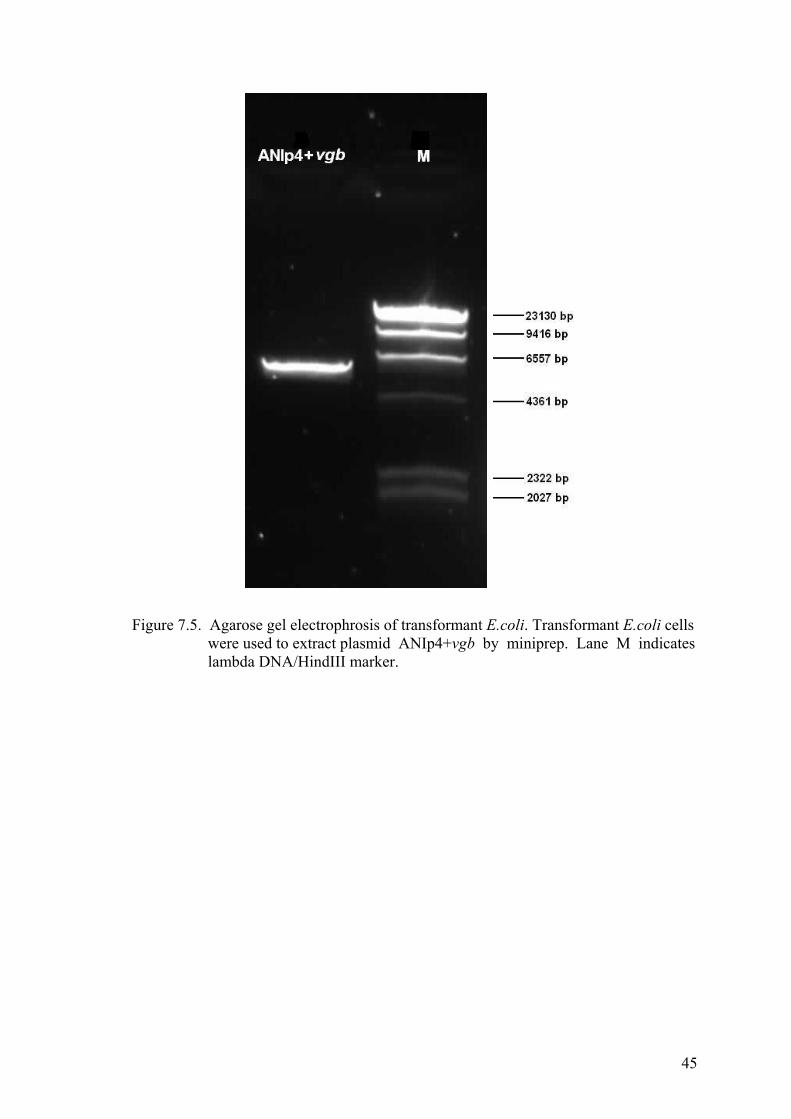
45
Figure 7.5. Agarose gel electrophrosis of transformant E.coli. Transformant E.coli cells were used to extract plasmid ANIp4+vgb by miniprep. Lane M indicates lambda DNA/HindIII marker.

46
Figure 7.6. Agarose gel electrophrosis of PCR product from transformant E.coli. Plasmid samples extracted from DH5 α E. coli were used. FseI and NheI restriction enzymes were used to cut ANIp4+vgb plasmid to obtain vgb gene. After double digestion, samples were subjected to PCR and later agarose gel electrophoresis was performed. Lane 1 was the PCR product of double digested plasmid ANIp4+vgb. Lane1 indicates vgb gene band from transformed E. coli. Lane 2 indicates negative control. (In negative control water is used instead of DNA when PCR run is carried out.) Lane M indicates 100bp DNA ladder.

47
Figure 7.7. Agarose gel electrophrosis of PCR product from transformant A.sojae. After transformation of Mutant 7/2, the plasmid extraction was subjected to PCR procedure. Lane M is lambda DNA marker.

48
Figure 7.8. Agarose gel analysis of transformant A. sojae and M7/2. Lane 1 and 2 indicates the PCR product of TraGlux, lane 3 and 4 indicate PCR product of M7/2 (contamination observed), lane 5 and 6 PCR product of TraElectro.
7.3. Results of Sequence Analysis
In order to confirm and verify that both of the transformations performed by
using Glucanex and electroporation technique were successful, sequence analysis of the
transformed strains were performed. Sequence analysis was performed by Izmir
Institute of Technology, Biotechnology and Bioengineering Central Research
Laboratories with Applied Biosystems 3130xl DNA Sequencer with the extracted
plasmid samples of TraGlux, TraElectro, M7/2 and vgb gene stock coming from Illinois
Institute of Technology Department of Biological Sciences, USA. Plasmid samples
were later subjected to PCR procedure and the PCR products were used as templates for
sequence analysis.
Figures 7.9. the coding sequence for the PCR product of vgb stock coming from
Illinois Institute of Technology Department of Biological Sciences, USA was given for
comparison. The alignments of vgb sequence analysis results and output of the blast
results from Finch TV program presented in Figures 7.10.and 7.11., respectively,
confirmed that the stock vgb has an alignment of 99% with Vitreoscilla sp. hemoglobin
gene (vgb), complete cds; and UvrA (uvrA) gene, partial cds. Similarly, in the figures
7.12. and 7.13. the transformation of A. sojae with vgb via glucanex has an alignment
with Vitreoscilla sp. hemoglobin gene (vgb), complete cds; and UvrA (uvrA) gene,
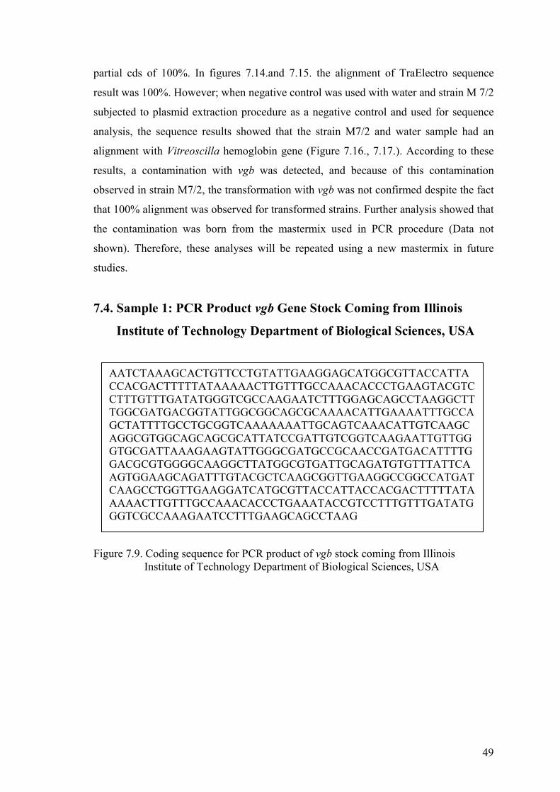
49
partial cds of 100%. In figures 7.14.and 7.15. the alignment of TraElectro sequence
result was 100%. However; when negative control was used with water and strain M 7/2
subjected to plasmid extraction procedure as a negative control and used for sequence
analysis, the sequence results showed that the strain M7/2 and water sample had an
alignment with Vitreoscilla hemoglobin gene (Figure 7.16., 7.17.). According to these
results, a contamination with vgb was detected, and because of this contamination
observed in strain M7/2, the transformation with vgb was not confirmed despite the fact
that 100% alignment was observed for transformed strains. Further analysis showed that
the contamination was born from the mastermix used in PCR procedure (Data not
shown). Therefore, these analyses will be repeated using a new mastermix in future
studies.
7.4. Sample 1: PCR Product vgb Gene Stock Coming from Illinois
Institute of Technology Department of Biological Sciences, USA
Figure 7.9. Coding sequence for PCR product of vgb stock coming from Illinois Institute of Technology Department of Biological Sciences, USA
AATCTAAAGCACTGTTCCTGTATTGAAGGAGCATGGCGTTACCATTACCACGACTTTTTATAAAAACTTGTTTGCCAAACACCCTGAAGTACGTCCTTTGTTTGATATGGGTCGCCAAGAATCTTTGGAGCAGCCTAAGGCTTTGGCGATGACGGTATTGGCGGCAGCGCAAAACATTGAAAATTTGCCAGCTATTTTGCCTGCGGTCAAAAAAATTGCAGTCAAACATTGTCAAGCAGGCGTGGCAGCAGCGCATTATCCGATTGTCGGTCAAGAATTGTTGGGTGCGATTAAAGAAGTATTGGGCGATGCCGCAACCGATGACATTTTGGACGCGTGGGGCAAGGCTTATGGCGTGATTGCAGATGTGTTTATTCAAGTGGAAGCAGATTTGTACGCTCAAGCGGTTGAAGGCCGGCCATGATCAAGCCTGGTTGAAGGATCATGCGTTACCATTACCACGACTTTTTATAAAAACTTGTTTGCCAAACACCCTGAAATACCGTCCTTTGTTTGATATGGGTCGCCAAAGAATCCTTTGAAGCAGCCTAAG

50
Table 7.2. Output of blast results from Finch TV program by using the sample of vgb gene stock. Alignment with Vitreoscilla sp. hemoglobin gene (vgb) , complete cds; and UvrA (uvrA) gene, partial cds is 99%.
Accession Description Max score
Total score
Query Coverage
E value
Max ident
EU363512.1
Synthetic construct hemoglobin/glycolate oxidase fusion protein gene 731 902 95% 0.0 99%
L21670.1
Vitreoscilla sp. hemoglobin (vgb) gene, complete cds; and UvrA (uvrA) gene, partial cds 728 898 95% 0.0 99%
AF292694.1 Vitreoscilla sp. HG1 hemoglobin (vgb) gene, complete cds 728 898 95% 0.0 99%
Figure 7.10. Alignments of vgb gene sequence analysis result. The query sample sent for sequence analysis was for positive control since it is the original vgb gene stock coming from Illinois Institute of Technology Department of Biological Sciences, USA. Alignment with Vitreoscilla sp. hemoglobin gene (vgb) , complete cds; and UvrA (uvrA) gene, partial cds is 99%.

51
7.5. Sample 2: Transformed M 7/2 A. sojae Strain with Glucanex
(TraGlux)
Figure 7.11.Coding sequence for strain TraGlux
Table 7.3. Alignments of strain TraGlux extracted plasmid sequence analysis result.
Accession Description Max score
Total score
Query Coverage
E value
Max ident
EU363512.1
Synthetic construct hemoglobin/glycolate oxidase fusion protein gene 731 1019 96% 0.0 100%
L21670.1
Vitreoscilla sp. hemoglobin (vgb) gene, complete cds; and UvrA (uvrA) gene, partial cds 728 1016 95% 0.0 100%
AF292694.1
Vitreoscilla sp. HG1 hemoglobin (vgb) gene, complete cds 728 1016 95% 0.0 100%
AACTCTCAAGCACTGTTCCTGTATTGAAGGAGCATGGCGTTACCATTACCACGACTTTTTATAAAAACTTGTTTGCCAAACACCCTGAAGTACGTCCTTTGTTTGATATGGGTCGCCAAGAATCTTTGGAGCAGCCTAAGGCTTTGGCGATGACGGTATTGGCGGCAGCGCAAAACATTGAAAATTTGCCAGCTATTTTGCCTGCGGTCAAAAAAATTGCAGTCAAACATTGTCAAGCAGGCGTGGCAGCAGCGCATTATCCGATTGTCGGTCAAGAATTGTTGGGTGCGATTAAAGAAGTATTGGGCGATGCCGCAACCGATGACATTTTGGACGCGTGGGGCAAGGCTTATGGCGTGATTGCAGATGTGTTTATTCAAGTGGAAGCAGATTTGTACGCTCAAGCGGTTGAAGGCCGGCCATGATCAAGCCTGTATTGAAGGAGCATGGCGTTCCATTACCACCACTTTTTATAAAAACTTGTTTGCCAAACCCCCTGAACTACCTCCTTTGTTTGATATGGGTCGCCCAACAATCTTTGGAGCACCTAAGGCTTTGGCGATGACGGTATTGGCGGCAGCCCAAAACATTGAAAATTTTGCCAGTTATTTTTGCTGCGGTCAAAAAA

52
Figure 7.12. Out put of blast results from Finch TV program by using the sample of strain TraGlux. Alignment with Vitreoscilla sp. hemoglobin gene (vgb), complete cds; and UvrA (uvrA) gene, partial cds is 100%.
7.6. Sample3: Transformed M 7/2 A. sojae Strain with Electroporation
(TraElectro)
Figure 7.13 Alignments of strain TraElectro extracted plasmid sequence analysis result.
AATACTCAAGCACTGTTCCTGTATTGAAGGAGCATGGCGTTACCATTACCACGACTTTTTATAAAAACTTGTTTGCCAAACACCCTGAAGTACGTCCTTTGTTTGATATGGGTCGCCAAGAATCTTTGGAGCAGCCTAAGGCTTTGGCGATGACGGTATTGGCGGCAGCGCAAAACATTGAAAATTTGCCAGCTATTTTGCCTGCGGTCAAAAAAATTGCAGTCAAACATTGTCAAGCAGGCGTGGCAGCAGCGCATTATCCGATTGTCGGTCAAGAATTGTTGGGTGCGATTAAAGAAGTATTGGGCGATGCCGCAACCGATGACATTTTGGACGCGTGGGGCAAGGCTTATGGCGTGATTGCAGATGTGTTTATTCAAGTGGAAGCAGATTTGTACGCTCAAGCGGTTGAAGGCCGGCCATGATCA

53
Table 7.4. Coding sequence for the sample of transformed A. sojae with electroporation (TraElectro).
Accession Description Max score
Total score
Query Coverage
E value
Max ident
EU363512.1
Synthetic construct hemoglobin/glycolate oxidase fusion protein gene 731 731 94% 0.0 100%
L21670.1
Vitreoscilla sp. hemoglobin (vgb) gene, complete cds; and UvrA (uvrA) gene, partial cds 728 728 94% 0.0 100%
AF292694.1
Vitreoscilla sp. HG1 hemoglobin (vgb) gene, complete cds 728 728 94% 0.0 100%
Figure 7.14 Out put of blast results from Finch TV program by using TraElectro. Alignment with Vitreoscilla sp. hemoglobin (vgb) gene, complete cds; and UvrA (uvrA) gene, partial cds is 100%.

54
7.7. Sample 4: Mutant 7/2 Aspergillus sojae
Figure 7.15.Coding sequence for Mutant 7/2 Aspergillus sojae.
Table 7.5. Alignments of Mutant M7/2 Aspergillus sojae
Accession Description Max score
Total score
Query Coverage
E value
Max ident
EU363512.1
Synthetic construct hemoglobin/glycolate oxidase fusion protein gene 731 902 95% 0.0 99%
L21670.1
Vitreoscilla sp. hemoglobin (vgb) gene, complete cds; and UvrA (uvrA) gene, partial cds 728 898 95% 0.0 99%
AF292694.1 Vitreoscilla sp. HG1 hemoglobin(vgb) gene, complete cds 728 898 95% 0.0 99%
AATCTAAAGCACTGTTCCTGTATTGAAGGAGCATGGCGTTACCATTACCACGACTTTTTATAAAAACTTGTTTGCCAAACACCCTGAAGTACGTCCTTTGTTTGATATGGGTCGCCAAGAATCTTTGGAGCAGCCTAAGGCTTTGGCGATGACGGTATTGGCGGCAGCGCAAAACATTGAAAATTTGCCAGCTATTTTGCCTGCGGTCAAAAAAATTGCAGTCAAACATTGTCAAGCAGGCGTGGCAGCAGCGCATTATCCGATTGTCGGTCAAGAATTGTTGGGTGCGATTAAAGAAGTATTGGGCGATGCCGCAACCGATGACATTTTGGACGCGTGGGGCAAGGCTTATGGCGTGATTGCAGATGTGTTTATTCAAGTGGAAGCAGATTTGTACGCTCAAGCGGTTGAAGGCCGGCCATGATCAAGCCTGGTTGAAGGATCATGCGTTACCATTACCACGACTTTTTATAAAAACTTGTTTGCCAAACACCCTGAAATACCGTCCTTTGTTTGATATGGGTCGCCAAAGAATCCTTTGAAGCAGCCTAAG

55
Figure 7.16. Output of blast results from Finch TV program by using the sample of Mutant 7/2 Aspergillus sojae. There is alignment with Vitreoscilla hemoglobin gene (indicating contamination).
7.8. Sample 5: Negative Control with Water
Figure 7.17.Coding sequence for negative control with water.
ATCTCAAGCACTGTTCCTGTATTGAAGGAGCATGGCGTTACCATTACCACGACTTTTTATAAAAACTTGTTTGCCAAACACCCTGAAGTACGTCCTTTGTTTGATATGGGTCGCCAAGAATCTTTGGAGCAGCCTAAGGCTTTGGCGATGACGGTATTGGCGGCAGCGCAAAACATTGAAAATTTGCCAGCTATTTTGCCTGCGGTCAAAAAAATTGCAGTCAAACATTGTCAAGCAGGCGTGGCAGCAGCGCATTATCCGATTGTCGGTCAAGAATTGTTGGGTGCGATTAAAGAAGTATTGGGCGATGCCGCAACCGATGACATTTTGGACGCGTGGGGCAAGGCTTATGGCGTGATTGCAGATGTGTTTATTCAAGTGGAAGCAGATTTGTACGCTCAAGCGGTTGAAGGCCGGCCATGATCAAACCTGTATTGAAGGAGCATGGCGTTACCATTACCACGACTTTTTATAAAAACTTGTTTGCCAAACACCCTGAAGTACGTCCTTTGTTTT

56
Table 7.6. Alignments of negative control with water.
Accession Description Max score
Total score
Query Coverage
E value
Max ident
EU363512.1
Synthetic construct hemoglobin/glycolate oxidase fusion protein gene 731 889 95% 0.0 100%
L21670.1
Vitreoscilla sp. hemoglobin (vgb) gene, complete cds; and UvrA (uvrA) gene, partial cds 728 886 94% 0.0 100%
AF292694.1 Vitreoscilla sp. HG1 hemoglobin (vgb) gene, complete cds 728 886 94% 0.0 100%
Figure 7.18. Output of blast results from Finch TV program by using the sample of negative control with water.
7.9. Agar Diffusion Method for Detection of Pectinase Production
Using Polygalacturonic Acid
The reason in performing the agar diffusion test was to determine the effect of
the mutation on the PG production ability. The idea was either not to adversely effect or
to enhance the PG production caused by the mutations, which were necessary in order

57
to select the uridine negative mutants that could be used in the future transformation of
A. sojae with the vgb. Therefore, the best possible potent isolate was selected among the
mutants and coded as M7/2 and used as host train for the transformation. This strain
was compared with the other strains (control M5/6, wild type M7 and the transformed
strains TraGlux and TraElectro) used in the study. The PG production ability of the
strains was compared based on the average ratio of the diameters of clearance zones to
the colony diameters measured. According to the results, control strains M 5/6 and M 7
showed higher ratio (Table 7.7 and 7.8; Figure 7.19 and 7.20). It can be concluded, that
the mutation caused, resulted into a slight decrease in PG activity. Therefore, the actual
comparison among the strains should be done among M7 and M7/2 in order to observe
the effect of mutation and among M7/2, TraGlux and TraElectro in order to observe the
effect of transformation. Similarly, PG activity based on the clear zones indicated, that
strain TraElectro exhibited higher PG activity compared to vgb host strain M7/2 and
TraGlux strain.
In general agar diffusion method is considered as a fast, cheap and an easy
technique compared to analytical methods, in spite of the fact that it doesn’t give exact
activity results. As a result this technique is a simple technique, which should be only
used as prelimary method for the selection of isolates, where other techniques are
difficult to conduct under the same conditions. The individual pictures and their clear
zone measurements are given in Table 7.7.-7.11. and Figure 7.19-7.23., respectively.

58
Figure 7.19. Solid media containing polygalacturonic acid (PGA) for detection of enzyme production of the control strain M5/6.
Table 7.7. Ratio of the diameters of clearance zones of the control strain M 5/6.
Colony Clearance Zone/Colony
(cm)
Ratio
(cm)
Average Ratio
(cm)
1 3,23/0,924 3,49
2 3,20/0,62 5.13
3 3,35/0,95 3,53
4,05
1
2
3

59
Figure 7.20. Solid media containing polygalacturonic acid (PGA) for detection of enzyme production of the wild type strain M7.
Table 7.8. Ratio of the diameters of clearance zones of the wild type strain M7
Colony Clearance Zone/Colony
(cm) Ratio (cm)
Average Ratio (cm)
1 4,092/1,53 2,66
2 4,54/1,74 2,614
3 4,18/1,60 2,612
2,626
1
2
3

60
Figure 7.21. Solid media containing polygalacturonic acid (PGA) for detection of enzyme production of the mutated strain M7/2
Table 7.9. Ratio of the diameters of clearance zones of the mutated strain M7/2
Colony Clearance Zone/Colony
(cm)
Ratio
(cm)
Average Ratio
(cm)
1 3,11/2,15 1,44
2 3,20/2,67 1,19
3 2,84/1,39 2,03
4 3,11/1,13 2,73
1,88
1
2
3
4
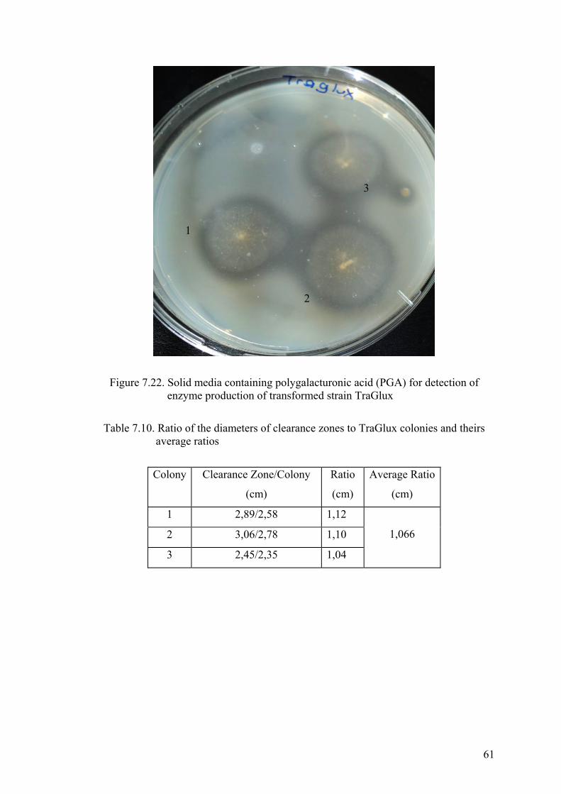
61
Figure 7.22. Solid media containing polygalacturonic acid (PGA) for detection of
enzyme production of transformed strain TraGlux
Table 7.10. Ratio of the diameters of clearance zones to TraGlux colonies and theirs average ratios
Colony Clearance Zone/Colony
(cm)
Ratio
(cm)
Average Ratio
(cm)
1 2,89/2,58 1,12
2 3,06/2,78 1,10
3 2,45/2,35 1,04
1,066
1
2
3

62
Figure 7.23. Solid media containing polygalacturonic acid (PGA) for detection of enzyme production from spore solution of strain TraElectro
Table 7.11. Ratio of the diameters of clearance zones to TraElectro colonies and their average ratios
Colony Clearance Zone/Colony
(cm)
Ratio
(cm)
Average Ratio
(cm)
1 4,18/2,14 1,95
2 4,85/1,98 2,44 2,19
Table 7.12. Average ratio of the diameters of clearance zones to colonies of strains used.
Colony Average Ratio
M 5/6 4,05
M 7 2,626
M 7/2 1,88
TraGlux 1,066
TraElectro 2,19
1
2

63
CHAPTER 8
CONCLUSION
In this study, the preliminary results of transformation of Aspergillus sojae with
vgb were obtained. ANIp4 shuttle vector was used as a vector in order to transform A.
sojae. For the selection of transformed A. sojae cells, uridine auxotrophic mutants were
tried to be selected after UV mutagenesis. However, using a procedure based on the
selection of uridine supported growth did not result in A. sojae pyrG mutants. Further
studies will need to be performed in order to obtain pyrG mutants based on FOA direct
selection in addition to uridine and uracil supplements in the growth medium. Also gene
replacement with homolog recombination will be used in order to obtain uridine
auxotroph strains as an alternative method to UV mutation.
In this study, the transformation of A. sojae with vgb was tried using both
methods of electroporation and protoplasting and tested by sequence analysis and
agarose gel electrophoresis. (Alignment with Vitreoscilla sp. hemoglobin gene (vgb),
complete cds; and UvrA (uvrA) gene, partial cds was for strains TraElectro and TraGlux
were 100%). Protoplast preparation is a problematic method because in literature fungal
protoplasting via enzymes were usually prepared by using the enzyme Novozyme 234
however, this enzyme is no longer available for sale (De Bekker, et al., 2009).
According to this knowledge a less common used enzyme glucanex was used in order to
prepare A. sojae protoplasts.
The A. sojae strains used in this study were: Strain M5/6 and M7 which were
initially mutated in order to improve the production of exo-polygalacturonase using the
wild type Aspergillus sojae. Strain M7/2 was UV mutated from strain M7 in order to
obtain pyrG negative strains within the scope of this thesis project. Strain TraGlux was
obtained by transformation of M7/2 with vgb via protoplasting method. Strain
TraElectro was obtained by transformation of M7/2 with vgb gene via electroporation
method; however both strains could not be confirmed due to the contamination occurred
in PCR process which carried the contamination to the sequence analysis. In order to
obtain the effect of vgb gene, M7/2 was compared with TraElectro and TraGlux by
PGA zone measurements since this strain was the untransformed strain with the rest of

64
the characteristics similar to the transformed ones. Other strains (M5/6 and M7) were
subjected to PGA zone mearurement analysis as potential control strains in order to
compare the effectiveness of the mutation and the transformation process, as they were
studied routinely in our laboratuvary. This indicated that vgb has a positive affect on PG
activity. If TraGlux and TraElectro were compared separately, it was seen that
TraElectro had about 2 times higher PG activity compared to TraGlux.
In the future studies the expession of vgb will be tested using the methods CO-
difference spectra, SDS-PAGE and western blotting analysis. The effect of vgb
expression will be evaluated in submerged fermentation and the optimum conditions
under microaerophilic situations will be determined for the maximum exo-
polygalacturonase production.

65
REFERENCES
Acuna-Arguelles, M.E., Gutierrez-Rajas, M., Viniegra-Gonzalez, G., Favela-Toress,
E., (1995). Production and properties of three pectinolytic activities produced by A. niger in submerged and solid state fermentaion. Appl. Microbiol. Biotechnol. (43), 808-814.
Alana, A., Alkorta, I., Dominguez, J.B., Llama, M.J., Serra, J.L., (1990). Pectin lyase
activity in a Penicillium italicum strain. Appl. Environ. Microbiol. 56 (12), 3755-3759.
Al-Obaidi, Z.S., Aziz, G.M., Al-Bakir, A.Y., 1987. Screening of fungal strains for
polygalacturonase production. J. Agric. Water Resour. Res. 6, 125-182. Akins, R. A., Lambowitz, A. M. (1985). General ethod for cloning Neurospora crassa
nuclear genes by complementation of mutants. Mol. Cell. Biol. 5, 2227-2278. Appleby, CA. (1992). The origin and functions of haemoglobins in plants. Sci Prog ,
76,365–398. Arredondo-Peter, R., Hargrove, M.S., Sarath, G., Moran, J.F., Lohrman, J. S., Olson,
J.S., Klucas, R.V. (1997). Rice hemoglobins. Gene cloning, analysis, and O2-binding kinetics of a recombinant protein synthesized in Escherichia coli. Plant Physiol, 115,1259–1266.
A.T. Still University, (2010). Asexual fruiting structure of Aspergillus species,
illustrating septate hyphae, conidiophore, vesicle, phialides and conidospores. www.atsu.edu, December, 2010
Borin, M.D.F., Said, S., Fonseca, M.J.V., 1996. Purification and biochemical
characterization of an extracellular endopolygalacturonase from Penicillium frequentans. J. Agric. Food Chem. 44, 1616-1620.
Bou, G., Cervero, G., Dominguez, M. A., Quereda, C., Martinez-Beltran, J. (2000). The
characterization of a nosocomial outbreak caused by a multiresistant Actinetobacter baumannii strain with a carbapenem-hydrolysing enzyme: high level carbapenem resistance in A. baumannii is not due solely to the presence of beta-lactamases. J. Clin. Microbiol., 38(9), 3299-3305.

66
Bruhlmann, F., Kim, K.S., Zimmerman, W., Fiechter, A., (1994). Pectinolytic enzymes from actinomycetes for the degumming of ramie bast fibers. Appl. Environ. Microbiol. 60 (6), 2107-2112.
Brünker, P., Minas, W., Kallio, P. T., Bailey, J. E. (1998). Genetic engineering of an
industrial strain of Saccharopolyspora erythraea for stable expression of the Vitreoscilla hemoglobin gene (vhb). Microbiology, 144, 2441-2448.
Cao, J., Zheng, L., Chen, S., 1992. Screening of pectinase producer from alkalophilic
bacteria and study on its potential application in degumming of rammie. Enz. Microbiol. Technol. 14, 1013-1016.
Channe, P.S., Shewal, J.G., 1995. Pectinase production by Sclerotium rolfsii: Effect of
culture conditions. Folia Microbiol. 40,111-117. Chen, W., Hughes, D. E., Bailey, J. E. (1994). Intracellular expression of Vitreoscilla
hemoglobin alters the aerobic metabolism of Saccharomyces cerevisia. Biotechnology Progress, 10(3), 308-312.
Chesson, A., Codner, R.C., (1978). Maceration of vegetable by a strain of Bacillus
subtilis. J. Appl. Bacteriol. 44, 347-364. Collins, R. A., Stohl, L. L., Cole, M. D., Lambowitz, A. M. (1981). Characterization of
a novel plasmid DNA found in mitochondria of N. crassa. Cell, 24, 443-452. Cullen, D., Leong, S.A. (1987). Transformation of Aspergillus nidulans with the
hygromcin-resistance gene, hph. Gene, 57, 21-26. Dave, B.A., Vaughn, R.H., (1971). Purification and properties of a polygalacturonic
acid trans-eliminase produced by Bacillus pumilus. J. Bacteriol. 108, 166-174. De Bekker, C., Wiebenga, A., Aguilar, G., Wösten, H. A. B., (2009). An enzyme
cocktail for efficient protoplast formation in Aspergillus niger. Journal of Microbiological Methods, 76(3), 305-306.
Dikshit, K. L., Dikshit, R. P., Webster, D. A. (1990). Study of Vitreoscilla globin(vgb)
gene expression and promoter activity in E. coli through transcriptional fusion. Nucleic Acids Research, 18(14), 4149-4155.

67
Dikshit, R. P., Dikshit, K. L., Liu, Y. Webster, D. A. (1992). The bacterial hemoglobin from Vitreoscilla can support the aerobic growth of Escherichia coli lacking terminal oxidases. Archives of Biochemistry and Biophysics, 293(2), 241-245.
Dogan, I, Pagilla, K., Webster, D.A., Stark, B., (2006). Expression of Vitreoscilla
hemoglobin in Gordonia amarae enhances biosurfactant production. J. Industrial Microbiology and Biotechnology, 33(8), 693-700.
Eichhorn, G. L., Marzill L. G. (Ed.). (1987). Advances in Inorganic Biochemistry (Vol.
7). Faure, M., Camonis, J. H., Jacquet, M. (1989). Molecular characterization of a
Dictyostelium discoideum gene encoding a multifunctional enzyme of the pyrimidine pathway. European Journal of Biochemistry, 179(2), 345-358.
Fincham, J. R. (1989). Transformation in fungi. Microbiol. Mol. Biol. Rev., 53(1), 148-
170. Fogarty, M.V., Kelly, C.T., (1983). Pectic enzymes. In: Fogarty, M.W. (Ed.), Microbial
Enzymes and Biotechnology. Applied Science Publishers, London, 131-182. Gaisson, L., Lalonde, M. (1987). Analysis of a linear plasmid isolated from the
pathogenic fungus Ceratocystis fimbriata. Curr. Genet. 11, 332-334. Gao, G., Nara, T., Nakajima-Shimada, J., Aoki, T. (1999). Novel organization and
sequences of five genes encoding all six enzymes for de novo pyrimidine biosynthesis in Trypanosoma cruzi. Journal of Molecular Biology, 285(1), 149-161.
Gardner P.R, Gardner A.M, Martin L.A, Dou Y, Li T.S, Olson J.S, Zhu H., Riggs, A.F.
(2000) Nitric-oxide dioxygenase activity and function of flavohemoglobins. Sensitivity to nitric oxide and carbon monoxide inhibition. J Biol Chem, 275, 31581–31587.
Geçkil, H., Gencer, S. (2004). Production of L-asparaginase in Enterobacter aerogenes
expressing Vitreoscilla hemoglobin. Applied Microbiology and Biotechnology, 63(6), 691-697.
Gellisen, G. (Ed.). (2005). Production of Recombinant Proteins: Wiley-VCH.

68
Goldman, G. H., Osmani, S. A., (Ed.). (2008). The Aspergilli: Genomics, medical aspects and biotechnology, and research methods: CRC Press.
Griffiths, A. (1995). Natural plasmids of filamentous fungi. Microbiol. Rev., 59(4), 673-
685. Hankin, L., Anagnostakis, S. L. (1975). The use of solid media for detection of enzyme
production by fungi. Mycologia,67 Henriksson, J., Åslund, L., Macina, R. A., Franke de Cazzulo, B. M., Cazzulo, J. J.,
Frasch, A. C. C., Pettersson, U. (1990). Chromosomal localization of seven cloned antigen genes provides evidence of diploidy and further demonstration of karyotype variability in Trypanosoma cruzi. Molecular and Biochemical Parasitology, 42(2), 213-223.
Hoffman, G., Diano, A., Nielson, J., (2009) Recombinant bacterial hemoglobin alters
the metabolism of Aspergillus niger.Metabolic Enngineering, 11(1), 8-12. Honeyman, A. L., Currier, T. C. (1986). Isolation and characterization of linear DNA
elements from the mitochondria of Gaeumannomyces graminis. Appl. Environ. Microbiol. 52, 924-929.
Horikoshi, K., (1990). Enzymes of alkalophiles. In: Fogarty, W.M., Kelly, C.T. (Eds.),
Microbial Enzymes and Biotechnology, second ed. Elsevier Applied Science, London, pp. 275-294.
Kallio, P. T., Jin Kim, D., Tsai, P. S., Bailey, J. E. (1994). Intracellular expression of
Vitreoscilla hemoglobin alters E. coli energy metabolism under oxygen-limited conditions. Europian Journal of Biochemistry , 219, 201-208.
Kallio, P. T., Bailey, J. E. (1996). Intracellular expression of Vitreoscilla hemoglobin
(Vhb) enhances total protein secretion and improves the production of α-amylase and neutral protease in Bacillus subtilis. Biotechnology Progress, 12(1), 31-39.
Kanamasa, S. Y., K.; Kawaguchi, T.; Sumitani, J.; Arai, M. (2003). Transformation of
Aspergillus aculeatus using the drug resistance gene of Aspergillus oryzae and the pyrG gene of Aspergillus nidulans. Bioscience, Biotechnology, and Biochemistry, 67, 2661-2663.

69
Kanamasa, S., Tadaka, G., Kawaguchi T., Sumitani J., Arai, M. (2001). Overexpression and Purification of Aspergillus aculeatus β-Mannosidase and Analysis of the Integrated Gene in Aspergillus oryzae. Journal of Bioscience and Bioengineering, 92.
Karbassi, A., Vaughn, R.H., (1980). Purification and properties of polygalacturonic acid
trans-eliminase from Bacillus stearothermophilus. Can. J. Microbiol. 26, 377-384.
Kashyap, D. R., Vohra, P. K., Chopra, S., Tewari, R. (2001). Applications of pectinases
in the commercial sector: A review. Bioresource Technology, 77(3), 215-227. Kashyap, D.R., Chandra, S., Kaul, A., Tewari, R., (2000). Production purification and
characterization of pectinase from a Bacillus sp DT7. World J. Microbiol. Biotechnol. 16, 277-282.
Katsuya, S., Kaneko, I., Owaki, M., Ishikawa, K., Tsujimoto, T., Tsuge, T. (1997).
Circular DNA Plasmid in the Phytopathogenic Fungus Alternaria Alternata: Its Temperature-Dependent Curing and Association with Pathogenicity. Genetics, 146(1), 111-120
Khleifat, K., Abboud M. M. (2003). Correlation between bacterial hemoglobin ggene
(vgb) and aeration : Their effect on the growth and α-amylase activity in transformed Enterobacter aerogenes. J. Applied Microbiology, 94(6), 1052-1058.
Khosravi, M., Webster, D. A., Stark, B. C. (1990). Presence of the bacterial hemoglobin
gene improves α-amylase production of a recombinant Escherichia coli strain. Plasmid, 24(3), 190-194.
Kistler, H. C., Leong, S. A. (1986). Linear plasmidlike DNA in the plant pathogenic
fungus Fusiarum oxysporum f. sp. conglutinans. J. Bacteriol. 167, 587-593. Klich, M. A., Montalbano, B., Ehrlich, K. (1997). Northern analysis of aflatoxin
biosynthesis genes in Aspergillus parasiticus and Aspergillus sojae. Applied Microbiology and Biotechnology, 47(3), 246-249.
Koukaki, M., Giannoutsou, E., Karagouni, A., Diallinas, G. (2003). A novel improved
method for Aspergillus nidulans transformation. Journal of Microbiological Methods, 55(3), 687-695.

70
Kroneck, P. M. H., Jakob, W., Webster, D. A., DeMaio, R. (1991). Studies on the
bacterial hemoglobin from Vitreoscilla. BioMetals, 4(2), 119-125. Liang, F., Shouwen, C., Ming, S., Ziniu, Y. (2007). Expression of Vitreoscilla
hemoglobin in Bacillus thuringiensis improve the cell density and insecticidal cyrstal proteins yield. Applied Microbiology and Biotechnology,74 (2), 390-397.
Lin, L-H., Li, Y-F., Huang, M-C. Tsai, Y-C. (2004).Intracellular expression of
Vitreoscilla hemoglobin in Aspergillus terreus to alleviate the effect of short break in aeration during culture. Biotechnology Letters, 26(13), 1067-1072.
Liu, S. C., Liu, Y. X., Webster, D. A., Stark, B. C. (1994). Sequence of the region
downstream of the Vitreoscilla hemoglobin gene: vgb is not part of a multigene operon. Applied Microbiology and Biotechnology, 42(2), 304-308.
Liu, S. C., Webster, D. A.,Wei, M. L., Stark, B. C. (1995a). Genetic engineering to
contain the Vitreoscilla hemoglobin gene enhanced degradation of benzoic acid by Xanthomonas maltophilia. Biotechnology and bioengineering, 49(1), 101-105.
Liu, S. C., Webster, D. A., Stark, B. C. (1995b). Cloning and expression of the
Vitreoscilla hemoglobin gene in preudomonads: Effects on cell growth. Applied Microbiology and Biotechnology, 44(3), 419-424.
Liu, Y., Li, Q., Yuan, Q., (2009). The Construction of pDH25-pcpC-Vgb as a
Recombinant DNA System for the Intracellular Expression of Vitreoscilla Hemoglobin in Cephalosporium Acremonium. Modern Applied Science, 3, 75-82.
Long, H., Wang, T.-H., Zhang, Y.-K., (2008). Isolation of Trichoderma reesei pyrG
Negative Mutant by UV Mutagenesis and Its Application in Transformation. Chemical Research in Chinese Universities, 24(5), 565-569.
Lubertozzi, D., and Keasling, J. D. (2009). Developing Aspergillus as a host for
heterologous expression. Biotechnology Advances, 27(1), 53-75. Machida, M., Gomi, K. (Ed.). (2010). Aspergillus: Molecular Biology and Genomics:
Caister Academic Press.

71
Magro, P., Varvaro, L., Chilosi, G., Avanzo, C., Balestra, G.M., (1994). Pectinolytic enzymes produced by Pseudomonas syringae pv. Glycinea. FEMS Microbiol. Lett. 117, 1-6.
Mattern, I. E., Unkles, S., Kinghorn, J. R., Pouwels, P. H., Hondel, C. A. M. J. J.,
(1987). Transformation of Aspergillus oryzae using the A. niger pyrG gene. Molecular and General Genetics MGG, 210(3), 460-461.
Mohan, M., Meyer, R., J., Anderson, J. B., and Horgen, P. A. (1984). Plasmid-like
DNAs in the commercially important mushroom genus Agaricus. Curr. Genet., 8, 441-453.
Molbak, L., Tett, A., Ussery, D. W., Wall, K., Turner, S., Bailey, M., Field, D., (2003).
The Plasmid Genome Database. Microbiology, 149(11), 3043-3045. Nagel, C.W., Vaughn, R.H., (1961). The characterisitc of a polygalacturonase produced
by Bacillus polymyxa. Arc. Biochem. Biophys. 93, 344-352. Nara, T., Hshimoto, T., Aoki, T., (2000). Evolutionary implications of the mosaic
pyrimidine-biosynthetic pathway in eukaryotes. Gene, 257(2), 209-222. Nasumo, S., Starr, M.P., (1967). Polygalacturonic acid trans-eliminase of Xanthomonas
compestris. Biochem. J. 104, 178-184. Normand, P., Simonet, P., Giasson, L., Ravel-Chapius, P., Fontin, J., A., Lalonde, M.
(1987). Presence of a linear plasmid-like DNA molecule in the fungal pathogen Ceratocystis fimbriata. Curr. Genet. 11, 335-338.
Orii, Y., Webster, D.A., Photodissociation of oxygenated cytochrome o(s) (Vitreoscilla)
and kinetic studies of reassociation. (1986) J Biol Chem 261,3544–3547. Osmani, S. A., May, G. S., Morris, N. R. (1987). Regulation of the mRNA levels of
nimA, a gene required for the G2-M transition in Aspergillus nidulans. The Journal of Cell Biology, 104(6), 1495-1504.
Oza, K., Kafer, E. (1990). Cloning of the DNA Repair Gene, uvsF, by Transformation
of Aspergillus nidulans. Genetics, 125(2), 341-349.

72
Peberdy, J. F. (1979). Fungal Protoplasts: Isolation, Reversion, and Fusion. Annual Review of Microbiology, 33, 21-39.
Pendse, G. J., Bailey, J. E. (1994). Effect of vitreoscilla hemoglobin expression on
growth and specific tissue plasminogen activator productivity in recombinant chinese hamster ovary cells. Biotechnology and Bioengineering, 44(11), 1367-1370.
Rijssel, M.W., Gerwig, J.G.J., Hausen, T.A., 1993. Isolation and characterization of an
extracellular glycosylated protein complex from Clostridium thermosaccharolyticum with pectin methylesterase and polygalacturonate hydrolase activity. Appl. Environ. Microbiol. 59 (3), 828-836.
Rubitge, T. (1986). Survey of Fusarium species for plasmid-like DNA and some
evidence for its occurrence in a strain of F. merismoides. Mycol. Soc. 87, 463-466.
Ruiz-Díez, B. (2002). Strategies for the transformation of filamentous fungi. Journal of
Applied Microbiology, 92(2), 189-195. Runco, R., Navarro, A. R., Maldonado, M. C. (2001). Regulation of the production of
polygalacturonase by Aspergillus terreus. World Journal of Microbiology and Biotechnology, 17(5), 487-491.
Samac, D. A., Leong, S. A., (1989). Mitochondrial plasmids of filamentous fungi:
characteristics and use in transformation vectors. Molecular plant-microbe interactions, 2, 155-159.
Samac, D. A., Leong, S. A. (1986). Lack of association of mitochondrial plasmids and
pathogenity in Nectria haematococca Fusiarum solani f. sp. cucurbitae. Plasmid 19, 57-67.
Sambrook, J., Fritsch E. F., Maniatis, T. (1989). Molecular Cloning: A laboratory
Manual. Cold Spring Harbor Laboratory Press, Cold Spring Harbor. Samson, R. A., Hong, S.-B., Frisvad, J. C. (2006). Old and new concepts of species
differentiation in Aspergillus. Medical Mycology, 44, 133-148.

73
Sanchez, O. and Aguirre, J. (1996). Efficient transformation of Aspergillus nidulans by electroporation of germinated conidia. Fungal Genetics Nexsletter, 43, 48-51.
Sgorbati, B., Scardovi, V., Leblanc, D. J., (1982). Plasmids in the Genus
Bifidobacterium. J Gen Microbiol, 128(9), 2121-2131. Skory, C. D., Horng, J. S., Pestka, J. J., Linz, J. E. (1990). Transformation of
Aspergillus parasiticus with a homologous gene (pyrG) involved in pyrimidine biosynthesis. Appl. Environ. Microbiol., 56(11), 3315-3320.
Solís, S., and Flores, M. E., (1997). Improvement of pectinase production by
interspecific hybrids of Aspergillus strains. Letters in Applied Microbiology, 24(2), 77-81.
Sthol, L. L., Collins, R. A., Cole, M. D., Lambowitz, A. M. (1982). Characterization of
two new plasmid DNAs found in mitochondria of wild-type Neurospora intermedia strains. Nucleic Acids Res. (10),1439-1458.
Storms, R., Zheng, Y., Hongshan L., Sillaots, S., Martinez-Perez, A., Tsang, A. (2004).
Plasmid vectors for protein production, gene expression and molecular manipulations in Aspergillus niger. Elsevier, 53, 191–204.
Su, Y., Li, X., Liu, Q., Hou, Z., Zhu, X., Guo, X., Ling, P. (2010). Improved poly-
gamma-glutamic acid production by chromosomal integration of the Vitreoscilla hemoglobin gene (vgb) in Bacillus subtilis. Bioresour Technol., 101(12), 4733-4736.
Sun, C.-B., Kong, Q.-L., Xu, W.-S. (2002). Efficient transformation of Penicillium
chrysogenum mediated by Agrobacterium tumefaciens LBA4404 for cloning of Vitreoscilla hemoglobin gene. 5, 29-41.
Takahashi, T., Chang, P.-K., Matsushima, K., Yu, J., Abe, K., Bhatnagar, D.,
Cleveland, T. E., Koyama, Y. (2002). Nonfunctionality of Aspergillus sojae aflR in a Strain of Aspergillus parasiticus with a Disrupted aflR Gene. Appl. Environ. Microbiol., 68(8), 3737-3743.
Takahashi, T., Jin, F. J., Sunagawa, M., Machida, M., Koyama, Y. (2008). Generation
of Large Chromosomal Deletions in Koji Molds Aspergillus oryzae and Aspergillus sojae via a Loop-Out Recombination. Appl. Environ. Microbiol., 74(24), 7684-7693. Gen. Genet. 201, 161-167.

74
Taylor, J. W., Smolich, B. D., May, G. (1985). An evolutionary comparison of
homologous mitocondrial plasmid DNAs from three Neurospora species. Mol. Gen. Genet. 201, 161-167.
Tilburn, J., Scazzocchio, G., Taylor, G. G., Zabicky-Zissman, J. H., Lockington, R. A.,
Davies, R. W. (1983). Transformation by integration in Aspergillus nidulans Gene, 26(2-3) 205-221.
Trevaskis, B, Watts, R. A, Andersson, C. R, Llewellyn, D. J, Hargrove, M. S, Olson, J.
S., Dennis, E. S., Peacock, W. J. (1997). Two hemoglobin genes in Arabidopsis thaliana: The evolutionary origins of leghemoglobins. Proc Natl Acad Sci ,94,12230–4.
Tudzynski, P., Esser, K. (1986). Extrachromosomal genetics of Claviceps purpurea. II.
Plasmids in various wild strains and integrated plasmid sequences in mitochondrial genomic DNA. Curr. Genet. 10, 463-467.
Van Hartingsveldt, W., Mattern, I. E., Zeijl, C. M. J., Pouwels, P. H., Hondel, C. A. M.
J. J. (1987). Development of a homologous transformation system for Aspergillus niger based on the pyrG gene. Molecular and General Genetics MGG, 206(1), 71-75.
Varavallo, M. A., Queiroz, M. V. d., Lana, T. G., Brito, A. T. R. d., Gonçalves, D. B., &
Araújo, E. F. d. (2007). Isolation of recombinant strains with enhanced pectinase production by protoplast fusion between Penicillium expansum and Penicillium griseoroseum. Brazilian Journal of Microbiology, 38, 52-57.
Vary, P. H., Andersen, P. R., Green, E., Hermon-Taylor, J., McFadden, J. J. (1990). Use
of highly specific DNA probes and the polymerase chain reaction to detect Mycobacterium paratuberculosis in Johne's disease. J. Clin. Microbiol., 28(5), 933-937.
Wainwright, M. (Ed.). (1992). An Inrtoduction to Fungal Biotechnology: Wiley. Wei, M. L., Webster, D. A., Stark, B. C., (1998). Metabolic engineering of Serratia
marcescens with the bacterial hemoglobin gene: Alterations in fermentation pathways. Biotechnology and Bioengineering, 59(5), 640-646.

75
Weidner, G. d'Enfert, C., Koch, A., Mol, P. C., Brakhage, A. A. (1998). Development of a homologous transformation system for the human pathogenic fungus Aspergillus fumigatus based on the pyrG gene encoding orotidine 5′′-monophosphate decarboxylase. Curr Genet., 33(5), 378-385.
Wu, J.-M., Hsu, T.-A., Lee, C.-K. (2003). Expression of the gene coding for bacterial
hemoglobin improves β-galactosidase production in a recombinant Pichia pastoris. Biotechnology Letters, 25(17), 1457-1462.
Yelton, M. M., Hamer, J. E., Timberlake, W. E. (1983). Transformation of Aspergillus
nidulans by Using trpC Plasmid. Genetics, 81,1470-1474. Yuzuru Ilmura, K. G., Hiroshi Uzu and Shodo Hara. (1986). Transformation of
Aspergillus oryzae through plasmid-mediated complementation of the methionine-auxotrophic mutation. Agricultural Biolological Chemistry, 51, 323-328.
Zhang, L., Li, Y., Wang, Z., Xia, Y., Chen, W., Tang, K. (2007). Recent developments
and future prospects of Vitreoscilla hemoglobin application in metabolic engineering. Biotechnology Advances 25, 123-136.

76
APPENDIX A
CHEMICAL USED IN EXPERIMENTS
CHEMICAL CODE
Velvet Pad for Replica Plating MP Biomedicals 5000-007
Ammonium Sulphate ((NH4)2SO4) Applichem A1032,0500
Agar-Agar Applichem A0949
Agarose Low EEO Applichem A2114,0250
Ammonium Heptamolypbdate Tetrahydrate Merck 1.01182
Ampicillin Sigma A 9393-5g
Brain heart infusion agar(BHI) Fluka 70138
Calcium Choloride Dihydrate (CaCl2 ) Riedel-De Haën 31307
Coomassie brilliant blue G-250 Fluka 27815
Copper (II) sulfate-5-hydrate Riedel-De Haën 12849
CuSO4.5H2O Copper (II) sulphate-5-hydrate Riedel 1kg 12849
D (+)-Glucose Fluka 49150 1 kg
Disodium Hydrogen Arsenate Heptahydrate Applichem A3666
EcoRI Fermentas FD0274
Disodiumsalt(EDTA) (Standart Volumetric Solution 0,1M)
Applichem A 1505,0500
Ethanol Applichem A2937
FeSO4.7H2O (Iron (11) Sulfate Heptahydrate ) Riedel-De Haën 1 kg
FseI Fermentas
D-Galacturonic acid Fluka 48280

77
Gel Stain Gel Star 10000 A Concent Lonza Zx 250 ml
Gene Ruler 100bp DNA Ladder Plus (Fermentas 50 mg.sm 1F3
D-(+)-Galacturanicacid Fluka 48280 25g
Glucanex (Lysing Enzymes from Trichoderma harzianum)
Sigma L 1412-10g
Glycerol Sigma G5516
Boric Acid (H3BO3) Sigma B 6768-500g
Hydrochloric acid (HCl) Riedel-De Haën
HindIII Enzyme Fermantas FD0504
Isopropanol Riedel-De Haën
Potassium Chloride (KCI) Riedel-De Haën 31248
Potassium Phosphate Monobasic (KH2PO4) Riedel-De Haën 1 KG
Lambda DNA Fermentas
Loading Dye Fermentas
Maltrin Pendik Nişasta (Nişkoz H.
Maltoz 41521)
PCR Maxtermix (2X) Fermentas 00050331
Magnesium chloride anhydrous (MgCI2) Fluka 63063
Magnesium sulphate heptahydrate (MgSO4.7H2O) Merck 1.05886
Manganese (II) Chloride Tetrahydrate (MnCI2.4H2O)
Merck 1.05886
Manganese (II) Sulphate Monohydrate (MnSO4.4H2O)
Riedel-De Haën
Molasses Merck 1.02786
Molybdenium (IV) Oxyde (MoO3) Merck 1.00401.0250
Sodium Carbonate Anhydrous(Na2CO3) Applichem A3900.1000
di-Sodium Hydrogen Phosphate (Na2HPO4.2H2O) Fluka 71662 1 kg
Sodium Sulphate (Na2SO4) Merck 1.06649.1000

78
Sodium Chloride (NaCI) Riedel-De Haën 13423
NaH2PO4.2H2O Riedel-De Haën
Sodium bicarbonate (NaHCO3) Sigma S 8875 500g
Sodium Nitrate (NaNO3) Applichem A 3911.1000
Sodium Hydroxide (NaOH) Merck 1 kg 1.06462.1000
NheI Fermentas FD0973
PEG 4000 Applichem
Peptone Merck 1.07214.9999
Polygalacturonic Acid Fluka 81325 50g
Potassium Sodium Tartate tetrahydrate (C4H4KNaO6.4H2O)
Sigma S6170
Qiagen Miniprep Kit Quiagen
Trizma Base Sigma T-8404 1kg
Tryptone Applichem A1553.1000
Tween-80 Merck 8.22187.0500
Uridine Applichem A0666.0010
Yeast Extract Fluka 70161
ZnSO4.7H2O Merck 1.03753

79
APPENDIX B
MEDIA
B.1. LB Agar Medium
Tryptone (10 g/l)
Yeast extract (5 g/l)
NaCI (10 g/l)
Deionized water (1000 ml)
Agar (15 g/ml)
Ingredients were dissolved in 900 ml distilled water by stirring with heating and
made up to 1000ml. pH was adjusted to 7,5. Medium was sterilised by autoclaving at
121°C for 15 minutes.
B.2. LB Broth Medium
Tryptone (10 g/l)
Yeast extract (5 g/l)
NaCI (10 g/l)
Deionized water (1000 ml)
Ingredients were dissolved in 900 ml distilled water by stirring with gentle
heating and made up to 1000 ml. pH was adjusted to 7,5. Medium was sterilized by
autoclaving at 121°C for 15 minutes.

80
B.4. LB Agar Medium Supplemented with Ampicillin
Tryptone (10 g/l)
Yeast extract (5 g/l)
NaCI (10 g/l)
Ampicillin (50 μg/ml)
Deionized water (1000 ml)
Agar (15 g/ml)
Ingredients except ampicillin were dissolved in 900 ml distilled water by stirring
with heating and made up to 1000 ml. pH was adjusted to 7,5. Medium was sterilized by
autoclaving at 121°C for 15 minutes. Ampicillin 100 mg/ml stock solution (see
Appendix C) was added to 50μg/ml final concentration after autoclaving.
B.5. LB Broth Medium Supplemented with Ampicillin
Tryptone (10 g/l)
Yeast extract (5 g/l)
NaCI (10 g/l)
Ampicillin (50 μg/ml)
Deionized water (1000 ml)
Ingredients except ampicillin were dissolved in 900 ml distilled water by stirring
with gentle heating and made up to 1000ml. pH was adjusted to 7,5. Medium was
sterilized by autoclaving at 121°C for 15 minutes. Ampicillin 100 mg/ml stock solution
(see Appendix C) was added to 50μg/ml final concentration after autoclaving.
B.6. YME (Yeast Malt Extract)
Malt extract (10 g/l)
Yeast extract (4 g/l)

81
Glucose (4 g/l)
Agar (20 g/l)
Ingredients were dissolved in 900 ml distilled water by stirring with gentle
heating and made up to 1000ml. Medium was sterilized by autoclaving at 121°C for 15
minutes.
B.7.Minimal Agar Medium
NaNO3 ( 6 g/l)
KCI (0.52 g/l)
MgSO4.7H2O (0.52 g/l)
KH2SO4 (1.52 g/l)
D-Glucose (10 g/l)
Hunter’s Trace Elements Solution (2 ml/l)
Agar (15 g/l)
Hunter’s Trace Elements Solution (In 100 ml of water)
ZnSO4.7H2O (2.2 g)
H3BO3 (1.1 g)
MnCI2.4H2O (0.5 g)
FeSO4.7H2O (0.5 g)
CoCI2.6H2O (0.16 g)
CuSO4.5H2O (0.16 g)
NH4Mo7O24.4H2O (0.11 g)
EDTA. (5.0 g)
In order to prepare Hunter’s Trace Elements Solution, all ingredients were
mixed and pH was adjusted to 6.5-6.5 using KOH. (Solution was disolved after
reaching right pH.) Solution was sterilized by autoclaving at 121°C for 15 minutes.
Ingredients except Hunter’s Trace Elements Solution and D-Glucose were
dissolved in 900 ml distilled water by stirring with gentle heating and filled to 948ml.

82
Medium was sterilized by autoclaving at 121°C for 15 minutes. Hunter’s Trace
Elements Solution and D-Glucose were separately prepared and autoclaved (D-Glucose
was dissolved in 50 ml of distilled water) and added to 948ml solution after cooling
50°C and made up to 1000ml.
B.8. Minimal Broth Medium
NaNO3 (6 g/l)
KCI (0.52 g/l)
MgSO4.7H2O (0.52 g/l)
KH2SO4 (1.52 g/l)
D-Glucose (10 g/l)
Hunter’s Trace Elements Solution (2 ml/l)
Hunter’s Trace Elements Solution (In 100 ml of water)
ZnSO4.7H2O (2.2 g)
H3BO3 (1.1 g)
MnCI2.4H2O (0.5 g)
FeSO4.7H2O (0.5 g)
CoCI2.6H2O (0.16 g)
CuSO4.5H2O (0.16 g)
NH4Mo7O24.4H2O (0.11 g)
EDTA. (5.0 g)
In order to prepare Hunter’s Trace Elements Solution, all ingredients were
mixed and pH was adjusted to 6.5-6.5 using KOH. (Solution was disolved after
reaching right pH.) Solution was sterilized by autoclaving at 121°C for 15 minutes.
Ingredients except Hunter’s Trace Elements Solution and D-Glucose were
dissolved in 900 ml distilled water by stirring with gentle heating and filled to 948ml.
Medium was sterilized by autoclaving at 121°C for 15 minutes. Hunter’s Trace
Elements Solution and D-Glucose were separately prepared and autoclaved (D-Glucose

83
were dissolved in 50 ml of distilled water) and added to 948 ml solution after cooling
50°C and made up to 1000ml.
B.9. Minimal Agar Medium Supplemented with Uridine
NaNO3 ( 6 g/l)
KCI (0.52 g/l)
MgSO4.7H2O (0.52 g/l)
KH2SO4 (1.52 g/l)
D-Glucose (10 g/l)
Hunter’s Trace Elements Solution (2 ml/l)
Uridine (10ml stock 2,4% /l) (0,24 mg/ml)
Agar (15 g/l)
Hunter’s Trace Elements Solution (In 100 ml of water)
ZnSO4.7H2O (2.2 g)
H3BO3 (1.1 g)
MnCI2.4H2O (0.5 g)
FeSO4.7H2O (0.5 g)
CoCI2.6H2O (0.16 g)
CuSO4.5H2O (0.16 g)
NH4Mo7O24.4H2O (0.11 g)
EDTA. (5.0 g)
In order to prepare Hunter’s Trace Elements Solution, all ingredients were
mixed and pH was adjusted to 6.5-6.5 using KOH. (Solution was disolved after
reaching right pH.) Solution was sterilized by autoclaving at 121°C for 15 minutes.
Ingredients except Hunter’s Trace Elements Solution D-Glucose and Uridine
(0.24mg/ml) were dissolved in 900 ml distilled water by stirring with gentle heating and
filled to 948ml. Medium was sterilized by autoclaving at 121°C for 15 minutes.
Hunter’s Trace Elements Solution and D-Glucose were separately prepared and
autoclaved (D-Glucose was dissolved in 50 ml of distilled water), 10 ml stock solution
84
of uridine was filter sterilized and added to 948ml solution after cooling 50°C and
made up to 1000ml.
B.10. Minimal Broth Medum Supplemented with Uridine
NaNO3 ( 6 g/l)
KCI (0.52 g/l)
MgSO4.7H2O (0.52 g/l)
KH2SO4 (1.52 g/l)
D-Glucose (10 g/l)
Hunter’s Trace Elements Solution (2 ml/l)
Uridine (10ml stock 2.4% /l) (0,24 mg/ml)
Hunter’s Trace Elements Solution (In 100 ml of water)
ZnSO4.7H2O (2.2 g)
H3BO3 (1.1 g)
MnCI2.4H2O (0.5 g)
FeSO4.7H2O (0.5 g)
CoCI2.6H2O (0.16 g)
CuSO4.5H2O (0.16 g)
NH4Mo7O24.4H2O (0.11 g)
EDTA. (5.0 g)
In order to prepare Hunter’s Trace Elements Solution, all ingredients were
mixed and pH was adjusted to 6.5-6.5 using KOH. (Solution was disolved after
reaching right the pH.) Solution was sterilized by autoclaving at 121°C for 15 minutes.
Ingredients except Hunter’s Trace Elements Solution D-Glucose and uridine
(0.24 mg/ml) were dissolved in 900 ml distilled water by stirring with gentle heating
and filled to 948ml. Medium was sterilized by autoclaving at 121°C for 15 minutes.
Hunter’s Trace Elements Solution and D-Glucose were separately prepared and
autoclaved (D-Glucose are dissolved in 50 ml of distilled water), 10 ml stock solution of

85
2.4% uridine was filter sterilized and added to 948 ml solution after cooling 50°C and
made up to 1000ml.
B.11. Molasses Agar Slants
Glycerol (45 g/l)
Peptone (18 g/l)
Molasses (45 g/l)
NaCI (5 g/l)
FeSO4.7H2O (15 mg/l)
Agar (20 g/l)
Stock Solutions (5ml/l of each)
Stock Solutions
KH2P04 (1.2%) 5 ml/l
MgSO4.7H2O (1%) 5 ml/l
CuS04.5H2O (0.24%) 5 ml/l
MnSO4.5H20 (0.3%) 5 ml/l
KCI (10%) 5ml/l
5ml/l of each below listed stock solutions are used, and stored at 4C.
Ingredients were dissolved in 900 ml distilled water by stirring with gentle
heating and filled to 1000 ml. While medium was warm enough, 10 ml of this media
was added to 18x180mm glass tubes. Medium was sterilized by autoclaving at 121°C
for 15 minutes and tubes were left to solidify in slant position.
B.12. PGA
Mineral salts solution (500 ml/l)
Yeast extract (1 g /l)
Agar (15 g /l)

86
Polygalacturonic acid (PGA) (5g 500 ml/l)
Distilled water (500 ml/l)
The mineral salts solution
(NH4)SO4 (2 g/l)
KH2PO4 (4 g/l)
Na2HPO4 (6 g/l)
FeSO4.7H2O (0,2 g/l)
1000 times concentrated salt solution
The 1000 times concentrated salt solution
CaCI2 (1,32 g/l)
H3BO3 (0,01 g/l)
MnSO4 (0,011 g/l)
ZnSO4.7H2O (0.125 g/l)
CuSO4.5H2O (0,078 g/l)
MoO3 (0,01 g/l)
FeSO4.7H2O had to be dissolved in d-H2O before adding to the mineral salt
solution. Mineral salt solution and PGA were autoclaved separately from the yeast
extract and agar. After autoclaving, all the components of the media were mixed
together. pH of the media was adjusted to pH 5 by using 5N HCI.

87
APPENDIX C
STOCK SOLUTIONS
C.1. Ampicillin 100 mg/ml Stock Solution
1 g of ampicillin was weight and added to a 15 ml tube to which 10 ml of
sterilized deionized was added. This solution was inverted and vortexed to mix it
completely. Final volume was adjusted to 10 ml. Stock solution was filtered through a
0.2 syringe filter into a new tube. 1 ml portions or less were filled into in sterile 1.5 ml
tubes and stored at -20C.
C.2. 50X TAE
242 g of Tris base was dissolved in deionized water. 57.1 ml glacial acetic acid
and 100 ml 0.5 M EDTA (pH 8.0) were added. The final volume was adjusted to 1000
ml with deionized water.
C.3. 1X TAE
20 ml of 50X TAE buffer was taken and 980 ml of deionized water was added to
obtain 1X TAE buffer.