transgene therapy for rat anti-thy1.1 glomerulonephritis via mesangial cell vector with a...
TRANSCRIPT
NANO EXPRESS Open Access
Transgene therapy for rat anti-Thy1.1glomerulonephritis via mesangial cell vector witha polyethylenimine/decorin nanocomplexJian-Yong Sun1, Yu Sun1, Hui-Juan Wu1,2, Hong-Xia Zhang1, Zhong-Hua Zhao1, Qi Chen1 and Zhi-Gang Zhang1,2*
Abstract
Polyethylenimine (PEI), a cationic polymer, is one of the most efficient non-viral vectors for transgene therapy.Decorin (DCN), a leucine-rich proteoglycan secreted by glomerular mesangial cells (MC), is a promising anti-fibroticagent for the treatment of glomerulonephritis. In this study, we used PEI–DCN nanocomplexes with different N/Pratios to transfect MC in vitro and deliver the MC vector with PEI–DCN expressing into rat anti-Thy1.1 nephritiskidney tissue via injection into the left renal artery in vivo. The PEI–plasmid DNA complex at N/P 20 had the highestlevel of transfection efficiency and the lowest level of cytotoxicity in cultured MC. Following injection, the ex vivogene was transferred successfully into the glomeruli of the rat anti-Thy1.1 nephritis model by the MC vector withthe PEI–DCN complex. The exogenous MC with DCN expression was located mainly in the mesangium and theglomerular capillary. Over-expression of DCN in diseased glomeruli could result in the inhibition of collagen IVdeposition and MC proliferation. The pathological changes of rat nephritis were alleviated following injection of thevector. These findings demonstrate that the DCN gene delivered by the PEI–DNA nanocomplex with the MC vectoris a promising therapeutic method for the treatment of glomerulonephritis.
Keywords: Polyethylenimine, Decorin, Nanocomplex, Mesangial cell, Rat anti-Thy1.1 nephritis
BackgroundGene therapy is a promising strategy for treatment of in-heritable or acquired diseases [1-3]. One of the key fac-tors for successful gene therapy is the development ofsafe and efficient gene delivery vectors. Accumulatingdata show that one of the most common using non-viralvectors in vivo and in vitro is polyethylenimine (PEI)[4,5]. PEI is a highly water-soluble cationic polymer inwhich every two carbon atoms are followed by a nitro-gen atom. Under physiological conditions, approximately20% of the nitrogens of PEI are protonated and, as a re-sult, the ionization state of the polymer can be changedover a broad pH range [6]. The positive charge of PEIresults in effective binding to the negatively chargedplasmid DNA (pDNA), forming a nanoparticle of thepDNA complex that is small enough to be transported
easily through the cytoplasm and to protect the pDNAfrom nuclease degradation [7,8]. PEI has two structuralforms, linear and branched, with various molecularmasses. Some studies have shown that 25 kDa branchedPEI can be used as a gene vector that has many advan-tages, such as high transfection efficiency, low cytotox-icity, no immune reaction and easy modification in vitro[9-11]. PEI has been used successfully in many experi-ments and clinical trials [4,12]. For example, the intra-peritoneal injection route of administration of PEI–DNAis considered to be promising for the treatment of a var-iety of diseases, including carcinoma of the stomach,pancreas and ovary [13,14]. Whether by aerosol inhal-ation or by the intranasal route, the PEI–DNA complexis potentially useful as a therapeutic approach for manylung diseases, such as cancer and asthma [15]. Deliveryof the p53 tumor suppressor gene and PEI complex byaerosol can inhibit lung metastasis induced by a melan-oma [16]. When administered by the intranasal route,the IL-4 antisense oligodeoxynucleotides–PEI complexesare capable of inhibiting IL-4 secretion in a sensitized
* Correspondence: [email protected] of Pathology, Shanghai Medical College, Fudan University,Shanghai, China2Key Laboratory of Molecular Medicine, Ministry of Education of China,Shanghai Medical College, Fudan University, Shanghai, China
© 2012 Sun et al.; licensee Springer. This is an Open Access article distributed under the terms of the Creative CommonsAttribution License (http://creativecommons.org/licenses/by/2.0), which permits unrestricted use, distribution, and reproductionin any medium, provided the original work is properly cited.
Sun et al. Nanoscale Research Letters 2012, 7:451http://www.nanoscalereslett.com/content/7/1/451
murine model of airway inflammation, which is thoughtto be an effective therapy for asthma [17].Glomerulosclerosis, the end stage of various progressive
glomerular diseases, is characterized by over-expressionof TGF-β1 and extracellular matrix deposition. Decorin(DCN) is a leucine-rich proteoglycan secreted by glom-erular mesangial cells (MC) in kidney. DCN can bind toTGF-β and neutralize its biological activity, inhibit thematrix deposition in glomeruli induced by over-expression of TGF-β1 and, thus, inhibit the developmentof glomerulosclerosis. Earlier, we showed that the over-expression of DCN could inhibit MC growth, promotecell apoptosis, and down-regulate the protein levels ofTGF-β1 and collagen type IV in vitro [18]. Using thehemagglutinating virus of Japan; Sendao virus (HVJ)-liposome method to transfer a DCN plasmid vector intorat skeletal muscle, Isaka and colleagues found increasedamounts of DCN protein in skeletal muscle and in kid-ney with a marked therapeutic effect on rat glomerulo-nephritis [19]. Therefore, DCN is a promising anti-fibrotic agent for the treatment of glomerulonephritis.Successful gene therapy is dependent on the choice of
a sensitive gene and a suitable gene vector, and identifi-cation of the correct target. Therefore, it is essential ingene therapy research to explore the way of directionalgene delivery to target organ. In a study of glomerulardisease, it is found that the exogenous mesangial celltransferred into kidney by renal artery injection has afeature of “homing instinct” and can migrate to themesangium of glomeruli. Kim et al. transferred rat MCwith β-galactosidase expressing plasmid as a cell vectorinto a rat left kidney. Cells expressing enzyme activitywere located in the glomerular capillary and mesangiumin 80% of the glomeruli 1 day later [20]. This study pro-vides evidence that transfer of an ex vivo gene to glom-eruli is feasible and might prove to be a useful tool forfuture investigation of glomerular diseases. We thusused the MC cell vector to deliver a PEI–DCN expres-sion plasmid into an animal nephritis model for genetherapy research.In this study, we used a PEI–DCN nanocomplex as a
gene vector to transfect cultured mesangial cells andinvestigated the effect of DCN in vitro. Subsequently, wetransferred the PEI–DCN expressing MC into the leftkidney of a rat anti-Thy1.1 glomerulonephritis model vialeft renal artery injection and assessed the feasibility ofgene therapy delivered by a PEI nanocomplex in glom-erular diseases.
MethodsPolyethylenimine (PEI, branched, 25 kDa) and themonoclonal antibody to β-actin were purchased fromSigma-Aldrich (St. Louis, MO). The monoclonal anti-DCN antibody was from R&D Systems (Minneapolis,
MN). The monoclonal anti-vimentin antibody was fromChamngdao Biotechnology (Shanghai, China). The mono-clonal antibody to transforming growth factor (TGF) β1and the rabbit polyclonal antibody to collagen IV were fromAbcam (Cambridge, UK). The monoclonal antibody togreen fluorescent protein (GFP) was from Cell SignalingTechnology (Danvers, MA). The monoclonal antibody toproliferating cell nuclear antigen (PCNA) was from SantaCruz Biotechnology (Santa Cruz, CA). Peroxidase-conjugated goat anti-mouse IgG and goat anti-rabbit IgGwere from Jackson ImmunoResearch (West Grove, PA).
Cell culture and plasmidsMesangial cells were prepared from the kidney cortex ofmale Sprague–Dawley rats as described [21]. Cells main-tained in Dulbecco’s modified Eagle medium supplementedwith 10% (v/v) fetal bovine serum at 37°C in a humidified5% (v/v) CO2 atmosphere were used between passages 6and 10. Plasmid pcDNA3.1A-DCN made in our laboratory[22] and plasmid pEGFP (Clontech, Mountain View, CA)were amplified in Escherichia coli and purified by the His-peed Plasmid Midi Kit (QIAGEN) according to the manu-facturer’s protocol.
Preparation of the PEI–pDNA nanocomplexBranched 25 kDa PEI and pDNA were each dissolved inphosphate-buffered saline (PBS, pH 7.4). Equal volumes ofthe PEI and pDNA solutions were mixed together to givePEI nitrogen to pDNA phosphate (N/P) ratios of 1, 5, 10,15 and 20, shaken for 30 s and then kept at roomtemperature for 30 min. The diameter and zeta potential ofthe complexes were determined by a laser particle sizeanalyzer (Nicomp™ 380 PSS, CA).
Transfection of PEI–pEGFP or PEI–DCN into MCpEGFP was used as a reporter gene to monitor gene ex-pression. The preparation of PEI–pEGFP complexes at vari-ous N/P ratios is described in the preceding section. At24 h before transfection, mesangial cells were seeded into6-well plates at a density of 3× 10⁵ cells/well. At the time oftransfection, the cells were washed twice with PBS, 200 μlmixture solution containing 4 μg PEI–pEGFP complex wasadded to each well followed by incubation for 6 h at 37°C,the transfection agent then was replaced by fresh DMEMcontaining 10% (v/v) fetal bovine serum and the cells wereincubated for 48 h at 37°C. Transfection of pEGFP withLipofectamine™ 2000 (Invitrogen, Carlsbad) preparedaccording to the manufacturer’s protocol was used as apositive control. At 48 h following transfection, expressionof GFP in transfected cells was observed by inverted fluor-escence microscopy. Cells were harvested and used toquantify the transfection efficiency of PEI by flow cytometry
Sun et al. Nanoscale Research Letters 2012, 7:451 Page 2 of 9http://www.nanoscalereslett.com/content/7/1/451
(FACScan, BD Biosciences, MD, USA). All transfectionexperiments were done in triplicate.MC were grown as described in the preceding section
and treated with PEI–pDCN complex at an N/P ratio of20. At 3 days after transfection, geneticin (G418, Sigma)at a concentration of 0.6 mg/ml was added to the cells.Three weeks later, positive clones were detected byWestern blotting after culture expansion.
CytotoxicityMC cells were seeded in 96-well plates at a density of8 × 103 cells/well before transfection and incubation for24 h at 37°C. The cells were transfected by PEI–pEGFPat different N/P ratios as described above and incubatedfor another 48 h at 37°C. Cells treated with naked plas-mid were used as a negative control. The cells weretransfected with Lipofectamine™ 2000 as a positive con-trol. Cytotoxicity was assessed using the Cell CountingKit8 (Dojindo, Tokyo, Japan) according to the manufac-turer’s instructions. Relative cell viability was calculatedas:
Cell viability %ð Þ ¼ OD450ðsampleÞ=OD450ðcontrolÞ � 100
Western blottingCells were harvested in lysis buffer (20 mM Tris at pH8.0, 150 mM NaCl, 1% (v/v) Nonidet P-40, 1 mM phe-nylmethylsulfonyl fluoride (PMSF), 1 mM Na3VO4,1 mM NaF and 1 μg/ml each of aprotinin, leupeptin andpepstatin) on ice. Proteins (700 μg) of were separated bySDS-PAGE (10% (w/v) polyacrylamide gel) and trans-ferred onto a polyvinylidene fluoride membrane. Afterblocking with 5% non-fat milk, the membrane wasprobed with the appropriate primary antibody overnighton a shaker at 4°C and proteins were detected byenzyme-linked chemiluminescence using an ECL kit(Thermo Scientific, Rockford, IL).
Cell cycleAnalysis of the cell cycle was done by flow cytometry.MC were digested from culture plates by 0.05% (w/v)trypsin and treated with citrate buffer for 30 min, thenincubated with RNase for a few minutes followed byaddition of propidium iodide for 15 min. The resultswere analyzed by CellQuest software (BD Bioscience,Mississauga, Ontario, Canada) at the Institute of Bio-chemistry and Cell Biology, Chinese Academy ofSciences.
In vivo experimental designA rat anti-Thy1.1 glomerulonephritis model was madeby injection of rabbit anti-rat thymocyte serum (ATS)into the tail vein of male Sprague–Dawley rats as
described [23]. At day 3, the rats were anesthetized byan intraperitoneal injection of 10% (w/v) chloral hydrate(0.4 ml/100 g body weight). The left kidney was exposedthrough a left flank incision, the renal artery was sepa-rated and blood flow was interrupted by vessel clamping.MC with PEI–PEGFP or PEI–DCN were trypsinized,suspended in 800 μl of PBS with cell concentration1 × 106, and injected into the left renal artery using a 29-gauge needle at a rate of 70 μl/s. The cells remained inthe left kidney for 5 min and then the blood was allowedto re-perfuse. For the control group, normal mesangialcells were injected into the left renal artery. After 1, 3and 5 days, 10 rats from the control group and 10 ratsfrom each time group of the experimental group weresacrificed, and both left and right kidneys from each ratwere removed separately.
ImmunohistochemistryParaffin-embedded (5 μm thick) sections were deparaffi-nized and endogenous peroxidase was quenched with3% (v/v) H2O2. The antigen retrieval was done by micro-wave at 600 power 10 min, in 0.1 M citric acid-sodiumcitrate buffer, pH 6.0. Non-specific binding was blockedusing 5% (v/v) normal sheep serum for 30 min at 37°C.Sections were then incubated with primary antibodies(anti-EGFP 1:100, anti-DCN 1:200, anti-vimentin 1:50,anti-PCNA 1:100, anti-collagen IV 1:50) at 4°C over-night. Immobilized antibodies were detected by theavidin/biotin/peroxidase technique (Vectastain ABCKits, Vector Laboratories, UK). Diaminobenzidine wasused as the chromogen and hematoxylin was used as thenuclear counterstain. The primary antibody was replacedby PBS in the negative control.
Statistical analysisWe selected five different glomeruli from each slide atrandom for the evaluation of staining results. The posi-tive area of each glomerulus was measured bycomputer-assisted image analysis (KS400, Zeiss,Germany). All values are expressed as mean ± SD.Results were analyzed by Student’s t-test with statisticallysignificant difference set at P ≤0.05.
Results and discussionIt has been reported that the N/P ratio can influence theefficiency of nanoparticle transfection in vitro [24]. Toidentify the optimal transfection conditions, pEGFP wasused as a reporter and PEI–pEGFP nanocomplexes withdifferent N/P ratios were prepared. The particle size andthe zeta potential of PEI–pEGFP nanocomplexes wereexamined (Figure 1). We found the diameter of thenanocomplex increased from N/P 1 to N/P 5 and thendecreased from N/P 5 to N/P 20, while the zeta potentialof nanocomplexes indicated positive charges increased
Sun et al. Nanoscale Research Letters 2012, 7:451 Page 3 of 9http://www.nanoscalereslett.com/content/7/1/451
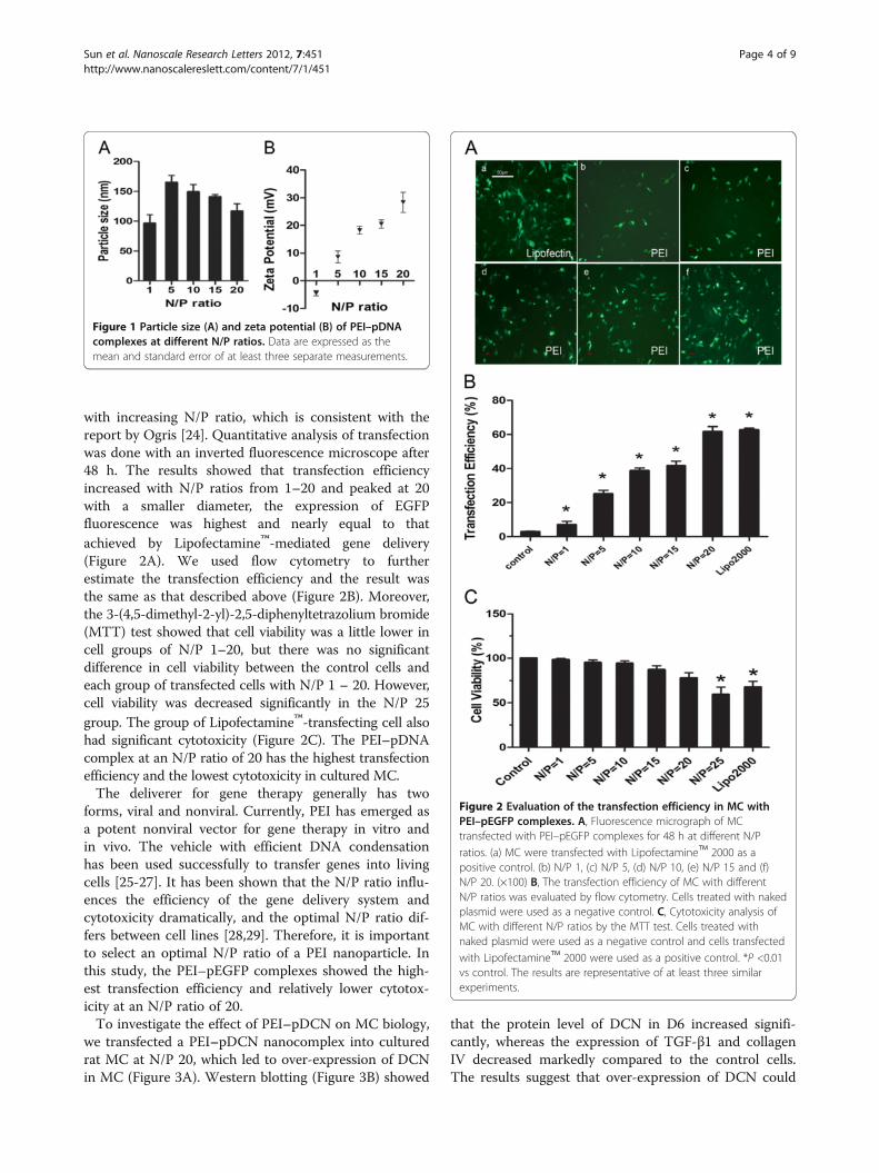
with increasing N/P ratio, which is consistent with thereport by Ogris [24]. Quantitative analysis of transfectionwas done with an inverted fluorescence microscope after48 h. The results showed that transfection efficiencyincreased with N/P ratios from 1–20 and peaked at 20with a smaller diameter, the expression of EGFPfluorescence was highest and nearly equal to thatachieved by Lipofectamine™-mediated gene delivery(Figure 2A). We used flow cytometry to furtherestimate the transfection efficiency and the result wasthe same as that described above (Figure 2B). Moreover,the 3-(4,5-dimethyl-2-yl)-2,5-diphenyltetrazolium bromide(MTT) test showed that cell viability was a little lower incell groups of N/P 1–20, but there was no significantdifference in cell viability between the control cells andeach group of transfected cells with N/P 1 – 20. However,cell viability was decreased significantly in the N/P 25group. The group of Lipofectamine™-transfecting cell alsohad significant cytotoxicity (Figure 2C). The PEI–pDNAcomplex at an N/P ratio of 20 has the highest transfectionefficiency and the lowest cytotoxicity in cultured MC.The deliverer for gene therapy generally has two
forms, viral and nonviral. Currently, PEI has emerged asa potent nonviral vector for gene therapy in vitro andin vivo. The vehicle with efficient DNA condensationhas been used successfully to transfer genes into livingcells [25-27]. It has been shown that the N/P ratio influ-ences the efficiency of the gene delivery system andcytotoxicity dramatically, and the optimal N/P ratio dif-fers between cell lines [28,29]. Therefore, it is importantto select an optimal N/P ratio of a PEI nanoparticle. Inthis study, the PEI–pEGFP complexes showed the high-est transfection efficiency and relatively lower cytotox-icity at an N/P ratio of 20.To investigate the effect of PEI–pDCN on MC biology,
we transfected a PEI–pDCN nanocomplex into culturedrat MC at N/P 20, which led to over-expression of DCNin MC (Figure 3A). Western blotting (Figure 3B) showed
that the protein level of DCN in D6 increased signifi-cantly, whereas the expression of TGF-β1 and collagenIV decreased markedly compared to the control cells.The results suggest that over-expression of DCN could
Figure 1 Particle size (A) and zeta potential (B) of PEI–pDNAcomplexes at different N/P ratios. Data are expressed as themean and standard error of at least three separate measurements.
Figure 2 Evaluation of the transfection efficiency in MC withPEI–pEGFP complexes. A, Fluorescence micrograph of MCtransfected with PEI–pEGFP complexes for 48 h at different N/P
ratios. (a) MC were transfected with Lipofectamine™ 2000 as apositive control. (b) N/P 1, (c) N/P 5, (d) N/P 10, (e) N/P 15 and (f)N/P 20. (×100) B, The transfection efficiency of MC with differentN/P ratios was evaluated by flow cytometry. Cells treated with nakedplasmid were used as a negative control. C, Cytotoxicity analysis ofMC with different N/P ratios by the MTT test. Cells treated withnaked plasmid were used as a negative control and cells transfected
with Lipofectamine™ 2000 were used as a positive control. *P <0.01vs control. The results are representative of at least three similarexperiments.
Sun et al. Nanoscale Research Letters 2012, 7:451 Page 4 of 9http://www.nanoscalereslett.com/content/7/1/451
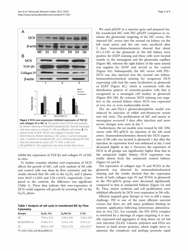
inhibit the expression of TGF-β1 and collagen IV of MCin vitro.To further examine whether over-expression of DCN
affects the growth of MC, cell cycle analysis of D6 cellsand control cells was done by flow cytometry and theresults showed that D6 cells in the G0/G1 and S phaseswere 84.47 ± 1.05% and 5.26 ± 0.51%, respectively. Com-pared to the controls, the difference was significant(Table 1). These data indicate that over-expression ofDCN could suppress cell growth by arresting MC in theG0/G1 phase.
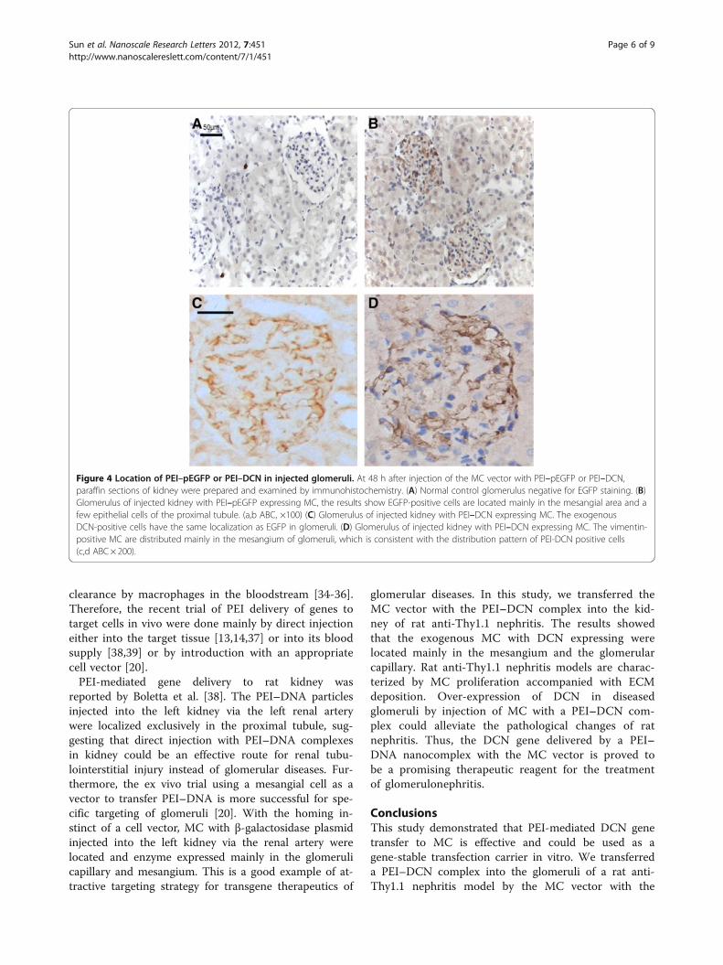
We used pEGFP as a reporter gene and prepared sta-bly transfected MC with PEI–pEGFP complexes to es-timate the glomerular targeting of the MC vector. Weinjected MC vector into the normal rat kidney via theleft renal artery and the rats were sacrificed after2 days. Immunohistochemistry showed that about65 ± 2.13% of the glomeruli in the left kidney werepositive for EGFP staining and these cells were locatedmainly in the mesangium and the glomerular capillary(Figure 4B), whereas the right kidney of the same animalwas negative for EGFP and served as the control(Figure 4A). Subsequently, the MC vector with PEI–DCN was also injected into the normal rats kidney.Immunohistochemical staining for exogenous DCNexpressing cells had the same localization in glomerulias EGFP (Figure 4C), which is consistent with thedistribution pattern of vimentin-positive cells that isrecognized as a mesangial cell marker in glomeruli(Figure 4D) [30]. By contrast, DCN staining was nega-tive in the normal kidney where DCN was expressedat very low or even undetectable levels.The rat anti-Thy1.1 glomerulonephritis model was
induced by injection of rabbit anti-thymocyte seruminto tail veins. The proliferation of MC and matrix inmesangium occurred 3 days after injection and moresevere changes were seen at day 7.Furthermore, the rat models were treated by MC (D6)
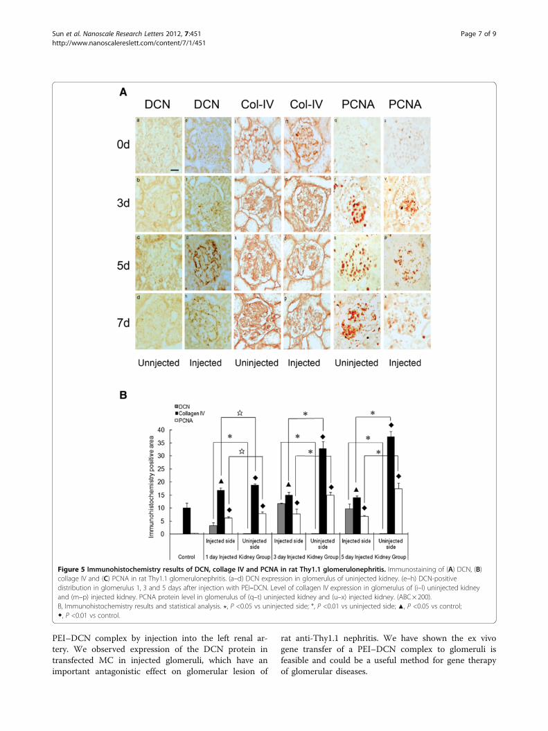
vector with PEI–pDCN via injection of the left renalartery. Immunohistochemistry showed the DCN expres-sion of D6 cells was located in glomeruli 1 day after theinjection; its expression level was enhanced at day 3 anddecreased slightly at day 5. However, the expression ofDCN in all groups was significantly higher than that inthe uninjected (right) kidney; DCN expression wastotally absent from the uninjected control kidneys(Figure 5A and B).The expression of collagen type IV and PCNA in the
glomeruli was detected by immunohistochemicalstaining and the results showed that the expressionlevels of both collagen type IV and PCNA in glomeruliin the PEI–pDCN group were decreased significantlycompared to that in uninjected kidneys (Figure 5A andB). Thus, matrix synthesis and cell proliferation wereinhibited effectively by DCN over-expression in D6 cells.Efficient targeted gene therapy in vivo is still a major
challenge. PEI is one of the most efficient nonviralvectors but there are still many problems limiting itssystemic application following intravenous administra-tion in vivo [31]. For example, the PEI–DNA complexis restricted by a shortage of organ targeting; it is usu-ally expressed and aggregates in lung tissue via rat tailvein injection [32,33]. Cationic polymers and DNA areknown to bind serum proteins, which might serve toopsonize the complexes and perhaps promote rapid
Figure 3 DCN over-expression inhibited expression of TGF-β1and collagen IV in MC. A, The protein level of DCN was assessedby Western blotting in PEI–pDCN transfected MC. Normal mesangialcells were used as a control. D1–D6 are different cell clones. B, Therelative levels of DCN, TGF-β1 and collagen IV protein weredetermined by Western blotting in D6. Lanes 1, normal mesangialcells; 2, pcDNA3.1A-transfected MC; and 3, DCN-transfected MC (D6).Data are presented as mean± SD. *P <0.01 vs MC andMC/pcDNA3.1A. The experiment was done at least three times.
Table 1 Analysis of cell cycle in transfected MC by FlowCytometry
Groups G0/G1 (%) G2/M (%) S (%)
MC 74.61 ± 3.76 12.06 ± 0.92 13.32 ± 2.03
MC/pcDNA3.1A 75.48 ± 4.17 12.14 ± 1.43 12.37 ± 2.1
D6 84.47 ± 1.05* 10.53 ± 2.79 5.26 ± 0.51*
*P < 0.05 vs MCs and MC/pcDNA3.1A.
Sun et al. Nanoscale Research Letters 2012, 7:451 Page 5 of 9http://www.nanoscalereslett.com/content/7/1/451
clearance by macrophages in the bloodstream [34-36].Therefore, the recent trial of PEI delivery of genes totarget cells in vivo were done mainly by direct injectioneither into the target tissue [13,14,37] or into its bloodsupply [38,39] or by introduction with an appropriatecell vector [20].PEI-mediated gene delivery to rat kidney was
reported by Boletta et al. [38]. The PEI–DNA particlesinjected into the left kidney via the left renal arterywere localized exclusively in the proximal tubule, sug-gesting that direct injection with PEI–DNA complexesin kidney could be an effective route for renal tubu-lointerstitial injury instead of glomerular diseases. Fur-thermore, the ex vivo trial using a mesangial cell as avector to transfer PEI–DNA is more successful for spe-cific targeting of glomeruli [20]. With the homing in-stinct of a cell vector, MC with β-galactosidase plasmidinjected into the left kidney via the renal artery werelocated and enzyme expressed mainly in the glomerulicapillary and mesangium. This is a good example of at-tractive targeting strategy for transgene therapeutics of
glomerular diseases. In this study, we transferred theMC vector with the PEI–DCN complex into the kid-ney of rat anti-Thy1.1 nephritis. The results showedthat the exogenous MC with DCN expressing werelocated mainly in the mesangium and the glomerularcapillary. Rat anti-Thy1.1 nephritis models are charac-terized by MC proliferation accompanied with ECMdeposition. Over-expression of DCN in diseasedglomeruli by injection of MC with a PEI–DCN com-plex could alleviate the pathological changes of ratnephritis. Thus, the DCN gene delivered by a PEI–DNA nanocomplex with the MC vector is proved tobe a promising therapeutic reagent for the treatmentof glomerulonephritis.
ConclusionsThis study demonstrated that PEI-mediated DCN genetransfer to MC is effective and could be used as agene-stable transfection carrier in vitro. We transferreda PEI–DCN complex into the glomeruli of a rat anti-Thy1.1 nephritis model by the MC vector with the
Figure 4 Location of PEI–pEGFP or PEI–DCN in injected glomeruli. At 48 h after injection of the MC vector with PEI–pEGFP or PEI–DCN,paraffin sections of kidney were prepared and examined by immunohistochemistry. (A) Normal control glomerulus negative for EGFP staining. (B)Glomerulus of injected kidney with PEI–pEGFP expressing MC, the results show EGFP-positive cells are located mainly in the mesangial area and afew epithelial cells of the proximal tubule. (a,b ABC, ×100) (C) Glomerulus of injected kidney with PEI–DCN expressing MC. The exogenousDCN-positive cells have the same localization as EGFP in glomeruli. (D) Glomerulus of injected kidney with PEI–DCN expressing MC. The vimentin-positive MC are distributed mainly in the mesangium of glomeruli, which is consistent with the distribution pattern of PEI-DCN positive cells(c,d ABC× 200).
Sun et al. Nanoscale Research Letters 2012, 7:451 Page 6 of 9http://www.nanoscalereslett.com/content/7/1/451
PEI–DCN complex by injection into the left renal ar-tery. We observed expression of the DCN protein intransfected MC in injected glomeruli, which have animportant antagonistic effect on glomerular lesion of
rat anti-Thy1.1 nephritis. We have shown the ex vivogene transfer of a PEI–DCN complex to glomeruli isfeasible and could be a useful method for gene therapyof glomerular diseases.
Figure 5 Immunohistochemistry results of DCN, collage IV and PCNA in rat Thy1.1 glomerulonephritis. Immunostaining of (A) DCN, (B)collage IV and (C) PCNA in rat Thy1.1 glomerulonephritis. (a–d) DCN expression in glomerulus of uninjected kidney. (e–h) DCN-positivedistribution in glomerulus 1, 3 and 5 days after injection with PEI–DCN. Level of collagen IV expression in glomerulus of (i–l) uninjected kidneyand (m–p) injected kidney. PCNA protein level in glomerulus of (q–t) uninjected kidney and (u–x) injected kidney. (ABC× 200).B, Immunohistochemistry results and statistical analysis. ?, P <0.05 vs uninjected side; *, P <0.01 vs uninjected side; ▲, P <0.05 vs control;◆, P <0.01 vs control.
Sun et al. Nanoscale Research Letters 2012, 7:451 Page 7 of 9http://www.nanoscalereslett.com/content/7/1/451
AbbreviationsPEI: polyethylenimine; DCN: decorin; MC: mesangial cell; pDNA: plasmid DNA;HVJ: hemagglutinating virus of Japan; PBS: phosphate-buffered saline;GFP: green fluorescent protein; PMSF: phenylmethylsulfonyl fluoride;ATS: anti-rat thymocyte serum.
Competing interestsThe authors declare that they have no competing interest.
Authors’ contributionsJYS performed the experiments and drafted the manuscript. YS, HJW andHXZ carried out the partial experiments and participated in the mechanismanalysis. ZHZ and QC helped in the technical support for the experiments.ZGZ designed the experiments and revised the manuscript. All authors readand approved the final manuscript.
AcknowledgementsThis project was supported by a grant from the National Natural ScienceFoundation of China (NSFC: 30570859). We thank Professor Muyi Guo andDr Xiurong Zhang for advice and help with techniques, and Mr HuakangCao for his kind instructions for animal experiments.
Received: 20 March 2012 Accepted: 9 July 2012Published: 9 August 2012
References1. Merdan T, Kopecek J, Kissel T: Prospects for cationic polymers in gene and
oligonucleotide therapy against cancer. Adv Drug Deliv Rev 2002,54:715–758.
2. Song S, Goudy K, Campbell-Thompson M, Wasserfall C, Scott-Jorgensen M,Wang J, Tang Q, Crawford JM, Ellis TM, Atkinson MA, Flotte TR:Recombinant adeno-associated virusmediated alpha-1 antitrypsin genetherapy prevents type I diabetes in NOD mice. Gene Ther 2004,11:181–186.
3. Olefsky JM: Diabetes: Gene therapy for rats and mice. Nature 2000,408:420–421.
4. Lungwitz U, Breunig M, Blunk T, Göpferich A: Polyethylenimine-based non-viral gene delivery systems. Eur J Pharm Biopharm 2005, 60:247–266.
5. Lemkine GF, Demeneix BA: Polyethylenimines for in vivo gene delivery.Curr Opin Mol Ther 2001, 3:178–182.
6. Tang MX, Szoka FC: The influence of polymer structure on theinteractions of cationic polymers with DNA and morphology of theresulting complexes. Gene Ther 1997, 4:823–832.
7. Intra J, Salem AK: Characterization of the transgene expression generatedby branched and linear polyethylenimine-plasmid DNA nanoparticlesin vitro and after intraperitoneal injection in vivo. J Control Release 2008,130:129–138.
8. Zhang XQ, Intra J, Salem AK: Comparative study of poly (lactic-co-glycolicacid)-polyethyleneimine-plasmid DNA microparticles prepared usingdouble emulsion methods. J Microencapsul 2008, 25:1–12.
9. Fischer D, Bieber T, Li Y, Elsässer HP, Kissel T: A novel non-viral vector forDNA delivery based on low molecular weight, branchedpolyethylenimine: effect of molecular weight on transfection efficiencyand cytotoxicity. Pharm Res 1999, 16:1273–1279.
10. Huh SH, Do HJ, Lim HY, Kim DK, Choi SJ, Song H, Kim NH, Park JK, ChangWK, Chung HM, Kim JH: Optimization of 25 kDa linear polyethyleniminefor efficient gene deliver. Biologicals 2007, 35:165–171.
11. Kichler A: Gene transfer with modified polyethylenimines. J Gene Med2004, 6(Suppl 1):S3–S10.
12. Morille M, Passirani C, Vonarbourg A, Clavreul A, Benoit JP: Progress indeveloping cationic vectors for non-viral systemic gene therapy againstcancer. Biomaterials 2008, 29:3477–3496.
13. Aoki K, Furuhata S, Hatanaka K, Maeda M, Remy JS, Behr JP, Terada M,Yoshida T: Polyethylenimine-mediated gene transfer into pancreatictumor dissemination in the murine peritoneal cavity. Gene Ther 2001,8:508–514.
14. Louis MH, Dutoit S, Denoux Y, Erbacher P, Deslandes E, Behr JP, Gauduchon P,Poulain L: Intraperitoneal linear polyethylenimine (L-PEI)-mediatedgene delivery to ovarian carcinoma nodes in mice. Cancer Gene Ther2006, 13:367–374.
15. Di Gioia S, Conese M: Polyethylenimine-mediated gene delivery to thelung and therapeutic applications. Drug Des Devel Ther 2009, 2:163–188.
16. Gautam A, Densmore CL, Waldrep JC: Inhibition of experimental lungmetastasis by aerosol delivery of PEI-p53 complexes. Mol Ther 2000,2:318–323.
17. Seong JH, Lee KM, Kim ST, Jin SE, Kim CK: Polyethylenimine-basedantisense oligodeoxynucleotides of IL-4 suppress the production of IL-4in a murine model of airway inflammation. J Gene Med 2006, 8:314–323.
18. Wu H, Wang S, Xue A, Liu Y, Liu Y, Wang H, Chen Q, Guo M, Zhang Z:Overexpression of decorin induces apoptosis and cell growth arrest incultured rat mesangial cells in vitro. Nephrology 2008, 13:607–615.
19. Isaka Y, Brees DK, Ikegaya K, Kaneda Y, Imai E, Noble NA, Border WA: Genetherapy by skeletal muscle expression of decorin prevents fibroticdisease in rat kidney. Nat Med 1996, 2:418–423.
20. Kim HJ, Kim SI, Yun IJ, Kwak JH, Yu SH: Gene delivery into rat glomerulususing a mesangial cell vector. Mol Cells 2000, 10:662–668.
21. Zhang M, Guo MY, Chen Q, Jin HM: The culture of rat glomerularmesangial cells. Journal of Shanghai Medical University 1995, 22:207–209. inChinese.
22. Wang H, Zhang Z, Liu X, Chen G, Chen Qi-Guo M: Influence on the Growthand Changes of Several Phenotypes of Cultured Rat MsC Transfected byDCN Gene. Journal of Shanghai Medical University 2003, 30:414–417. inChinese.
23. Chen G, Guo M, Zhang Y, Zhang J, Zhang X, Chen Q: Preparation of anti-thy1 serum and establishment of rat mesangioproliferativeglomerulonephritis model. Chinese Journal of Clinical and experimentalPathology 1996, 12:241–245. in Chinese.
24. Ogris M, Steinlein P, Kursa M, Mechtler K, Kircheis R, Wagner E: The size ofDNA/transferrin-PEI complexes is an important factor for geneexpression in cultured cells. Gene Ther 1998, 5:1425–1433.
25. Boussif O, Lezoualc’h F, Zanta MA, Mergny MD, Scherman D, Demeneix B,Behr JP: A versatile vector for gene and oligonucleotide transfer intocells in culture and in vivo: polyethylenimine. Proc Natl Acad Sci 1995,92:7297–7301.
26. Aoki K, Furuhata S, Hatanaka K, Maeda M, Remy JS, Behr JP, Terada M,Yoshida T: Polyethylenimine-mediated gene transfer into pancreatictumor dissemination in the murine peritoneal cavity. Gene Ther 2001,8:508–514.
27. Nimesh S, Chandra R: Polyethylenimine nanoparticles as an efficientin vitro siRNA delivery system. Eur J Pharm Biopharm 2009, 73:43–49.
28. Godbey WT, Wu KK, Mikos AG: Poly(ethylenimine)-mediated gene deliveryaffects endothelial cell function and viability. Biomaterials 2001,22:471–480.
29. Gharwan H, Wightman L, Kircheis R, Wagner E, Zatloukal K: Nonviral genetransfer into fetal mouse livers (a comparison betneen cationic polymerPEI and naked DNA). Gene Ther 2003, 10:810–817.
30. Yuasa T, Izawa T, Kuwamura M, Yamate J: Thy-1 Expressing MesenchymalCells in Rat Nephrogenesis in Correlation with Cells Immunoreactive forα-Smooth Muscle Actin and Vimentin. J Toxicol Pathol 2010, 23:1–10.
31. Kircheis R, Blessing T, Brunner S, Wightman L, Wagner E: Tumor targetingwith surface-shielded ligand–polycation DNA complexes. J Control Release2001, 72:165–170.
32. Bragonzi A, Boletta A, Biffi A, Muggia A, Sersale G, Cheng SH, Bordignon C,Assael BM, Conese M: Comparison between cationic polymers and lipidsin mediating systemic gene delivery to the lungs. Gene Ther 1999,6:1995–2004.
33. Rudolph C, Lausier J, Naundorf S, Müller RH, Rosenecker J: In vivo genedelivery to the lung using polyethylenimine and fracturedpolyamidoamine dendrimers. J Gene Med 2000, 2:269–278.
34. Ogris M, Brunner S, Schuller S, Kircheis R, Wagner E: PEGylated DNA/transferring PEI complexes: reduced interaction with blood components,extended circulation in blood and potential for systemic gene delivery.Gene Ther 1999, 6:595–605.
35. Dash PR, Read ML, Fisher KD, Howard KA, Wolfert M, Oupicky D, Subr V,Strohalm J, Ulbrich K, Seymour LW: Decreased binding to proteins andcells of polymeric gene delivery vectors surface modified with amultivalent hydrophilic polymer and retargeting through attachment oftransferrin. J Biol Chem 2000, 275:3793–3802.
36. Kircheis R, Wightman L, Wagner E: Design and gene delivery activity ofmodified polyethylenimines. Adv Drug Deliv Rev 2001,53:341–358.
Sun et al. Nanoscale Research Letters 2012, 7:451 Page 8 of 9http://www.nanoscalereslett.com/content/7/1/451
37. Chen G, Zhang X, Yang F, Mu L: Disposition of nanoparticle-baseddelivery system via inner ear administration. Curr Drug Metab 2010,11:886–897.
38. Boletta A, Benigni A, Lutz J, Remuzzi G, Soria MR, Monaco L: Nonvival genedelivery to the rat kidney with polyethylenimine. Hum Gene Ther 1997,8:1243–1251.
39. Liu M, Li ZH, Xu FJ, Lai LH, Wang QQ, Tang GP, Yang WT: An oligopeptideligand-mediated therapeutic gene nanocomplex for liver cancer-targeted therapy. Biomaterials 2012, 33:2240–2250.
doi:10.1186/1556-276X-7-451Cite this article as: Sun et al.: Transgene therapy for rat anti-Thy1.1glomerulonephritis via mesangial cell vector with a polyethylenimine/decorin nanocomplex. Nanoscale Research Letters 2012 7:451.
Submit your manuscript to a journal and benefi t from:
7 Convenient online submission
7 Rigorous peer review
7 Immediate publication on acceptance
7 Open access: articles freely available online
7 High visibility within the fi eld
7 Retaining the copyright to your article
Submit your next manuscript at 7 springeropen.com
Sun et al. Nanoscale Research Letters 2012, 7:451 Page 9 of 9http://www.nanoscalereslett.com/content/7/1/451