transient expression of cc chemokine teck in the ovary during ovulation: its potential role in...
TRANSCRIPT
Transient Expression of CC ChemokineTECK in the Ovary during Ovulation:Its Potential Role in Ovulation
INTRODUCTION
It has long been speculated that several physiologicalprocesses of normal ovaries, such as ovulation, mayresemble the inflammatory process.1,2 The presence ofdifferent subsets of leukocytes at different phases ofthe ovarian life cycle in various animal species has beenwell documented.3–6 A growing body of evidence
suggests that ovulation and the post-ovulatory proces-ses may indeed constitute a local inflammatory reac-tion. It has been shown that the large influx ofleukocytes, including macrophages and neutrophils,into the ovary during the ovulatory process appearedto be a response to the luteinizing hormone (LH)surge.7 An equally important influx of monocytes intothe corpus luteum characterizes the late post-ovulatory
American Journal of Reproductive ImmunologyAJRI 2005; 53: 238–248Copyright � Blackwell Munksgaard, 2005
Zhou C, Wu J, Borillo J, Torres L, McMahon J, Bao Y, Lou Y-H.Transient expression of CC chemokine TECK in the ovary duringovulation: its potential role in ovulation. AJRI 2005; 53:238–248� Blackwell Munksgaard, 2005
PROBLEM: Chemokine thymus-expressed chemokine (TECK), whichis expressed exclusively in the thymus and small intestine, plays acritical role in T-cell development. Our previous study revealed itsexpression in the ovary also. This study investigated its ovarianexpression during ovulatory process.METHOD OF STUDY: Super-ovulation was induced in young femaleCD1 mice by equine chorionic gonadotropin (eCG) and humanchorionic gonadotropic (hCG). Ovarian TECK expression duringovulation was determined by: (1) reverse transcriptase-polymerasechain reaction (RT-PCR) at mRNA level, (2) Western blot andimmunohistology at the protein level, and (3) leukocyte infiltrationassay at the bioactive level.RESULTS: A transient, high-level expression of TECK in murineovaries at the mRNA level during hCG-induced ovulation wasdetected. Sequencing of directly cloned PCR product confirmed theovarian expression of TECK. The peak expression of TECK wasobserved at 10–12 hr post-hCG injection; real-time PCR revealed an800-fold increase during its expression peak over 0 hr. The expressedovarian TECK protein was readily detectable by Western blot.Immunohistochemistry localized TECK expression to the ovarianinterstitial tissue surrounding, or in the theca layer of the maturefollicles undergoing ovulatory process. Expression of TECK receptor,the CC chemokine receptor (CCR9) was also detected in the ovulatingovaries. Using in vitro leukocyte infiltration assay, we firstdemonstrated that ovaries undergoing the ovulatory process were ableto selectively chemoattract mononuclear cells. Importantly,neutralization of TECK by the antibody resulted in a 85% reduction inthe chemotactic activities of the ovaries.CONCLUSION: This study suggested that ovarian expression ofTECK is under a tight hormonal regulation, and expressed TECK maybe responsible for recruitment of mononuclear cells into the ovary toparticipate in the ovulatory process.
Cindy Zhou1, Jean Wu1, JasonBorillo1, Lisa Torres1, JohnMcMahon2, Yongde Bao3, Ya-HuanLou1
Departments of 1Diagnostic Sciences, Dental Branch, and2Integrative Biology and Pharmacology, School ofMedicine, University of Texas Health Science Center atHouston, Houston, TX, USA; 3Department of Microbiology,School of Medicine, University of Virginia, Charlottesville,VA, USA
Key words: Chemokines, ovary, superovulation, TECK
Address reprint request to Dr Ya-Huan Lou, Department ofDiagnostic Sciences, Dental Branch, University of TexasHealth Science Center at Houston, Houston, TX 77030,USA.E-mail: [email protected]
Submitted November 1, 2004;revised January 13, 2005;accepted January 21, 2005.
� BLACKWELL MUNKSGAARD, 2005
phase.8 Thus, it is likely that leukocytes and theirassociated cytokines may be required for ovulationand/or post-ovulation functions through an inflamma-tion-like process.1,2 As a key step in the inflammatoryprocess, certain groups of leukocytes must be recruitedby locally released chemokines. Hence, it is reasonableto hypothesize that leukocytes must be attracted awayfrom circulation and into the ovary, or migrate fromone location to another presumably by way of local,possibly intra-ovarian, chemoattractants.
Chemokines, which are the most likely candidatesfor local chemoattractants during ovulation, constitutea family of structurally related, small, inducible,secretary proinflammatory proteins involved in avariety of immune responses especially as chemoat-tractants and activators of specific types of leukocytes.9
Approximately 40 chemokines are known, which aregrouped into four subfamilies based on the numberand position of cysteine residues at their C-terminus.CC and CXC chemokines are two major groups.Generally speaking, CC chemokines attract mononu-clears, while CXC show chemotactic activity towardpolymorphonuclears. Each subfamily of chemokineshas its distinct receptors.10 Each receptor may serve asa ligand for multiple chemokines, usually from thesame subfamily.
Several chemokines have been implicated in ovula-tion, corpus luteum function, and even in folliculardevelopment.11–17 The CXC chemokine interleukin(IL)-8 is probably the best-described chemokine forits involvement in ovulation.12–15 A recent study hasalso shown the elevated expression of several CCchemokines, which included monocyte chemotacticprotein (MCP)-1 and MCP-3, during the ovulatoryprocess at the mRNA level. This suggests a potentialrole of those chemokines in the ovulatory process.16,17
However, constitutive expression of those chemokineshas also been observed and thus, they might beinvolved in ovarian functions other than ovula-tion.18–20 It is clear that chemokines are involved inseveral ovarian functions.
We first examined ovarian expression of more than20 chemokines.21 Unique expression pattern of a CCchemokine thymus-expressed chemokine (TECK orCCL25) led to a more detailed investigation of thischemokine. TECK is a relatively new member of theCC chemokine family.22 TECK, which plays a criticalrole in T-cell development by directing trafficking ofpre-T cells into primary lymphoid organs, is reportedto express exclusively in the thymus and small intes-tine.22–24 We report in the present study a transient,high level of TECK expression and expression of itsreceptor CCR9 in the ovaries during hCG-inducedovulatory process at both mRNA and protein levels.More importantly, the expressed ovarian TECK was
biologically active, and responsible for 85% of totalchemotactic activity of the ovaries for mononuclearcells. Thus, our study suggests that TECK, in additionto several other candidate chemokines, may play a rolein ovulation by directing leukocytes into ovariantissue.
MATERIALS AND METHODS
Superovulation Induction and Mouse Ovary SamplingYoung female CD1 mice (6–8 weeks old) were pur-chased from Harlan (Indianapolis, IN, USA). Themice were maintained in the animal facility at theUniversity of Texas Health Science Center at Houston,and allowed to acclimate for a minimum of 1 week. Awell-established method was used for induction ofsuper-ovulation in young females. Briefly, the animalswere injected with pregnant mare’s serum gonadotro-pin (eCG; Sigma, St Louis, MO, USA) at 5 IU/mouseintraperitoneally (i.p.). The mice were injected i.p. withhCG (human chorionic gonadotropin, 5 IU/mouse;Sigma) after 48 hr. The mice without the secondinjection of hCG were used as a control. Anothergroup of mice received two phosphate-buffered saline(PBS) injections and served as an additional control.Ovaries were collected at designated time points forvarious purposes. At the same time, Fallopian tubeswere removed and examined under a dissectingmicroscope for the presence of ovulated eggs todetermine the time of ovulation. Ovaries were pro-cessed for total RNA extraction or for Western blots.In some cases, ovaries were fixed in 4% paraformal-dehyde for immunohistochemistry.
RT-PCR Detection of TECK and CCR9Total RNA was isolated from the ovaries at designatedtime points, using a commercial kit (Ambion, Austin,TX, USA), and cDNA was synthesized using 1 lg oftotal RNA through an RT reaction (RNA-PCR CoreKit; Applied Biosystems, Foster City, CA, USA). PCRwas carried out to detect TECK mRNA using a pair ofprimers (5¢-TGGAATGTTCTCCGGCATGCTAGG-3¢, 5¢-TGGCACTGGCATGCCTAGAAGACG-3¢),which resulted in a 396-bp product. PCR was carriedout under the following conditions: pre-heating at94�C for 3 min followed by 35 cycles of PCR (94�C1 min, 69�C 30 s, 72�C 1 min)(GeneAmp9700, AppliedBiosystems). For CCR9, a pair of primers (5¢-AAGAACCTGGGATGCATTAGCCAGGC-3¢, 5¢-CCAGATCTGAAGTAACAGAAACTGGG-3¢) wasused, which resulted in a 475-bp product. The productswere separated by electrophoresis in 1.5% agarose gel,stained with ethidium-bromide, and visualized underUV light illumination. RNA isolated from thymus,
OVARIAN EXPRESSION OF CHEMOKINE TECK / 239
AMERICAN JOURNAL OF REPRODUCTIVE IMMUNOLOGY VOL. 53, 2005
peripheral blood leukocytes (PBL), or other organswere used as controls. A housekeeper gene HPRT wasused as a control.25 In order to rule out genomic DNAcontamination in the samples, absence of PCR productin RNA samples without reverse transcriptase (i.e.under identical RT conditions without reverse tran-scriptase) was confirmed. This group was referred to asRT).Selected samples were also used for real-time PCR
based on the identical conditions described above.Briefly, after RT reaction, cDNA concentrations weredetermined by Microplate Spectrophotometer (SPEC-TRAmax; Molecular Devices, Sunnyvale, CA, USA).Real-time PCR were carried out at two concentrationsof the cDNA (1.5 ng/50 lL and 0.75 ng/50 lL) induplicate in iCycler iQ real-Time PCR detection systemusing iQ SYBR Green Supermix (Bio-Rad, Hercules,CA, USA). Housekeeper gene HPRT was used as thestandard.25 Relative quantification of TECK expres-sion was carried based on the threshold cycle of the twogenes following the 2�DDC
T method,26 where
DDCT ¼ ðCT:TECK � CT:HPRTÞtime x
� ðCT:TECK � CT:HPRTÞtime 0:
Cloning and Sequencing of TECK cDNA,and Expression of Recombinant Mouse TECKAn identical protocol was used for RT-PCR amplifi-cation of ovarian TECK cDNA with a high-fidelityDNA polymerase (Platinum pfx; Invitrogen, Carlsbad,CA, USA). PCR product of ovarian TECK waspurified on an agarose gel after electrophoresis, anddirectly cloned into pCR4TOPO-TA vector (Invitro-gen). Multiple independent clones of recombinantvectors were chosen and sequenced by an automatedDNA sequencer in a core facility at the University ofVirginia. The DNA sequence was compared withpublished mouse TECK cDNA sequence.22
Recombinant TECK (rTECK) was expressed, andused as an antigen. Briefly, cDNA encoding the open-reading frame of mouse TECK [excluding 23 aminoacid (a.a.) signal sequence] was generated by RT-PCRof total ovarian RNA using a pair of primers (5¢-TGGAATGTTCTCCGGCATGCTAGG-3¢, 5¢-TGGCACTGGCATGCCTAGAAGACG-3¢) with a high-fidelity DNA polymerase (Platinum pfx). The PCRproduct, which was 375 bp, was directly cloned intoexpression vector pCR/T7/CT/TOPO (Invitrogen),and the sequence was verified by an automatedsequencer. Non-fusion rTECK (121 a.a.) wasexpressed under T7 promoter by DE3 Escherichiacoli. Although rTECK should have a calculatedmolecular mass of 13.1 kDa, it migrated to 12.5 kDaon SDS-PAGE, which has also been reported by others(technical sheet for recombinant TECK from R&D,
refer to Fig. 6A, B). The rTECK in insoluble form waspurified by preparative sodium dodecyl sulfate-poly-acrylamide gel electrophoresis (SDS-PAGE) (15%)following a previously established method.27 Thepurified rTECK (2 mg/mL) was mixed with completeFreund adjuvant (CFA), and subcutaneously injectedinto one hind footpad and the base of the tail(30 lL/site) of mice. The mice were boosted oncewith rTECK in incomplete Freund adjuvant (IFA)3 weeks later. Blood was sampled from the tail vein ofimmunized mice, and sera were used for detectingcirculating antibody to rTECK by Western blot aspreviously described.27 The antisera with high titer(>64 000) were selected, stored at )80�C, and used forexperiments as indicated.
Leukocyte Infiltration AssayChemotactic activity of ovaries undergoing the ovula-tory process for PBL was measured by a leukocyteinfiltration assay using a B-D BioCoat Control CellCulture Inserts system (pore size 3 lm; BD, San Diego,CA, USA) with some modifications. Briefly, theovaries collected from mice at 8 hr post-hCG injectionwere teased open as a chemoattractant source andplaced at the lower chamber in a DMEM-basedmedium supplemented with 10% fetal calf serum,2 mm l-glutamine, non-essential and essential aminoacids, sodium pyruvate, 100 U/mL penicillin,100 lg/mL streptomycin (BioWhittaker, Walkersville,MD, USA), 5 lm b-mercaptoethanol, and 25% of thesera from the same donors. The medium with ran-domly sampled ovaries was used as a negative control.The ovary donor was bled through the axillary arteryunder anesthesia before removal of ovaries. The PBLswere isolated with standardized Ficoll gradient cen-trifugation following manufacturer’s instruction (Hist-opaque 1.119, Sigma). PBLs were suspended in aserum-free DMEM medium (see above), and addedinto insert wells (upper chamber) at 2 · 106 cells/wellin 500 lL. In some cases, the ovaries were pre-incubated with anti-TECK antisera (1:10, see above)or the sera from CFA-immunized mice before place-ment in the lower chamber. The assembled sets wereincubated at 37�C, 5% CO2, for 8 hr, and the cells inthe insert wells were gently and thoroughly washedaway with cold PBS. After fixation in acetone/meth-anol (1:1) for 15 min at room temperature, the wellswere stained by H-E. The bottom membranes were cutfrom the insert, dried and mounted on a slide. Theleukocytes, which had migrated through the mem-brane, were counted according to their morphology.
ImmunohistochemistryA well-established method was applied for immuno-peroxidase staining.25 Ovaries, either sampled at 11 hr
240 / ZHOU ET AL.
� BLACKWELL MUNKSGAARD, 2005
post-hCG injection, or from non-ovulating mice, werefixed in a 4% paraformaldehyde solution in PBS for24 hr, washed with cold PBS and immersed in 30%sucrose solution in PBS at 4�C for 3 days. The ovarywas embedded in OCT and 3-lm frozen sections wereprepared. The sections were permeablized with 0.3%of Triton X-100/PBS for 20 min and blocked with 3%bovine serum albumin (BSA)/PBS for 30 min at roomtemperature. The sections were further treated withavidin and biotin to avoid non-specific binding,according to manufacturer’s recommendations(Vector Laboratories, Burlingham, CA, USA). Thetreated sections were incubated overnight at 4�C with abiotin-labeled goat antibody to mouse TECK (1:500 inBSA/PBS; R&D, Minneapolis, MN, USA) or normalgoat IgG (1:500), followed by incubation with perox-idase-conjugated avidin–biotin complex (ABC, VectorLaboratories). Brown color was developed using DABas a substrate and nickel as an enhancer. The sectionwas counterstained by methyl green.
Western Blot Detection of TECKOne ovary, either sampled from an eCG/hCG-treatedor non-ovulating mouse, was immersed in 200 lL ofcold 0.15 m NH4HCO3 buffer containing proteaseinhibitor cocktail (Roche Applied Science, Indianapo-lis, IN, USA) and carefully teased into small pieces.The tissue fragments were incubated on ice for 5 minwith constant stirring and followed by centrifugation(25 800 g, 10 min at 4�C). The process was repeatedtwo more times. The resultant supernatants werepooled and lyophilized. The lyophilized powder wasre-dissolved in 30 lL H2O, and re-lyophilized, whichresulted in a small protein pellet. The protein wasdissolved in 20 lL of 1x SDS loading buffer, heated at90�C for 5 min, and 10 lL was loaded on to 15%PAGE gel. A recombinant mouse TECK with bioac-tivity from R&D was used as a standard. After
electrophoresis, the proteins were transferred onto anitrocellulose membrane. The membrane was blockedwith skim milk, and incubated by the same antibodyused for immunohistochemistry at 4�C overnight.After intensive washing with PBS containing Tween-20, the membrane was further incubated with ABC(see above). Enhanced chemiluminescence (ECL) wasused to visualize the TECK band, and the result wasrecorded on an X-ray film. A recombinant TECK withbiological activity, obtained from R&D was used as apositive control.
RESULTS
Detection of TECK Expression in Normal OvariesWe have previously reported that among 20 chemok-ines tested, which included 17 CC chemokines and twoCXC chemokines, constitutive expression of two CCtypes (MCP-1 and RANTES) and one CXC type(IP-10) were detected at significant levels in randomlysampled normal ovaries, while other chemokinesshowed variable or no expression.21
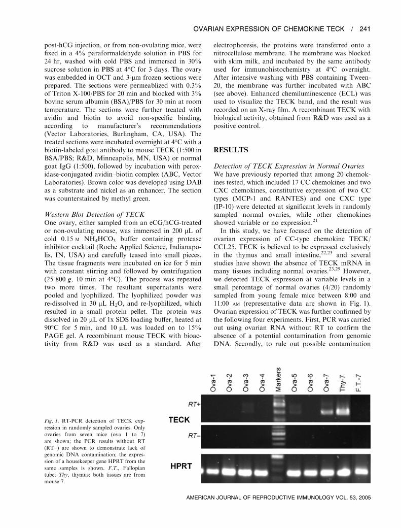
In this study, we have focused on the detection ofovarian expression of CC-type chemokine TECK/CCL25. TECK is believed to be expressed exclusivelyin the thymus and small intestine,22,23 and severalstudies have shown the absence of TECK mRNA inmany tissues including normal ovaries.23,29 However,we detected TECK expression at variable levels in asmall percentage of normal ovaries (4/20) randomlysampled from young female mice between 8:00 and11:00 am (representative data are shown in Fig. 1).Ovarian expression of TECK was further confirmed bythe following four experiments. First, PCR was carriedout using ovarian RNA without RT to confirm theabsence of a potential contamination from genomicDNA. Secondly, to rule out possible contamination
Fig. 1. RT-PCR detection of TECK exp-
ression in randomly sampled ovaries. Only
ovaries from seven mice (ova 1 to 7)
are shown; the PCR results without RT
(RT)) are shown to demonstrate lack of
genomic DNA contamination; the expres-
sion of a housekeeper gene HPRT from the
same samples is shown. F.T., Fallopian
tube; Thy, thymus; both tissues are from
mouse 7.
OVARIAN EXPRESSION OF CHEMOKINE TECK / 241
AMERICAN JOURNAL OF REPRODUCTIVE IMMUNOLOGY VOL. 53, 2005
from other tissues, the Fallopian tube and other fattytissue attached to the ovary were dissected out andtested for TECK expression; no detectable TECK wasobserved (Fig. 1). Thirdly, the PCR products weredirectly cloned into a sequencing vector; sequencing ofmultiple independent clones showed that the cDNAwas identical to that of thymic TECK. Finally, in orderto rule out the possibility that expression of TECKmight originate from PBL which were circulatingthrough ovarian tissue, PBL expression of TECKwas examined by RT-PCR. RNA was isolated from anequivalent number of PBL in an ovary and tested forTECK expression (approximately 5 · 104 cells). Nodetectable TECK expression was observed (data notshown). Based on the results from the above experi-ments, we concluded that a small percentage of normalovaries did express a low level of TECK.
Transient Expression of TECK at High Levels in theOvary during OvulationBased on the observation that TECK was expressedonly in a few mouse ovaries, we hypothesized thatTECK expression might be related to reproductivecycles. We decided to investigate ovarian TECKexpression during ovulation. Superovulation wasinduced in young female mice by injections of eCGand hCG at a 48-hr interval. Ovaries were sampled at1-hr intervals after hCG injection, and the occurrenceof ovulation was determined by isolation of ovulatedeggs from Fallopian tubes. In parallel, the ovaries weresampled from mice that only received eCG injections,or that only received PBS, and were used as controls.The time course for TECK expression post-hCG
injection was established. Identical quantity of RNAfrom each sample was used for RT-PCR. PCR onhousekeeper gene HPRT was first carried out to
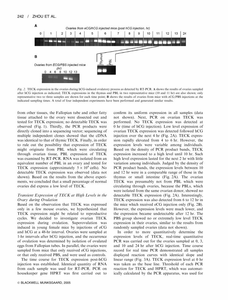
confirm its uniform expression in all samples (datanot shown). Next, PCR on ovarian TECK wasperformed. No TECK expression was detected at0 hr (time of hCG injection). Low level expression ofovarian TECK expression was detected followed hCGinjection over the next 4 hr (Fig. 2A). TECK expres-sion rapidly elevated from 4 to 6 hr. However, theexpression levels were variable among individuals.Based on the density of PCR product bands, TECKexpression increased to a high level until 10 hr. Suchhigh level expression lasted for the next 2 hr with littlevariation among individuals. Judged by the density ofPCR product bands, the expression levels between 10and 12 hr were in a comparable range of those in thethymus or small intestine (Fig. 2A). The ovarianTECK was presumably not from PBL which werecirculating through ovaries, because the PBLs, whichwere isolated from the same ovarian donor, showed nodetectable TECK expression (Fig. 2A). Interestingly,TECK expression was also detected from 6 to 12 hr inthe mice which received eCG injection only (Fig. 2B).However, the expression levels were much lower, andthe expression became undetectable after 12 hr. ThePBS group showed no or extremely low level TECKexpression in their ovaries, similar to the results fromrandomly sampled ovaries (data not shown).
In order to more quantitatively determine theexpression levels of TECK, real-time quantitativePCR was carried out for the ovaries sampled at 0, 3,and 10 and 24 hr after hCG injection. Time courserecord for real time PCR demonstrated all samplesdisplayed reaction curves with identical slope andlinear range (Fig. 3A). TECK expression level at 0 hrwas taken as the base line. Threshold of each PCRreaction for TECK and HPRT, which was automat-ically calculated by the PCR apparatus, was used for
Fig. 2. TECK expression in the ovaries during hCG-induced ovulatory process as detected by RT-PCR. A shows the results of ovaries sampled
after hCG injection as indicated; TECK expressions in the thymus and PBL in two representative mice (10 and 11 hr) are also shown; only
representative two to three samples are shown for each time point; B shows the results of ovaries from mice with eCG/PBS injections at the
indicated sampling times. A total of four independent experiments have been performed and generated similar results.
242 / ZHOU ET AL.
� BLACKWELL MUNKSGAARD, 2005

calculation of the relative expression level. Ovariessampled at 10 hr showed nearly an 800-fold increase inTECK expression when compared with those at 0 hr(Fig. 3B). While the ovaries sampled at 3 hr showedslightly over 10-fold increase, those sampled at 24 hrbelow 10-fold. Although ovarian expression levels at10 hr seem slightly lower than those of the thymus inreal-time PCR testing, statistical analysis (t-test) sug-gested no significant difference between the two groups(P ¼ 0.2). Real-time PCR again demonstrated that theovarian TECK expression level at 10 hr was in therange of those in the thymus (Fig. 3B). Thus, thoseresults from quantitative real time PCR also supportedour previous observation based on conventional PCRthat TECK expressed transiently at a high level afterhCG injection.
Although a transient, high-level expression of TECKduring ovulation was observed at the mRNA level, itwas essential to demonstrate its expression at theprotein level. Three ovaries each were sampled at 0, 6,10 and 12 hr post-hCG injection and used for detec-tion of TECK proteins by Western blot (Fig. 4). Witha detection limit of 0.25 ng TECK/lane, TECK proteinwas undetectable in the ovaries sampled at 0 hr.However, TECK protein was readily detectable inthe ovaries sampled at 6, 10 and 12 hr. It was difficultto determine the amount of TECK in the ovarybecause of the different band pattern of TECKbetween standard and ovarian TECK in Western
blot. It was estimated, by calculation of total densityof the band, approximately between 0.2 to 0.6 ngTECK was present in 1 mg ovarian tissue during itsexpressing peak (12 hr) (Fig. 4). In contrast, TECKprotein was not detected in any ovaries from thecontrol groups including randomly sampled ovaries orovaries without hCG injection. Thus, ovarian expres-sion of TECK during ovulation was detectable at bothmRNA and protein levels.
Tissue Location of TECK Expression duringOvulationImmunoperoxidase staining was used to localizeTECK expression during ovulation in the ovarieswith a commercial goat anti-TECK antibody labeledwith biotin. Ovaries were sampled at 11 hr after hCGinjection, at which time, ovaries appeared to expressthe highest level of TECK. Specific TECK staining wascarefully confirmed by (1) a clear cytoplasmic staining(inset in Fig. 5) and (2) absence of any stainings whenbiotin-labeled normal goat IgG was used. OvarianTECK expression was localized to two tissue sites(Fig. 5). First, the majority of TECK expression cellswere clustered in the interstitial tissue surroundingmature follicles undergoing ovulation. Secondly, asignificantly smaller number of TECK-expressing cellswere localized to the theca layers of those follicles. TheTECK-expressing cells usually had an elongated,irregular shape (inset in Fig. 5). The actual type of
A B
3000
PC
R b
asel
ine
subt
ract
edC
F R
FU
TE
CK
exp
ress
ion
leve
l
(2∆∆
CT)2000
1000
Ovary (10 hr)
Ovary (3 hr)
Thymus
0
10 15 20 25
Cycle
30 35 40 0 3 10
Ovaries post hCG injection (hr)
24 thymus
215
210
25
20
Fig. 3. Real-time PCR detection of ovarian TECK expression during ovulation. A, a representative time record of a real-time PCR on the
cDNA of ovaries (3 and 10 hr after hCG injection) and the thymus; B, summary of real-time PCR of ovarian samples or thymus (three samples
for each group); TECK expression levels are shown as increasing folds relative to 0-hr level (for calculation, see Materials and Methods).
Fig. 4. Detection of TECK expression at
the protein level by Western blot. Proteins
extracted from ovaries sampled at the indi-
cated times are used for Western blot. Left
panel shows Western blot on bioactive
TECK of known quantity. Western blot on
b-actin from the same ovaries is also shown
in the lower panel.
OVARIAN EXPRESSION OF CHEMOKINE TECK / 243
AMERICAN JOURNAL OF REPRODUCTIVE IMMUNOLOGY VOL. 53, 2005

cell has not yet been determined. However, F4/80+
macrophages were not the source of TECK expression,because F4/80+ staining failed to co-localize withTECK expression (data not shown). No TECKexpressing cells were found in the ovaries eitherrandomly sampled or sampled at 0 hr post-hCGinjection (Fig. 5B).
Chemoattracting of Mononuclear Cells by OvulatingOvariesIt is postulated that ovulation resembles an inflamma-tory process, in which recruitment of leukocytes bychemokines is a key step.30 We decided to determinewhether ovaries undergoing the ovulatory processpossessed the leukocyte chemotactic activity, by usingin vitro leukocyte infiltration assay. As ovarian TECKexpression reached its peak from 10 to 12 hr, theovaries were sampled at 8 hr after hCG injection.Thus, the entire process of leukocyte migration, whichmay take more than 4 hr, would be exposed to thehighest concentration of ovarian TECK. These ovar-ies, sampled at 8 hr after hCG injection, showeda dramatic increase in attracting PBLs when com-pared with ovaries from randomly sampled mice(P < 0.001)(Fig. 6A). Correlation analysis showed a
significant correlation between numbers of the migra-ting PBL and the amount of ovarian tissues (r ¼ 0.945,P ¼ 0.016) (Fig. 6A). The sera from the ovarian donorwere also tested, and showed no chemotactic activityfor any leukocytes (migrating leukocyte number <20).Thus, ovaries undergoing the ovulation possessedchemotactic activity. Morphological observation afterH-E staining suggested that more than 95% of themigrated leukocytes were monocytes or other mono-nuclear cells (Fig. 6B), when compared with only 25%of mononuclear cells in total PBL. This result suggeststhat the chemoattractant released by the ovary mayselectively act on mononuclear cells, enriching themononuclear cell population during migration. Enrich-ment of the mononuclear cell population suggests thatovulating ovaries may release CC-type chemokinesduring the ovulatory process.31
TECK Antibody Inhibits Leukocyte Migrationinto the Ovary during OvulationAlthough we had observed a dramatic increase in CCchemokine TECK expression at both RNA andprotein levels during ovulation, elevated expressionof several other chemokines such as MCP-1 andRANTES have been reported by other groups.17 As
Fig. 5. Immunohistochemical localization
of TECK expression in the ovary. A, an
ovary sampled at 11 hr post-hCG injection
(·400). TECK-positive cells are indicated by
arrows. Inset, TECK-expressing cells at
high magnification showing clear cytoplas-
mic staining (·800). B, a randomly sampled
ovary stained by TECK antibody (·400). th,Theca; gr, granulose; IT, interstitium.
4000A B
3000
Mononuclear
50%
8Random hCG treated
Number of ovaries
Mig
rate
d le
ukoc
ytes
4 6 9 16
95%
2000
1000
0
Fig. 6. Chemotactic activity of ovaries undergoing the hCG-induced ovulation as determined by leukocyte infiltration assay. A, ovaries from
hCG-treated mice or untreated randomly selected mice are used as a chemoattractant source; chemotactic activities are expressed as total
leukocytes that have migrated through the membrane; mononuclear cells are expressed as a % of the total PBL; B, micrograph showing
migrated leukocytes responding to hCG-treated ovaries (·200); inset, high magnification of migrated leukocytes (·400).
244 / ZHOU ET AL.
� BLACKWELL MUNKSGAARD, 2005

those three chemokines belong to CC-type chemo-kines, all of them have the potential to chemoattractmononuclear cells. We next asked whether TECKreleased by the ovulating ovaries was biologicallyactive as a chemoattractant for mononuclear cells. Inorder to test this, recombinant mouse TECK (rTECK)was expressed in E. coli, and purified (Fig. 7A). Anti-TECK anti-sera were generated in mice using purifiedrTECK as an antigen. The quality of the anti-sera fromimmunized mice was verified for their reactivity toTECK by Western blot (Fig. 7B). Neutralizationactivity of the TECK antisera was confirmed by aleukocyte infiltrating assay using bioactive TECK aschemoattractant source (50 ng/mL). In the assay, anti-TECK sera, diluted at 1:20, inhibited approximately95% of leukocyte migration. With the identical leuko-cyte infiltration assay system as described above, weexamined whether neutralization of TECK with anti-TECK antisera would result in inhibition of leukocytemigration. Ovarian tissues were either mixed with anti-TECK antiserum (1:20), or with the serum from CFA-immunized mice (1:20). In addition, ovaries alone wereused as a positive control, while randomly sampledovaries were used as a negative control. The anti-TECK antisera group showed a 85% reduction innumber of migrating PBLs when compared with theCFA serum group or the group with ovaries alone(Fig. 7C).
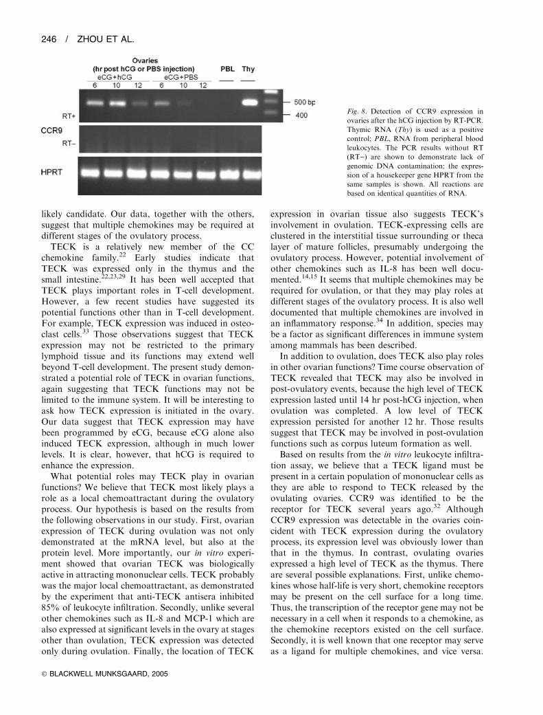
Detection of Expression of TECK Receptor CCR9We demonstrated transient expression of biologicallyactive TECK in the ovary during ovulation. As CCR9has been identified to be a ligand for TECK,32 weasked whether CCR9 was also expressed in the ovaryby RT-PCR. Ovaries, sampled from the experimentalmice at 6–12 hr post-hCG injection, expressed low
levels of CCR9 when compared with those in thethymus (Fig. 8). On the contrary, both ovaries andthymus expressed a similar level of the housekeepergene HPRT. Interestingly, ovaries from the controlmice with eCG injection only also expressed CCR9 at alevel comparable with the experimental mice at 6 hr(Fig. 8). Unlike the experimental mice, however, theCCR9 expression in the control mice fell and becameundetectable at 10 and 12 hr. As expected, CCR9expression was not detected in randomly sampledovaries. PBL isolated from the mice under superovu-lation induction were also used, and no CCR9 expres-sion was observed (Fig. 8). As a positive control,thymus expressed a very high level of CCR9 (Fig. 8).All RNA samples were tested for potential genomicDNA contamination. As shown in Fig. 8, PCR onRNA samples without RT showed completely negativefor CCR9.
DISCUSSION
Our study demonstrated a transient, high-level expres-sion of a CC chemokine TECK in the ovary duringovulation at both the mRNA and protein level. Moresignificantly, the expressed ovarian TECK was biolo-gically active, and may be a major source of chemo-attractant in recruiting mononuclear leukocytes duringovulation. A growing body of evidence showed thatovulation may resemble the inflammatory process, inwhich chemokines play a critical role in recruitingleukocytes. Many previous studies have searched forpotential candidate chemokines as the chemoattractantduring ovulation. IL-8, MCP-1 and several otherchemokines have been suspected to be the key chemo-kines.13,17 Our data suggest that TECK is another
1200A B C
800
M
19.0 kd
14.1
6.7
1 2 1 2
Mig
rate
d le
ukoc
ytes
400
0AntiserahCG ovary
untreated ovary
––4
6 6 6CFA anti-TECK –
–––
Fig. 7. TECK antibody inhibits leukocyte chemotactic activity of ovulating ovaries. A, whole cell lysate of DE3 E. coli which expresses
recombinant TECK (rTECK) (lane 1), and rTECK after purification (lane 2); rTECK is indicated by an arrow; protein markers (M) are
indicated; proteins are stained by Coomassie blue; B, Western blots demonstrating that anti-TECK antiserum from an rTECK-immunized
mouse react with both rTECK (lane 1) and bioactive TECK from R&D (lane 2). C, effect of TECK antibody (TECK) on chemotactic activity of
hCG-treated ovaries; serum from CFA-immunized mice (CFA) is used as a control; conditions for each assay are indicated; the ovaries were
sampled at 8 hr post-hCG injection.
OVARIAN EXPRESSION OF CHEMOKINE TECK / 245
AMERICAN JOURNAL OF REPRODUCTIVE IMMUNOLOGY VOL. 53, 2005
likely candidate. Our data, together with the others,suggest that multiple chemokines may be required atdifferent stages of the ovulatory process.TECK is a relatively new member of the CC
chemokine family.22 Early studies indicate thatTECK was expressed only in the thymus and thesmall intestine.22,23,29 It has been well accepted thatTECK plays important roles in T-cell development.However, a few recent studies have suggested itspotential functions other than in T-cell development.For example, TECK expression was induced in osteo-clast cells.33 Those observations suggest that TECKexpression may not be restricted to the primarylymphoid tissue and its functions may extend wellbeyond T-cell development. The present study demon-strated a potential role of TECK in ovarian functions,again suggesting that TECK functions may not belimited to the immune system. It will be interesting toask how TECK expression is initiated in the ovary.Our data suggest that TECK expression may havebeen programmed by eCG, because eCG alone alsoinduced TECK expression, although in much lowerlevels. It is clear, however, that hCG is required toenhance the expression.What potential roles may TECK play in ovarian
functions? We believe that TECK most likely plays arole as a local chemoattractant during the ovulatoryprocess. Our hypothesis is based on the results fromthe following observations in our study. First, ovarianexpression of TECK during ovulation was not onlydemonstrated at the mRNA level, but also at theprotein level. More importantly, our in vitro experi-ment showed that ovarian TECK was biologicallyactive in attracting mononuclear cells. TECK probablywas the major local chemoattractant, as demonstratedby the experiment that anti-TECK antisera inhibited85% of leukocyte infiltration. Secondly, unlike severalother chemokines such as IL-8 and MCP-1 which arealso expressed at significant levels in the ovary at stagesother than ovulation, TECK expression was detectedonly during ovulation. Finally, the location of TECK
expression in ovarian tissue also suggests TECK’sinvolvement in ovulation. TECK-expressing cells areclustered in the interstitial tissue surrounding or thecalayer of mature follicles, presumably undergoing theovulatory process. However, potential involvement ofother chemokines such as IL-8 has been well docu-mented.14,15 It seems that multiple chemokines may berequired for ovulation, or that they may play roles atdifferent stages of the ovulatory process. It is also welldocumented that multiple chemokines are involved inan inflammatory response.34 In addition, species maybe a factor as significant differences in immune systemamong mammals has been described.
In addition to ovulation, does TECK also play rolesin other ovarian functions? Time course observation ofTECK revealed that TECK may also be involved inpost-ovulatory events, because the high level of TECKexpression lasted until 14 hr post-hCG injection, whenovulation was completed. A low level of TECKexpression persisted for another 12 hr. Those resultssuggest that TECK may be involved in post-ovulationfunctions such as corpus luteum formation as well.
Based on results from the in vitro leukocyte infiltra-tion assay, we believe that a TECK ligand must bepresent in a certain population of mononuclear cells asthey are able to respond to TECK released by theovulating ovaries. CCR9 was identified to be thereceptor for TECK several years ago.32 AlthoughCCR9 expression was detectable in the ovaries coin-cident with TECK expression during the ovulatoryprocess, its expression level was obviously lower thanthat in the thymus. In contrast, ovulating ovariesexpressed a high level of TECK as the thymus. Thereare several possible explanations. First, unlike chemo-kines whose half-life is very short, chemokine receptorsmay be present on the cell surface for a long time.Thus, the transcription of the receptor gene may not benecessary in a cell when it responds to a chemokine, asthe chemokine receptors existed on the cell surface.Secondly, it is well known that one receptor may serveas a ligand for multiple chemokines, and vice versa.
Fig. 8. Detection of CCR9 expression in
ovaries after the hCG injection by RT-PCR.
Thymic RNA (Thy) is used as a positive
control; PBL, RNA from peripheral blood
leukocytes. The PCR results without RT
(RT)) are shown to demonstrate lack of
genomic DNA contamination; the expres-
sion of a housekeeper gene HPRT from the
same samples is shown. All reactions are
based on identical quantities of RNA.
246 / ZHOU ET AL.
� BLACKWELL MUNKSGAARD, 2005
Existence of possible multiple ligands for TECK hasbeen documented.35–37 For example, a recent study hasidentified a novel chemokine receptor CCX-CKR to bea ligand for TECK as well as several other chemo-kines.35 Thirdly, there may be unidentified ligands forTECK other than CCR9. It is evidenced by the findingthat T-cell development is not significantly impaired inCCR9-knockout mice.28 Because both TECK andCCR9 were discovered only recently, it is difficult atpresent to test the possibilities described above. Nev-ertheless, it will be the first essential step to determinethe presence of CCR9 on the cell surface of mononu-clear cells, which migrated in response to ovarianTECK during ovulation.
AcknowledgmentsThis study was supported by the NIH grant RO1HD35993. C. Zhou is a post-doctoral trainee suppor-ted by NIH T32 HD07324.
REFERENCES
1. Espey LL: Ovulation as an inflammatory reaction—a hypothesis. Biol Reprod 1980; 22:73–106.
2. Adashi EY: The potential relevance of cytokines toovarian physiology: the emerging role of resident ovariancells of the white blood cell series. Endocr Rev 1990;11:454–464.
3. Brannstrom M, Pascoe V, Norman RJ, McClure N:Localization of leukocyte subsets in the follicle wall andin the corpus luteum throughout the human menstrualcycle. Fertil Steril 1994; 61:488–495.
4. Brannstrom M, Mayrhofer G, Robertson SA: Localiza-tion of leukocyte subsets in the rat ovary during theperiovulatory period. Biol Reprod 1993; 48:277–286.
5. Standaert FE, Zamora CS, Chew BP: Quantitative andqualitative changes in blood leukocytes in the porcineovary. Am J Reprod Immunol 1991; 25:163–168.
6. Suzuki T, Sasano H, Takaya R, Fukaya T, Yajima A,Date F, Nagura H: Leukocytes in normal-cycling humanovaries: immunohistochemical distribution and charac-terization. Hum Reprod 1998; 13:2186–2191.
7. Brannstrom M, Norman RJ: Involvement of leukocytesand cytokines in the ovulatory process and corpusluteum function. Hum Reprod 1993; 8:1762–1775.
8. Brannstrom M, Giesecke L, Moore IC, van den HeuvelCJ, Robertson SA: Leukocyte subpopulations in the ratcorpus luteum during pregnancy and pseudopregnancy.Biol Reprod 1994; 50:1161–1167.
9. Luster AD: Chemokines – chemotactic cytokines thatmediate inflammation. N Engl J Med 1998; 338:436–445.
10. Horuk R: Chemokine receptors. Cytok Grow Fact Rev2001; 12:313–335.
11. Garcia-Velasco JA, Arici A: Chemokines and humanreproduction. Fertil Steril 1998; 71:983–993.
12. Runesson E, Bostrom EK, Janson PO, Brannstrom M:The human pre-ovulatory follicle is a source of thechemotactic cytokine interleukin-8. Mol Hum Reprod1996; 2:245–250.
13. Arici A, Oral E, Bukulmez O, Buradagunta S, Engin O,Olive DL: Interleukine-8 expression and modulation inhuman preovulatory follicles and ovarian cells. Endo-crinology 1996; 137:3762–3769.
14. Ujioka T, Matsukawa A, Tanaka N, Matsuura K,Yoshonaga M, Okamura H: Interleukine-8 as an essen-tial factor in the human chorionic gonadotropin-inducedrabbit ovulatory process: interleukine-8 induces neutro-phil accumulation and activation. Biol Reprod 1998;58:526–530.
15. Ujioka T, Matsukawa A, Tanaka N, Matsuura K, Yo-shonaga M, Okamura H: Analysis of the cytokineinteraction among interleukine-1b, interleukine-8 andinterleukine-1 receptor antagonist in rabbit ovulatoryprocess. Fertil Steril 1998; 70:759–765.
16. Arici A, Oral E, Bukulmez O, Buradagunta S, BahtiyarO, Jones EE: Monocyte chemotactic protein-1 expres-sion in human preovulatory follicles and ovarian cells.J Reprod Immunol 1997; 32:201–219.
17. Wong KH, Negishi H, Adashi EY: Expression, hormo-nal regulation, and cyclic variation of chemokines in therat ovary: key determinants of the intraovarian residenceof representatives of the white blood cell series. Endo-crinology 2002; 143:784–791.
18. Bowen JM, Keyes PL, Waren JS, Townson DH: Pro-lactin-induced regression of the rat corpus luteum:expression of monocyte chemoattractant protein-1 andinvasion of macrophages. Biol Reprod 1996; 54:1120–1127.
19. Townson DH, Warren JS, Flory CM, Naftalin DM,Keyes PL: Expression of monocyte chemoattractantprotein-1 in the corpus luteum of the rat. Biol Reprod1996; 54:513–520.
20. Senturk LM, Seli E, Gutierrez LS,MorG, ZeyneloguHB,Arici A: Monocyte chemotactic protein-1 expression inhuman corpus luteum.MolHumReprod 1999; 5:697–702.
21. Zhou C, Borillo J, Wu J, Torres L, Lou YH: Ovarianexpression of chemokines and their receptors. J ReprodImmunol 2004; 63:1–9.
22. Vicari AP, Figueroa DJ, Hedrick JA, Foster JS, SinghKP, Menon S, Copeland NG, Gilbert DJ, Jenkins NA,Bacon KB, Zlotnik A: TECK: a novel CC chemokinespecifically expressed by thymic dendritic cells andpotentially involved in T cell development. Immunity1997; 7:291–301.
23. Kunkel EJ, Campbell JJ, Haraldsen G, Pan J, Boisvert J,Roberts AI, Ebert EC, Vierra MA, Goodman SB,Genovese MC, Wardlaw AJ, Greenberg HB, ParkerCM, Butcher EC, Andrew DP, Agace WW: LymphocyteCC chemokine receptor 9 and epithelial thymus-expressed chemokine (TECK) expression distinguish thesmall intestinal immune compartment: epithelial expres-sion of tissue-specific chemokines as an organizingprinciple in regional immunity. J Exp Med 2000;192:761–768.
24. Mora JR, Bono MR, Manjunath N, Weninger W,Cavanagh LL, Rosemblatt M, von Andrian UH:Selective imprinting of gut-homing T cells by Peyer’spatch dendritic cells. Nature 2003; 424:88–93.
25. Lou YH, Borillo J: Migration of T cells from nearbyinflammatory foci into antibody bound tissue: a relay ofT cell and antibody actions in targeting native auto-antigen. J Autoimmun 2003; 21:27–35.
OVARIAN EXPRESSION OF CHEMOKINE TECK / 247
AMERICAN JOURNAL OF REPRODUCTIVE IMMUNOLOGY VOL. 53, 2005
26. Livak KJ, Schmittgen TD: Analysis of relative geneexpression data using real-time quantitative PCR and the2�DDCT method. Methods 2001; 25:402–408.
27. Wu J, Hicks J, Ou CN, Singleton D, Borillo J, LouYH: Glomerulonephritis induced by recombinantCol4a3NC1 is not associated with GBM antibody: apotential T cell mediated mechanism. J Immunol 2001;167:2388–2395.
28. Wurbel MA, Philippe JM, Nguyen C, Victorero G,Freeman T, Wooding P, Miazek A, Mattei MG, Malis-sen M, Jordan BR, Malissen B, Carrier A, Naquet P:The chemokine TECK is expressed by thymic andintestinal epithelial cells and attracts double- and single-positive thymocytes expressing the TECK receptorCCR9. Eur J Immunol 2000; 30:262–271.
29. Premack BA, Schall TJ: Chemokine receptors: gatewaysto inflammation and infection. Nat Med 1996; 2:1174–1178.
30. Wells TN, Power CA, Lusti-Narasimhan M, HoogewerfAJ, Cooke RM, Chung CW, Peitsch MC, Proudfoot AE:Selectivity and antagonism of chemokine receptors.J Leuk Biol 1996; 59:53–60.
31. Zaballos A, Gutierrez J, Varona R, Ardavin C, MarquezG: Cutting edge: identification of the orphan chemokinereceptor GPR-9–6 as CCR9, the receptor for the chem-okine TECK. J Immunol 1999; 162:5671–5675.
32. Lean JM, Murphy C, Fuller K, Chambers TJ: CCL9/MIP-1c and its receptor CCR1 are the major chemokine
ligand/receptor species expressed by osteoclasts. J CellBiochem 2002; 87:386–393.
33. Schluger NW, Rom WN: Early responses to infection:chemokines as mediators of inflammation. Curr OpinImmunol 1997; 9:504–508.
34. Townson JR, Nibbs RJ: Characterization of mouseCCX-CKR, a receptor for the lymphocyte-attractingchemokines TECK/mCCL25, SLC/mCCL21 and MIP-3beta/mCCL19: comparison to human CCX-CKR. EurJ Immunol 2002; 32:1230–1241.
35. Gosling JD, Dairaghi J, Wang Y, Hanley M, Talbot D,Miao Z, Schall TJ: Cutting edge: identification of a novelchemokine receptor that binds dendritic cell- and T cell-active chemokines including ELC, SLC, and TECK.J Immunol 2000; 164:2851–2856.
36. Yu CR, Peden KW, Zaitseva MB, Golding H, FarberJM: CCR9A and CCR9B: two receptors for the chem-okine CCL25/TECK/Ck beta-15 that differ intheir sensitivities to ligand. J Immunol 2000; 164:1293–1305.
37. Wurbel MA, Malissen M, Guy-Grand D, Meffre E,Nussenzweig MC, Richelme M, Carrier A, Malissen B:Mice lacking the CCR9 CC-chemokine receptor show amild impairment of early T- and B-cell development anda reduction in T-cell receptor cd+ gut intraepitheliallymphocytes. Blood 2001; 98:2626–2632.
248 / ZHOU ET AL.
� BLACKWELL MUNKSGAARD, 2005