translation of schnarf, k. 1929. “fertilization.” pages
TRANSCRIPT

Translation by C. J. Mellor of Schnarf, 1929. “Fertilization.” In Embryologie der Angiospermen
Page 1
Translation of Schnarf, K. 1929. “Fertilization.” pages 262-279, In Schnarf, K., Embyrologie der Angiospermen. Handbuch der Pflanzenan Anatomie/
Band 10.2. Gebrüder Borntraeger, Berlin.
Translation by Chauncey J. Mellor, Department Modern Foreign Languages, University of Tennessee, Knoxville, TN. Email: [email protected].
Assisted by Joseph H. Williams. Department of Ecology and Evolution, University
of Tennessee, Knoxville, TN. Email: [email protected].
November 23, 2007 Dear reader, The following is a translation of Karl Schnarf’s chapter on fertilization biology of angiosperms from his classic 1929 review of their embryology. We have tried to stick to as literal a translation as possible, however, I (J. Williams) changed several words to stay in keeping with current botanical language. For example, we used “transmitting tissue” instead of “guiding tissue,” and “callose plugs” instead of “callose clots.” In addition, the long and very useful table on fertilization timing has not as yet been translated entirely – only the relevant data on timing is included here. I have used brackets [ ] to indicate notes or interpretations by the translators. Please feel free to contact me should you see any errors or room for improvement. Respectfully, Joe Williams Email: [email protected]
Translation by C. J. Mellor of Schnarf, 1929. “Fertilization.” In Embryologie der Angiospermen
Page 2
C. Fertilization 1. The Pollen Tube
The life of the male gametophyte falls into two segments through the circumstance that a relatively long or relatively short dormant phase follows upon the maturation of the pollen grain. In this [dormant] phase the pollen grain possesses a certain resistance to external influences. It can tolerate relatively high temperatures without losing its ability to germinate (Rittinghaus 1886b) and is likewise extraordinarily resistant to desiccation, whereas the pollen tube possesses no resistance to it whatsoever (Pfundt 1910). The duration of pollen dormancy is quite different in various plants. According to Rittinghaus the time period within which dry pollen retains its germination ability varies between 17 and 66 days and may amount on average to 30-40 days. Mangin (1886) found in the plants he investigated that the pollen remains capable of development for from one to 80 days. Holman and Brubaker (1926), who also consider very thoroughly the literature on this aspect of pollen physiology, found that the pollen of Typha latifolia, /pp. 262-263/ preserved via calcium chloride, still germinated after 336 days. By contrast, Graminae evidence a strikingly short-lived pollen. The second stage in the life of the male gametophyte, the pollen tube,1 begins with the germination of the pollen grain. This tube is characterized by a significant lengthening, which in some cases amounts to 20 cm or more; in addition it is characterized by a peculiarly progressing growth process in the course of which always only a relatively small portion of the front end is filled with living cytoplasm whereas the region further back, ie. back towards the pollen grain, is only empty membrane. When therefore the pollen tube has arrived at the ovule, no connection via living cytoplasm exists to the pollen grain on the stigma--a fact already stressed by Hofmeister (1861) (cf. also Weatherwax 1919, p. 78). In most cases in the pollen tube there occurs a closing off of its living portion from the portion departing from [containing] the cytoplasm by the occasional origination of
1 In general, the discovery of the pollen tube is ascribed to G. Amici since this researcher described it in a Portulaca in 1823. Certainly Amici was the first to deal more thoroughly with the question of fertilization and saw the penetration of the pollen tube into the seed structure. Svedelius (1924, p. 5) nevertheless points out that Linné describes in detail the fertilization process in an Amaryllis species already in his treatise “Sexus plantarum” presented to the imperial academy in Petersburg in 1760. Namely, he had observed that after the emptying of the pollen on the stigma finally fine channels or opaque stripes slowly creep from the stigma to the seed structures. According to Stenar (1925b) the Amaryllis species investigated by Linné was Sprekia formosissima.

Translation by C. J. Mellor of Schnarf, 1929. “Fertilization.” In Embryologie der Angiospermen
Page 3
callose (polysaccharide) plugs which arise as a rule on the inner wall of the tube as a ring-shaped thickening which gradually widens into a complete closing off (cf. Kirkwood 1907a). The formation of the callose plugs, however, can also take place in such a way that a one-sided deposit occurs in the inner wall of the pollen tube, which increases in size until it reaches the opposite wall, as observed by Guéguen (1901, p. 288) in Convallaria majalis and von Bobiloff-Preisser (1917) in Narcissus. Cf. also Ill. 30, Figs. 6-10. The membrane of the pollen tube is always only a protrusion of the intine (endosporium) of the pollen tube. In many respects there is still a lack of clarity on its material composition, even though most authors assume [it to be] cellulose. Even so, one can still find other statements on this point. Tomaschek (1889) for example cites for Colchicum, that the thickening layers only begin to show a cellulose reaction when the pollen tube is briefly heated in potassium hydroxide; he further concludes [there is] a formation of cutin which is supposed to make the pollen tube more resistant to destructive influences on its long path and thus to protect the cytoplasm; heated in potassium hydroxide and then treated with iodine and sulfuric acid the medial layer turns dark purple, the layer adjacent to the protoplasm turns a weak blue and the original membrane of the pollen tube turns dark blue. Palla (1890) who investigated pollen tubes of Leucojum, Galanthus, Scilla, Hyacinthus (among others) under cultivation, by contrast found everywhere a positive cellulose reaction with “Chlorzinkjod” [a chemical that presumably contains chlorine, zinc and iodine]. According to Biourge (1892) /263-264/ the wall of the pollen tube consists generally of cellulose, at least in its inner layer, if it is layered. The external layer often consists of pectin, sometimes of cellulose. It is probable a priori that the tip of the pollen tube would exhibit a special behavior, on the one hand considering the fact that the growth occurs here (Acqua 1890), and on the other because the emptying of the contents takes place. This emptying

Translation by C. J. Mellor of Schnarf, 1929. “Fertilization.” In Embryologie der Angiospermen
Page 4
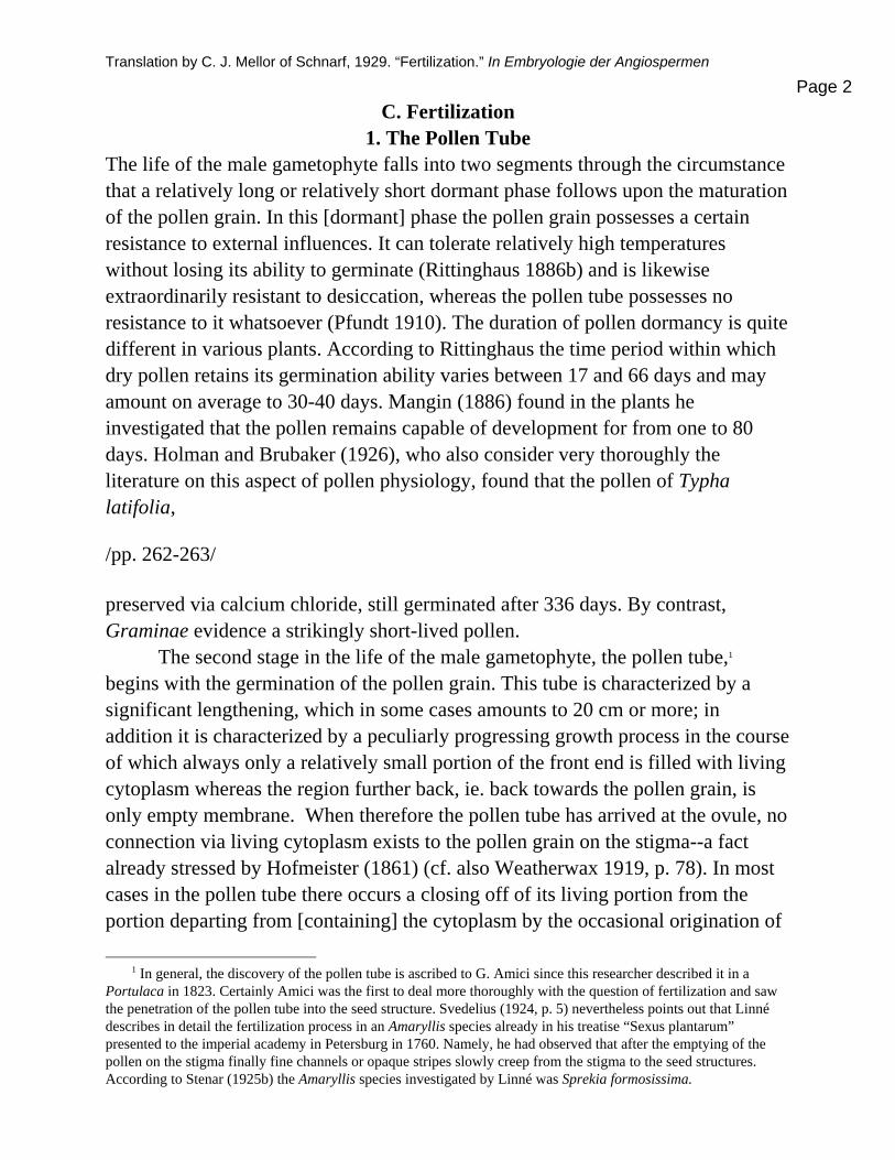
Ill. 30. Fig. 1 Germinating pollen on a stigmatic papilla in Primula officinalis. Figs. 2-5. Cyclanthera explodens. Fig. 2 Longitudinal section through the micropylar region of the ovule, exhibiting the pollen tube (Ps) in the extension of the nucellus; Fig. 3 Transverse section through the extension of the nucellus before pollination; Fig. 4 likewise after pollination; Fig. 5 Transmitting tissue in the placental folds, pollen tubes in transverse section. Figs. 6-10. Plug formation in the pollen tube of Sarcodes sanguinea. -- Fig. 1. after Dahlgren. Figs. 2-5 after Kirkwood. Figs. 6-10 after N. Oliver. Magnification: Fig. 1 880x, Fig. 2 slightly enlarged, Figs 3-5 440x, Figs. 6-10 400x. normally takes place in the embryo sac, but in artificial cultures, as various observers report, [it occurs] spontaneously, i.e. through very slight chemical or mechanical stimuli; “often a slight shaking of the preparation” (Palla 1890; cf. also Bobilioff-Preisser 1917). According to Ishikawa (1918) now the wall of the pollen tube at the tip consists of cellulose and pectin materials. The cytoplasm of the pollen tube is generally fine-grained (for its

Translation by C. J. Mellor of Schnarf, 1929. “Fertilization.” In Embryologie der Angiospermen
Page 5
distribution in crooked portions of the pollen tube /264-265/ see Mitschka 1898)2 In cultures the cytoplasm frequently exhibits vigorous streaming (on this see Bobilioff-Preisser 1917 among others) in which various content bodies can appear, above all, starch. Mangin (1886) saw the significance of this in the fact that starch-containing pollen is not dependent on nutrient uptake from outside. Green (1894) describes small, strongly refractive content bodies in the pollen tube which are continuously expelled into the liquid culture medium. In Narcissus this author observed that this expulsion takes places through a pore with a clearly defined margin at the tip of the pollen tube.3 In the pollen grains and pollen tubes of the Asclepiadaceae according to Guignard (1922b) spindle-shaped content bodies occur that are protein crystalloids. According to their origin they arise from plastids which are developed in the young microspores. In the pollen tube either free nuclei appear or the generative nucleus and sperm nuclei surrounded by clearly delineated cytoplasm of their own. Concerning first of all the number of these nuclei, a group of three is usual; the appearance of more than three does occur, but must be considered an anomaly. Already Strasburger (1884a) described a relevant case in Ornithogalum sp. where occasionally four nuclei appeared in the pollen tube and Schniewind-Thies (1901) saw an exception with five nuclei in a germinated pollen grain on Scilla sibirica. Suessenguth (1923) observed in pollen tubes of Spathiglottis occasionally four to five nuclei; in one case he even observed eight nuclei, i.e. the number which the female gametophyte normally reaches. Likewise in Vincetoxicum nigrum pollen tubes occasionally appear which, apart from a vegetative nucleus, also contain four sperm nuclei. Guignard (1922a), to whom we owe this observation, presumes that in this case the generative nucleus has passed through two rounds of division. It has occasionally been observed in Asclepias, Eichhornia, Hemerocallis and Lilium that the vegetative nucleus divides in the pollen tube (On this see Coulter and Chamberlain 1903, p. 135, and Strasburger 1908, p. 544.) More recently Dahlgren (1916) observed in Primula officinalis a doubling of the vegetative nucleus that presumably arose amitotically and Lagerberg (1909) observed in Adoxa an
2 According to Seifriz (1921, p. 291) it is extremely liquid and has therefore very little viscosity. Cytoplasm emerging from pollen tubes in many cases proves to be water soluble and therefore constitutes an exception to the rule that cytoplasm is insoluble in water.
3 Cf. also Hofmeister (1858, p. 173) “On the other hand the pollen tube tips of some plants showed themselves to be equipped with spots: with narrow channels leading through the thickened layers to the primary, thin, closed, external skin of the pollen tube (Godetia, Oenothera, Crocus).”

Translation by C. J. Mellor of Schnarf, 1929. “Fertilization.” In Embryologie der Angiospermen
Page 6
incomplete interlacing of this. In Lilium candidum according to Herrig (1922) a fragmentation of the vegetative nucleus frequently takes places in the pollen tube. As already mentioned in another connection, the generative nucleus divides either already in the pollen grain or only later in the pollen tube. In general the behavior is presumably constant within the same species. But the appearance of variable behavior /265-266/ was shown by Frisendahl (1912, p. 47) for Myricaria mermanica, where the division of the generative cell occurs just as frequently in the pollen grain as in the pollen tube. In this plant the sperm nuclei in the pollen tube are usually naked, but can also be surrounded with distinct cytoplasm of their own which can only be weakly stained. In addition Dahlgren (1916), who pursued [the issue of] the content of the pollen grain of Primula officinalis on the stigma, saw that the two sperm nuclei either form within the pollen grain or in the pollen tube. The sperm nuclei that arise in the pollen grain almost always lack their own cytoplasm, but in the pollen tube cells were observed a couple of times. Rather contradictory information on the appearance of sperm cells in pollen tubes is reported. In Lilium martagon Guignard (1889) saw that the generative cell divides into two clear sperm cells in the pollen tube through the aid of a cell plate. In contrast to this finding, however, Koernicke (1906), Strasburger (1908) and Nawaschin (1910) established that during the division of the generative kernel the surrounding cytoplasm loses its definition in relation to the cytoplasm of the pollen tube, so that after the division only naked sperm nuclei can be identified. In details, however, the findings of these three authors diverge somewhat. According to Koernicke the loss of definition occurs in prophase, according to Strasburger in metaphase and according to Nawaschin in telophase. By contrast on the other hand, Welsford (1914, p. 267) has more recently asserted that in Lilium martagon and [L.] auratum the division occurring in the pollen tube can lead to the formation of sperm cells. We see therefore that even in one and the same species no complete clarity exists on the [matter of] the existence and regeneration [?] of the sperm cells in the pollen tube. But perhaps these findings can be interpreted to mean that at some times sperm cells are formed, at other times their formation does not take place, and in the latter case the cytoplasm of the generative cell at times loses its independence earlier and at times somewhat later. By the way, the process of nuclear division in the generative cell likewise points to some somewhat variable relationships. The achromatic spindle shape is often poorly developed or is lacking

Translation by C. J. Mellor of Schnarf, 1929. “Fertilization.” In Embryologie der Angiospermen
Page 7
entirely, so that the thought has been expressed that perhaps the distribution of the chromosomes into the daughter cells occurs by means of independent movements of the chromosomes (cf. Nawaschin 1910, Welsford 1914). With regard to the presence of sperm nuclei or sperm cells in the pollen tube in general Strasburger (1908, p. 534) expressed the supposition that the presence of individualized sperm cells in the pollen tube may be tied to the division of the generative cell in the pollen grain. As will become evident in what follows, we are dealing here simply with a rule that has exceptions. The quite variable behavior of the nuclei in the pollen tube, especially the gamete nuclei or gamete cells might be amplified in what follows by a number of examples from a variety of related groups. In Chlorophytum sternbergianum the division of the generative nucleus occurs in the pollen tube and during this process “the elongated cell sequestering it [i.e. the generative nucleus]” disappears (Strasburger 1888). Nicotiana tabacum behaves similarly (Guignard 1902a), /266-267/ where the generative cell divides in the pollen tube during its penetration into the transmitting tissue of the style. At first [early pollen tube growth], the vegetative nucleus at the tip proceeds first and the generative cell follows at variously great intervals, whereas [in pollen tubes] nearer the ovule the vegetative nucleus is preceded by the sperm cells. In Datura laevis, however, the timing of the nuclear division in the generative cell is less fixed and at the time when the division occurs, the vegetative nucleus has often lost its original appearance and has assumed the shape of a rather long knotted piece of string. Subsequently it has disintegrated and the generative nuclei are no longer surrounded by their own cytoplasm. Such a loss of its own cytoplasm in the pollen tube can come about even when the division of the generative cell into two sperm cells has already taken place, as for example is the case with Najas major according to Guignard (1899d) and probably also in Silphium (Merrell 1900). Among the numerous cases in which exclusively naked sperm nuclei have been observed in the pollen tube the following examples may be cited: Alisma plantago (Schaffner 1896), Sagittaria (Schaffner 1897a), Malvastrum peruvianum (Stenar 1925b, Fig. 50), Thismia luetzelbergii (Goebel and Suessenguth 1924), Lobelia erinus (Armand 1912), Clivia nobilis (Herrig 1922). Especially in more recent times, however, the cases seem to become more numerous in which well-defined sperm cells were observed in the pollen tube.

Translation by C. J. Mellor of Schnarf, 1929. “Fertilization.” In Embryologie der Angiospermen
Page 8
Thus Ishikawa (1918) observed in Oenothera nutans and [O.] pycnocarpa the generative nucleus and both sperm nuclei are embedded in a well-defined cytoplasmic sheath [Plasmascheide]. His observation is possibly also of special interest that this proper cytoplasm was bunched up more densely at the front end than at the back end. On the other hand, neither Modilewski (1909a) nor Geerts (1909) have described nor illustrated such a cytoplasm sheath in Oenothera biennis or [O.] Lamarckiana. Herrig (1919) found in Butomus umbellatus and Echeveria desmatiana well-defined sperm cells in the pollen tube and in Lilium candidum he observed either a sperm cell with two nuclei or two sperm cells or even two naked sperm nuclei. In Elodea canadensis Wylie (1904) was able to trace sperm cells in the pollen tube as far as the micropyle. In Asclepias cornuti according to Finn (1925) both sperm cells appear at times quite close together, at times more or less distant from one another in the pollen tube; in so doing they preserve their structure (cf. p. 45, Ill. 4, Fig. 7 & 8) together with their tail-like extensions and their staining response (i.e. response to stain). Further worthy of mention are the sperm cells in the pollen tubes of Lupinus luteus, Narcissus incomparabilis and Crocus vernus, in which Ruhland and Wetzel (1924) were able to positively identify chloroplasts of extremely small size as well as [confirm] the older observation regarding Endymion nutans, where Guignard (1899c) was able to establish in the tip of the pollen tube arriving at the embryo sac, well-defined sperm cells, which only lose their own cytoplasm in the embryo sac. In Ulmus montana Lagerberg (1909) observed very well defined sperm cells which had originated in the pollen tube and retained their individuality during their transport through the pollen tube. This fact, and moreover the conclusion that a weakly staining vegetative nucleus is demonstrable at the tip of the advancing pollen tube, makes the observation of Shattuck (1905) regarding Ulmus americana somewhat uncertain, in which the sperm nuclei lose their own cytoplasm upon entering the pollen tube and the vegetative nucleus is not at all said to enter into it. Possibly Shattuck overlooked the existence of sperm cells in the pollen tube; since after entering the embryo sac the sperm nuclei seem to possess something akin to their own cytoplasm, as the remark of Shattuck suggests: “After entering the sac the nuclei ... begin to gather a small amount of cytoplasm around them.” In addition the observations of Lagerberg on a Viola sp., where the division of the generative cell occurs in the pollen tube and clearly delineated sperm cells are formed, are of interest here. In contrast to the above-mentioned rule of Strasburger that the presence of individualized sperm cells in the pollen tube is connected to the division of the generative cell in the pollen kernel, it is evident in Viola, “that the

Translation by C. J. Mellor of Schnarf, 1929. “Fertilization.” In Embryologie der Angiospermen
Page 9
individuality of generative cells or the sperm cells respectively is not necessarily lost when the division of the generative cells is displaced into the pollen tube.” Finally the peculiar behavior of Myosurus minimus might be cited. In this plant according to Tschernojarow (1926) the nuclear division creating the two sperm nuclei takes place in the generative cell of the pollen grain. The generative cell is preserved and even appears in the pollen tube and even in the embryo sac without the occurrence of a cell division; it undergoes only a stricture in the middle and takes on a biscuit shape. To be sure, a peculiar cell. Its cytoplasm remains “always colorless with the application of triple stain according to Flemming and has the appearance of the completely homogeneous hyaline substance with rather strong refractive properties. Attempts to stain this cytoplasm in any other manner were without success” (Tschernojarow 1926). Such peculiar two-nucleate generative cells have by the way also been observed in Juglans (Nawaschin and Finn 1913). Its existence could be denied because of the invisibility of the cytoplasm and the bright areas surrounding the nuclei [could be] regarded as a shrinking phenomenon, except for the indisputable fact, that the two sperm nuclei are joined by them and always appear at a definite distance from one another. The total picture emerging from these observations is not very satisfying. Sperm cells and naked sperm nuclei were observed in pollen tubes and, in part, within the same genus contradictory reports are found. Future observations must create clarity here. Perhaps it is only dependent on the technique used, whether we observe sperm nuclei or sperm cells. The vegetative nucleus which is almost always clearly identifiable by its size and its appearance shows variable behavior in the pollen tube. Its position with regard to the generative cells or the sperm cells is variable by the fact that it sometimes precedes them and sometimes follows. In general the first behavior [preceding] appears to be the more numerous one (cf. on this Strasburger 1877, 1884a, Elfving 1879). The vegetative nucleus can in addition be preserved for a very long time or degenerate in the course of the pollen tube growth. This latter circumstances occurs in Lobelia erinus relatively early when it disintegrates as soon as the pollen tube becomes twenty times longer than the diameter /268-269/ of the pollen tube. In Myricaria germanica it is either subject to early disintegration or it can be retained unchanged Frisendahl 1912).

Translation by C. J. Mellor of Schnarf, 1929. “Fertilization.” In Embryologie der Angiospermen
Page 10
In general only a single pollen tube emerges from a pollen kernel (monosiphonic pollen; Goebel 1923, p 1707). However for pollen kernels of various Malvaceae, Cucurbitaceae and Campanulaceae the emergence of several pollen tubes from a pollen kernel (polysiphonic pollen) seems to be typical. For the first-named family this behavior has already been described by Strasburger (1884a, p. 44, Table II, Figs. 57-59). In Althaea rosea according to Guignard (1904) up to ten pollen tubes can shoot forth, and Stenar (1925b, p. 37) found at least the same number in Malva pusilla. In Mala neglecta he even found 14. Polysiphonic behavior was observed more occasionally in Valerianaceae where according to Asplund (1920, p. 45) up to three tubes, corresponding to the number of pores, can occur, and Armand (1912) advances similar results for Lobelia. Among the monocotyledons only monosiphonic behavior is observed, but Coulter and Rose (1886) saw occasional emergence of two tubes from a pollen kernel in Tradescantia virginica. What is more commonly mentioned in the literature than the emergence of several tubes is the appearance of branchings of the pollen tube in various portions of its extent. Even here the Malvaceae are to be named in which researchers as early as Hofmeister (1858, p. 91) point out the brachiation of the pollen tube before it impinges on the embryo sac, a fact which was later described by Guignard (1904) for Hibiscus trionum. Moreover, knot-shaped swellings and branchings occur quite commonly in Oenotheraceae (Ishikawa 1918, Beer 1906). Dichotomous branching of the pollen tube in the ovary and in the micropyle were further observed by Tschernojarow (1926) in Myosurus minimus, whereby it was also determined that the vegetative nucleus and the generative cell appear always only in one and the same branch, namely in the stronger one. Further instances are: Casuarina (Treub 1891), Corylus, Quercus, Carpinus (Benson 1894), Juglans (Nawaschin 1895), Carya (Billings 1903), Crotalaria sagittalis (Cook 1924), Asclepias cornuti (Gager 1902, Hugo Fischer (18904), Anthericum liliago (Elfving 1879), Hippeastrum aulicum (Hofmeister 1859), Iris (Sawyer 1925), Pothos longifolia (Hofmeister 1859). Waldersdorff (1924) saw the appearance of branched pollen tubes in cultures of Epilobium angustifolium, [E.] montanum, [E.] roseum, Clarkia pulchella, [C.] elegans, in species of Circaea, Lopezia, Fuchsia, Oenothera, but also in representatives of others families: Trifolium, Saponaria, Nymphaea, Morisia, Tropaeolum and others. In the observations of Waldersdorff the circumstance is of interest on the one hand, that certain culture conditions favor the appearance of branches, and on the other hand, the observations that these
4 Observed by the former in ovaries and by the latter in cultures.

Translation by C. J. Mellor of Schnarf, 1929. “Fertilization.” In Embryologie der Angiospermen
Page 11
branchings occur by the formation of forks at the tip of the pollen tube. From an already finished piece of tube no bulgings come forth. /269-270/ The pollen tube normally emerges from the pollen grain on the stigma growing through the transmitting structures of the style to the ovule. The physiological questions relating to this process can only be touched upon briefly. We can artificially create the conditions for the germination of the pollen grain by providing the pollen a suitable nutritional medium. The numerous investigations on this point have shown that sugar solutions of a certain concentration are suitable in most plants to bring the pollen to germination.5 Some pollen however germinates in water, humid air (cf. also Waldersdorff 1924); in some plants however special stimulants are required (Ericaceae pollen according to Molisch 1893). The conditions that suffice for germination are not adequate to nourish the pollen tubes on a lasting basis. Up to a certain point the tubes grow at the cost of the reserve materials contained in the pollen grain. Cane sugar alone is not a full-value nourishment. With regard to nourishment the pollen tubes exhibit a very strict specificity, but they behave less specifically with regard to stimulants since it has been shown that protein and various sugars generally act enticingly (Tokugawa 1904; cf. also Waldersdorff 1924, Rotmistrow 1925). The fact that the conditions for the germination of the pollen by and large are rather similar and undistinguished is harmonious with the fact that pollen can generate tubes on foreign stigmata. Strasburger (1886) demonstrated that this capability is not limited by boundaries of relatedness and the dicotyledonous pollen (e.g. Lathyrus montanus) can not only germinate on stigma of Convallaria latifolia, but can also project pollen tubes on into the ovaries. Miyoshi (1894) and Tokugawa (1904) present examples of the germination of pollen on foreign stigmata. That the conditions for the germination of the pollen do not coincide with those for the nourishment of the pollen tubes is shown by the experiments which Jost (1907) conducted with pollen of Hippeastrum aulicum and Lilium martagon. Whereas in cultures the pollen tubes grew to a maximum length of 2 cm, they were able to attain a length in the transmitting tissue of the style that was significantly longer than that necessary in nature.6 This was shown by the above named
5 Of newer papers on this subject, cf., in addition to those cited, also Katz (1926). 6 On the other hand, Bobilioff-Preisser (1917) succeeded in achieving in Vincva minor tubes on an artificial
substrate of a length exceeding that necessary for fertilization. Lidforss (1909, p. 458) conjectures: “By a

Translation by C. J. Mellor of Schnarf, 1929. “Fertilization.” In Embryologie der Angiospermen
Page 12
researcher /270-271/ in the following manner: he cultured pollen tubes to grow through a number of sectioned pieces of style. It was also shown that the growth of the pollen tube is not unlimited even on suitable transmitting tissue, as distinguished from the fungal hyphae with which the pollen tubes are occasionally compared in the literature. The rapidity at which the pollen tube grows is demonstrated by a few examples. Tokugawa (1904) found in various Lilium species which were dusted with pollen of their own species the following average rates of growth (in mm per hour): Lilium speciosum H. ..................1.376 Lilium speciosum S. ..................1.400 Lilium Hansoni ..................1.681 Lilium auratum ..................2.125 Pollen set onto the stigmata of other species showed a slower growth, as the following table indicates: Pollen of Lilium auratum on the stigmata of: Lilium auratum ..................2.125 Lilium Hansoni ..................1.000 Lilium speciosum S. ..................0.833 Lilium speciosum H. ..................1.000 Sawyer (1917) measured the added growth in the length of the pollen tubes on the stigmata of Iris versicolor within the first seven hours after pollination and obtained the following measurements: Elapsed time since pollination | Length of the pollen tube 1 hours | 0.1-0.6 mm 3 hours | 2-2.5 mm 5 hours | 4.5-5 mm 7 hours | 8-9.5 mm He found therefore that within the first seven hours the growth rate increases with time. However, there are also data indicating that the pollen tube in certain plants grows at a slowed rate. Kirkwood (1906) found that in various Cucurbitaceae the combination of protein substances with various sugars and possibly also lipoid materials ... one might succeed in preparing nourishing substances which provide pollen tubes the same material that the nourishing solutions of Pfeffer, Sachs, v.d. Crone etc. provide for the roots of the higher plants.”

Translation by C. J. Mellor of Schnarf, 1929. “Fertilization.” In Embryologie der Angiospermen
Page 13
greatest portion of the distance between the stigma and the embryo sac is traversed in the first three to five hours. In the vicinity of the micropyle the rate is slower. The author attributes this peculiarity to the circumstance that the amount of reserve materials is greatest at the beginning of pollen tube growth. However, it is much more probable that the anatomical differences of transmitting tracts are to be cited in the differences of growth rate. Jost (1907) established the following growth rates: Zizania sp. ..................1.7 mm/hour Zea mays ..................3 mm/hour Secale cereale ..................0.8 mm/hour /271-272/ The pollen tubes of the Gramineae are among those, as were already given by Hofmeister (1861) and Strasburger (1878, p. 221), which grow the fastest. Let us cite here verbatim what Jost (1907) reports on the behavior of the germinating pollen on the stigma of Secale cereale: “The rupture of the cuticle and the splitting of the central lamella, first between two epidermal cells, then between the four cell rows in the axis of the hair, occurs at an incredible speed. The growth proceeds so uniformly as if there were no obstacles whatever to be overcome. Just five minutes after the application of the pollen I have seen tubes inside the hairs. One can at first easily follow the growth process by the starch laden stream of protoplasm which is propelled forward toward the tip of the pollen tube. I once observed a stigma which 15 minutes after pollination was so engorged with pollen tubes that it almost looked like a plasmodium because of the streaming of plasma in them.” The speed at which the pollen tube grows in the style varies with temperature. Thus Heribert-Nilsson (1910) found by experiment that the style averaging 85 mm in length of Oenothera lamarckiana in mid-July is traversed in 19 hours (average growth 4.47 mm/hour), whereas at the end of July (with a somewhat lower summer temperature) 23 hours were necessary. Most recently, Buchholtz and Blakeslee (1927) recently showed how surprisingly great the influence of temperature on the rate of growth of the pollen tube is by precise experiments on Datura stramonium. In this case at 11.1°C the average growth in the style was 1.28 mm in the first 12 hours; at a higher temperature it rose very significantly up to the optimum temperature of 33.3°C, where it amounted to 5.86 mm, i.e. four and a half times faster. Data regarding the time passing from the pollination to the arrival of the pollen tube at the ovule are of special interest. Such data are relatively frequent in

Translation by C. J. Mellor of Schnarf, 1929. “Fertilization.” In Embryologie der Angiospermen
Page 14
the literature. Certainly they are of variable value. Often it is unclear from the statements of the author whether by his indications of time he means the time from pollination to fertilization or to the intrusion of the pollen tube into the micropyle. It should further be noted that some of the values were established by the fixing of the material at certain time intervals after pollination, and the greater these intervals are, the less the precision of the observation is. Finally the duration given by the observers can not be generalized. It holds for a certain species or variety at a certain location under quite specific weather conditions. Nevertheless, the following data are of significance in a variety of aspects. Betula. The pollen tube arrives at the embryo sac in one month in the middle of June (Benson 1894), also Hofmeister (1858, p. 96 f.) Corresponding results in Nawaschin (1894). Carpinus. The pollen tube arrives at the embryo sac at the end of June in almost 2 months (Benson 1894). Alnus. Almost three months (Benson 1894). The corresponding time is much smaller in Alnus alnobetula: On 31 May the blossoms /pages 273 through 276/ literal translation incomplete.
Species Family Time Translation Reference p. 272 (see also above) Betula alba Betulaceae 1 month See above Carpinus betulus Betulaceae almost 2 months Benson 1894 (and
others) Alnus glutinosa Betulaceae almost 3 months Benson 1894 Alnus alnobetula Betulaceae may 31 in pollination
period, june 24 pt nr fmg; fert before june 29
Wolpert 1910
/pp. 272-273/ Corylus avellana Betulaceae over 4 months Benson 1894 (and
others) Quercus pedunculata Fagaceae 2 months Hofmeister 1858,
S. 96 Quercus rubra Fagaceae 13-14 months Hofmeister 1858,
S. 96 Quercus robur Fagaceae 4 months in 1 yr variets,
11 month in 2 yr vars. Benson 1894
Quercus velutina Fagaceae over 1 year (13 mo.) Conrad 1900; Coulter and Chamberlain 1915

Translation by C. J. Mellor of Schnarf, 1929. “Fertilization.” In Embryologie der Angiospermen
Page 15
Quercus cerris Fagaceae 3 months Fraulein Mathilde Demant, pers comm.
Hicoria pecan (=Carya olivaeformis)
Juglandaceae 5-7 months Woodroof and Woodroof 1927
Viscum album Viscaceae **more than 5, less than 10 days
Pisek 1923
Arceuthobium oxycedri Santalaceae **>5 months T. Johnson 1888 Ulmus montana U. pedunculata
Ulmaceae 3-4 dap Nawaschin 1898b, also cited in Schnarf 1929
Celtis australis Fabaceae **6-7 weeks Modilewski 1908a
Humulus japonicus Cannabaceae 70 hours in greenhouse Winge 1914 Humulus lupulus Cannabaceae Ca. 140 hap Winge 1914 Cynomorium coccineum Cynomoriaceae 4 days Juel 1903b,
coulter an d chamberlain 1915
Polygonum aviculare Polygonaceae > 7d Lonay 1922a Fagopyrum esculentum Polygonaceae legit poll, 18 hap; illegit
> 72 hap Stevens 1912
Mercurialis annua Euphorbiaceae **48 hap Strasburger 1909 Cereus tortuosus Cactaceae 3 weeks Guignard 1886e Cereus nycticalus Cactaceae 1 month Guignard 1886e Cereus triangularis Cactaceae 3 weeks D’Hubert 1896,
Coulter and Chamberlain 1915
Cereus spinosissimus Cactaceae 1 week Strasburger 1908 Phyllocactus-Arten Cactaceae 12-15 hap D’Hubert 1896,
Coulter and Chamberlain 1915
Hevea brasiliensis Euphorbiaceae ** C. Heusser 1919 /273-274/ Hamamelis virginiana Hamamelidaceae 5-7 months Shoemaker 1905 Platanus orientalis Platanaceae at least 3 wks Cytinus hyposcistis Cytinaceae 10 hap Hofmeister 1858
S. 109 Cardamine pentaphylla Cardamine polyphylla
Brassicaceae 2-3 days Schwarzenbach 1922
Pirus (=Pyrus) Rosaceae various varieties w times from 4d 2h, 4d 7h, and 2d 4h.
Osterwalder 1910
Phaeseolus vulgaris Fabaceae 8-9 h to mp Weinstein 1926 Trifolium pretense Fabaceae 18 hap in July; 35-50 h in
October = poll to egg cell div.
Martin 1914
Oenothera nutans Oenothera pycnocarpa
Onagraceae pts to enter fmg 48 hap Ishikawa 1918
Oenothera rubrinervis Onagraceae 36 hap O’Neal 1923 Citrus trifoliata Rubiaceae ca. 4 weeks Osawa 1912 Citrus aurantium Rubiaceae ca. 4 weeks Strasburger 1878 Rhus toxicodendron Anacardiaceae **30-40 hours Grimm 1912 Acer negundo Sapindaceae **40-72 hours Taylor 1920

Translation by C. J. Mellor of Schnarf, 1929. “Fertilization.” In Embryologie der Angiospermen
Page 16
Anthriscus silvestris Apiaceae 14 days btwn poll and emb dev
Hakansson 1923
Carum carvi Apiaceae fertilz bef 5 days Hakansson 1923 Statice bahusiense (=Limonium)
Plumbaginaceae
few hours Dahlgren 1916
Primula officinalis (longistyl)
Primulaceae 42 hours, bt longer w illeg pollination
Dahlgren 1916
Monotropa uniflora Ericaceae normally 5 days aft. poll. Shibata 1902 Convolvulus arvensis Convolvulaceae **few hours Peters 1908, S. 51 Nicotiana tabacum Solanaceae 2 days @ 20-25 degrees Guignard 1902a Datura laevis Solanaceae about 24 hours Guignard 1902a Torenia asiatica Scrophulariaceae 36 hours Strasburger 1884a /274-275/ Pedicularis silvatica Orobanchaceae 6-10 hours Hofmeister 1858,
1859 Lathraea squamaria Orobanchaceae few hours Hofmeister 1858 Gloxinia hybrida Gesneriaceae Strasburger 1878,
cited in Brink 1926
Gloxinia hybrida Gesneriaceae 60 hours Strasburger 1884a Gloxinia hybrida Gesneriaceae 36 h to pass through 40
cm long style see footnote 1 Strasburger 1878,
S. 22 Fraxinus excelsior Oleaceae Hofmeister 1858,
S. 109 Coffea liberica Rubiaceae OC 3-4 dap; 5-6 hap self Faber 1912 Melothria pendula L. Cucurbitaceae 26 hours Kirkwood 1906 Micrampelis lobata (=Echinocystis lobata (Michx.) Torr. & Gray)
Cucurbitaceae 19 hours Kirkwood 1906
Cyclanthera explodens Cucurbitaceae 41 hours Kirkwood 1906 Lactuca muralis Asteraceae < 6.5 hours Dahlgren 1920 Zostera marina Zosteraceae 10 hours Hofmeister 1861 Limnocharis emarginata Limnocharitaceae 18 hours Hall 1912 Lilium martagon Lilium candidum
Liliaceae pt in style 18-20hap; gamete fusion 65-72 hap
Mottier 1898
Lilium martagon Lilium auratum
Liliaceae 5 and 7 dap, respectively Welsford 1914
Lilium martagon Liliaceae 20 hap see footnote 2 Overton 1891 Lilium philadelphicum Liliaceae spm in contct w egg 60-
72 hap; Weniger 1918
Lilium longiflorum Liliaceae spm in contct w egg 120 hap
Weniger 1918
Lilium pyrenaicum Liliaceae fert starts 2-3 dap Strasburger 1884a Merendera caucasica Colchicaceae **btwn 16 hap and 7
days dep on envir condts Hofmeister 1861
Hippeastrum aulicum Amaryllidaceae 2-4 days Jost 1907 Tulipa gesneriana Liliaceae 8-10 hap Ernst 1901 Cyrtanthus parviflorus Amaryllidaceae 36-50 hap in greenhouse Taylor 1921 Leucojum vernum Amaryllidaceae 26-36 hap Hofmeister 1858 Crocus vernus Iridaceae pt to mp 24 hap, bt drier,
cooler temp 48-72 hap Hofmeister 1861
Iris versicolor Iridaceae 79 h btwn pollin and spm in fmg
Sawyer 1917
Zea mays Poaceae ca. 25 h Weatherwax 1919

Translation by C. J. Mellor of Schnarf, 1929. “Fertilization.” In Embryologie der Angiospermen
Page 17
Triticum vulgare Poaceae 1-2 days Brenchley 1909 Triticum vulgare Poaceae 32-40 hours Jensen 1918 /275-276/ Secale cereale Poaceae 7 hap see footnote 1 Jost 1907 Vanda tricolor pallens Orchidaceae 6 months Guignard 1886b Vanda tricolor pallens Orchidaceae 5 months Treub 1879 Vanda tricolor superba Orchidaceae ca. 5 months Guignard 1886b Vanda suavis Rollissonii Orchidaceae 6-10 months Guignard 1886b Angraecum superbum Orchidaceae ca. 4 months Guignard 1886b Phaius grandifolius Orchidaceae 2 months Guignard 1886b Cypripedium barbatum Orchidaceae 2.5 months Guignard 1886b Paphiopedilum insigne Orchidaceae 3.5 months Afzelius 1916 Orchis morio Orchidaceae 15 days Guignard 1886b Orchis latifolia Orchidaceae 20 days Guignard 1886b Orchis simia Orchidaceae 13 days Guignard 1886b Orchis ustulata Orchidaceae 8-10 days Guignard 1886b Orchis pyramidalis Orchidaceae 8-10 days Guignard 1886b Gymnadenia conopsea Orchidaceae 15 days Guignard 1886b Ophrys arachnites Orchidaceae 3 months Guignard 1886b Epipactis rubra Orchidaceae 3 months Guignard 1886b Listera ovata Orchidaceae 10 days Guignard 1886b Limodorum abortivum Orchidaceae 24 days Guignard 1886b Himantoglossum hircinum Orchidaceae “something fewer” (than
what?) see footnote 2 Guignard 1886b
Himantoglossum hircinum Orchidaceae pt to ov 36 hap see footnote 2 K. Heusser 1915 Vanilla aromatica Orchidaceae 1.5 months Guignard 1886f Sobralia micrantha Orchidaceae 4.5 months Treub 1879 Stanhopea oculata Orchidaceae 3-4 months Treub 1879 Phalaenopsis grandiflora Orchidaceae over 4 months Treub 1879 Epidendrum ciliare Orchidaceae 5 months Treub 1879 Laelia brysiana Orchidaceae 4 months Treub 1879 Cypripedium venustum Orchidaceae 5 months Treub 1879 Goodyera discolor Orchidaceae less than 1 month Treub 1879 Phajus wallichi Orchidaceae less than 6 months Treub 1879 Gastrodia elata Orchidaceae ** see footnote 3 Kusano 1915 Arum maculatum Araceae ** Hofmeister 1861 Pothos longifolia Araceae ** Hofmeister 1861 Footnotes: 1 Not translated yet 2 Not translated yet
3 Below: Dendrobium nobile Orchidaceae 4 months Hildebrand 1863 Eria stellata Orchidaceae 2 months Hildebrand 1863 Bletia tankervilliae (=Bletilla?)
Orchidaceae 2 months Hildebrand 1863
Cymbidium sinense Orchidaceae 6 (?)months Hildebrand 1863 Cypripedium insigne Orchidaceae 4 months Hildebrand 1863 Orchis mascula Orchidaceae 3 weeks Hildebrand 1863 Orchis morio Orchidaceae 2 weeks Hildebrand 1863 Orchis latifolia Orchidaceae almost 3 wks Hildebrand 1863 Orchis militaris Orchidaceae 4 weeks Hildebrand 1863 Orchis maculata Orchidaceae 17-18 days Hildebrand 1863 Orchis coriophora Orchidaceae 9 days Hildebrand 1863

Translation by C. J. Mellor of Schnarf, 1929. “Fertilization.” In Embryologie der Angiospermen
Page 18
Orchis pyramidalis Orchidaceae 8-9 days Hildebrand 1863 Cypripedium calceolus Orchidaceae 5 weeks Hildebrand 1863 Cypripedium parviflorum Orchidaceae 5 weeks Hildebrand 1863 Cephalanthera grandiflora Orchidaceae 5-6 weeks Hildebrand 1863 Neottia nidus avis Orchidaceae less than 9 days Hildebrand 1863 Listera ovata Orchidaceae 8-9 days Hildebrand 1863 Gymnadenia conopea Orchidaceae 2 weeks Hildebrand 1863 Ophyrys myodes Orchidaceae 3 weeks Hildebrand 1863 Platanthera chlorantha Orchidaceae 3.5 weeks Hildebrand 1863 /pp. 276-277/ That these data are not of equal value was already emphasized. Apart from the imprecision which in such time data can never be entirely avoided, external conditions influence very significantly the period of time that the pollen tube needs to reach the embryo sac. The observations of Shibata (1902b) on Montrovia uniflora above all revealed the significant influence of temperature in this regard whereas other influences, such as air pressure and even mechanical injuries, were of little significance. The influence of temperature is shown also by the above-mentioned observation of Martin (1914) on Trifolium pratense as well as by the older observations of Hofmeister (1861) on Crocus vernus, Merendera caucasica, Iris pumila, Lilium species, Colchicum autumnale, Puschkinia aloides and similar species. Even Weinstein (1926), who observed in Phaeseolus grown in a glass-house [that] fertilization [occurred] as early as eight or nine hours after pollination and even in other cases a very rapid process of development, is inclined to attribute this rapidity [of development] to the higher temperature. The cases, in which the time between pollination and fertilization is very large, are of special interest. We see that they arise in groups of the most varied systematic positions, especially in Monocyclamydeae, in Cactaceae, and in Orchidaceae. The cause of the long duration is not known: we only know individual phenomena that are associated with it. In Betula the pollination occurs at a time when the ovary is still completely undeveloped. The pollen tubes grow to the base of the stigma and tarry there,7 until the development of the ovary and the seed arrangement is complete. This rest period, which amounts to about four weeks, is determined according to Nawaschin (1894) by the structure of the stigma base whose tissue places an obstacle difficult to breach for the pollen tubes. Benson (1894) cites experiments on pollen cultures of Carpinus for an explanation of this. The tubes which extend at the germination of the pollen grew for two days,
7 This phenomenon also appears perhaps in other contexts. Ernst (1901) considers it probably that in Tulipa gesneria, where the fertilization only takes place 8-10 days after the pollination, the pollen tubes rest in the cracks between the placentas for a relatively long period.

Translation by C. J. Mellor of Schnarf, 1929. “Fertilization.” In Embryologie der Angiospermen
Page 19
then their ends fattened and broadened. In the extensions two nuclei appeared and a couple times it was observed that a wall of separation appeared at the border between the pollen tube and the extension. Benson is inclined to consider this formation not as a malformation produced by the culture8 but as a “secondary pollen grain” since she saw ball-shaped swollen pollen tube ends even in macerated stigmata. In favor of this view, rather similar observations made by Shoemaker (1905) on cultivated pollen tubes of Hamamelis virginiana, may be cited: “In the course of about three days growth the pollen tube frequently ‘encysted,’ that is a spherical swelling developed at the tip or near the tip of the tube into which nearly all the contents of the tube were withdrawn, including /277-278/ one or both nuclei. A wall was then formed completely closing off the swelling, which was often as large as the original grain.” The behavior of the pollen tube in the blossom [probably pistil is meant] conforms with this behavior of the pollen tube as well. In the first growth period it presses forward to the base of the funiculus; then it overwinters, whereby it is remarkably fat and also its wall is thicker than earlier; only in the second growth period does it grow through the micropyle to the egg cell, carrying out the fertilization approximately five to seven months after the pollination. Regrettably the verification of these interesting data on the formation of a secondary pollen grain in the style or ovary has yet to be established. The strikingly long period that elapses between the pollination and the fertilization of many Orchidaceae is accompanied in part by different conditions than in the just discussed Monochlamydeae. The pollen tubes grow down into the ovary, in which at this point in some species not even the placentae are fully formed (cf. on this Hildebrand 1863), and serve as a stimulus according to the findings of Treub (1883c) and Guignard (1886b) for the further development of the ovary and in many cases also for the formation of the seed structure.9 In the evaluation of the partially extraordinarily long duration of fertilization, the fact must however also be taken into consideration that the relevant observations were made on plants grown in hothouses, i.e. under conditions differing quite
8 By the way, the swelling of the ends of the pollen tubes in cultures is according to Elfving (1879) a common phenomenon. Following the swelling there is generally a bursting.
9 K. Heusser (1915) is of the opinion at least for Himantoglossum that the pollination only effects an acceleration of the further development of the seed structures. –– Sharp (1912, p. 379) proved by reciprocal pollinations between species of various genera that the stimulus necessary for the development of the seed structures can also be generated by foreign pollen that is not capable of effecting fertilization.

Translation by C. J. Mellor of Schnarf, 1929. “Fertilization.” In Embryologie der Angiospermen
Page 20
substantially from natural conditions, a fact that Guignard also emphasizes. Moreover, the behavior of the Orchidaceae is not isolated in the plant world. Solms-Laubach (1898) pointed it out also for Rafflesia and Brugmansia and the observations set down in Ernst and Schmid (1913, p. 27) confirmed this information. At this point the Larzidabalaceae might also be mentioned, since in Akebia quinata the seed structures come to full development according to Velser (1913) only after pollination and, if pollination is absent, at most only the two-nucleate stage of the embryo sac is reached. The information in Vesque (1879, p. 332) suggests the fact that similar behavior is more broadly distributed in the family. It is at least notable that in families so distant from one another the same economic principle has developed independently: a large number of fertilizable seed structures is only then generated when the certainty is given by pollination that seeds can develop from them. A number of the cited observations on the rapidity at which the pollen tube progresses indicates that this rapidity depends also in part on the pollen. Information regarding the influence of the age of pollen seems to be absent from the literature. But it is repeatedly reported that the pollen of another individual or another variety develops more rapidly than self pollen, such as by Osterwalder (1910) for Pirius species and by Faber (1912) for Coffea. The weakness /278-279/ of self pollen can at times go so far that its growth ceases before reaching the seed structure, as Osterwalder determined for various varieties of pears: “Königin Luise,” [Queen Louisa] “Erzbischof Hons,” [Archbishop Hons] and “Regentin” [Regent (fem.)] and for the apple variety “Böhmischer Rosenapfel.” The pollen of other varieties behaved differently; sometimes it showed better growth, in some cases it was just as incapable as self pollen. Ishikawa (1918) considers self-sterility to rely on weak growth rate of the pollen tubes in the case of self-sterile Oenothera crossings and this phenomenon plays a great role in the discussion of the cause of self-sterility. By Renner (1919b) and Hiorth (1926) it was further shown, that in certain Oenothera forms pollen grains with various genetic factors generate pollen tubes that grow at quite different rates. In a similar manner, Correns found that the pollen tubes determining female progeny in Melandrium grow more quickly than those determining male progeny. Cf. also Heribert-Nilsson (1910, 1920). Concerning the behavior of legitimate and illegitimate pollen tubes, cf. the data from Stevens (1912), Dahlgren (1916) and others.

Translation by C. J. Mellor of Schnarf, 1929. “Fertilization.” In Embryologie der Angiospermen
Page 21
The idea that “pollen tube competition” in general deserves overarching significance has more recently been advanced by Buchholz (1922). In a provisional communication Henckel (1924) further represents the view: “The entire structure of the gynoecium of the zoidophilic angiosperms has the purpose of lengthening the fertilization pathway, in order to provide single pollen grains the opportunity of revealing their greatest rapidity and strength.”
2. Behavior of the Pollen Grain and of the Pollen Tube on the Stigma
The tissue of the stigma of the angiosperms is well characterized in a variety of respects. Generally it is an epidermis with numerous papillae. If in some cases, e.g. in Sambucus, Alangium (Schnarf 1922b), Entelea, Sparmannia, Tilia (Stenar 1925b) no papillae on the stigma appear, then the epidermis has a different appearance than the usual epidermal cells: their cells are in general richer in protoplasm and their nuclei larger. The appearance of the papillae on the stigma is very variable: usually they are just outward protrusions of the epidermal cells, though they are occasionally separated from them as independent cells; their size is just as rich in variety as their form: branched, forked, bottle-shaped, drumstick-shaped, ball-shaped, stave-shaped etc. (cf. on this Snow 1893, Behrens 1875, Dalmer 1800, Guéguen 1901-1902 and others)10 Through these designations only the peripheral portions appear to be characterized, though the basal portion of the papilla can [also] exhibit a special construction; thus in Rafflesia, as Hunziger (1920) indicates, it is irregularly branched. The stigma papillae contain in general no chlorophyll; a small amount of it was observed by Rittinghaus (1886a) in Lythrum virgatum. //end//
10 In heterostylous plants various sizes of stigma papillae were observed as well as a connection between them
and the size of the legitimate pollen; cf. however [see] also the observations of Tischler (1918) on Lythrum salicaria.