translocation of porphyromonas gingivalis gingipain adhesin
TRANSCRIPT
INFECTION AND IMMUNITY, Aug. 2010, p. 3616–3624 Vol. 78, No. 80019-9567/10/$12.00 doi:10.1128/IAI.00187-10Copyright © 2010, American Society for Microbiology. All Rights Reserved.
Translocation of Porphyromonas gingivalis Gingipain Adhesin PeptideA44 to Host Mitochondria Prevents Apoptosis�
Heike Boisvert* and Margaret J. DuncanDepartment of Molecular Genetics, The Forsyth Institute, Boston, Massachusetts 02115
Received 23 February 2010/Returned for modification 17 March 2010/Accepted 3 June 2010
Porphyromonas gingivalis, a Gram-negative oral anaerobe, is associated with periodontal diseases that, insome form, affect up to 80% of the U.S. population. The organism is highly proteolytic, and noncatalyticadhesin domains of the major proteases, gingipains, are involved in bacterium-host interactions. Recently, weshowed that gingipain adhesin peptide A44 hijacks the host’s clathrin-dependent endocytosis system, allowingthe peptide and whole bacteria to be internalized by epithelial cells. In the present study, we found by cellfractionation assays and confocal microscopy that peptide A44 translocated to host mitochondria. Cell viabilityassays and quantitative real-time PCR showed that the peptide interacted with the cell death machinery bytriggering upregulation of antiapoptotic factors bcl-2 and bcl-XL and prevented staurosporine-induced apop-tosis for up to 12 h. We confirmed these findings with Western blot analyses of caspase-9 activation in timecourse experiments with staurosporine. Finally, we verified a similar antiapoptotic effect for P. gingivalis,showing for the first time that the organism manipulated mitochondrial functions during the first hours ofinfection, thus resisting host cell clearance by apoptosis of infected cells. This mechanism may enable thebacteria to persist in the protected cellular environment until the next step in pathogenesis, progression orresolution of infection.
The Gram-negative oral anaerobe Porphyromonas gingivalisis a causative agent of chronic periodontitis in adults. Thedisease is episodic in nature, characterized by periods of acuteinfection accompanied by a strong host inflammatory response.Following treatment, the organism chronically persists in lownumbers in subgingival plaque and has also been detected invivo within epithelial cells (44). While the latter sequesterbacteria from the host immune response, in the intracellularenvironment bacteria must adjust to and modulate conditionsto ensure their survival.
Many in vitro studies have demonstrated P. gingivalis adher-ence to and entry into host cells mediated by bacterial surfacefimbriae (24, 46) and gingipain cysteine proteases (4, 34, 35).Gingipains are a family of cysteine proteases found on the cellsurface, in membrane vesicles, and in culture supernatants ofPorphyromonas gingivalis. Arg-gingipain (Rgp) and Lys-gingi-pain (Kgp) both have catalytic domains that autoprocess theadhesin domains into smaller adhesin peptides. Recently, weshowed that peptide A44 from the adhesin domain of RgpAand live P. gingivalis cells were internalized by HEp-2 cells bya clathrin-dependent mechanism (4). Analyses of host cell sig-naling and survival following P. gingivalis infection have dem-onstrated the induction of both pro- and antiapoptotic events(25, 26, 45), suggesting direct or indirect involvement with hostmitochondria. In addition to generating most of the cell’s en-ergy, mitochondria play a role in other cell functions such asdifferentiation, cell signaling, control of the cell cycle andgrowth, and cell death (11, 32). Apoptosis, or programmed celldeath, occurs in selected cells and is characterized by DNA
fragmentation and chromatin condensation and shrinkage,while sparing neighboring cells from damage. Bacteria manip-ulate host cell pathways to ensure their own survival, andpathogens have the ability to induce or to block apoptosis (13,14, 41). Mitochondria, therefore, are prime targets for patho-gens to manipulate to ensure their survival (reviewed in refer-ence 2).
In the present study, we investigated the events that followedentry of RgpA adhesin peptide A44 into epithelial cells. Usingseveral approaches we first determined that A44 rapidly traf-ficked to mitochondria, suggesting that the peptide interacteddirectly with the organelle and its pathways. In the first hoursafter entry and targeting, A44 appeared to have a protectiveeffect and also protected cells from staurosporine-inducedapoptosis, but after prolonged incubation, A44 was nolonger effective at preventing the pathway.
MATERIALS AND METHODS
Bacterial strains, cell culture, and chemicals. Bacterial strains and HEp-2 cellswere cultured as described previously (4). All chemicals were purchased fromeither Sigma-Aldrich (MO) or Fisher Scientific (PA), unless specifically stated.
Purification and FITC labeling of recombinant peptide A44. Construction andpurification of recombinant hexahistidyl-tagged A44 peptide (r-A44) was de-scribed previously (4). For fluorescein isothiocyanide (FITC) labeling, r-A44 wasdialyzed overnight against 50 mM Na2CO3 buffer (pH 9.3), followed by additionof a 10 molar ratio of FITC in dimethyl sulfoxide (DMSO) and incubation for 1 hin the dark. To purify labeled peptide from free FITC, dialysis was performed at4°C with several changes of phosphate-buffered saline (PBS; pH 7.4).
Binding of recombinant FITC-labeled A44 to HEp-2 cells. To quantify bindingof FITC-labeled r-A44, HEp-2 cells were seeded to 96-well plates (5 � 103 cellsper well) and cultivated overnight in Dulbecco’s modified Eagle’s medium(DMEM) supplemented with 10% fetal calf serum. After the addition of freshmedium, FITC-labeled A44 was added at concentrations of 0 to 50 �g/well,followed by incubation for 3 to 48 h at 37°C with 5% CO2. Cells were washedthree times with PBS and lysed with 100 �l distilled water. Lysates were trans-ferred to 96-well UV-compatible plates, and fluorescence was measured at 490/520 nm.
* Corresponding author. Mailing address: Department of MolecularGenetics, The Forsyth Institute, Boston, MA 02115. Phone: (617)892-8292. Fax: (617) 262-4021. E-mail: [email protected].
� Published ahead of print on 14 June 2010.
3616
Dow
nloa
ded
from
http
s://j
ourn
als.
asm
.org
/jour
nal/i
ai o
n 10
Dec
embe
r 20
21 b
y 21
9.10
0.37
.239
.
Fractionation of HEp-2 cells. Western blot assays to detect the association ofr-A44 with HEp-2 epithelial cells were described previously (4). Briefly, tissueculture plates (petri dishes) were seeded with approximately 1 � 107 HEp-2 cellsand incubated overnight at 37°C with 5% CO2. After the addition of fresh culturemedium, r-A44 (2 �g/ml) was added and the plates were incubated for 3 to 48 h.Supernatants were retained as controls to monitor the presence and/or degra-dation of peptides. Plates were washed three times with PBS, and cells werescraped from wells and transferred to sample tubes. HEp-2 cytoplasmic, cytoskel-etal, and nuclear protein fractions were isolated using a ProteoJET kit (Fermen-tas, MD). The insoluble pellet remaining at the end of the procedure wasdesignated the cytoskeletal fraction. To confirm the correct compartmental sep-aration of proteins, each fraction was size fractionated by SDS-PAGE, thenblotted to nitrocellulose membranes. After being blocked overnight with 5%(wt/vol) milk, blots were probed with rabbit primary antibodies to cytoskeletalprotein early endosome antigen 1 (EEA1) and cytoplasmic protein glyceralde-hyde 3-phosphatase dehydrogenase (GAPDH). Mouse primary antibodies wereused to detect mitochondrial protein cytochrome c (CytC; Biolegend, CA).Horseradish peroxidase (HRP)-labeled anti-rabbit and anti-mouse secondaryantibodies were obtained from Sigma-Aldrich (MO).
Purification of A44-specific antibodies. A44-specific antibodies were purifiedusing a modified immunoblotting technique (39). Briefly, purified r-A44 wasfractionated by SDS-PAGE and blotted to nitrocellulose. The membrane wasstained with Ponceau S (0.5% [vol/vol] in acetic acid) for 5 min and washed withdistilled water. Strips containing proteins were excised, rinsed with water, andblocked for 2 h at room temperature with 5% (wt/vol) milk in PBS. After beingwashed three times with PBS, the strips were incubated overnight at 4°C withanti-RgpA polyclonal antibody that recognizes all peptides within the RgpAadhesin domain (6). Unbound antibody was removed by washing the strips threetimes with PBS, and anti-A44-specific antibody was eluted with 100 mM glycine(pH 5.2). The antibody solution was neutralized with 1/10 volume of 1 M Tris(pH 8.0) and stored at �20°C.
Colorimetric cell viability assay. Viability of HEp-2 cells was measured using3-(4,5-dimethylthiazol-2H)-2,5-diphenyl tetrazolium bromide (MTT). Purifiedr-A44 was incubated at different concentrations with HEp-2 epithelial cells (3 �104 cells/well) in 24-well plates for 1 to 24 h at 37°C and 5% CO2. For someexperiments 2 �M staurosporine was added for an additional 24 h. After incu-bation, wells were washed three times with PBS, MTT stock solution (5 mg/ml inPBS, 10 �l per 100 �l medium) was added, and the plates were incubated for anadditional 1 h in a CO2 chamber. Following the addition of 100 �l acid-isopro-panol (0.04 N HCl in isopropanol) to each well to solubilize reduced tetrazoliumcrystals, the solution was transferred to a 96-well plate and absorption wasmeasured at 570 nm in an HTS 7000 Plus bioassay reader (Perkin Elmer, MA).
Confocal laser scanning and electron microscopy. All fluorescent images wereobserved with a TCS SP2 microscope (Leica Microsystems GmbH, Germany). Incertain experiments latex beads were coated with r-A44 and incubated withHEp-2 epithelial cells; these interactions were observed by double immunoflu-orescence, as described previously (4). F-actin in HEp-2 cells was stained withrhodamine-phalloidin (Molecular Probes, CA) diluted 1:50 in PBS and incu-bated for 20 min at room temperature. In certain assays we used an A44-greenfluorescent protein (GFP)-expressing HEp-2 cell line. To construct the pEGFP-A44 plasmid, primers 5� CCGCTCGAGATGAGCGGTCAGGCCGAG and 5�CGCGTCGACCTTGCCATTGGCCTTGATCT, containing XhoI and SalI re-striction sites (underlined), were used to amplify the A44 coding sequence fromP. gingivalis 33277 chromosomal DNA. Amplification products were cloned intothe pJet vector (Fermentas), and the XhoI and SalI fragment from a correctplasmid was isolated. The insert was purified using the Qiaquick kit (Qiagen,CA), ligated into XhoI/SalI-digested plasmid pEGFP-N3, and transformed intoEscherichia coli DH5�. Plasmid pEGFP-A44 was purified, and transient trans-fection was carried out using the FuGENE HD transfection reagent (Roche, IN)according to the manufacturer’s protocol.
Immunogold-labeling experiments used either HEp-2 epithelial cells grown onplates and incubated with purified r-A44 (2 �g/ml) for 90 min at 37°C and 5%CO2 or the A44-GFP-expressing HEp-2 cell line. Cells were fixed with 3%paraformaldehyde in 0.1 M cacodylate buffer (pH 7.2), gradually dehydrated withethanol, and embedded in LR White (Electron Microscopy Sciences, PA). Sec-tions were transferred to carbon-Formvar grids for immunolabeling with anti-Histag- or anti-A44-specific antibodies. Following treatment with gold-labeled (10nm) anti-rabbit secondary antibodies, grids were counterstained with lead citrateand uranyl acetate. Sections were viewed with a 1200 EX electron microscope(JEOL, MA) operating at 100 kV.
Real-time qRT-PCR. We used quantitative reverse transcription-PCR (qRT-PCR) to analyze changes in the expression of host cell proteins Bcl-2, Bcl-XL,Bax, Bak, tumor necrosis factor alpha (TNF-�), NF-�B, RhoA, Mcl-1, Stat3, and
cIAP in the presence of peptide A44 or live P. gingivalis cells. Expression of�-globin was used as the host cell gene internal control. Primer sequences usedfor qRT-PCR can be found in Table 1. To isolate RNA, HEp-2 cells were seededon tissue culture plates (1 � 107 cells/plate) and cultivated overnight at 37°C and5% CO2. After addition of fresh medium, cells were incubated with either r-A44(2 �g/ml) or P. gingivalis (multiplicity of infection [MOI], 1:100) for 1 to 24 h or1 to 6 h, respectively, as above. RNA was isolated using the Epicenter Master-pure RNA isolation kit according to the manufacturer’s protocol. Total RNA (2�g) was transcribed to cDNA with oligo(dT) primers using the RevertAid cDNAsynthesis kit (Fermentas) according to the manufacturer’s protocol. cDNA (1 �l)was used in a 20-�l reaction mixture with 1 �l each of forward and reverseprimers and 10 �l 2� SYBR green PCR mix (Bio-Rad, CA). Amplifications werecarried out in an iCycler (Bio-Rad) under the following conditions: predenatur-ation at 95°C for 2 min 30 s followed by 50 cycles each of denaturation at 95°Cfor 15 s, annealing at 55°C for 15 s, and elongation at 72°C for 15 s. Expressionratios were calculated according to the method of Pfaffl (28) for relative quan-tification using �-globin values as internal controls.
Detection of activated caspase by Western blotting. HEp-2 cells were seededin tissue culture plates (1 � 107 cells per plate) and incubated for 16 h withpeptide A44 (2 �g/ml) at 37°C and 5% CO2; control plates were incubated withDMEM alone. Apoptosis was induced with staurosporine (2 �M) for 6 to 24 h,and then cells were washed and harvested. In samples that were incubated withmedium alone, cells were collected from the supernatant after staurosporineinduction for 12, 16, and 24 h since they detached from the plates. For incuba-tions with peptide A44 and with staurosporine for 24 h, there were also detachedcells in the supernatant, and these were collected and combined with attachedcells from the same plate. Cell lysates were size separated by SDS-PAGE andtransferred to nitrocellulose. Blots were blocked overnight with 5% milk in PBSand probed with anti-caspase-9 antibody (Cell Signaling Technologies, MA). Theantibody recognizes procaspase and the activated cleaved caspase form. HRP-labeled anti-rabbit antibody and an ECL kit (Pierce, IL) were used to detectbinding of primary antibody.
TABLE 1. Primers used for qRT-PCR
GenePrimer
Direction Sequence
�-globin gene Forward TCAGGATCCACGTGCTTGTCAReverse TACCCTTGGACCCAGAGGTTCTTTGA
bcl-2 Forward GGTGGAGGAGCTCTTCAGGReverse ATAGTTCCACAAAGGCATCC
bcl-XL Forward CTGAATCGGAGATGGAGACCReverse TGGGATGTCAGGTCACTGAA
bax Forward TCTGACGGCAACTTCAACTGReverse CGTCCCAAAGTAGGAGAGGA
bak Forward TCTGGCCCTACACGTCTACCReverse ACAAACTGGCCCAACAGAAC
NF-�B gene Forward GCACGACAACATCTCATTGGReverse TCCCAAGAGTCATCCAGGTC
TNF-� gene Forward CGTCTCCTACCAGACCAAGGReverse CCAAAGTAGACCTGCCCAGA
rhoA Forward CCGGCGCGAAGAGGCTGGAReverse GCACATACACCTCTGGGAACT
mcl-1 Forward TAAGGACAAAACGGGACTGGReverse ACCAGCTCCTACTCCAGCAA
stat3 Forward TGGCACTTGTAATGGCGTCTTCReverse CAGCAGGGAGGAGTCACCAG
cIAP gene Forward TGTTCCAGTTCAGCCTGAGCAGReverse CACCTCAAGCCACCATCACAAC
VOL. 78, 2010 GINGIPAIN ADHESIN PEPTIDE PREVENTS APOPTOSIS 3617
Dow
nloa
ded
from
http
s://j
ourn
als.
asm
.org
/jour
nal/i
ai o
n 10
Dec
embe
r 20
21 b
y 21
9.10
0.37
.239
.
RESULTS
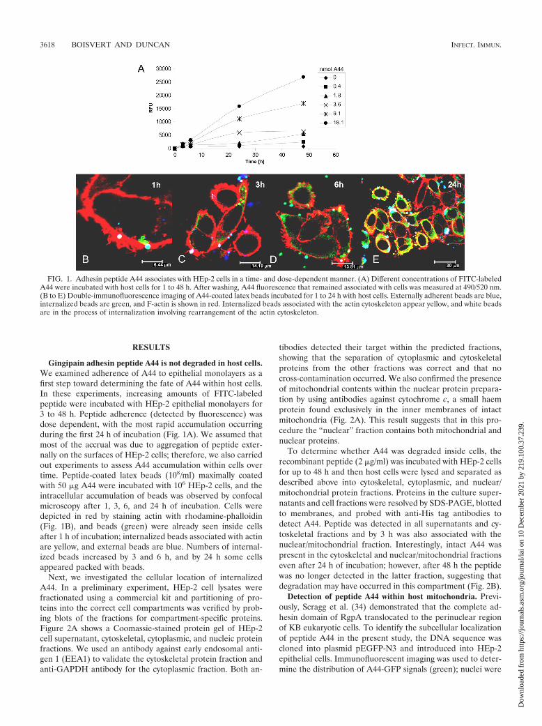
Gingipain adhesin peptide A44 is not degraded in host cells.We examined adherence of A44 to epithelial monolayers as afirst step toward determining the fate of A44 within host cells.In these experiments, increasing amounts of FITC-labeledpeptide were incubated with HEp-2 epithelial monolayers for3 to 48 h. Peptide adherence (detected by fluorescence) wasdose dependent, with the most rapid accumulation occurringduring the first 24 h of incubation (Fig. 1A). We assumed thatmost of the accrual was due to aggregation of peptide exter-nally on the surfaces of HEp-2 cells; therefore, we also carriedout experiments to assess A44 accumulation within cells overtime. Peptide-coated latex beads (108/ml) maximally coatedwith 50 �g A44 were incubated with 106 HEp-2 cells, and theintracellular accumulation of beads was observed by confocalmicroscopy after 1, 3, 6, and 24 h of incubation. Cells weredepicted in red by staining actin with rhodamine-phalloidin(Fig. 1B), and beads (green) were already seen inside cellsafter 1 h of incubation; internalized beads associated with actinare yellow, and external beads are blue. Numbers of internal-ized beads increased by 3 and 6 h, and by 24 h some cellsappeared packed with beads.
Next, we investigated the cellular location of internalizedA44. In a preliminary experiment, HEp-2 cell lysates werefractionated using a commercial kit and partitioning of pro-teins into the correct cell compartments was verified by prob-ing blots of the fractions for compartment-specific proteins.Figure 2A shows a Coomassie-stained protein gel of HEp-2cell supernatant, cytoskeletal, cytoplasmic, and nucleic proteinfractions. We used an antibody against early endosomal anti-gen 1 (EEA1) to validate the cytoskeletal protein fraction andanti-GAPDH antibody for the cytoplasmic fraction. Both an-
tibodies detected their target within the predicted fractions,showing that the separation of cytoplasmic and cytoskeletalproteins from the other fractions was correct and that nocross-contamination occurred. We also confirmed the presenceof mitochondrial contents within the nuclear protein prepara-tion by using antibodies against cytochrome c, a small haemprotein found exclusively in the inner membranes of intactmitochondria (Fig. 2A). This result suggests that in this pro-cedure the “nuclear” fraction contains both mitochondrial andnuclear proteins.
To determine whether A44 was degraded inside cells, therecombinant peptide (2 �g/ml) was incubated with HEp-2 cellsfor up to 48 h and then host cells were lysed and separated asdescribed above into cytoskeletal, cytoplasmic, and nuclear/mitochondrial protein fractions. Proteins in the culture super-natants and cell fractions were resolved by SDS-PAGE, blottedto membranes, and probed with anti-His tag antibodies todetect A44. Peptide was detected in all supernatants and cy-toskeletal fractions and by 3 h was also associated with thenuclear/mitochondrial fraction. Interestingly, intact A44 waspresent in the cytoskeletal and nuclear/mitochondrial fractionseven after 24 h of incubation; however, after 48 h the peptidewas no longer detected in the latter fraction, suggesting thatdegradation may have occurred in this compartment (Fig. 2B).
Detection of peptide A44 within host mitochondria. Previ-ously, Scragg et al. (34) demonstrated that the complete ad-hesin domain of RgpA translocated to the perinuclear regionof KB eukaryotic cells. To identify the subcellular localizationof peptide A44 in the present study, the DNA sequence wascloned into plasmid pEGFP-N3 and introduced into HEp-2epithelial cells. Immunofluorescent imaging was used to deter-mine the distribution of A44-GFP signals (green); nuclei were
FIG. 1. Adhesin peptide A44 associates with HEp-2 cells in a time- and dose-dependent manner. (A) Different concentrations of FITC-labeledA44 were incubated with host cells for 1 to 48 h. After washing, A44 fluorescence that remained associated with cells was measured at 490/520 nm.(B to E) Double-immunofluorescence imaging of A44-coated latex beads incubated for 1 to 24 h with host cells. Externally adherent beads are blue,internalized beads are green, and F-actin is shown in red. Internalized beads associated with the actin cytoskeleton appear yellow, and white beadsare in the process of internalization involving rearrangement of the actin cytoskeleton.
3618 BOISVERT AND DUNCAN INFECT. IMMUN.
Dow
nloa
ded
from
http
s://j
ourn
als.
asm
.org
/jour
nal/i
ai o
n 10
Dec
embe
r 20
21 b
y 21
9.10
0.37
.239
.

stained blue after treatment with the TO-PRO-3 DNA stain,and rhodamine-phalloidin (red) was used to reveal host actinand cell structure. Figure 3A and B show several cells express-ing A44-GFP in close proximity to TO-PRO-3-stained nuclei;
however, the blue DNA stain appeared to colocalize with A44-GFP in the perinuclear region. Expression of GFP can be toxicto cells, and in these experiments relatively few A44-GFP-expressing cells were observed within fields of view. Therefore,
FIG. 2. Translocation of peptide A44. (A) Coomassie-stained gel and Western blots of HEp-2 protein fractions: supernatant (SN), cytoskeletal (Sk),cytoplasmic (Cp), and nuclear (Nuc). Correct separation of fractions is shown using antibodies against cytoskeletal protein early endosome antigen 1(EEA1), cytoplasmic protein glyceraldehyde-3-phosphate dehydrogenase (GAPDH), and mitochondrial marker cytochrome c (CytC). �, anti. (B) De-tection of peptide A44 in HEp-2 fractions on prolonged incubation. Peptide A44 was detected in host cell fractions with anti-His tag antibodies.
FIG. 3. Confocal microscopy reveals that peptide A44 translocates to mitochondria. (A, B) A44-GFP expressed in host cells colocalizes withnucleic acid stain TO-PRO-3 outside host nuclei. (C) MitoTracker (red) was used to label mitochondria. The merged image shows FITC-labeledA44 (green) translocated to host mitochondria (colocalization is shown in yellow).
VOL. 78, 2010 GINGIPAIN ADHESIN PEPTIDE PREVENTS APOPTOSIS 3619
Dow
nloa
ded
from
http
s://j
ourn
als.
asm
.org
/jour
nal/i
ai o
n 10
Dec
embe
r 20
21 b
y 21
9.10
0.37
.239
.

the experiments were repeated with FITC-labeled A44 (green)and MitoTracker (red) to stain membranes of mitochondria,the only other DNA-containing organelles in eukaryotic cells.As shown in Fig. 3C, FITC-labeled A44 colocalized with mi-tochondria in the perinuclear region, implying that A44 traf-ficked to mitochondria and not cell nuclei.
As an additional approach, we used immunogold labelingand transmission electron microscopy to confirm the translo-cation of peptide A44 to mitochondria. First, we treated prep-arations of the A44-GFP-expressing HEp-2 cell line with A44-specific antibodies, followed by 10-nm-gold-labeled anti-rabbitsecondary antibodies. As shown in Fig. 4A, gold particles canbe seen within mitochondrial structures, confirming the traf-ficking of A44 to and localization within mitochondria. In asecond set of experiments to determine whether exogenouslyadded A44 also translocates to host mitochondria, we addedA44 (2 �g/ml) to HEp-2 epithelial cells, then used the sameimmunogold-labeling technique and transmission electron mi-croscopy to visualize the peptide (Fig. 4B). There were nodifferences in localization of endogenous transiently expressedA44 and exogenously added peptide, as both migrated to hostmitochondria. However, as measured previously, the amountof A44 detected within cells was much larger when the peptidewas added exogenously. These data suggest that even though
A44 is intimately associated with mitochondria there are notoxic effects on cell viability.
Peptide A44 blocks apoptosis in host epithelial cells. Toinvestigate whether uptake of A44 into mitochondria affectedhost cell viability, we used a quantitative colorimetric assaywith MTT as an indicator of mitochondrial dehydrogenase(oxidoreductase) activities (23). Since the tetrazolium salt iscleaved only by hydrogenases produced from active mitochon-dria, the formation of a colored product (reduced tetrazolium)is an indicator of living cells. Monolayers of epithelial cellswere incubated with A44 (2 �g/ml) for up to 24 h, and therewas no reduction in indicator color compared to the negativecontrol (monolayers to which no peptide was added) (Fig. 5,�STS group), indicating that A44 does not damage host mi-tochondria and affect viability.
Next, we investigated whether A44 could protect host cellsfrom staurosporine-induced apoptosis. As above, cells werepreincubated with A44 (2 �g/ml) for 1 to 24 h, followed by theaddition of fresh medium containing 2 �M staurosporine toinduce apoptosis, and incubation continued for a further 24 h.As shown in Fig. 5 (�STS group), cells that were not pre-treated with A44 before the staurosporine challenge showedthe lowest viability (10 to 15%), and a similar result was ob-tained when cells had only a short (1-h) pretreatment with
FIG. 4. Localization of A44 within mitochondria. Shown is transmission electron microscopy of sections of host cells either expressing A44-GFP(A) or incubated with exogenous peptide A44 (B). A44 was detected with A44-specific (A) or anti-His tag (B) antibodies. Secondary antibodieswere 10-nm-gold-labeled anti-rabbit antibodies. Arrows indicate internalized, gold-labeled A44. Scale bars, 100 nm.
FIG. 5. Peptide A44 prevents staurosporine-induced apoptosis in host cells. Apoptosis was induced with 2 �M staurosporine for 24 h in cellspreincubated with peptide A44. Percentages of live cells were extrapolated from amounts of MTT indicator taken up by cells. Student’s t test fromat least three different experiments revealed statistical significance of viable cells (*, P 0.05).
3620 BOISVERT AND DUNCAN INFECT. IMMUN.
Dow
nloa
ded
from
http
s://j
ourn
als.
asm
.org
/jour
nal/i
ai o
n 10
Dec
embe
r 20
21 b
y 21
9.10
0.37
.239
.

peptide A44 (15 to 20% live cells). As shown in Fig. 5, longerpretreatments of host cells with A44 afforded greater protec-tion against staurosporine-induced cell death; for example,host cell cultures pretreated for 3 and 6 h showed higherpercentages of live cells, 43% and 57%, respectively. Preincu-bation with peptide for 16 h gave the most protection, with allcells retaining viability. Lastly, the antiapoptotic effect of A44decreased over time since a 24-h pretreatment yielded approx-imately 59% cell viability.
A44 induces the expression of antiapoptotic factors. Apop-tosis is controlled by the balance of pro- and antiapoptoticfactors at the mitochondrial membrane, and the expression ofthese factors is regulated by several signal transduction sys-tems. Therefore, we investigated whether peptide A44 and liveP. gingivalis affect the expression of these factors in HEp-2epithelial cells. Following treatment of monolayers with eitherpurified peptide A44 (2 �g/ml) for up to 24 h or P. gingivalis forup to 6 h, HEp-2 cellular RNA (2 �g) was transcribed intocDNA using oligo(dT) primers, and the expression of severalpro- and antiapoptotic factors was quantified by qRT-PCR.We used the �-globin gene as the internal control host gene tocalculate the relative expression levels of 4 antiapoptotic genes,bcl-2, bcl-XL, mcl-1, and the gene encoding NF-�B, whichactivates inhibitor of apoptosis proteins (IAP). In addition, wemeasured the expression of 3 proapoptotic genes, bax, bak, andthe TNF-� gene, and of prosurvival factors rhoA, stat3, andcIAP1. As depicted in Fig. 6A, bcl-2 expression is over 2-foldhigher in cells incubated with peptide A44 for 1, 3, or 6 h than
in cells without A44. After 6 h a 2-fold induction for bcl-XLwas also detected. We detected up to 3-fold-higher expressionlevels of bcl-2 after 16 h of incubation compared to those forcells without A44. These results suggest that the induction ofantiapoptotic factors was an early effect of A44 treatment.When HEp-2 epithelial cells were incubated with A44 for 24 h,quantitative analysis did not show induced gene expression,and we concluded that the effect of A44 on antiapoptoticfactors Bcl-2 and Bcl-XL was transient and limited to the earlystages of infection.
Expression of the same genes was quantified after 1, 3, and6 h of incubation with P. gingivalis cells (Fig. 6B). Incubationfor 1 h did not change the expression profile in cells; however,a significant increase (2-fold) in bcl-2 expression was observedafter 3 and 6 h of incubation with bacteria. bcl-XL expressionwas also upregulated approximately 2-fold after 6 h. We alsodetected a 2-fold induction of mcl-1 expression and up to3-fold increases in NF-�B and TNF-� RNA transcripts after6 h of incubation. After longer incubations HEp-2 cells de-tached from plates, indicative of loss of viability due to apop-tosis. Thus, as with A44, the early events in P. gingivalis infec-tion appear to be antiapoptotic and temporary.
Peptide A44 delays activation of caspase-9. Overexpressionof antiapoptotic bcl-2 family members is known to preservemitochondrial membrane potential integrity and therefore pre-vent leakage of cytochrome c into the cytoplasm. Release ofcytoplasmic cytochrome c activates caspase-9, which in turnactivates caspase-3, an executioner caspase that initiates irre-
FIG. 6. Expression of apoptosis-related genes in host cells after incubation with A44 or live P. gingivalis. (A) After 1 to 24 h of incubation withpeptide A44, relative gene expression was measured by quantitative RT-PCR. (B) Relative gene expression in cells incubated for 1, 3, and 6 h withP. gingivalis (Pg) 33277 was measured by qRT-PCR. Using Student’s t test statistical significance in expression levels from at least three separateexperiments was obtained (*, P 0.05; **, P 0.1).
VOL. 78, 2010 GINGIPAIN ADHESIN PEPTIDE PREVENTS APOPTOSIS 3621
Dow
nloa
ded
from
http
s://j
ourn
als.
asm
.org
/jour
nal/i
ai o
n 10
Dec
embe
r 20
21 b
y 21
9.10
0.37
.239
.
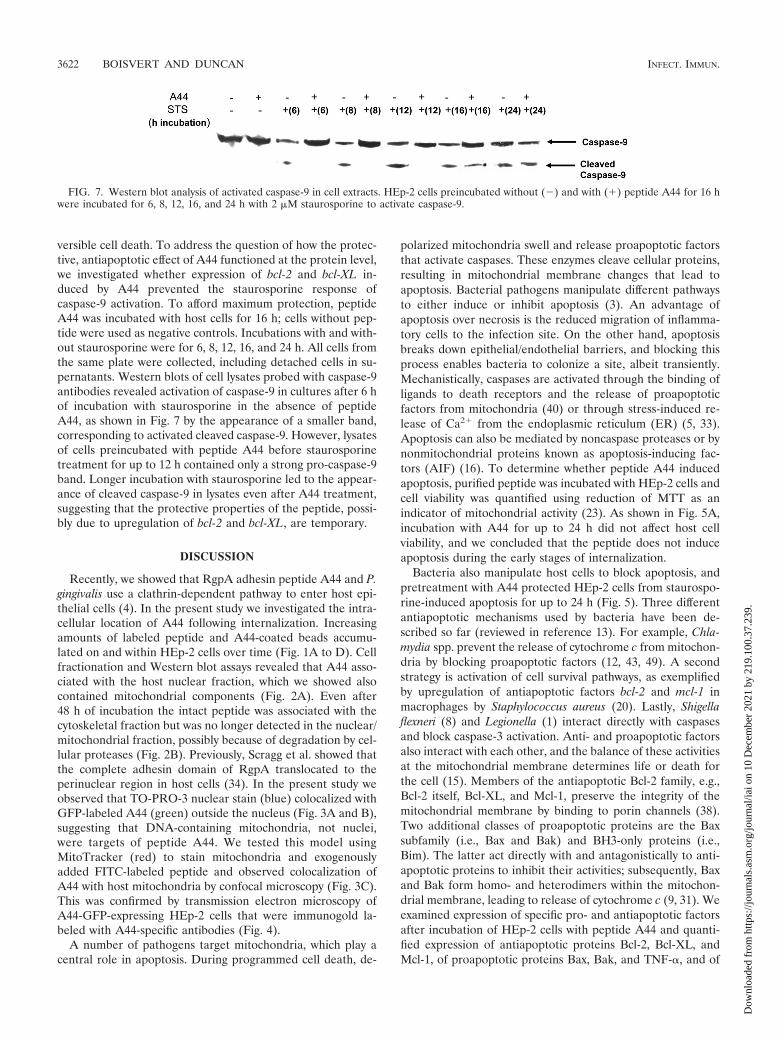
versible cell death. To address the question of how the protec-tive, antiapoptotic effect of A44 functioned at the protein level,we investigated whether expression of bcl-2 and bcl-XL in-duced by A44 prevented the staurosporine response ofcaspase-9 activation. To afford maximum protection, peptideA44 was incubated with host cells for 16 h; cells without pep-tide were used as negative controls. Incubations with and with-out staurosporine were for 6, 8, 12, 16, and 24 h. All cells fromthe same plate were collected, including detached cells in su-pernatants. Western blots of cell lysates probed with caspase-9antibodies revealed activation of caspase-9 in cultures after 6 hof incubation with staurosporine in the absence of peptideA44, as shown in Fig. 7 by the appearance of a smaller band,corresponding to activated cleaved caspase-9. However, lysatesof cells preincubated with peptide A44 before staurosporinetreatment for up to 12 h contained only a strong pro-caspase-9band. Longer incubation with staurosporine led to the appear-ance of cleaved caspase-9 in lysates even after A44 treatment,suggesting that the protective properties of the peptide, possi-bly due to upregulation of bcl-2 and bcl-XL, are temporary.
DISCUSSION
Recently, we showed that RgpA adhesin peptide A44 and P.gingivalis use a clathrin-dependent pathway to enter host epi-thelial cells (4). In the present study we investigated the intra-cellular location of A44 following internalization. Increasingamounts of labeled peptide and A44-coated beads accumu-lated on and within HEp-2 cells over time (Fig. 1A to D). Cellfractionation and Western blot assays revealed that A44 asso-ciated with the host nuclear fraction, which we showed alsocontained mitochondrial components (Fig. 2A). Even after48 h of incubation the intact peptide was associated with thecytoskeletal fraction but was no longer detected in the nuclear/mitochondrial fraction, possibly because of degradation by cel-lular proteases (Fig. 2B). Previously, Scragg et al. showed thatthe complete adhesin domain of RgpA translocated to theperinuclear region in host cells (34). In the present study weobserved that TO-PRO-3 nuclear stain (blue) colocalized withGFP-labeled A44 (green) outside the nucleus (Fig. 3A and B),suggesting that DNA-containing mitochondria, not nuclei,were targets of peptide A44. We tested this model usingMitoTracker (red) to stain mitochondria and exogenouslyadded FITC-labeled peptide and observed colocalization ofA44 with host mitochondria by confocal microscopy (Fig. 3C).This was confirmed by transmission electron microscopy ofA44-GFP-expressing HEp-2 cells that were immunogold la-beled with A44-specific antibodies (Fig. 4).
A number of pathogens target mitochondria, which play acentral role in apoptosis. During programmed cell death, de-
polarized mitochondria swell and release proapoptotic factorsthat activate caspases. These enzymes cleave cellular proteins,resulting in mitochondrial membrane changes that lead toapoptosis. Bacterial pathogens manipulate different pathwaysto either induce or inhibit apoptosis (3). An advantage ofapoptosis over necrosis is the reduced migration of inflamma-tory cells to the infection site. On the other hand, apoptosisbreaks down epithelial/endothelial barriers, and blocking thisprocess enables bacteria to colonize a site, albeit transiently.Mechanistically, caspases are activated through the binding ofligands to death receptors and the release of proapoptoticfactors from mitochondria (40) or through stress-induced re-lease of Ca2� from the endoplasmic reticulum (ER) (5, 33).Apoptosis can also be mediated by noncaspase proteases or bynonmitochondrial proteins known as apoptosis-inducing fac-tors (AIF) (16). To determine whether peptide A44 inducedapoptosis, purified peptide was incubated with HEp-2 cells andcell viability was quantified using reduction of MTT as anindicator of mitochondrial activity (23). As shown in Fig. 5A,incubation with A44 for up to 24 h did not affect host cellviability, and we concluded that the peptide does not induceapoptosis during the early stages of internalization.
Bacteria also manipulate host cells to block apoptosis, andpretreatment with A44 protected HEp-2 cells from staurospo-rine-induced apoptosis for up to 24 h (Fig. 5). Three differentantiapoptotic mechanisms used by bacteria have been de-scribed so far (reviewed in reference 13). For example, Chla-mydia spp. prevent the release of cytochrome c from mitochon-dria by blocking proapoptotic factors (12, 43, 49). A secondstrategy is activation of cell survival pathways, as exemplifiedby upregulation of antiapoptotic factors bcl-2 and mcl-1 inmacrophages by Staphylococcus aureus (20). Lastly, Shigellaflexneri (8) and Legionella (1) interact directly with caspasesand block caspase-3 activation. Anti- and proapoptotic factorsalso interact with each other, and the balance of these activitiesat the mitochondrial membrane determines life or death forthe cell (15). Members of the antiapoptotic Bcl-2 family, e.g.,Bcl-2 itself, Bcl-XL, and Mcl-1, preserve the integrity of themitochondrial membrane by binding to porin channels (38).Two additional classes of proapoptotic proteins are the Baxsubfamily (i.e., Bax and Bak) and BH3-only proteins (i.e.,Bim). The latter act directly with and antagonistically to anti-apoptotic proteins to inhibit their activities; subsequently, Baxand Bak form homo- and heterodimers within the mitochon-drial membrane, leading to release of cytochrome c (9, 31). Weexamined expression of specific pro- and antiapoptotic factorsafter incubation of HEp-2 cells with peptide A44 and quanti-fied expression of antiapoptotic proteins Bcl-2, Bcl-XL, andMcl-1, of proapoptotic proteins Bax, Bak, and TNF-�, and of
FIG. 7. Western blot analysis of activated caspase-9 in cell extracts. HEp-2 cells preincubated without (�) and with (�) peptide A44 for 16 hwere incubated for 6, 8, 12, 16, and 24 h with 2 �M staurosporine to activate caspase-9.
3622 BOISVERT AND DUNCAN INFECT. IMMUN.
Dow
nloa
ded
from
http
s://j
ourn
als.
asm
.org
/jour
nal/i
ai o
n 10
Dec
embe
r 20
21 b
y 21
9.10
0.37
.239
.

prosurvival factors NF-�B, Stat3, RhoA, and cIAP-1. Expres-sion of Bcl-2 was upregulated in cells incubated with A44 forup to 16 h (Fig. 6). Upregulation of antiapoptotic proteins suchas Bcl-2 prevents release of cytochrome c and has also beenshown to abrogate the function of proapoptotic proteins andprotect against cell death (18). We also detected a transient2-fold induction of bcl-XL after 6 h of incubation; however, by24 h expression of bcl-XL decreased to control levels, as mea-sured in cells without the peptide. The half-life of bcl-2 RNAis relatively short compared to the long half-life (up to 10 h) ofthe Bcl-2 protein, and differences in mRNA levels detected inour qRT-PCR experiments and the actual number of live cellsin MTT assays could be due to the different turnover rates forRNA and protein (30). Timelines for and the progression ofvarious apoptotic events vary greatly in cells, and the timerequired for an activity to peak in one cell line might differfrom that required in another. Several studies showed thatoverexpression of Bcl-2 was not effective in protecting cellsfrom staurosporine-induced apoptosis; however, overex-pressed Bcl-XL was able to protect neuroblastoma cells (47).In our experiments A44 delayed staurosporine-induced apop-tosis for up to 24 h, at which point there were still more livecells (35 to 40%) than in controls without A44 treatment.
When HEp-2 cells were incubated with live P. gingivalis forup to 6 h, we detected 2-fold increases in levels of Bcl-2 andBcl-XL (Fig. 6B). We also found significantly higher levels ofthe transcript for Mcl-1, which blocks proapoptotic factors andinhibits the oligomerization of Bak and Bax in the mitochon-drial membrane. Mcl-1 has a faster turnover rate then Bcl-2and is modulated and activated by several growth factors, in-cluding phosphorylated Akt and phosphatidylinositol 3-kinase(10, 29, 30). In addition, NF-�B and TNF-� levels were 2.5-and 3.3-fold higher, respectively, than those in uninfected cells.While NF-�B prevents apoptosis by blocking cytochrome crelease and activation of caspases-9 and -3, TNF-� inducesapoptosis by activating caspase-8 in the extrinsic apoptosispathway. In our assays, cells incubated with P. gingivalis formore than 6 h began detaching from tissue culture plates (datanot shown), indicative of loss of viability. Thus, it is possiblethat, upon longer incubation, P. gingivalis activation of deathreceptors via TNF-� cannot be offset by simultaneously acti-vated NF-�B, Bcl-2, Bcl-XL, and Mcl-1.
There are contradictory reports on whether P. gingivalis haspro- or antiapoptotic effects on host cells. In one study, the anal-ysis of the transcriptional profile of gingival epithelial cells ex-posed for 2 h to P. gingivalis showed an increase in bcl-2 and bif-1,which protected host cells from camptothecin-induced apoptosis(17). In an earlier study it was also shown that P. gingivalis 33277rescued primary gingival epithelial cells from camptothecin-in-duced apoptosis since early upregulation of bax expression pre-ceded upregulation of bcl-2 at 24 h, leading to the conclusion thatthe first cellular response of apoptosis was subsequently delayedby P. gingivalis (25). In another study with P. gingivalis 33277,staurosporine-induced apoptosis was blocked but restored in thepresence of a phosphatidylinositol 3-kinase inhibitor, implicatinga pathway in which phosphorylated Akt and activated NF-�Bprevented depolymerization of the mitochondrial membrane andcytochrome c release induced by staurosporine (45). This is con-sistent with our observation of higher NF-�B levels in cells ex-posed to the same P. gingivalis strain. Another antiapoptotic path-
way was detected when gingival epithelial cells were infected withP. gingivalis 33277 whereby, in addition to Stat3, Jak, an upstreamkinase, was also upregulated (21). In the present study we did notdetect differences in stat3 levels, possibly due to the different hostcell types used; however, Stat3-induced survival responses such asupregulated bcl-2, bcl-XL, and mcl-1 were observed, and the over-all antiapoptotic effect was the same. Recently, it was suggestedthat gingipains of P. gingivalis strain W50 mediate apoptosis in adose-dependent manner (26). Strain W83 induced apoptosis bygingipain-mediated cleavage of N- and VE-cadherins, resulting inloss of epithelial and endothelial cell adhesion by caspase-depen-dent and -independent mechanisms (7, 36, 37). Indeed, severalother studies demonstrated that on prolonged incubation pro-teases destroy cells and tissue and induce apoptosis, implying thatgingipain activity is decreased when P. gingivalis is internalized byhost cells, allowing survival (48). We also observed loss of cellviability after infection with P. gingivalis 33277, but only afterprolonged incubation of more than 6 h. In the studies cited above,concentrations of P. gingivalis and/or gingipains were much higherthan those used in the present study; however, we also found thatincubations with similarly high concentrations of bacteria or pro-teases led to host cell death (H. Boisvert, unpublished data).
Staurosporine-induced apoptosis results in the translocation ofBax from the cytosol to mitochondria and loss of membranepotential due to opening of mitochondrial pores. The subsequentreleases of cytochrome c and apoptosis-inducing factors initiatethe apoptotic cascade through activation of caspase-9 (19, 27, 42).Our hypothesis that A44 delays apoptosis was tested with stauro-sporine-treated cells, and we demonstrated that 16 h of preincu-bation with peptide A44 delayed activation of caspase-9 for up to12 h compared to activation in cells without peptide A44. How-ever, A44 could not prevent cleavage and activation of caspase-9after longer staurosporine exposure.
In summary, our previous work demonstrated, for the firsttime, that host cells internalized RgpA peptide A44 and live P.gingivalis via a clathrin-dependent mechanism. In the presentstudy we continued this mechanistic analysis and identified eventsfollowing A44 entry into cells. Our data showed that peptide A44entered host cells efficiently and was not degraded. We discoveredthat the peptide trafficked to mitochondria, where it has the po-tential to direct host cells toward survival or death by manipulat-ing apoptosis. By quantifying of transcription of pro- and anti-apoptotic factors produced by cells in response to A44, a modelemerged whereby the peptide induced expression of antiapop-totic factors, confirmed by the observation that A44 protectedagainst staurosporine-induced apoptosis. On the other hand, theinitial antiapoptotic effect was short-lived in cells infected with liveP. gingivalis, as indicated by observations of cell rounding anddetachment after 6 h of incubation. This result could be antici-pated since whole bacteria present to the cell a more complexmixture of surface proteins that potentially promote a more ef-fective apoptotic response.
ACKNOWLEDGMENTS
We thank Ziedonis Skobe and Justine Dobeck for advice and helpwith electron and confocal microscopy.
This work was supported by U.S. National Institutes of Healthgrants R01-DE015931 and T32-DE007327-08.
VOL. 78, 2010 GINGIPAIN ADHESIN PEPTIDE PREVENTS APOPTOSIS 3623
Dow
nloa
ded
from
http
s://j
ourn
als.
asm
.org
/jour
nal/i
ai o
n 10
Dec
embe
r 20
21 b
y 21
9.10
0.37
.239
.

REFERENCES
1. Abu-Zant, A., M. Santic, M. Molmeret, S. Jones, J. Helbig, and Y. Abu Kwaik.2005. Incomplete activation of macrophage apoptosis during intracellular rep-lication of Legionella pneumophila. Infect. Immun. 73:5339–5349.
2. Blanke, S. R. 2005. Micro-managing the executioner: pathogen targeting ofmitochondria. Trends Microbiol. 13:64–71.
3. Bohme, L., and T. Rudel. 2009. Host cell death machinery as a target forbacterial pathogens. Microbes Infect. 11:1063–1070.
4. Boisvert, H., and M. J. Duncan. 2008. Clathrin-dependent entry of a gingi-pain adhesin peptide and Porphyromonas gingivalis into host cells. Cell.Microbiol. 10:2538–2552.
5. Breckenridge, D. G., M. Germain, J. P. Mathai, M. Nguyen, and G. C. Shore.2003. Regulation of apoptosis by endoplasmic reticulum pathways. Onco-gene 22:8608–8618.
6. Chen, T., and M. J. Duncan. 2004. Gingipain adhesin domains mediatePorphyromonas gingivalis adherence to epithelial cells. Microb. Pathog. 36:205–209.
7. Chen, Z., C. A. Casiano, and H. M. Fletcher. 2001. Protease-active extracel-lular protein preparations from Porphyromonas gingivalis W83 induce N-cadherin proteolysis, loss of cell adhesion, and apoptosis in human epithelialcells. J. Periodontol. 72:641–650.
8. Clark, C. S., and A. T. Maurelli. 2007. Shigella flexneri inhibits staurospo-rine-induced apoptosis in epithelial cells. Infect. Immun. 75:2531–2539.
9. Cory, S., and J. M. Adams. 2002. The Bcl2 family: regulators of the cellularlife-or-death switch. Nat. Rev. Cancer 2:647–656.
10. Cuconati, A., C. Mukherjee, D. Perez, and E. White. 2003. DNA damageresponse and MCL-1 destruction initiate apoptosis in adenovirus-infectedcells. Genes Dev. 17:2922–2932.
11. Dimmer, K. S., and D. Rapaport. 2008. Proteomic view of mitochondrialfunction. Genome Biol. 9:209.
12. Dong, F., M. Pirbhai, Y. Xiao, Y. Zhong, Y. Wu, and G. Zhong. 2005.Degradation of the proapoptotic proteins Bik, Puma, and Bim with Bcl-2domain 3 homology in Chlamydia trachomatis-infected cells. Infect. Immun.73:1861–1864.
13. Faherty, C. S., and A. T. Maurelli. 2008. Staying alive: bacterial inhibition ofapoptosis during infection. Trends Microbiol. 16:173–180.
14. Grassme, H., V. Jendrossek, and E. Gulbins. 2001. Molecular mechanisms ofbacteria induced apoptosis. Apoptosis 6:441–445.
15. Green, D. R., and J. C. Reed. 1998. Mitochondria and apoptosis. Science281:1309–1312.
16. Hail, N., Jr., B. Z. Carter, M. Konopleva, and M. Andreeff. 2006. Apoptosiseffector mechanisms: a requiem performed in different keys. Apoptosis 11:889–904.
17. Handfield, M., J. J. Mans, G. Zheng, M. C. Lopez, S. Mao, A. Progulske-Fox,G. Narasimhan, H. V. Baker, and R. J. Lamont. 2005. Distinct transcrip-tional profiles characterize oral epithelium-microbiota interactions. Cell.Microbiol. 7:811–823.
18. Kempf, V. A., A. Schairer, D. Neumann, G. A. Grassl, K. Lauber, M. Leb-iedziejewski, M. Schaller, P. Kyme, S. Wesselborg, and I. B. Autenrieth.2005. Bartonella henselae inhibits apoptosis in Mono Mac 6 cells. Cell.Microbiol. 7:91–104.
19. Kluck, R. M., E. Bossy-Wetzel, D. R. Green, and D. D. Newmeyer. 1997. Therelease of cytochrome c from mitochondria: a primary site for Bcl-2 regula-tion of apoptosis. Science 275:1132–1136.
20. Koziel, J., A. Maciag-Gudowska, T. Mikolajczyk, M. Bzowska, D. E. Sturdevant,A. R. Whitney, L. N. Shaw, F. R. DeLeo, and J. Potempa. 2009. Phagocytosis ofStaphylococcus aureus by macrophages exerts cytoprotective effects manifestedby the upregulation of antiapoptotic factors. PLoS One 4:e5210.
21. Mao, S., Y. Park, Y. Hasegawa, G. D. Tribble, C. E. James, M. Handfield,M. F. Stavropoulos, O. Yilmaz, and R. J. Lamont. 2007. Intrinsic apoptoticpathways of gingival epithelial cells modulated by Porphyromonas gingivalis.Cell. Microbiol. 9:1997–2007.
22. Reference deleted.23. Mosmann, T. 1983. Rapid colorimetric assay for cellular growth and survival:
application to proliferation and cytotoxicity assays. J. Immunol. Methods65:55–63.
24. Nakagawa, I., A. Amano, M. Kuboniwa, T. Nakamura, S. Kawabata, and S.Hamada. 2002. Functional differences among FimA variants of Porphyromo-nas gingivalis and their effects on adhesion to and invasion of human epi-thelial cells. Infect. Immun. 70:277–285.
25. Nakhjiri, S. F., Y. Park, O. Yilmaz, W. O. Chung, K. Watanabe, A. El-Sabaeny, K. Park, and R. J. Lamont. 2001. Inhibition of epithelial cellapoptosis by Porphyromonas gingivalis. FEMS Microbiol. Lett. 200:145–149.
26. O’Brien-Simpson, N. M., R. D. Pathirana, G. D. Walker, and E. C. Reynolds.2009. Porphyromonas gingivalis RgpA-Kgp proteinase-adhesin complexespenetrate gingival tissue and induce proinflammatory cytokines or apoptosisin a concentration-dependent manner. Infect. Immun. 77:1246–1261.
27. Petit, P. X., M. Goubern, P. Diolez, S. A. Susin, N. Zamzami, and G.Kroemer. 1998. Disruption of the outer mitochondrial membrane as a resultof large amplitude swelling: the impact of irreversible permeability transi-tion. FEBS Lett. 426:111–116.
28. Pfaffl, M. W. 2001. A new mathematical model for relative quantification inreal-time RT-PCR. Nucleic Acids Res. 29:e45.
29. Rajalingam, K., M. Sharma, C. Lohmann, M. Oswald, O. Thieck, C. J.Froelich, and T. Rudel. 2008. Mcl-1 is a key regulator of apoptosis resistancein Chlamydia trachomatis-infected cells. PLoS One 3:e3102.
30. Reed, J. C. 1996. A day in the life of the Bcl-2 protein: does the turnover rateof Bcl-2 serve as a biological clock for cellular lifespan regulation? Leuk.Res. 20:109–111.
31. Roset, R., L. Ortet, and G. Gil-Gomez. 2007. Role of Bcl-2 family memberson apoptosis: what we have learned from knock-out mice. Front. Biosci.12:4722–4730.
32. Scheffler, I. E. 2001. A century of mitochondrial research: achievements andperspectives. Mitochondrion 1:3–31.
33. Scorrano, L., S. A. Oakes, J. T. Opferman, E. H. Cheng, M. D. Sorcinelli, T.Pozzan, and S. J. Korsmeyer. 2003. BAX and BAK regulation of endoplas-mic reticulum Ca2�: a control point for apoptosis. Science 300:135–139.
34. Scragg, M. A., A. Alsam, M. Rangarajan, J. M. Slaney, P. Shepherd, D. M.Williams, and M. A. Curtis. 2002. Nuclear targeting of Porphyromonasgingivalis W50 protease in epithelial cells. Infect. Immun. 70:5740–5750.
35. Scragg, M. A., S. J. Cannon, M. Rangarajan, D. M. Williams, and M. A.Curtis. 1999. Targeted disruption of fibronectin-integrin interactions in hu-man gingival fibroblasts by the RI protease of Porphyromonas gingivalisW50. Infect. Immun. 67:1837–1843.
36. Sheets, S. M., J. Potempa, J. Travis, C. A. Casiano, and H. M. Fletcher. 2005.Gingipains from Porphyromonas gingivalis W83 induce cell adhesion mole-cule cleavage and apoptosis in endothelial cells. Infect. Immun. 73:1543–1552.
37. Sheets, S. M., J. Potempa, J. Travis, H. M. Fletcher, and C. A. Casiano. 2006.Gingipains from Porphyromonas gingivalis W83 synergistically disrupt en-dothelial cell adhesion and can induce caspase-independent apoptosis. In-fect. Immun. 74:5667–5678.
38. Shimizu, S., M. Narita, and Y. Tsujimoto. 1999. Bcl-2 family proteins reg-ulate the release of apoptogenic cytochrome c by the mitochondrial channelVDAC. Nature 399:483–487.
39. Smith, D. E., and P. A. Fisher. 1984. Identification, developmental regula-tion, and response to heat shock of two antigenically related forms of a majornuclear envelope protein in Drosophila embryos: application of an improvedmethod for affinity purification of antibodies using polypeptides immobilizedon nitrocellulose blots. J. Cell Biol. 99:20–28.
40. Thornberry, N. A., and Y. Lazebnik. 1998. Caspases: enemies within. Science281:1312–1316.
41. Weinrauch, Y., and A. Zychlinsky. 1999. The induction of apoptosis bybacterial pathogens. Annu. Rev. Microbiol. 53:155–187.
42. Wolf, C. M., and A. Eastman. 1999. The temporal relationship betweenprotein phosphatase, mitochondrial cytochrome c release, and caspase acti-vation in apoptosis. Exp. Cell Res. 247:505–513.
43. Xiao, Y., Y. Zhong, W. Greene, F. Dong, and G. Zhong. 2004. Chlamydiatrachomatis infection inhibits both Bax and Bak activation induced by stauro-sporine. Infect. Immun. 72:5470–5474.
44. Yilmaz, O. 2008. The chronicles of Porphyromonas gingivalis: the micro-bium, the human oral epithelium and their interplay. Microbiology 154:2897–2903.
45. Yilmaz, O., T. Jungas, P. Verbeke, and D. M. Ojcius. 2004. Activation of thephosphatidylinositol 3-kinase/Akt pathway contributes to survival of primaryepithelial cells infected with the periodontal pathogen Porphyromonas gin-givalis. Infect. Immun. 72:3743–3751.
46. Yilmaz, O., K. Watanabe, and R. J. Lamont. 2002. Involvement of integrinsin fimbriae-mediated binding and invasion by Porphyromonas gingivalis.Cell. Microbiol. 4:305–314.
47. Yuste, V. J., I. Sanchez-Lopez, C. Sole, M. Encinas, J. R. Bayascas, J. Boix,and J. X. Comella. 2002. The prevention of the staurosporine-inducedapoptosis by Bcl-X(L), but not by Bcl-2 or caspase inhibitors, allows theextensive differentiation of human neuroblastoma cells. J. Neurochem.80:126–139.
48. Zhang, Y., T. Wang, W. Chen, O. Yilmaz, Y. Park, I. Y. Jung, M. Hackett,and R. J. Lamont. 2005. Differential protein expression by Porphyromonasgingivalis in response to secreted epithelial cell components. Proteomics5:198–211.
49. Zhong, Y., M. Weininger, M. Pirbhai, F. Dong, and G. Zhong. 2006. Inhi-bition of staurosporine-induced activation of the proapoptotic multidomainBcl-2 proteins Bax and Bak by three invasive chlamydial species. J. Infect.53:408–414.
Editor: S. M. Payne
3624 BOISVERT AND DUNCAN INFECT. IMMUN.
Dow
nloa
ded
from
http
s://j
ourn
als.
asm
.org
/jour
nal/i
ai o
n 10
Dec
embe
r 20
21 b
y 21
9.10
0.37
.239
.