transmission of infection by flexible gastrointestinal ...cmr.asm.org/content/26/2/231.full.pdf ·...
TRANSCRIPT
Transmission of Infection by Flexible Gastrointestinal Endoscopy andBronchoscopy
Julia Kovaleva,a Frans T. M. Peters,b Henny C. van der Mei,c John E. Degenera
Department of Medical Microbiology, University of Groningen, University Medical Center Groningen, Groningen, The Netherlandsa; Endoscopy Center, University ofGroningen, University Medical Center Groningen, Groningen, The Netherlandsb; Department of Biomedical Engineering, University of Groningen, University MedicalCenter Groningen, Groningen, The Netherlandsc
SUMMARY . . . . . . . . . . . . . . . . . . . . . . . . . . . . . . . . . . . . . . . . . . . . . . . . . . . . . . . . . . . . . . . . . . . . . . . . . . . . . . . . . . . . . . . . . . . . . . . . . . . . . . . . . . . . . . . . . . . . . . . . . . . . . . . . . . . . . . . . . . . . . . . . . .231INTRODUCTION . . . . . . . . . . . . . . . . . . . . . . . . . . . . . . . . . . . . . . . . . . . . . . . . . . . . . . . . . . . . . . . . . . . . . . . . . . . . . . . . . . . . . . . . . . . . . . . . . . . . . . . . . . . . . . . . . . . . . . . . . . . . . . . . . . . . . . . . . . . .231PRACTICAL ASPECTS OF FLEXIBLE ENDOSCOPE REPROCESSING . . . . . . . . . . . . . . . . . . . . . . . . . . . . . . . . . . . . . . . . . . . . . . . . . . . . . . . . . . . . . . . . . . . . . . . . . . . . . . . . . . . . . . . .232
Relevance of Cleaning and Disinfection . . . . . . . . . . . . . . . . . . . . . . . . . . . . . . . . . . . . . . . . . . . . . . . . . . . . . . . . . . . . . . . . . . . . . . . . . . . . . . . . . . . . . . . . . . . . . . . . . . . . . . . . . . . . . . . . . .232Relevance of Drying and Storage. . . . . . . . . . . . . . . . . . . . . . . . . . . . . . . . . . . . . . . . . . . . . . . . . . . . . . . . . . . . . . . . . . . . . . . . . . . . . . . . . . . . . . . . . . . . . . . . . . . . . . . . . . . . . . . . . . . . . . . . .234
EXOGENOUS INFECTIONS ASSOCIATED WITH FLEXIBLE ENDOSCOPY . . . . . . . . . . . . . . . . . . . . . . . . . . . . . . . . . . . . . . . . . . . . . . . . . . . . . . . . . . . . . . . . . . . . . . . . . . . . . . . . . . .234Bacteria . . . . . . . . . . . . . . . . . . . . . . . . . . . . . . . . . . . . . . . . . . . . . . . . . . . . . . . . . . . . . . . . . . . . . . . . . . . . . . . . . . . . . . . . . . . . . . . . . . . . . . . . . . . . . . . . . . . . . . . . . . . . . . . . . . . . . . . . . . . . . . . . . .235
Salmonella spp. . . . . . . . . . . . . . . . . . . . . . . . . . . . . . . . . . . . . . . . . . . . . . . . . . . . . . . . . . . . . . . . . . . . . . . . . . . . . . . . . . . . . . . . . . . . . . . . . . . . . . . . . . . . . . . . . . . . . . . . . . . . . . . . . . . . . . . .235Pseudomonas aeruginosa . . . . . . . . . . . . . . . . . . . . . . . . . . . . . . . . . . . . . . . . . . . . . . . . . . . . . . . . . . . . . . . . . . . . . . . . . . . . . . . . . . . . . . . . . . . . . . . . . . . . . . . . . . . . . . . . . . . . . . . . . . . . . .235Mycobacteria . . . . . . . . . . . . . . . . . . . . . . . . . . . . . . . . . . . . . . . . . . . . . . . . . . . . . . . . . . . . . . . . . . . . . . . . . . . . . . . . . . . . . . . . . . . . . . . . . . . . . . . . . . . . . . . . . . . . . . . . . . . . . . . . . . . . . . . . . .235Helicobacter pylori . . . . . . . . . . . . . . . . . . . . . . . . . . . . . . . . . . . . . . . . . . . . . . . . . . . . . . . . . . . . . . . . . . . . . . . . . . . . . . . . . . . . . . . . . . . . . . . . . . . . . . . . . . . . . . . . . . . . . . . . . . . . . . . . . . . . . .239Clostridium difficile . . . . . . . . . . . . . . . . . . . . . . . . . . . . . . . . . . . . . . . . . . . . . . . . . . . . . . . . . . . . . . . . . . . . . . . . . . . . . . . . . . . . . . . . . . . . . . . . . . . . . . . . . . . . . . . . . . . . . . . . . . . . . . . . . . . . .239Other microorganisms . . . . . . . . . . . . . . . . . . . . . . . . . . . . . . . . . . . . . . . . . . . . . . . . . . . . . . . . . . . . . . . . . . . . . . . . . . . . . . . . . . . . . . . . . . . . . . . . . . . . . . . . . . . . . . . . . . . . . . . . . . . . . . . . .239
Viruses . . . . . . . . . . . . . . . . . . . . . . . . . . . . . . . . . . . . . . . . . . . . . . . . . . . . . . . . . . . . . . . . . . . . . . . . . . . . . . . . . . . . . . . . . . . . . . . . . . . . . . . . . . . . . . . . . . . . . . . . . . . . . . . . . . . . . . . . . . . . . . . . . . .240Hepatitis B virus . . . . . . . . . . . . . . . . . . . . . . . . . . . . . . . . . . . . . . . . . . . . . . . . . . . . . . . . . . . . . . . . . . . . . . . . . . . . . . . . . . . . . . . . . . . . . . . . . . . . . . . . . . . . . . . . . . . . . . . . . . . . . . . . . . . . . . . .240Hepatitis C virus. . . . . . . . . . . . . . . . . . . . . . . . . . . . . . . . . . . . . . . . . . . . . . . . . . . . . . . . . . . . . . . . . . . . . . . . . . . . . . . . . . . . . . . . . . . . . . . . . . . . . . . . . . . . . . . . . . . . . . . . . . . . . . . . . . . . . . . .240HIV . . . . . . . . . . . . . . . . . . . . . . . . . . . . . . . . . . . . . . . . . . . . . . . . . . . . . . . . . . . . . . . . . . . . . . . . . . . . . . . . . . . . . . . . . . . . . . . . . . . . . . . . . . . . . . . . . . . . . . . . . . . . . . . . . . . . . . . . . . . . . . . . . . . . .240Enteroviruses . . . . . . . . . . . . . . . . . . . . . . . . . . . . . . . . . . . . . . . . . . . . . . . . . . . . . . . . . . . . . . . . . . . . . . . . . . . . . . . . . . . . . . . . . . . . . . . . . . . . . . . . . . . . . . . . . . . . . . . . . . . . . . . . . . . . . . . . . .240
Creutzfeldt-Jakob Disease (Prion Disease) . . . . . . . . . . . . . . . . . . . . . . . . . . . . . . . . . . . . . . . . . . . . . . . . . . . . . . . . . . . . . . . . . . . . . . . . . . . . . . . . . . . . . . . . . . . . . . . . . . . . . . . . . . . . . . . .241ENDOGENOUS INFECTION ASSOCIATED WITH FLEXIBLE ENDOSCOPY . . . . . . . . . . . . . . . . . . . . . . . . . . . . . . . . . . . . . . . . . . . . . . . . . . . . . . . . . . . . . . . . . . . . . . . . . . . . . . . . . .241
Flexible Gastrointestinal Endoscopy . . . . . . . . . . . . . . . . . . . . . . . . . . . . . . . . . . . . . . . . . . . . . . . . . . . . . . . . . . . . . . . . . . . . . . . . . . . . . . . . . . . . . . . . . . . . . . . . . . . . . . . . . . . . . . . . . . . . .241Diagnostic and therapeutic upper gastrointestinal endoscopy . . . . . . . . . . . . . . . . . . . . . . . . . . . . . . . . . . . . . . . . . . . . . . . . . . . . . . . . . . . . . . . . . . . . . . . . . . . . . . . . . . . . . . .241Colonoscopy and sigmoidoscopy . . . . . . . . . . . . . . . . . . . . . . . . . . . . . . . . . . . . . . . . . . . . . . . . . . . . . . . . . . . . . . . . . . . . . . . . . . . . . . . . . . . . . . . . . . . . . . . . . . . . . . . . . . . . . . . . . . . . .241Percutaneous endoscopic gastrostomy . . . . . . . . . . . . . . . . . . . . . . . . . . . . . . . . . . . . . . . . . . . . . . . . . . . . . . . . . . . . . . . . . . . . . . . . . . . . . . . . . . . . . . . . . . . . . . . . . . . . . . . . . . . . . . .241Endoscopic retrograde cholangiopancreaticography . . . . . . . . . . . . . . . . . . . . . . . . . . . . . . . . . . . . . . . . . . . . . . . . . . . . . . . . . . . . . . . . . . . . . . . . . . . . . . . . . . . . . . . . . . . . . . . . .241Antibiotic prophylaxis in flexible gastrointestinal endoscopy . . . . . . . . . . . . . . . . . . . . . . . . . . . . . . . . . . . . . . . . . . . . . . . . . . . . . . . . . . . . . . . . . . . . . . . . . . . . . . . . . . . . . . . . .242
Flexible Bronchoscopy . . . . . . . . . . . . . . . . . . . . . . . . . . . . . . . . . . . . . . . . . . . . . . . . . . . . . . . . . . . . . . . . . . . . . . . . . . . . . . . . . . . . . . . . . . . . . . . . . . . . . . . . . . . . . . . . . . . . . . . . . . . . . . . . . . .243IMPACT OF BIOFILM ON ENDOSCOPE REPROCESSING . . . . . . . . . . . . . . . . . . . . . . . . . . . . . . . . . . . . . . . . . . . . . . . . . . . . . . . . . . . . . . . . . . . . . . . . . . . . . . . . . . . . . . . . . . . . . . . . . . .243MICROBIOLOGICAL SURVEILLANCE OF ENDOSCOPE REPROCESSING . . . . . . . . . . . . . . . . . . . . . . . . . . . . . . . . . . . . . . . . . . . . . . . . . . . . . . . . . . . . . . . . . . . . . . . . . . . . . . . . . . .244CONCLUSION . . . . . . . . . . . . . . . . . . . . . . . . . . . . . . . . . . . . . . . . . . . . . . . . . . . . . . . . . . . . . . . . . . . . . . . . . . . . . . . . . . . . . . . . . . . . . . . . . . . . . . . . . . . . . . . . . . . . . . . . . . . . . . . . . . . . . . . . . . . . . . .245REFERENCES . . . . . . . . . . . . . . . . . . . . . . . . . . . . . . . . . . . . . . . . . . . . . . . . . . . . . . . . . . . . . . . . . . . . . . . . . . . . . . . . . . . . . . . . . . . . . . . . . . . . . . . . . . . . . . . . . . . . . . . . . . . . . . . . . . . . . . . . . . . . . . . .245AUTHOR BIOS . . . . . . . . . . . . . . . . . . . . . . . . . . . . . . . . . . . . . . . . . . . . . . . . . . . . . . . . . . . . . . . . . . . . . . . . . . . . . . . . . . . . . . . . . . . . . . . . . . . . . . . . . . . . . . . . . . . . . . . . . . . . . . . . . . . . . . . . . . . . . .254
SUMMARY
Flexible endoscopy is a widely used diagnostic and therapeuticprocedure. Contaminated endoscopes are the medical devices fre-quently associated with outbreaks of health care-associated infec-tions. Accurate reprocessing of flexible endoscopes involves clean-ing and high-level disinfection followed by rinsing and dryingbefore storage. Most contemporary flexible endoscopes cannot beheat sterilized and are designed with multiple channels, which aredifficult to clean and disinfect. The ability of bacteria to formbiofilms on the inner channel surfaces can contribute to failure ofthe decontamination process. Implementation of microbiologicalsurveillance of endoscope reprocessing is appropriate to detectearly colonization and biofilm formation in the endoscope and toprevent contamination and infection in patients after endoscopicprocedures. This review presents an overview of the infections andcross-contaminations related to flexible gastrointestinal endos-copy and bronchoscopy and illustrates the impact of biofilm onendoscope reprocessing and postendoscopic infection.
INTRODUCTION
The consequences of the use of contaminated endoscopes area recurrent topic in the endoscopy literature. Flexible en-
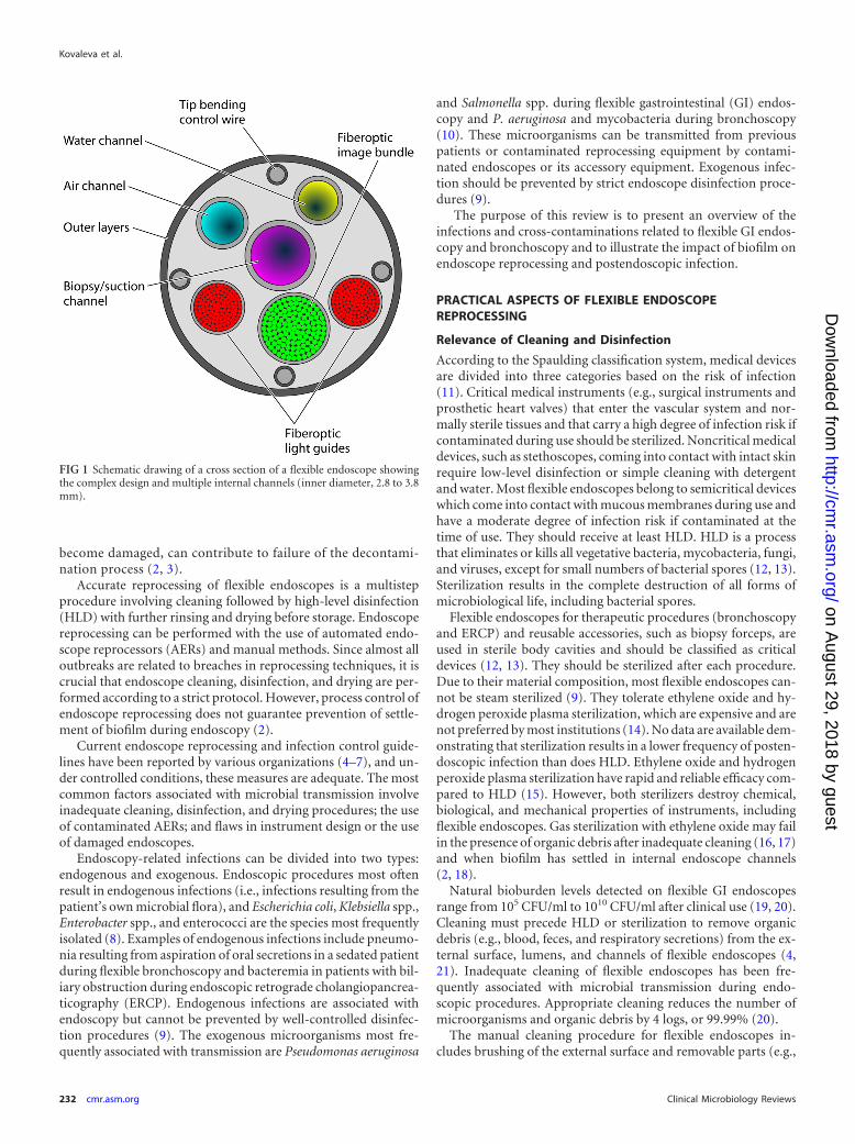
doscopes may become heavily contaminated with blood, secre-tions, and microorganisms during use. These instruments aredifficult to clean and disinfect and easy to damage because oftheir complex design, with narrow lumens and multiple inter-nal channels (Fig. 1) (1). If the instruments are not properlycleaned, the disinfection and drying procedures can fail andincrease the possibility of transmission of infection from onepatient to another (2). In addition, the ability of bacteria toform biofilms in the endoscope channels, especially when these
Address correspondence to Julia Kovaleva, [email protected].
Copyright © 2013, American Society for Microbiology. All Rights Reserved.
doi:10.1128/CMR.00085-12
April 2013 Volume 26 Number 2 Clinical Microbiology Reviews p. 231–254 cmr.asm.org 231
on August 29, 2018 by guest
http://cmr.asm
.org/D
ownloaded from
become damaged, can contribute to failure of the decontami-nation process (2, 3).
Accurate reprocessing of flexible endoscopes is a multistepprocedure involving cleaning followed by high-level disinfection(HLD) with further rinsing and drying before storage. Endoscopereprocessing can be performed with the use of automated endo-scope reprocessors (AERs) and manual methods. Since almost alloutbreaks are related to breaches in reprocessing techniques, it iscrucial that endoscope cleaning, disinfection, and drying are per-formed according to a strict protocol. However, process control ofendoscope reprocessing does not guarantee prevention of settle-ment of biofilm during endoscopy (2).
Current endoscope reprocessing and infection control guide-lines have been reported by various organizations (4–7), and un-der controlled conditions, these measures are adequate. The mostcommon factors associated with microbial transmission involveinadequate cleaning, disinfection, and drying procedures; the useof contaminated AERs; and flaws in instrument design or the useof damaged endoscopes.
Endoscopy-related infections can be divided into two types:endogenous and exogenous. Endoscopic procedures most oftenresult in endogenous infections (i.e., infections resulting from thepatient’s own microbial flora), and Escherichia coli, Klebsiella spp.,Enterobacter spp., and enterococci are the species most frequentlyisolated (8). Examples of endogenous infections include pneumo-nia resulting from aspiration of oral secretions in a sedated patientduring flexible bronchoscopy and bacteremia in patients with bil-iary obstruction during endoscopic retrograde cholangiopancrea-ticography (ERCP). Endogenous infections are associated withendoscopy but cannot be prevented by well-controlled disinfec-tion procedures (9). The exogenous microorganisms most fre-quently associated with transmission are Pseudomonas aeruginosa
and Salmonella spp. during flexible gastrointestinal (GI) endos-copy and P. aeruginosa and mycobacteria during bronchoscopy(10). These microorganisms can be transmitted from previouspatients or contaminated reprocessing equipment by contami-nated endoscopes or its accessory equipment. Exogenous infec-tion should be prevented by strict endoscope disinfection proce-dures (9).
The purpose of this review is to present an overview of theinfections and cross-contaminations related to flexible GI endos-copy and bronchoscopy and to illustrate the impact of biofilm onendoscope reprocessing and postendoscopic infection.
PRACTICAL ASPECTS OF FLEXIBLE ENDOSCOPEREPROCESSING
Relevance of Cleaning and Disinfection
According to the Spaulding classification system, medical devicesare divided into three categories based on the risk of infection(11). Critical medical instruments (e.g., surgical instruments andprosthetic heart valves) that enter the vascular system and nor-mally sterile tissues and that carry a high degree of infection risk ifcontaminated during use should be sterilized. Noncritical medicaldevices, such as stethoscopes, coming into contact with intact skinrequire low-level disinfection or simple cleaning with detergentand water. Most flexible endoscopes belong to semicritical deviceswhich come into contact with mucous membranes during use andhave a moderate degree of infection risk if contaminated at thetime of use. They should receive at least HLD. HLD is a processthat eliminates or kills all vegetative bacteria, mycobacteria, fungi,and viruses, except for small numbers of bacterial spores (12, 13).Sterilization results in the complete destruction of all forms ofmicrobiological life, including bacterial spores.
Flexible endoscopes for therapeutic procedures (bronchoscopyand ERCP) and reusable accessories, such as biopsy forceps, areused in sterile body cavities and should be classified as criticaldevices (12, 13). They should be sterilized after each procedure.Due to their material composition, most flexible endoscopes can-not be steam sterilized (9). They tolerate ethylene oxide and hy-drogen peroxide plasma sterilization, which are expensive and arenot preferred by most institutions (14). No data are available dem-onstrating that sterilization results in a lower frequency of posten-doscopic infection than does HLD. Ethylene oxide and hydrogenperoxide plasma sterilization have rapid and reliable efficacy com-pared to HLD (15). However, both sterilizers destroy chemical,biological, and mechanical properties of instruments, includingflexible endoscopes. Gas sterilization with ethylene oxide may failin the presence of organic debris after inadequate cleaning (16, 17)and when biofilm has settled in internal endoscope channels(2, 18).
Natural bioburden levels detected on flexible GI endoscopesrange from 105 CFU/ml to 1010 CFU/ml after clinical use (19, 20).Cleaning must precede HLD or sterilization to remove organicdebris (e.g., blood, feces, and respiratory secretions) from the ex-ternal surface, lumens, and channels of flexible endoscopes (4,21). Inadequate cleaning of flexible endoscopes has been fre-quently associated with microbial transmission during endo-scopic procedures. Appropriate cleaning reduces the number ofmicroorganisms and organic debris by 4 logs, or 99.99% (20).
The manual cleaning procedure for flexible endoscopes in-cludes brushing of the external surface and removable parts (e.g.,
FIG 1 Schematic drawing of a cross section of a flexible endoscope showingthe complex design and multiple internal channels (inner diameter, 2.8 to 3.8mm).
Kovaleva et al.
232 cmr.asm.org Clinical Microbiology Reviews
on August 29, 2018 by guest
http://cmr.asm
.org/D
ownloaded from
suction valves) and immersion in a detergent solution followed byirrigation of internal channels with a detergent. The endoscopeand accessories should be inspected for damage, and a leak testshould be performed before disinfection (4).
AERs are strongly recommended for reprocessing of flexibleendoscopes to document all steps and to minimize contaminationand contact with chemicals and contaminated instruments (5).However, contaminated and defective AERs can result in inade-quate reprocessing and contamination of endoscopes and havebeen associated with outbreaks of endoscopy-related infections(22–26). Presence of biofilms in the AERs has been detected dur-ing these outbreaks (23, 25, 26).
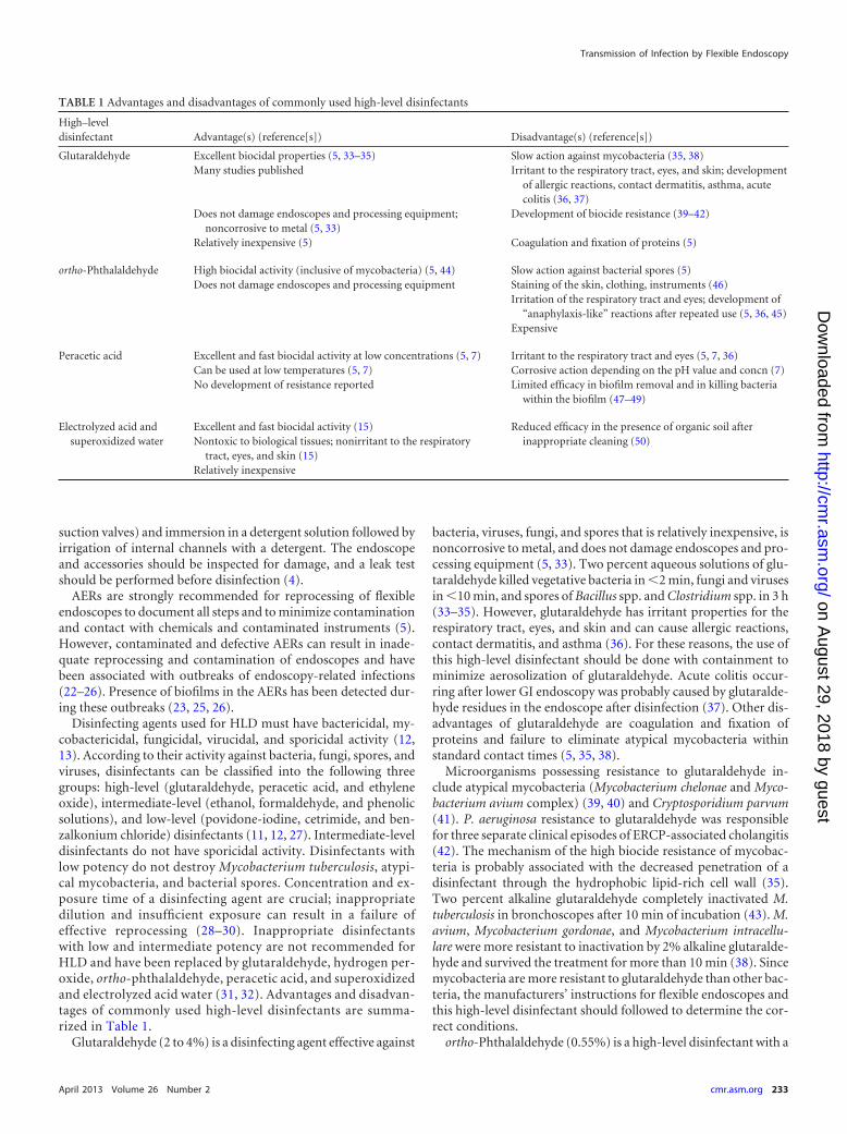
Disinfecting agents used for HLD must have bactericidal, my-cobactericidal, fungicidal, virucidal, and sporicidal activity (12,13). According to their activity against bacteria, fungi, spores, andviruses, disinfectants can be classified into the following threegroups: high-level (glutaraldehyde, peracetic acid, and ethyleneoxide), intermediate-level (ethanol, formaldehyde, and phenolicsolutions), and low-level (povidone-iodine, cetrimide, and ben-zalkonium chloride) disinfectants (11, 12, 27). Intermediate-leveldisinfectants do not have sporicidal activity. Disinfectants withlow potency do not destroy Mycobacterium tuberculosis, atypi-cal mycobacteria, and bacterial spores. Concentration and ex-posure time of a disinfecting agent are crucial; inappropriatedilution and insufficient exposure can result in a failure ofeffective reprocessing (28–30). Inappropriate disinfectantswith low and intermediate potency are not recommended forHLD and have been replaced by glutaraldehyde, hydrogen per-oxide, ortho-phthalaldehyde, peracetic acid, and superoxidizedand electrolyzed acid water (31, 32). Advantages and disadvan-tages of commonly used high-level disinfectants are summa-rized in Table 1.
Glutaraldehyde (2 to 4%) is a disinfecting agent effective against
bacteria, viruses, fungi, and spores that is relatively inexpensive, isnoncorrosive to metal, and does not damage endoscopes and pro-cessing equipment (5, 33). Two percent aqueous solutions of glu-taraldehyde killed vegetative bacteria in �2 min, fungi and virusesin �10 min, and spores of Bacillus spp. and Clostridium spp. in 3 h(33–35). However, glutaraldehyde has irritant properties for therespiratory tract, eyes, and skin and can cause allergic reactions,contact dermatitis, and asthma (36). For these reasons, the use ofthis high-level disinfectant should be done with containment tominimize aerosolization of glutaraldehyde. Acute colitis occur-ring after lower GI endoscopy was probably caused by glutaralde-hyde residues in the endoscope after disinfection (37). Other dis-advantages of glutaraldehyde are coagulation and fixation ofproteins and failure to eliminate atypical mycobacteria withinstandard contact times (5, 35, 38).
Microorganisms possessing resistance to glutaraldehyde in-clude atypical mycobacteria (Mycobacterium chelonae and Myco-bacterium avium complex) (39, 40) and Cryptosporidium parvum(41). P. aeruginosa resistance to glutaraldehyde was responsiblefor three separate clinical episodes of ERCP-associated cholangitis(42). The mechanism of the high biocide resistance of mycobac-teria is probably associated with the decreased penetration of adisinfectant through the hydrophobic lipid-rich cell wall (35).Two percent alkaline glutaraldehyde completely inactivated M.tuberculosis in bronchoscopes after 10 min of incubation (43). M.avium, Mycobacterium gordonae, and Mycobacterium intracellu-lare were more resistant to inactivation by 2% alkaline glutaralde-hyde and survived the treatment for more than 10 min (38). Sincemycobacteria are more resistant to glutaraldehyde than other bac-teria, the manufacturers’ instructions for flexible endoscopes andthis high-level disinfectant should followed to determine the cor-rect conditions.
ortho-Phthalaldehyde (0.55%) is a high-level disinfectant with a
TABLE 1 Advantages and disadvantages of commonly used high-level disinfectants
High–leveldisinfectant Advantage(s) (reference[s]) Disadvantage(s) (reference[s])
Glutaraldehyde Excellent biocidal properties (5, 33–35) Slow action against mycobacteria (35, 38)Many studies published Irritant to the respiratory tract, eyes, and skin; development
of allergic reactions, contact dermatitis, asthma, acutecolitis (36, 37)
Does not damage endoscopes and processing equipment;noncorrosive to metal (5, 33)
Development of biocide resistance (39–42)
Relatively inexpensive (5) Coagulation and fixation of proteins (5)
ortho-Phthalaldehyde High biocidal activity (inclusive of mycobacteria) (5, 44) Slow action against bacterial spores (5)Does not damage endoscopes and processing equipment Staining of the skin, clothing, instruments (46)
Irritation of the respiratory tract and eyes; development of“anaphylaxis-like” reactions after repeated use (5, 36, 45)
Expensive
Peracetic acid Excellent and fast biocidal activity at low concentrations (5, 7) Irritant to the respiratory tract and eyes (5, 7, 36)Can be used at low temperatures (5, 7) Corrosive action depending on the pH value and concn (7)No development of resistance reported Limited efficacy in biofilm removal and in killing bacteria
within the biofilm (47–49)
Electrolyzed acid andsuperoxidized water
Excellent and fast biocidal activity (15) Reduced efficacy in the presence of organic soil afterinappropriate cleaning (50)Nontoxic to biological tissues; nonirritant to the respiratory
tract, eyes, and skin (15)Relatively inexpensive
Transmission of Infection by Flexible Endoscopy
April 2013 Volume 26 Number 2 cmr.asm.org 233
on August 29, 2018 by guest
http://cmr.asm
.org/D
ownloaded from
higher mycobactericidal efficacy than glutaraldehyde (5, 44). Dis-advantages of this disinfectant include slow action against bacte-rial spores, irritation of the respiratory tract and eyes of the pa-tients and staff, and the possibility of causing “anaphylaxis-like”reactions after repeated use (5, 36, 45). ortho-Phthalaldehydestains skin, instruments, clothing, and surfaces and is more expen-sive than glutaraldehyde (46).
Peracetic acid is an oxidizing agent usually used for HLD offlexible endoscopes in AERs. It rapidly deactivates a large varietyof pathogenic microorganisms, viruses, and spores at low concen-trations (�0.3%) (5, 7). No development of microorganism resis-tance to peracetic acid has been reported. This disinfectant can beused at low temperatures and causes less irritation than glutaral-dehyde but has corrosive action depending on the pH value andconcentration (7). Several disinfectants that contain peracetic acidand hydrogen peroxide are available. Although the biocidal effectof peracetic acid on sessile microorganisms is well known, theeffect of this disinfecting agent on microbial biofilms has not beencompletely studied. A recent study demonstrated insufficient ef-ficacy of 1% peracetic acid disinfectant for 10 min against P.aeruginosa, Stenotrophomonas maltophilia, and Candida sp. bio-films (47). According to the literature, peracetic acid has the abil-ity to fixate biofilms, and therefore, it can show limited efficacy inbiofilm removal from the abiotic surfaces and in killing bacteriawithin the biofilm (48, 49). Vigorous cleaning with brushes oragitated liquids is important to remove as much biofilm as possi-ble (4).
Electrolyzed acid and superoxidized water are relatively newdisinfectants used for endoscope reprocessing (15). These disin-fectants are produced by the electrolysis of sodium chloride solu-tions. They have excellent biocidal activity and are inexpensive,nontoxic to biological tissues, and nonirritating to the respiratorytract, eyes, and skin (15). However, antimicrobial efficacy can bereduced in the presence of organic soil after inappropriate clean-ing (50).
After the disinfection phase, the remaining disinfectant must beremoved from the exterior and from the internal channels by rins-ing the endoscope with bacterium-free water. According to theGuideline Committee of the European Society of GastrointestinalEndoscopy, sterile water is preferable for the final rinse to preventrecontamination (5). Many outbreaks of endoscopy-related infec-tions and cross-contaminations due to P. aeruginosa, Serratiamarcescens, M. chelonae, Mycobacterium xenopi, and Methylobac-terium mesophilicum have been related to rinsing flexible endo-scopes after disinfection with nonsterile tap water (30, 51–57).The accepted procedures to produce bacterium-free water in dis-infectors for endoscopes include filtration, UV radiation, or heat-ing followed by cooling (5). However, major U.S. guidelines rec-ommend the use of sterile, filtered, or tap water to rinse theendoscope channels after HLD, followed by flushing of the endo-scope channels with 70% to 90% ethyl alcohol or isopropyl alco-hol (6, 7, 21). The rinse water must be discarded after each cycle.Water bottles used for irrigation during the procedure should behigh-level disinfected or sterilized at least daily. Sterile watershould be used to fill the water bottle.
Relevance of Drying and Storage
Accurate endoscope drying is crucial, whereas a humid environ-ment facilitates microbial growth during storage. The final dryingsteps greatly reduce the risk of remaining pathogens as well as the
possibility of recontamination of the endoscope by waterbornemicroorganisms such as Pseudomonas spp. and Acinetobacter spp.(58). Flexible endoscopes should be dried with filtered com-pressed air manually or in AERs between endoscopic procedures(a short drying cycle) and at the end of the day (an intensive finaldrying) (5, 59). Several guidelines recommend forced air drying,preceded by flushing of the internal channels with 70% to 90%ethanol or isopropanol at the end of a clinic day (6, 7, 21). Allen etal. (22) showed a high efficiency in preventing P. aeruginosa con-taminations by flushing 70% ethanol through endoscope chan-nels followed by drying with compressed air. Due to the fixativeproperties of ethanol, its use is not recommended in some coun-tries.
Storage is an important factor in the maintenance of bacterium-free endoscopes. It is recommended that a dust-free drying cabi-net be used for endoscope storage, in which the endoscopes arehung vertically (5, 60, 61). Noy et al. (62) noted that storing theendoscopes vertically in air after cleaning until the next use of theendoscope resulted in a drop of the contamination rate from 35 to0%. According to the literature, endoscopes stay bacterium freeafter prolonged storage if an adequate drying procedure is applied(60, 61). When stored in the drying cabinet with a laminar airflow,no growth of bacteria and Candida spp. was found in endoscopechannels 5 days after reprocessing (61). Low levels of microbialcontamination on endoscopes stored in the drying and storagecabinet were detected, compared with the stable or increased mi-crobial numbers on endoscopes stored outside the cabinet (60).
Many outbreaks of health care-associated infection after endos-copy and cross-contaminations due to P. aeruginosa, S. marc-escens, M. tuberculosis, and M. chelonae have been associated withdrying of flexible endoscopes without ethanol flushing or lack of adrying procedure (22–24, 30, 52, 57, 63–76).
EXOGENOUS INFECTIONS ASSOCIATED WITH FLEXIBLEENDOSCOPY
Despite the large number of endoscopic procedures that are per-formed annually, documented data suggest that postendoscopiciatrogenic infections are rare. In GI endoscopy, the estimated rateof health care-associated infection is approximately 1 out of 1.8million procedures (77). However, the true rate of transmissionduring endoscopy may go unrecognized because of technicallyinadequate surveillance, no surveillance at all, low frequency, orthe absence of clinical symptoms (2, 9). Two hundred eighty-onepatients infected after GI endoscopy were found in studies of en-doscopy-related infections between 1966 and 1992 in the UnitedStates (8). Gorse and Messner (14) reported 6% iatrogenic infec-tions after GI endoscopy in 116 hospitals. During the period of1974 to 2004, 30 outbreaks of endoscopy-related infections andcross-contaminations involving 251 patients infected after GI en-doscopic procedures were reported in the United States (78). In-adequate decontamination procedures and equipment malfunc-tion were two leading causes of postendoscopic infection andcontamination. More than 91% of the infections identified couldbe prevented if quality control systems were improved.
The important risk factors of infections in GI endoscopy arethe number of microorganisms present inside the endoscope orthe growth of a biofilm, invasive endoscopic procedures resultingin tissue damage, compromised immune status of the patient (hu-man immunodeficiency virus [HIV] infection, neoplastic dis-eases, transplant patients, and immunosuppressive treatment),
Kovaleva et al.
234 cmr.asm.org Clinical Microbiology Reviews
on August 29, 2018 by guest
http://cmr.asm
.org/D
ownloaded from

and the presence of infective foci (abscess and cholangitis) duringan endoscopic procedure (79–81).
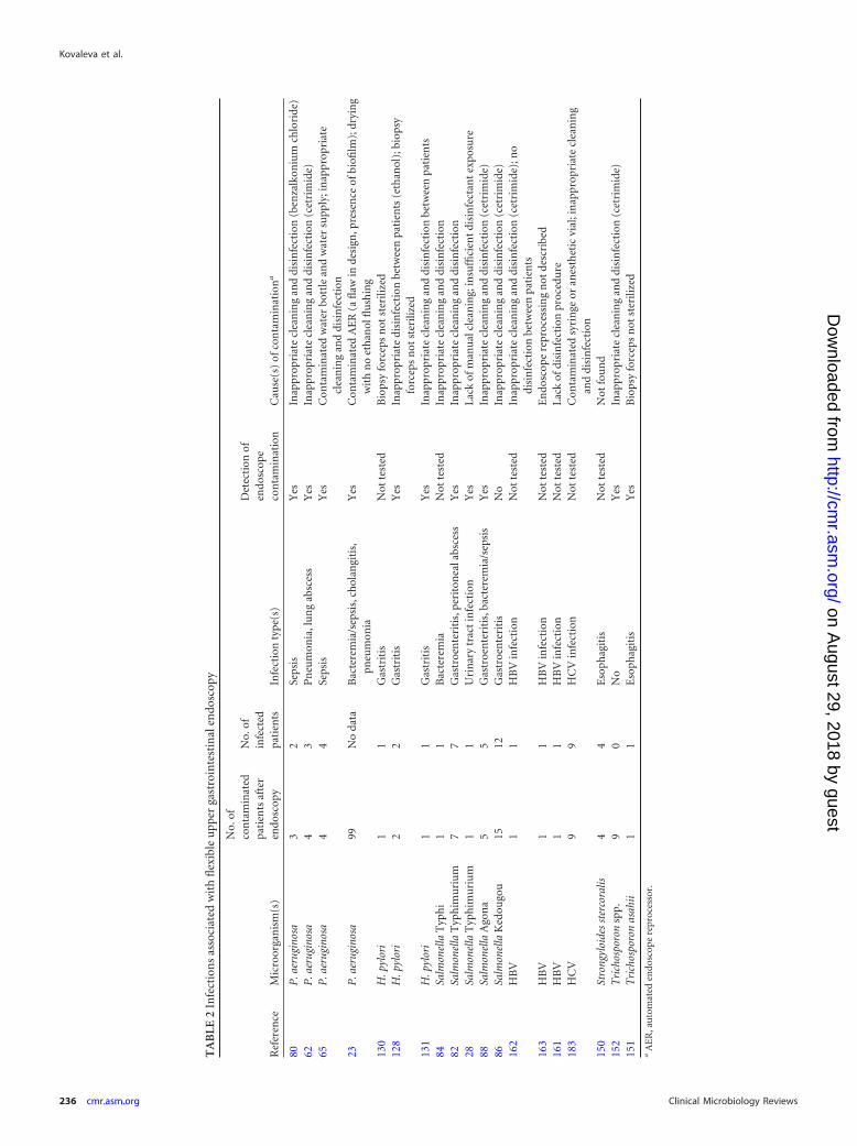
An overview of the exogenous endoscopy-related infectionsand cross-contaminations after flexible GI endoscopy and bron-choscopy is presented in Tables 2 to 5.
Bacteria
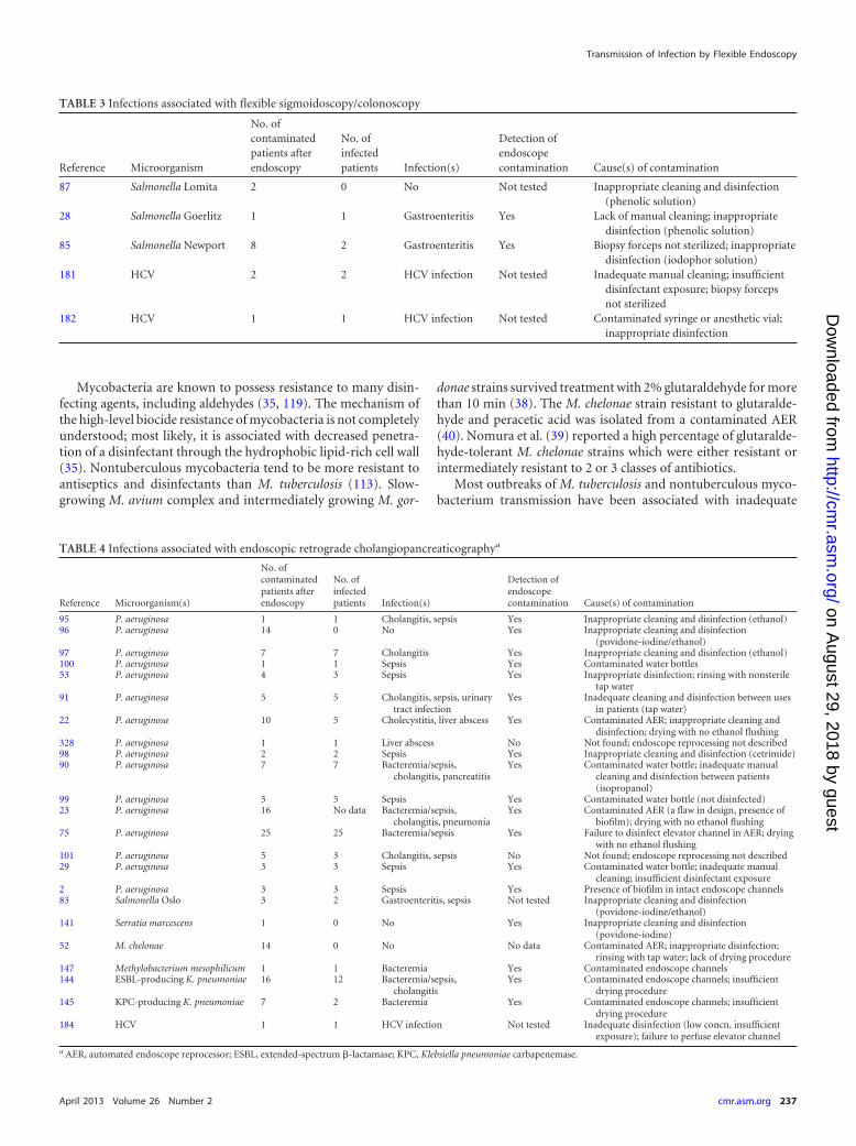
Salmonella spp. In the past, Salmonella spp. were the most com-mon microorganisms associated with infections transmitted by GIendoscopy (28, 82–88). Many Salmonella outbreaks were relatedto an inappropriate use of disinfectants with intermediate and lowpotency instead of high-level disinfecting agents (Tables 2 and 3).There have been no reports of Salmonella infection since the cur-rent guidelines for HLD have been followed.
The following Salmonella serotypes were isolated from con-taminated endoscopes or accessories: Salmonella enterica sero-types Typhi (84), Typhimurium (28, 82), Agona (88), Goerlitz(28), Newport (85), Oslo (83), Kedougou (86), and Lomita (87). Aminority of patients involved were asymptomatic carriers and hadstool or urine cultures positive for Salmonella spp. Salmonella in-fections developed within 1 to 9 days after a GI endoscopic proce-dure and included acute gastroenteritis (28, 82, 83, 85, 86), peri-toneal abscess (82), urinary tract infection (28), and bacteremia/sepsis (83, 84, 88).
Pseudomonas aeruginosa. P. aeruginosa, a Gram-negative op-portunistic pathogen, is the most commonly reported microor-ganism responsible for transmission of infection during GI endos-copy and bronchoscopy. It is known for its preference for a moistenvironment (hospital water supply and wet endoscope channelsafter reprocessing) (10). Pseudomonas is able to form biofilms, andthese biofilms are extremely difficult to remove from plumbing,AERs, and endoscope channels (2, 89). Serotype 10 of P. aerugi-nosa predominates in the published reports of Pseudomonas trans-mission (22, 23, 53, 73, 90, 91). It is possible that slime productionby P. aeruginosa serotype 10 in a biofilm can form a barrier toantibiotics and disinfectants and lead to antibacterial resistance,but no explanation was offered as to why specifically this serotypewas isolated.
Among healthy adults, P. aeruginosa can colonize many bodysites, as evidenced by isolation from throat, sputum, and stool(92). Hospitalized patients, as well as patients with certain chroniclung diseases, have higher colonization rates. During health care-related outbreaks, Pseudomonas transmission can result in coloni-zation of involved patients in the GI and respiratory tract with anabsence of clinical symptoms and negative blood cultures, whichwas determined by molecular typing (93). Severe health care-as-sociated postendoscopic infections due to P. aeruginosa includesepsis, liver abscess, and ascending cholangitis after flexible GIendoscopy (particularly after ERCP) and bloodstream infectionand pneumonia after bronchoscopy. Post-ERCP P. aeruginosa in-fectious complications occur most frequently in patients with bil-iary obstruction undergoing endoscopic biliary stenting (94).
Many outbreaks of P. aeruginosa infection after GI endoscopyand bronchoscopy have been associated with inadequate cleaningand the use of inappropriate intermediate-level and low-level dis-infectants (29, 62, 64, 73, 80, 90, 95–98), contaminated endoscopewater bottles and the water supply to the endoscope (29, 65, 90, 99,100), and drying of endoscope channels with no flushing with70% ethanol after disinfection (22, 30, 70, 73–75) or lack of adrying procedure (26, 64) (Tables 2 to 5). Two recent outbreaks of
multidrug-resistant P. aeruginosa post-ERCP infections have beenrelated to the remaining contamination of the endoscope despiteaccurate reprocessing followed by negative surveillance endo-scope cultures (2, 101). In an outbreak of post-ERCP sepsis de-scribed by Kovaleva et al. (2), the implicated endoscope was repet-itively found to be positive by culturing for multidrug-resistant P.aeruginosa after HLD and contained biofilm in undamaged innerchannels. The P. aeruginosa isolates from patients and the impli-cated endoscope underwent molecular typing and showed match-ing patterns.
Several postendoscopic P. aeruginosa outbreaks have been re-lated to contaminated or defective AERs (22, 23, 26, 74, 75, 102,103), the use of incorrect connectors between the endoscope andAER (103, 104), and defective endoscopes and accessories (93,105–107) (Tables 2 to 5). The presence of biofilm deposits on theinternal plumbing and detergent tank of AERs resulted in twooutbreaks (23, 26).
In most outbreaks, the Pseudomonas strains obtained from pa-tients were identical to those recovered from endoscopes, as de-termined by comparison of antimicrobial sensitivity patterns ofthe isolates, serotyping, and phage typing or by more recentlydeveloped molecular techniques (90, 97).
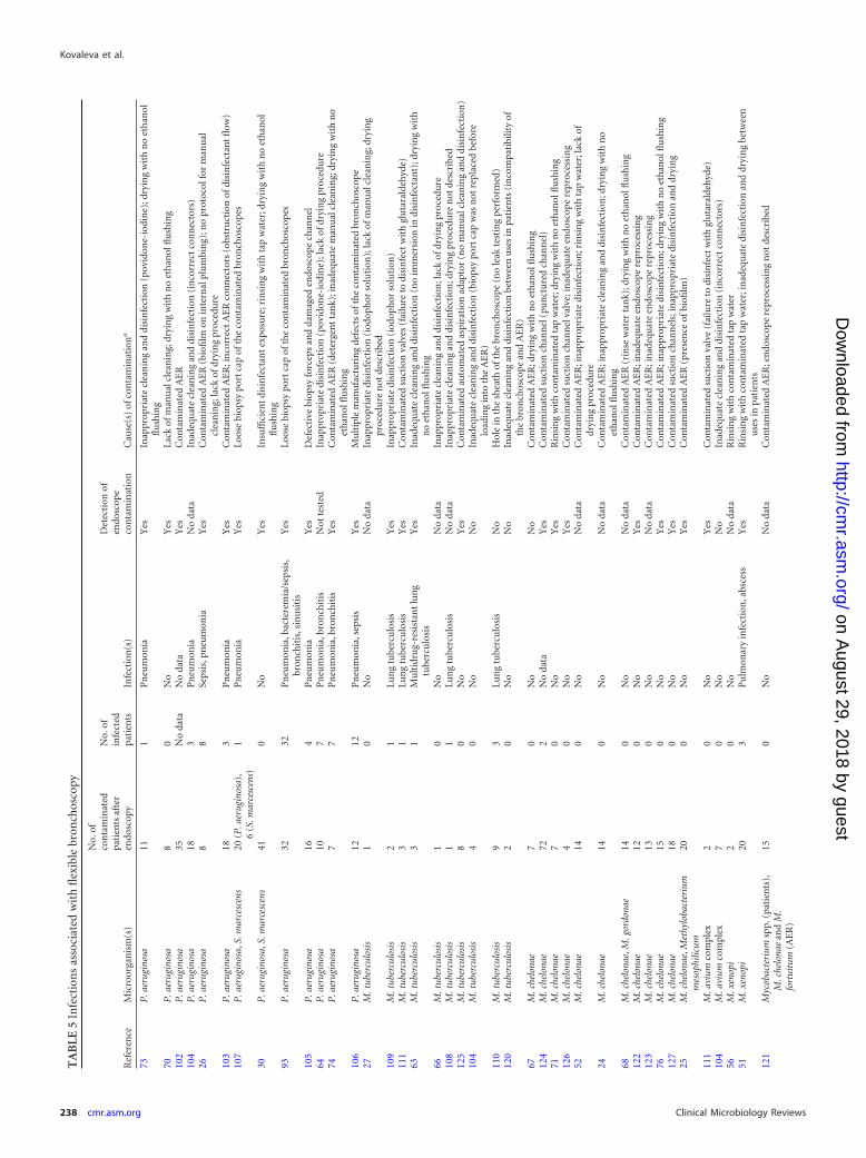
Mycobacteria. M. tuberculosis and nontuberculous mycobac-teria are microorganisms frequently associated with healthcare-associated transmission during endoscopic procedures,particularly during bronchoscopy. While transmission of non-tuberculous mycobacteria is usually associated with contami-nated AERs and rinsing water, contamination with M. tubercu-losis generally comes from an infected patient during anendoscopic procedure (8). Most outbreaks due to nontubercu-lous mycobacteria have involved rapidly growing M. chelonae,intermediately growing M. gordonae, and slowly growing M.xenopi and M. avium complex strains.
Most publications about mycobacterial outbreaks describedcross-contamination, a situation where the patient becomes noso-comially colonized with a strain from the endoscope in the ab-sence of infection, but reports of bronchoscopy-related pulmo-nary tuberculosis were documented (63, 108–111). The majorityof the patients was immunocompromised and had a history oflung cancer, HIV, or hematological malignancies. Most M. tuber-culosis isolates were susceptible to antituberculosis drugs, buthealth care-associated transmission of multidrug-resistant tuber-culosis has been reported (63). Nontuberculous mycobacteria,such as M. chelonae, M. avium, M. xenopi, M. gordonae, and M.fortuitum, can form the risk for development of colonization orinfection in severely immunocompromised patients (112, 113).
The effectiveness of disinfectants against mycobacteria de-pends on the composition and concentration of the active agent,contact time, and presence of organic material (27, 113, 114).Activities of different disinfectants against M. tuberculosis andnontuberculous mycobacteria were tested in several studies. vanKlingeren and Pullen (115) showed inadequate tuberculocidal ac-tivity of quaternary ammonium, good activity of phenolic disin-fectants, and rapid killing of M. tuberculosis with 70% ethanol and60% isopropanol. High-level effectiveness of 2% glutaraldehydeagainst M. tuberculosis and M. chelonae was demonstrated directlyafter cleaning and after 10- and 20-min exposures (43, 116, 117).Peracetic acid (0.26%) was effective against M. tuberculosis and M.avium complex strains within 10 to 20 min, with a 5-log reductionin viable bacteria (118).
Transmission of Infection by Flexible Endoscopy
April 2013 Volume 26 Number 2 cmr.asm.org 235
on August 29, 2018 by guest
http://cmr.asm
.org/D
ownloaded from
TA
BLE
2In
fect
ion
sas
soci
ated
wit
hfl
exib
leu
pper
gast
roin
test
inal
endo
scop
y
Ref
eren
ceM
icro
orga
nis
m(s
)
No.
ofco
nta
min
ated
pati
ents
afte
ren
dosc
opy
No.
ofin
fect
edpa
tien
tsIn
fect
ion
type
(s)
Det
ecti
onof
endo
scop
eco
nta
min
atio
nC
ause
(s)
ofco
nta
min
atio
na
80P
.aer
ugin
osa
32
Seps
isY
esIn
appr
opri
ate
clea
nin
gan
ddi
sin
fect
ion
(ben
zalk
oniu
mch
lori
de)
62P
.aer
ugin
osa
43
Pn
eum
onia
,lu
ng
absc
ess
Yes
Inap
prop
riat
ecl
ean
ing
and
disi
nfe
ctio
n(c
etri
mid
e)65
P.a
erug
inos
a4
4Se
psis
Yes
Con
tam
inat
edw
ater
bott
lean
dw
ater
supp
ly;i
nap
prop
riat
ecl
ean
ing
and
disi
nfe
ctio
n23
P.a
erug
inos
a99
No
data
Bac
tere
mia
/sep
sis,
chol
angi
tis,
pneu
mon
iaY
esC
onta
min
ated
AE
R(a
flaw
inde
sign
,pre
sen
ceof
biofi
lm);
dryi
ng
wit
hn
oet
han
olfl
ush
ing
130
H.p
ylor
i1
1G
astr
itis
Not
test
edB
iops
yfo
rcep
sn
otst
erili
zed
128
H.p
ylor
i2
2G
astr
itis
Yes
Inap
prop
riat
edi
sin
fect
ion
betw
een
pati
ents
(eth
anol
);bi
opsy
forc
eps
not
ster
ilize
d13
1H
.pyl
ori
11
Gas
trit
isY
esIn
appr
opri
ate
clea
nin
gan
ddi
sin
fect
ion
betw
een
pati
ents
84Sa
lmon
ella
Typ
hi
11
Bac
tere
mia
Not
test
edIn
appr
opri
ate
clea
nin
gan
ddi
sin
fect
ion
82Sa
lmon
ella
Typ
him
uri
um
77
Gas
troe
nte
riti
s,pe
rito
nea
labs
cess
Yes
Inap
prop
riat
ecl
ean
ing
and
disi
nfe
ctio
n28
Salm
onel
laT
yph
imu
riu
m1
1U
rin
ary
trac
tin
fect
ion
Yes
Lack
ofm
anu
alcl
ean
ing;
insu
ffici
ent
disi
nfe
ctan
tex
posu
re88
Salm
onel
laA
gon
a5
5G
astr
oen
teri
tis,
bact
erem
ia/s
epsi
sY
esIn
appr
opri
ate
clea
nin
gan
ddi
sin
fect
ion
(cet
rim
ide)
86Sa
lmon
ella
Ked
ougo
u15
12G
astr
oen
teri
tis
No
Inap
prop
riat
ecl
ean
ing
and
disi
nfe
ctio
n(c
etri
mid
e)16
2H
BV
11
HB
Vin
fect
ion
Not
test
edIn
appr
opri
ate
clea
nin
gan
ddi
sin
fect
ion
(cet
rim
ide)
;no
disi
nfe
ctio
nbe
twee
npa
tien
ts16
3H
BV
11
HB
Vin
fect
ion
Not
test
edE
ndo
scop
ere
proc
essi
ng
not
desc
ribe
d16
1H
BV
11
HB
Vin
fect
ion
Not
test
edLa
ckof
disi
nfe
ctio
npr
oced
ure
183
HC
V9
9H
CV
infe
ctio
nN
otte
sted
Con
tam
inat
edsy
rin
geor
anes
thet
icvi
al;i
nap
prop
riat
ecl
ean
ing
and
disi
nfe
ctio
n15
0St
rong
yloi
des
ster
cora
lis4
4E
soph
agit
isN
otte
sted
Not
fou
nd
152
Tri
chos
poro
nsp
p.9
0N
oY
esIn
appr
opri
ate
clea
nin
gan
ddi
sin
fect
ion
(cet
rim
ide)
151
Tri
chos
poro
nas
ahii
11
Eso
phag
itis
Yes
Bio
psy
forc
eps
not
ster
ilize
da
AE
R,a
uto
mat
eden
dosc
ope
repr
oces
sor.
Kovaleva et al.
236 cmr.asm.org Clinical Microbiology Reviews
on August 29, 2018 by guest
http://cmr.asm
.org/D
ownloaded from
Mycobacteria are known to possess resistance to many disin-fecting agents, including aldehydes (35, 119). The mechanism ofthe high-level biocide resistance of mycobacteria is not completelyunderstood; most likely, it is associated with decreased penetra-tion of a disinfectant through the hydrophobic lipid-rich cell wall(35). Nontuberculous mycobacteria tend to be more resistant toantiseptics and disinfectants than M. tuberculosis (113). Slow-growing M. avium complex and intermediately growing M. gor-
donae strains survived treatment with 2% glutaraldehyde for morethan 10 min (38). The M. chelonae strain resistant to glutaralde-hyde and peracetic acid was isolated from a contaminated AER(40). Nomura et al. (39) reported a high percentage of glutaralde-hyde-tolerant M. chelonae strains which were either resistant orintermediately resistant to 2 or 3 classes of antibiotics.
Most outbreaks of M. tuberculosis and nontuberculous myco-bacterium transmission have been associated with inadequate
TABLE 3 Infections associated with flexible sigmoidoscopy/colonoscopy
Reference Microorganism
No. ofcontaminatedpatients afterendoscopy
No. ofinfectedpatients Infection(s)
Detection ofendoscopecontamination Cause(s) of contamination
87 Salmonella Lomita 2 0 No Not tested Inappropriate cleaning and disinfection(phenolic solution)
28 Salmonella Goerlitz 1 1 Gastroenteritis Yes Lack of manual cleaning; inappropriatedisinfection (phenolic solution)
85 Salmonella Newport 8 2 Gastroenteritis Yes Biopsy forceps not sterilized; inappropriatedisinfection (iodophor solution)
181 HCV 2 2 HCV infection Not tested Inadequate manual cleaning; insufficientdisinfectant exposure; biopsy forcepsnot sterilized
182 HCV 1 1 HCV infection Not tested Contaminated syringe or anesthetic vial;inappropriate disinfection
TABLE 4 Infections associated with endoscopic retrograde cholangiopancreaticographya
Reference Microorganism(s)
No. ofcontaminatedpatients afterendoscopy
No. ofinfectedpatients Infection(s)
Detection ofendoscopecontamination Cause(s) of contamination
95 P. aeruginosa 1 1 Cholangitis, sepsis Yes Inappropriate cleaning and disinfection (ethanol)96 P. aeruginosa 14 0 No Yes Inappropriate cleaning and disinfection
(povidone-iodine/ethanol)97 P. aeruginosa 7 7 Cholangitis Yes Inappropriate cleaning and disinfection (ethanol)100 P. aeruginosa 1 1 Sepsis Yes Contaminated water bottles53 P. aeruginosa 4 3 Sepsis Yes Inappropriate disinfection; rinsing with nonsterile
tap water91 P. aeruginosa 5 5 Cholangitis, sepsis, urinary
tract infectionYes Inadequate cleaning and disinfection between uses
in patients (tap water)22 P. aeruginosa 10 5 Cholecystitis, liver abscess Yes Contaminated AER; inappropriate cleaning and
disinfection; drying with no ethanol flushing328 P. aeruginosa 1 1 Liver abscess No Not found; endoscope reprocessing not described98 P. aeruginosa 2 2 Sepsis Yes Inappropriate cleaning and disinfection (cetrimide)90 P. aeruginosa 7 7 Bacteremia/sepsis,
cholangitis, pancreatitisYes Contaminated water bottle; inadequate manual
cleaning and disinfection between patients(isopropanol)
99 P. aeruginosa 5 5 Sepsis Yes Contaminated water bottle (not disinfected)23 P. aeruginosa 16 No data Bacteremia/sepsis,
cholangitis, pneumoniaYes Contaminated AER (a flaw in design, presence of
biofilm); drying with no ethanol flushing75 P. aeruginosa 25 25 Bacteremia/sepsis Yes Failure to disinfect elevator channel in AER; drying
with no ethanol flushing101 P. aeruginosa 5 3 Cholangitis, sepsis No Not found; endoscope reprocessing not described29 P. aeruginosa 3 3 Sepsis Yes Contaminated water bottle; inadequate manual
cleaning; insufficient disinfectant exposure2 P. aeruginosa 3 3 Sepsis Yes Presence of biofilm in intact endoscope channels83 Salmonella Oslo 3 2 Gastroenteritis, sepsis Not tested Inappropriate cleaning and disinfection
(povidone-iodine/ethanol)141 Serratia marcescens 1 0 No Yes Inappropriate cleaning and disinfection
(povidone-iodine)52 M. chelonae 14 0 No No data Contaminated AER; inappropriate disinfection;
rinsing with tap water; lack of drying procedure147 Methylobacterium mesophilicum 1 1 Bacteremia Yes Contaminated endoscope channels144 ESBL-producing K. pneumoniae 16 12 Bacteremia/sepsis,
cholangitisYes Contaminated endoscope channels; insufficient
drying procedure145 KPC-producing K. pneumoniae 7 2 Bacteremia Yes Contaminated endoscope channels; insufficient
drying procedure184 HCV 1 1 HCV infection Not tested Inadequate disinfection (low concn, insufficient
exposure); failure to perfuse elevator channel
a AER, automated endoscope reprocessor; ESBL, extended-spectrum �-lactamase; KPC, Klebsiella pneumoniae carbapenemase.
Transmission of Infection by Flexible Endoscopy
April 2013 Volume 26 Number 2 cmr.asm.org 237
on August 29, 2018 by guest
http://cmr.asm
.org/D
ownloaded from
TA
BLE
5In
fect
ion
sas
soci
ated
wit
hfl
exib
lebr
onch
osco
py
Ref
eren
ceM
icro
orga
nis
m(s
)
No.
ofco
nta
min
ated
pati
ents
afte
ren
dosc
opy
No.
ofin
fect
edpa
tien
tsIn
fect
ion
(s)
Det
ecti
onof
endo
scop
eco
nta
min
atio
nC
ause
(s)
ofco
nta
min
atio
na
73P
.aer
ugin
osa
111
Pn
eum
onia
Yes
Inap
prop
riat
ecl
ean
ing
and
disi
nfe
ctio
n(p
ovid
one-
iodi
ne)
;dry
ing
wit
hn
oet
han
olfl
ush
ing
70P
.aer
ugin
osa
80
No
Yes
Lack
ofm
anu
alcl
ean
ing;
dryi
ng
wit
hn
oet
han
olfl
ush
ing
102
P.a
erug
inos
a35
No
data
No
data
Yes
Con
tam
inat
edA
ER
104
P.a
erug
inos
a18
3P
neu
mon
iaN
oda
taIn
adeq
uat
ecl
ean
ing
and
disi
nfe
ctio
n(i
nco
rrec
tco
nn
ecto
rs)
26P
.aer
ugin
osa
88
Seps
is,p
neu
mon
iaY
esC
onta
min
ated
AE
R(b
iofi
lmon
inte
rnal
plu
mbi
ng)
;no
prot
ocol
for
man
ual
clea
nin
g;la
ckof
dryi
ng
proc
edu
re10
3P
.aer
ugin
osa
183
Pn
eum
onia
Yes
Con
tam
inat
edA
ER
;in
corr
ect
AE
Rco
nn
ecto
rs(o
bstr
uct
ion
ofdi
sin
fect
ant
flow
)10
7P
.aer
ugin
osa,
S.m
arce
scen
s20
(P.a
erug
inos
a),
6(S
.mar
cesc
ens)
1P
neu
mon
iaY
esLo
ose
biop
sypo
rtca
pof
the
con
tam
inat
edbr
onch
osco
pes
30P
.aer
ugin
osa,
S.m
arce
scen
s41
0N
oY
esIn
suffi
cien
tdi
sin
fect
ant
expo
sure
;rin
sin
gw
ith
tap
wat
er;d
ryin
gw
ith
no
eth
anol
flu
shin
g93
P.a
erug
inos
a32
32P
neu
mon
ia,b
acte
rem
ia/s
epsi
s,br
onch
itis
,sin
usi
tis
Yes
Loos
ebi
opsy
port
cap
ofth
eco
nta
min
ated
bron
chos
cope
s
105
P.a
erug
inos
a16
4P
neu
mon
iaY
esD
efec
tive
biop
syfo
rcep
san
dda
mag
eden
dosc
ope
chan
nel
64P
.aer
ugin
osa
107
Pn
eum
onia
,bro
nch
itis
Not
test
edIn
appr
opri
ate
disi
nfe
ctio
n(p
ovid
one-
iodi
ne)
;lac
kof
dryi
ng
proc
edu
re74
P.a
erug
inos
a7
7P
neu
mon
ia,b
ron
chit
isY
esC
onta
min
ated
AE
R(d
eter
gen
tta
nk)
;in
adeq
uat
em
anu
alcl
ean
ing;
dryi
ng
wit
hn
oet
han
olfl
ush
ing
106
P.a
erug
inos
a12
12P
neu
mon
ia,s
epsi
sY
esM
ult
iple
man
ufa
ctu
rin
gde
fect
sof
the
con
tam
inat
edbr
onch
osco
pe27
M.t
uber
culo
sis
10
No
No
data
Inap
prop
riat
edi
sin
fect
ion
(iod
oph
orso
luti
on);
lack
ofm
anu
alcl
ean
ing;
dryi
ng
proc
edu
ren
otde
scri
bed
109
M.t
uber
culo
sis
21
Lun
gtu
berc
ulo
sis
Yes
Inap
prop
riat
edi
sin
fect
ion
(iod
oph
orso
luti
on)
111
M.t
uber
culo
sis
31
Lun
gtu
berc
ulo
sis
Yes
Con
tam
inat
edsu
ctio
nva
lves
(fai
lure
todi
sin
fect
wit
hgl
uta
rald
ehyd
e)63
M.t
uber
culo
sis
31
Mu
ltid
rug-
resi
stan
tlu
ng
tube
rcu
losi
sY
esIn
adeq
uat
ecl
ean
ing
and
disi
nfe
ctio
n(n
oim
mer
sion
indi
sin
fect
ant)
;dry
ing
wit
hn
oet
han
olfl
ush
ing
66M
.tub
ercu
losi
s1
0N
oN
oda
taIn
appr
opri
ate
clea
nin
gan
ddi
sin
fect
ion
;lac
kof
dryi
ng
proc
edu
re10
8M
.tub
ercu
losi
s1
1Lu
ng
tube
rcu
losi
sN
oda
taIn
appr
opri
ate
clea
nin
gan
ddi
sin
fect
ion
;dry
ing
proc
edu
ren
otde
scri
bed
125
M.t
uber
culo
sis
80
No
Yes
Con
tam
inat
edau
tom
ated
aspi
rati
onad
apto
r(n
om
anu
alcl
ean
ing
and
disi
nfe
ctio
n)
104
M.t
uber
culo
sis
40
No
No
Inad
equ
ate
clea
nin
gan
ddi
sin
fect
ion
(bio
psy
port
cap
was
not
repl
aced
befo
relo
adin
gin
toth
eA
ER
)11
0M
.tub
ercu
losi
s9
3Lu
ng
tube
rcu
losi
sN
oH
ole
inth
esh
eath
ofth
ebr
onch
osco
pe(n
ole
akte
stin
gpe
rfor
med
)12
0M
.tub
ercu
losi
s2
0N
oN
oIn
adeq
uat
ecl
ean
ing
and
disi
nfe
ctio
nbe
twee
nu
ses
inpa
tien
ts(i
nco
mpa
tibi
lity
ofth
ebr
onch
osco
pean
dA
ER
)67
M.c
helo
nae
70
No
No
Con
tam
inat
edA
ER
;dry
ing
wit
hn
oet
han
olfl
ush
ing
124
M.c
helo
nae
722
No
data
Yes
Con
tam
inat
edsu
ctio
nch
ann
el(p
un
ctu
red
chan
nel
)71
M.c
helo
nae
70
No
Yes
Rin
sin
gw
ith
con
tam
inat
edta
pw
ater
;dry
ing
wit
hn
oet
han
olfl
ush
ing
126
M.c
helo
nae
40
No
Yes
Con
tam
inat
edsu
ctio
nch
ann
elva
lve;
inad
equ
ate
endo
scop
ere
proc
essi
ng
52M
.che
lona
e14
0N
oN
oda
taC
onta
min
ated
AE
R;i
nap
prop
riat
edi
sin
fect
ion
;rin
sin
gw
ith
tap
wat
er;l
ack
ofdr
yin
gpr
oced
ure
24M
.che
lona
e14
0N
oN
oda
taC
onta
min
ated
AE
R;i
nap
prop
riat
ecl
ean
ing
and
disi
nfe
ctio
n;d
ryin
gw
ith
no
eth
anol
flu
shin
g68
M.c
helo
nae,
M.g
ordo
nae
140
No
No
data
Con
tam
inat
edA
ER
(rin
sew
ater
tan
k);d
ryin
gw
ith
no
eth
anol
flu
shin
g12
2M
.che
lona
e12
0N
oY
esC
onta
min
ated
AE
R;i
nad
equ
ate
endo
scop
ere
proc
essi
ng
123
M.c
helo
nae
130
No
No
data
Con
tam
inat
edA
ER
;in
adeq
uat
een
dosc
ope
repr
oces
sin
g76
M.c
helo
nae
150
No
Yes
Con
tam
inat
edA
ER
;in
appr
opri
ate
disi
nfe
ctio
n;d
ryin
gw
ith
no
eth
anol
flu
shin
g12
7M
.che
lona
e18
0N
oY
esC
onta
min
ated
suct
ion
chan
nel
s;in
appr
opri
ate
disi
nfe
ctio
nan
ddr
yin
g25
M.c
helo
nae,
Met
hylo
bact
eriu
mm
esop
hilic
um20
0N
oY
esC
onta
min
ated
AE
R(p
rese
nce
ofbi
ofilm
)
111
M.a
vium
com
plex
20
No
Yes
Con
tam
inat
edsu
ctio
nva
lve
(fai
lure
todi
sin
fect
wit
hgl
uta
rald
ehyd
e)10
4M
.avi
umco
mpl
ex7
0N
oN
oIn
adeq
uat
ecl
ean
ing
and
disi
nfe
ctio
n(i
nco
rrec
tco
nn
ecto
rs)
56M
.xen
opi
20
No
No
data
Rin
sin
gw
ith
con
tam
inat
edta
pw
ater
51M
.xen
opi
203
Pu
lmon
ary
infe
ctio
n,a
bsce
ssY
esR
insi
ng
wit
hco
nta
min
ated
tap
wat
er;i
nad
equ
ate
disi
nfe
ctio
nan
ddr
yin
gbe
twee
nu
ses
inpa
tien
ts12
1M
ycob
acte
rium
spp.
(pat
ien
ts),
M.c
helo
nae
and
M.
fort
uitu
m(A
ER
)
150
No
No
data
Con
tam
inat
edA
ER
;en
dosc
ope
repr
oces
sin
gn
otde
scri
bed
Kovaleva et al.
238 cmr.asm.org Clinical Microbiology Reviews
on August 29, 2018 by guest
http://cmr.asm
.org/D
ownloaded from

cleaning, disinfection, and drying procedures (24, 27, 51, 52, 63,66, 68, 71, 76, 104, 108, 109, 120), contaminated AERs and con-taminated tap water used to rinse bronchoscopes after disinfec-tion (24, 25, 51, 52, 56, 68, 71, 76, 121–123), and defective orcontaminated endoscopes and accessories (104, 110, 111, 120,124–127) (Tables 4 and 5). The presence of M. chelonae and M.mesophilicum biofilms was found in a contaminated AER duringone outbreak (25).
Helicobacter pylori. Although Helicobacter pylori is a commonpathogen in patients with chronic gastritis, peptic ulcer, and gas-tric cancer, transmission of H. pylori by GI endoscopy is rare.Langenberg et al. (128) documented a 1.1% risk of endoscopictransmission of H. pylori in patients. Tytgat (129) estimated thefrequency of transmission to be approximately 4 per 1,000 endo-scopic procedures when the infection rate in the population wasabout 60%. However, the true incidence of H. pylori transmissionmay be underestimated because of the high prevalence of Helico-bacter infection in the examined patient population and theasymptomatic or nonspecific clinical presentation of H. pylori in-fection (129).
H. pylori transmission was recognized with the introduction ofnew molecular techniques and was first demonstrated by usingrestriction enzyme DNA analysis (128, 130). Three H. pylori out-breaks after upper GI endoscopy were related to inadequate re-processing of endoscopes and not-sterilized biopsy forceps (128,130, 131). Four patients involved in these outbreaks developedpostendoscopic gastritis.
Although H. pylori is readily killed by most disinfectants, in-cluding glutaraldehyde, povidone-iodine, and benzalkoniumchloride, within 15 to 30 s (132), 70% ethanol failed to disinfectendoscopes between uses in patients (128). Disinfection with 2%glutaraldehyde for 5 and 10 min was effective in eliminating H.pylori DNA from flexible endoscopes (133, 134).
Clostridium difficile. C. difficile, an obligatory anaerobic,spore-forming, Gram-positive rod, is responsible for a number ofdifferent intestinal diseases and is transmitted from patient to pa-tient through the oral ingestion of its vegetative cells or en-dospores (135). Only one report of possible C. difficile transmis-sion with development of fulminant pseudomembranous colitisafter colonoscopy has been published (136). Thus, the risk of de-velopment of C. difficile-associated diarrhea after GI endoscopy isvery low (137).
Commonly used high-level disinfectants have been studied toassess whether the vegetative cells and endospores of C. difficile aredestroyed during different exposure times. Two percent glutaral-dehyde and peracetic acid are capable of destroying large numbersof C. difficile endospores using exposure times of 5 to 20 min(138–140).
Other microorganisms. Many health care-associated S. marc-escens outbreaks have been related to inadequate disinfection anddrying procedures (30, 55, 57, 72, 141, 142) and rinsing of endo-scope channels with nonsterile tap water after disinfection (30, 55,57) (Tables 4 and 5). A postbronchoscopic outbreak involved 117patients who were colonized with different Enterobacteriaceae andwas associated with two specific bronchoscopes contaminatedwith Klebsiella pneumoniae and Proteus vulgaris (143).
Duodenoscope-related nosocomial infections due to extend-ed-spectrum-�-lactamase (ESBL)-producing and K. pneumoniaecarbapenemase (KPC-2)-producing K. pneumoniae have been de-tected in two French hospital outbreaks (144, 145). K. pneumoniae14
2S.
mar
cesc
ens
33
Pn
eum
onia
Yes
Inap
prop
riat
ecl
ean
ing
and
disi
nfe
ctio
n(e
than
ol)
55S.
mar
cesc
ens
40
No
Yes
Inap
prop
riat
edi
sin
fect
ion
(eth
anol
);ri
nsi
ng
wit
hta
pw
ater
57S.
mar
cesc
ens
65
Bac
tere
mia
/sep
sis,
pneu
mon
ia,
wou
nd
infe
ctio
nY
esIn
appr
opri
ate
clea
nin
gan
ddi
sin
fect
ion
;rin
sin
gw
ith
tap
wat
er;l
ack
ofdr
yin
gpr
oced
ure
72S.
mar
cesc
ens
53(n
oda
taab
out
endo
scop
y)Y
esB
acte
rem
ia/s
epsi
s,pn
eum
onia
,w
oun
din
fect
ion
Yes
Dry
ing
wit
hn
oet
han
olfl
ush
ing
153
R.r
ubra
300
No
No
Con
tam
inat
edcl
ean
ing
bru
shes
69R
.rub
ra11
0N
oY
esC
onta
min
ated
suct
ion
chan
nel
;dry
ing
wit
hn
oet
han
olfl
ush
ing;
disi
nfe
ctio
npr
oced
ure
not
desc
ribe
d15
6B
acill
ussp
.10
0N
oY
esC
onta
min
ated
suct
ion
valv
es(n
odi
sass
embl
ing
prio
rto
clea
nin
g);i
nap
prop
riat
ecl
ean
ing
and
disi
nfe
ctio
n(e
than
ol)
54M
.mes
ophi
licum
70
No
No
Rin
sin
gw
ith
con
tam
inat
edta
pw
ater
;en
dosc
ope
repr
oces
sin
gn
otde
scri
bed
154
Bla
stom
yces
derm
atit
idis
20
No
Not
test
edIn
adeq
uat
em
anu
alcl
ean
ing;
dryi
ng
proc
edu
ren
otde
scri
bed
155
Legi
onel
lapn
eum
ophi
la3
0N
oN
otte
sted
Rin
sin
gw
ith
con
tam
inat
edta
pw
ater
(in
adeq
uat
em
ain
ten
ance
ofth
efi
lter
s)14
3K
lebs
iella
pneu
mon
iae,
Pro
teus
mir
abili
s,M
orga
nella
mor
gani
i,P
rote
usvu
lgar
is
117
0N
oY
esLo
ose
biop
sypo
rtca
pof
the
con
tam
inat
edbr
onch
osco
pe
aA
ER
,au
tom
ated
endo
scop
ere
proc
esso
r.
Transmission of Infection by Flexible Endoscopy
April 2013 Volume 26 Number 2 cmr.asm.org 239
on August 29, 2018 by guest
http://cmr.asm
.org/D
ownloaded from

producing ESBL type CTX-M-15 was isolated from patients withpost-ERCP sepsis and cholangitis (144). One duodenoscope wasindicated as the source of patient-to-patient transmission. Envi-ronmental cultures from the AERs and surfaces of the endoscopyrooms and routine surveillance cultures from endoscopes werenegative. In the second outbreak, transmission of KPC-2-produc-ing K. pneumoniae was associated with the use of a contaminatedduodenoscope that had previously been used to examine an indexpatient transferred from a Greek hospital (145). An incompletedrying procedure was detected during endoscope reprocessing.
Methylobacterium is a slow-growing, pink-pigmented, Gram-negative rod and is a common contaminant in water (146). Cross-contaminations of Methylobacterium in patients by contaminatedbronchoscopes have been related to contaminated tap water in thebronchoscopy unit and to biofilm-containing AERs (25, 54). Itwas considered a colonizer because no patient manifested trueinfection with this bacterium. Only one case of Methylobacteriumbacteremia in a patient after ERCP and removal of a biliary tractprosthesis has been reported (147). Methylobacterium has a strongbiofilm-producing ability and is highly resistant to dehydration,elevated temperatures, and ionizing radiation, which can explainthe frequent occurrence and colonization of Methylobacterium inthe hospital environment (148, 149).
A wide variety of other pathogens can be transmitted duringendoscopic procedures. Health care-associated transmission ofStrongyloides stercoralis (150), Trichosporon spp. (151, 152), andthe yeast Rhodotorula rubra (69, 153) have been reported. Cross-contaminations of Blastomyces dermatitidis (154), Legionellapneumophila (155), and Bacillus spp. (156) after flexible bron-choscopy were related to inadequate cleaning and disinfection anda contaminated suction valve of the instrument.
The sensitivity of many unusual pathogens to disinfectingagents is mainly unknown. La Scola et al. (157) reported that HLDwith 2% glutaraldehyde or peracetic acid disinfectants for 20 minmay be ineffective to prevent transmission of Tropheryma whip-plei by GI endoscopes. According to Muscarella (158), 2% glutar-aldehyde and gas sterilization with ethylene oxide are able to de-stroy the vegetative cells and endospores of Bacillus anthracis.Transmissible cysts of intestinal protozoa such as Giardia intesti-nalis and Cryptosporidium are highly resistant to many disinfec-tants, including chlorine (159) and 2% glutaraldehyde (160).
Viruses
Hepatitis B virus. Hepatitis B virus (HBV) is a highly infectiousDNA virus that is easily transmitted through contact with blood orbody fluids of an infected person. Despite the high infectivity ofhepatitis B, only one case of endoscopic HBV transmission con-firmed by molecular analysis has been documented after gastros-copy (161). Two other reports described HBV transmission in twopatients after GI endoscopy with an instrument used in HBV-positive patients (162, 163). Both patients became HBsAg positive9 months after endoscopy. Subtyping of the virus was not per-formed. The implicated endoscopes were inadequately disinfectedbetween procedures.
Several clinical studies monitored patients after GI endoscopicprocedures performed with an endoscope used during a precedingprocedure in HBV-positive patients (164–171). No evidence ofsubsequent HBV infection related to the previous endoscopy wasfound, confirming that HBV transmission is not associated with
GI endoscopy when appropriate disinfection procedures are per-formed.
Two studies have examined the sensitivity of HBV to disinfec-tants, including 2% glutaraldehyde at 20°C for 10 min and 80%ethanol at 11°C for 2 min, and heating at 98°C for 2 min (172,173). All treatments were shown to be effective, indicating that theresistance level of HBV is not extreme.
Hepatitis C virus. Hepatitis C virus (HCV) is a small, envelopedRNA virus that can be transmitted through contact with infectedblood or body fluids during diagnostic and therapeutic invasiveprocedures (174). The overall risk for HCV transmission throughGI endoscopy is controversial. Several studies found that GI en-doscopic procedures were associated with HCV infection (175–178). Other clinical studies monitored HCV-negative patientswho underwent GI endoscopy with the same endoscopes as thoseused on HCV-positive patients (179, 180). It was concluded thatthe risk for HCV transmission by endoscopy is low when adequateendoscope reprocessing is used.
Several cases of patient-to-patient HCV transmission havebeen related to inadequate cleaning and disinfection of GI endo-scopes and accessories (181–184) and to the use of contaminatedanesthetic vials or syringes (182, 183). In two cases, genotypingand nucleotide sequencing of the viral isolates showed the sameHCV strain (181, 182).
Ciesek et al. (185) examined the sensitivity of HCV to handantiseptics, high-level disinfectants, and high temperatures. Glu-taraldehyde (0.5%) and 0.05% peracetic acid were able to com-pletely inactivate HCV within 1 min of incubation. A �100-foldreduction of infectivity was observed after 5 min of exposure of thevirus to 75°C.
HIV. Recognition of the viral etiology of AIDS in 1983 led togreat concern about possible health care-associated transmissionof the virus. HIV might be inoculated during trauma into GI mu-cosa, for example, during insertion of a contaminated endoscope.
No cases of HIV transmission attributed to endoscopy havebeen reported so far. The virus is sensitive to many disinfectants,including 70% ethanol and 2% glutaraldehyde (186). Sampling of20 gastroscopes immediately after use in patients with AIDS andbefore disinfection found contamination with commensal bacte-ria, P. aeruginosa, Candida albicans, HBV, and, in 35% of cases,HIV (187). Bronchoscopes were less heavily contaminated: HIV,HBV, commensal bacteria, and Pneumocystis jirovecii were de-tected by culturing, immunofluorescence tests, and PCR afterbronchoscopy of patients with pulmonary manifestations of AIDS(187). Disinfection with 2% glutaraldehyde for 2 min completelyeliminated HIV from the endoscopes artificially contaminatedwith high levels of virus (188).
Enteroviruses. Enteroviruses are nonenveloped viruses that aremore resistant to chemical disinfectants than enveloped viruses.No cases of endoscopic transmission of enteroviruses have beenreported. Narang and Codd (189) found that 2% glutaraldehydereduced poliovirus titers by at least 6 logs within a 30-min testperiod. Hanson et al. (190) studied elimination of enterovirus by2% glutaraldehyde from endoscopes artificially contaminatedwith high virus levels. Samples were virus free after 2 min of dis-infection. Virus dried on surfaces was inactivated in 1 min by 2%glutaraldehyde, with a reduction of �6 logs. Thus, disinfectionwas effective against a heavy contamination of endoscopes withenterovirus.
Kovaleva et al.
240 cmr.asm.org Clinical Microbiology Reviews
on August 29, 2018 by guest
http://cmr.asm
.org/D
ownloaded from
Creutzfeldt-Jakob Disease (Prion Disease)
Creutzfeldt-Jakob disease (CJD) and other transmissible spongi-form encephalopathies are lethal degenerative neurological disor-ders with symptoms including dementia, ataxia, myoclonus, andpyramidal and extrapyramidal damage (191). These degenerativeencephalopathies are transmitted by infectious agents called pri-ons (protein particles without nucleic acid) and are characterizedby accumulation and different distribution of the specific prionprotein in the human body. CJD occurs in new-variant and clas-sical (sporadic and iatrogenic) forms. Iatrogenic CJD followedadministration of cadaveric human pituitary hormones (192,193), dural graft transplants (194), corneal transplants (195), andthe use of contaminated neurosurgical instruments (196).
In classical CJD, prion protein is concentrated in the centralnervous system and is found less often in other organs (191).Intestinal tissue, blood, and saliva have a low risk for transmissionof classical CJD. In new-variant CJD, large amounts of the prionprotein are accumulated in lymphoid tissue, including the GI tract(197). Therefore, new-variant CJD transmission via a GI endo-scopic procedure remains theoretically possible (198), but no re-ports of such transmission have been noted in the literature (199).
Prions are highly resistant to routine methods of decontamina-tion and sterilization and can remain infectious for years (200).Dry heat, glutaraldehyde, and ethylene oxide were concluded to beineffective disinfection and sterilization methods for medical de-vices (201, 202). Recommended chemical methods include a de-contamination step with concentrated sodium hydroxide, sodiumhypochlorite, or formic acid and prolonged steam sterilization(200, 203, 204). Most contemporary flexible endoscopes cannotbe heat sterilized and disinfected with high concentrations of dis-infectants without severe damage (1, 9). Therefore, flexible endo-scopes should be discarded after endoscopy in patients with CJD(205).
ENDOGENOUS INFECTION ASSOCIATED WITH FLEXIBLEENDOSCOPY
Flexible Gastrointestinal Endoscopy
Diagnostic and therapeutic upper gastrointestinal endoscopy.Bacteremia occurs when the mucosal membrane is damaged (forexample, during tooth cleaning). Microscopic tissue trauma oc-curring during endoscope insertion can result in transient bacte-remia. In this case, microorganisms isolated from blood culturesbelong to the oropharyngeal commensal microflora and are gen-erally of low pathogenicity (206).
The reported incidence of bacteremia after diagnostic upper GIendoscopy, with or without biopsies, was less than 8% (207–215).The isolated microorganisms included mainly Staphylococcus epi-dermidis and Streptococcus spp. No infectious complications weredetected in patients with bacteremia in a follow-up study 6months to 2 years after endoscopy.
Therapeutic upper GI endoscopy, including esophageal sclero-therapy, variceal ligation, and esophageal dilatation, is associatedwith significantly more tissue trauma than diagnostic endoscopy(216). These endoscopic procedures are frequently performed inthe setting of acute bleeding or a benign or malignant stricture ofthe esophagus and have a higher rate of bacteremia (30%) thandiagnostic endoscopic procedures (12.5%) (214). The incidenceof transient bacteremia ranges from 0% to 53% after esophagealsclerotherapy (65, 217–225), from 1% to 25% after endoscopic
variceal ligation (211, 217, 219, 226), and from 2% to 54% afteresophageal dilatation (227–230).
Other reported infectious complications after upper GI endos-copy include endocarditis of native and prosthetic valves (231–239), meningitis and/or cerebral abscess (240–245), and bacterialperitonitis (222, 246, 247). The majority of isolated organismswere Streptococcus viridans and Staphylococcus spp.
Colonoscopy and sigmoidoscopy. Microorganisms found inthe colon include anaerobic bacteria (Bacteroides fragilis and Clos-tridium spp.), Enterobacteriaceae (E. coli, Klebsiella spp., Entero-bacter spp., and Proteus spp.), and enterococci (248). The reportedincidence of bacteremia after colonoscopy, with or without biop-sies and polypectomies, ranges from 0% to 25% (249–257). Inimmunocompetent patients, bacteremia during or after lower GIendoscopic procedures is usually transient and asymptomatic.Duration of colonoscopy with or without biopsy and/or polypec-tomy does not appear to increase the rate of bacteremia (253, 254).The incidence of transient bacteremia after flexible sigmoidos-copy ranges from 0% to 1% (258–260).
Other infectious complications after colonoscopy and sig-moidoscopy include acute appendicitis (261–267), bacterial peri-tonitis (268, 269), endocarditis (270–273), and septicemia (274–277). Two cases of Listeria monocytogenes meningitis andsepticemia (278, 279) and one case of Fournier’s gangrene of theperineum have been described (280). The most commonly iso-lated microorganisms are enterococci, Enterobacteriaceae, andBacteroides spp.
Percutaneous endoscopic gastrostomy. The most commoncomplication of percutaneous endoscopic gastrostomy is peristo-mal wound infection, with the rate varying between 3% and 32%(281–286). The high risk of wound infection can be explained bycontamination of the wound with oropharyngeal microflora dur-ing placement of a tube. More severe complications include ne-crotizing fasciitis (287–289), abdominal abscess, peritonitis, andsepticemia (281, 290–295). The most common infecting organ-isms are Staphylococcus aureus, Gram-negative bacteria (Klebsiellaspp., Enterobacter spp., and P. aeruginosa), enterococci, and C.albicans (291, 295).
Endoscopic retrograde cholangiopancreaticography. ERCP isan endoscopic procedure associated with an incidence of severeinfectious complications of between 2% and 4%, including sepsis,ascending cholangitis, liver abscess, acute cholecystitis, and in-fected pancreatic pseudocyst (296–299). A complication of ERCPcan be defined as any event following the 30-day period after en-doscopy that changed the health status of a patient negatively(300). Post-ERCP complications can be divided into mild (up to 3days in the hospital), moderate (4 to 10 days of hospitalization),and severe (more than 10 days of hospitalization, surgical inter-vention, and/or death) complications. The important risk factorsfor post-ERCP infections are exogenous (because of contamina-tion of endoscopes and accessory equipment) and endogenouspatient-related factors, including obstruction of the bile or pan-creatic duct, tissue damage during the procedure, and compro-mised immune status (2, 18, 75, 81, 99).
The rate of occurrence of bacteremia ranges from 0% to 15%after ERCP of unobstructed pancreatic or bile ducts (96, 301–304)and from 0% to 27% in patients with biliary obstruction by stonesor a tumor (302–307). Not all bacteremic episodes result in aclinically relevant complication. ERCP-related sepsis is one of themost serious complications of this procedure, with a reported
Transmission of Infection by Flexible Endoscopy
April 2013 Volume 26 Number 2 cmr.asm.org 241
on August 29, 2018 by guest
http://cmr.asm
.org/D
ownloaded from
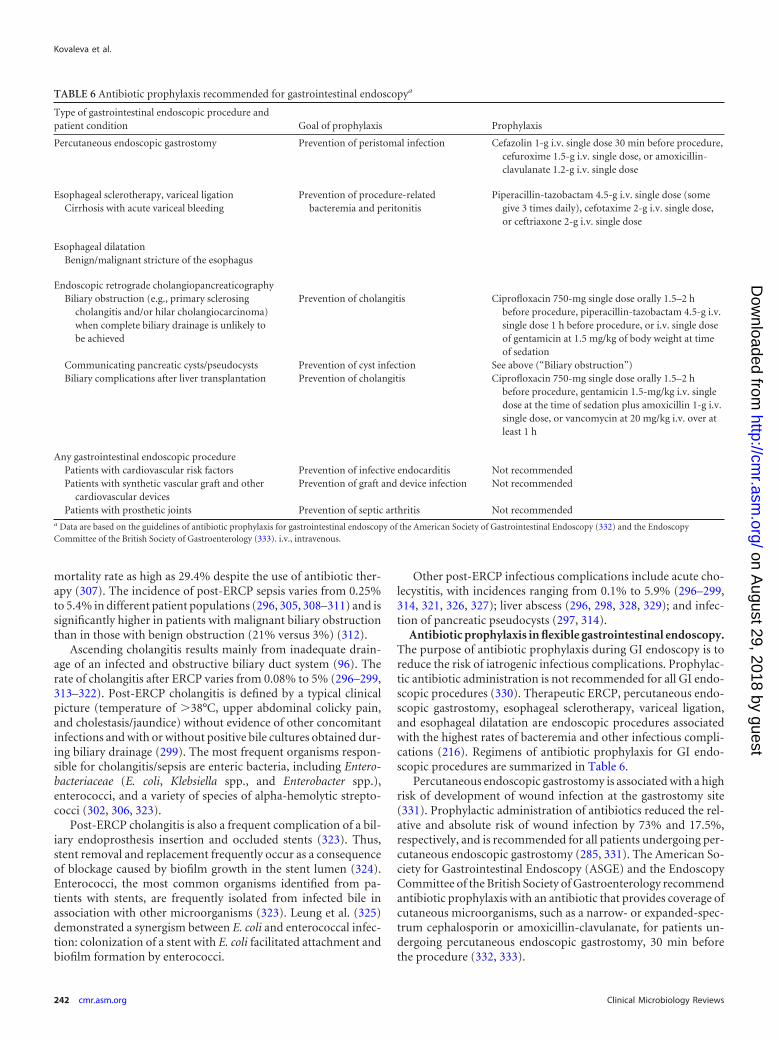
mortality rate as high as 29.4% despite the use of antibiotic ther-apy (307). The incidence of post-ERCP sepsis varies from 0.25%to 5.4% in different patient populations (296, 305, 308–311) and issignificantly higher in patients with malignant biliary obstructionthan in those with benign obstruction (21% versus 3%) (312).
Ascending cholangitis results mainly from inadequate drain-age of an infected and obstructive biliary duct system (96). Therate of cholangitis after ERCP varies from 0.08% to 5% (296–299,313–322). Post-ERCP cholangitis is defined by a typical clinicalpicture (temperature of �38°C, upper abdominal colicky pain,and cholestasis/jaundice) without evidence of other concomitantinfections and with or without positive bile cultures obtained dur-ing biliary drainage (299). The most frequent organisms respon-sible for cholangitis/sepsis are enteric bacteria, including Entero-bacteriaceae (E. coli, Klebsiella spp., and Enterobacter spp.),enterococci, and a variety of species of alpha-hemolytic strepto-cocci (302, 306, 323).
Post-ERCP cholangitis is also a frequent complication of a bil-iary endoprosthesis insertion and occluded stents (323). Thus,stent removal and replacement frequently occur as a consequenceof blockage caused by biofilm growth in the stent lumen (324).Enterococci, the most common organisms identified from pa-tients with stents, are frequently isolated from infected bile inassociation with other microorganisms (323). Leung et al. (325)demonstrated a synergism between E. coli and enterococcal infec-tion: colonization of a stent with E. coli facilitated attachment andbiofilm formation by enterococci.
Other post-ERCP infectious complications include acute cho-lecystitis, with incidences ranging from 0.1% to 5.9% (296–299,314, 321, 326, 327); liver abscess (296, 298, 328, 329); and infec-tion of pancreatic pseudocysts (297, 314).
Antibiotic prophylaxis in flexible gastrointestinal endoscopy.The purpose of antibiotic prophylaxis during GI endoscopy is toreduce the risk of iatrogenic infectious complications. Prophylac-tic antibiotic administration is not recommended for all GI endo-scopic procedures (330). Therapeutic ERCP, percutaneous endo-scopic gastrostomy, esophageal sclerotherapy, variceal ligation,and esophageal dilatation are endoscopic procedures associatedwith the highest rates of bacteremia and other infectious compli-cations (216). Regimens of antibiotic prophylaxis for GI endo-scopic procedures are summarized in Table 6.
Percutaneous endoscopic gastrostomy is associated with a highrisk of development of wound infection at the gastrostomy site(331). Prophylactic administration of antibiotics reduced the rel-ative and absolute risk of wound infection by 73% and 17.5%,respectively, and is recommended for all patients undergoing per-cutaneous endoscopic gastrostomy (285, 331). The American So-ciety for Gastrointestinal Endoscopy (ASGE) and the EndoscopyCommittee of the British Society of Gastroenterology recommendantibiotic prophylaxis with an antibiotic that provides coverage ofcutaneous microorganisms, such as a narrow- or expanded-spec-trum cephalosporin or amoxicillin-clavulanate, for patients un-dergoing percutaneous endoscopic gastrostomy, 30 min beforethe procedure (332, 333).
TABLE 6 Antibiotic prophylaxis recommended for gastrointestinal endoscopya
Type of gastrointestinal endoscopic procedure andpatient condition Goal of prophylaxis Prophylaxis
Percutaneous endoscopic gastrostomy Prevention of peristomal infection Cefazolin 1-g i.v. single dose 30 min before procedure,cefuroxime 1.5-g i.v. single dose, or amoxicillin-clavulanate 1.2-g i.v. single dose
Esophageal sclerotherapy, variceal ligation Prevention of procedure-relatedbacteremia and peritonitis
Piperacillin-tazobactam 4.5-g i.v. single dose (somegive 3 times daily), cefotaxime 2-g i.v. single dose,or ceftriaxone 2-g i.v. single dose
Cirrhosis with acute variceal bleeding
Esophageal dilatationBenign/malignant stricture of the esophagus
Endoscopic retrograde cholangiopancreaticographyBiliary obstruction (e.g., primary sclerosing
cholangitis and/or hilar cholangiocarcinoma)when complete biliary drainage is unlikely tobe achieved
Prevention of cholangitis Ciprofloxacin 750-mg single dose orally 1.5–2 hbefore procedure, piperacillin-tazobactam 4.5-g i.v.single dose 1 h before procedure, or i.v. single doseof gentamicin at 1.5 mg/kg of body weight at timeof sedation
Communicating pancreatic cysts/pseudocysts Prevention of cyst infection See above (“Biliary obstruction”)Biliary complications after liver transplantation Prevention of cholangitis Ciprofloxacin 750-mg single dose orally 1.5–2 h
before procedure, gentamicin 1.5-mg/kg i.v. singledose at the time of sedation plus amoxicillin 1-g i.v.single dose, or vancomycin at 20 mg/kg i.v. over atleast 1 h
Any gastrointestinal endoscopic procedurePatients with cardiovascular risk factors Prevention of infective endocarditis Not recommendedPatients with synthetic vascular graft and other
cardiovascular devicesPrevention of graft and device infection Not recommended
Patients with prosthetic joints Prevention of septic arthritis Not recommendeda Data are based on the guidelines of antibiotic prophylaxis for gastrointestinal endoscopy of the American Society of Gastrointestinal Endoscopy (332) and the EndoscopyCommittee of the British Society of Gastroenterology (333). i.v., intravenous.
Kovaleva et al.
242 cmr.asm.org Clinical Microbiology Reviews
on August 29, 2018 by guest
http://cmr.asm
.org/D
ownloaded from
Therapeutic upper GI endoscopic procedures associated with ahigh rate of postprocedure bacteremia and peritonitis includeesophageal sclerotherapy, variceal ligation, and esophageal dilata-tion. Prophylactic administration of antibiotics is recommendedfor all patients with cirrhosis and acute variceal bleeding beforeesophageal sclerotherapy and variceal ligation and for patientswith benign or malignant esophageal strictures before esophagealdilatation (333). A recent meta-analysis of 12 trials indicated asignificant beneficial effect of antibiotic prophylaxis comparedwith placebo and no prophylaxis in decreasing the incidence ofbacterial infections, bacteremia, pneumonia, spontaneous perito-nitis, and mortality from bacterial infections in cirrhotic patientswith GI bleeding (334). These benefits were observed indepen-dently of the antibiotic used. Administration of a broad-spectrumcephalosporin or piperacillin-tazobactam in a single dose or threedoses during 1 day is recommended by the ASGE and the BritishSociety of Gastroenterology (332, 333).
Recent studies examining antibiotic prophylaxis prior to ERCPconcluded that it may reduce the incidence of bacteremia but didnot reduce the incidence of clinical sepsis or cholangitis, andtherefore, routine use of antibiotic prophylaxis cannot be recom-mended (300, 335, 336). Antibiotic prophylaxis is recommendedbefore an ERCP in patients with biliary obstruction (e.g., primarysclerosing cholangitis and/or hilar cholangiocarcinoma) whencomplete biliary drainage is unlikely to be achieved and in patientswith communicating pancreatic cysts or pseudocysts for preven-tion of cyst infection (332, 333). An adequate biliary drainage willprevent postprocedure cholangitis or sepsis without prophylacticadministration of antibiotics. Continuation of antibiotics afterERCP in patients with biliary complications after liver transplan-tation may be beneficial, even when drainage is achieved (332,333). Prophylactic antibiotics should cover biliary flora such asEnterobacteriaceae, Bacteroides spp., P. aeruginosa, and entero-cocci. An optimum benefit of antibiotics is obtained if therapeuticlevels are present in the tissues at the time of the endoscopic pro-cedure. Antibiotic prophylaxis should be started orally or intrave-nously at least 1 to 2 h before the procedure. Oral ciprofloxacin,intravenous piperacillin-tazobactam, or gentamicin is recom-mended (332, 333). Increasing microbial resistance (ciprofloxa-cin), poor penetration into bile (gentamicin), and limited activityagainst enterococci (gentamicin and ciprofloxacin) are disadvan-tages of these antibiotics. According to the British Society of Gas-troenterology, for patients undergoing ERCP with biliary compli-cations after liver transplantation, these antibiotics should becombined with amoxicillin or vancomycin to target Enterococcusspp. (333). However, local incidence of amoxicillin-resistant, van-comycin-sensitive enterococci and known colonization with van-comycin-resistant enterococci need to be considered. In hospitalswith an increasing frequency of vancomycin-resistant entero-cocci, teicoplanin is recommended in preference to vancomycinfor two reasons: first, it is simpler and quicker to administer, andsecond, more sustained blood levels occur following a single dose(337).
Antibiotic administration for prevention of infective endocar-ditis (in patients with cardiovascular risk factors) and graft anddevice infections (in patients with synthetic vascular graft andother cardiovascular devices) before GI endoscopy is no longerrecommended by the ASGE and the American Heart Association(332, 338). This recommendation is based on an absence of clin-ically significant evidence of infective endocarditis and device in-
fection being associated with GI procedures and on an absence ofa conclusive link between GI endoscopy and development of anyother type of infection (332). Antibiotic prophylaxis of septic ar-thritis during GI endoscopy is not recommended for patients withorthopedic prostheses. However, prophylactic antibiotic admin-istration can be considered prior to any therapeutic GI endoscopicprocedure in patients with a seriously impaired immune status(e.g., in cases of neutropenia and hematological malignancy)(333).
Flexible Bronchoscopy
Oropharyngeal microorganisms from the upper respiratory tractcan be carried down into the lower respiratory tract during inser-tion of a bronchoscope into the lung through the mouth and canpenetrate into the bloodstream. These microorganisms includeviridans group streptococci, staphylococci, Moraxella spp., Neis-seria spp., and anaerobic bacteria (103).
Bacteremia following bronchoscopy, with or without biopsy, istransient and occurs in �5% of patients (339–343). The microor-ganisms most frequently isolated from positive blood cultures arestaphylococci and beta-hemolytic and viridans group strepto-cocci, which are part of the normal upper airway flora. Fever afterbronchoscopic procedures has been reported in many studies witha wide range of frequencies, from 0% to 27% (341, 343–349).Reported fevers were transient, developed during the first day af-ter bronchoscopy, and were not associated with septic complica-tions. One possible mechanism of development of fever afterbronchoscopy is the release of proinflammatory cytokines fromalveolar macrophages (350).
Pneumonia following bronchoscopic procedures has been re-ported, with a frequency of 0.6% to 6% (345, 346). Postbroncho-scopy pneumonia was defined as the development of fever and anew or progressive infiltrate on a chest radiograph with peripheralleukocytosis and elevated levels of the C-reactive protein after theprocedure. Several cases of fatal pneumonia, long abscess, andsepsis following bronchoscopy with isolation of Streptococcuspneumoniae, E. coli, Haemophilus influenzae, and Cryptococcus sp.from positive blood cultures and bronchial specimens have beendescribed (351–355).
Antibiotic prophylaxis is not recommended during flexiblebronchoscopy with or without biopsy because of the low inci-dence of bacteremia and endocarditis (356, 357). Only one casereport of infective endocarditis following bronchoscopy in anHIV-positive patient with mitral valve prolapse has been pub-lished (358). The American Heart Association does not recom-mend antibiotic prophylaxis for routine flexible bronchoscopy(357). However, administration of 2.0 g amoxicillin orally 1 hprior to bronchoscopy is recommended for rigid bronchoscopy,particularly for high-risk patients, including patients with pros-thetic cardiac valves, previous bacterial endocarditis, and complexcyanotic congenital heart disease (e.g., tetralogy of Fallot) (357).The British Thoracic Society Bronchoscopy Guidelines Commit-tee recommends prophylactic administration of antibiotics beforebronchoscopy for patients who are asplenic, have a heart valveprosthesis, or have a history of endocarditis (356).
IMPACT OF BIOFILM ON ENDOSCOPE REPROCESSING
Microorganisms in nature do not generally grow in nutrient-richsuspensions (the so-called planktonic state) as in the laboratorybut prefer to grow in surface-associated communities called bio-
Transmission of Infection by Flexible Endoscopy
April 2013 Volume 26 Number 2 cmr.asm.org 243
on August 29, 2018 by guest
http://cmr.asm
.org/D
ownloaded from
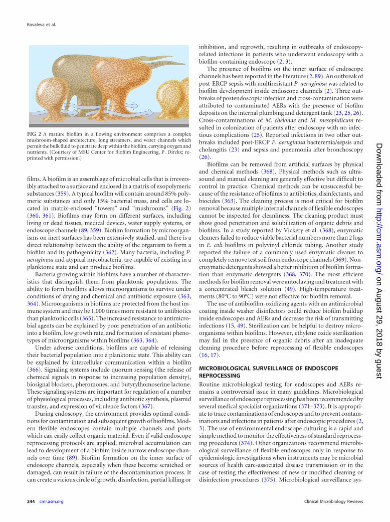
films. A biofilm is an assemblage of microbial cells that is irrevers-ibly attached to a surface and enclosed in a matrix of exopolymericsubstances (359). A typical biofilm will contain around 85% poly-meric substances and only 15% bacterial mass, and cells are lo-cated in matrix-enclosed “towers” and “mushrooms” (Fig. 2)(360, 361). Biofilms may form on different surfaces, includingliving or dead tissues, medical devices, water supply systems, orendoscope channels (89, 359). Biofilm formation by microorgan-isms on inert surfaces has been extensively studied, and there is adirect relationship between the ability of the organism to form abiofilm and its pathogenicity (362). Many bacteria, including P.aeruginosa and atypical mycobacteria, are capable of existing in aplanktonic state and can produce biofilms.
Bacteria growing within biofilms have a number of character-istics that distinguish them from planktonic populations. Theability to form biofilms allows microorganisms to survive underconditions of drying and chemical and antibiotic exposure (363,364). Microorganisms in biofilms are protected from the host im-mune system and may be 1,000 times more resistant to antibioticsthan planktonic cells (365). The increased resistance to antimicro-bial agents can be explained by poor penetration of an antibioticinto a biofilm, low growth rate, and formation of resistant pheno-types of microorganisms within biofilms (363, 364).
Under adverse conditions, biofilms are capable of releasingtheir bacterial population into a planktonic state. This ability canbe explained by intercellular communication within a biofilm(366). Signaling systems include quorum sensing (the release ofchemical signals in response to increasing population density),biosignal blockers, pheromones, and butyrylhomoserine lactone.These signaling systems are important for regulation of a numberof physiological processes, including antibiotic synthesis, plasmidtransfer, and expression of virulence factors (367).
During endoscopy, the environment provides optimal condi-tions for contamination and subsequent growth of biofilms. Mod-ern flexible endoscopes contain multiple channels and portswhich can easily collect organic material. Even if valid endoscopereprocessing protocols are applied, microbial accumulation canlead to development of a biofilm inside narrow endoscope chan-nels over time (89). Biofilm formation on the inner surface ofendoscope channels, especially when these become scratched ordamaged, can result in failure of the decontamination process. Itcan create a vicious circle of growth, disinfection, partial killing or
inhibition, and regrowth, resulting in outbreaks of endoscopy-related infections in patients who underwent endoscopy with abiofilm-containing endoscope (2, 3).
The presence of biofilms on the inner surface of endoscopechannels has been reported in the literature (2, 89). An outbreak ofpost-ERCP sepsis with multiresistant P. aeruginosa was related tobiofilm development inside endoscope channels (2). Three out-breaks of postendoscopic infection and cross-contamination wereattributed to contaminated AERs with the presence of biofilmdeposits on the internal plumbing and detergent tank (23, 25, 26).Cross-contaminations of M. chelonae and M. mesophilicum re-sulted in colonization of patients after endoscopy with no infec-tious complications (25). Reported infections in two other out-breaks included post-ERCP P. aeruginosa bacteremia/sepsis andcholangitis (23) and sepsis and pneumonia after bronchoscopy(26).
Biofilms can be removed from artificial surfaces by physicaland chemical methods (368). Physical methods such as ultra-sound and manual cleaning are generally effective but difficult tocontrol in practice. Chemical methods can be unsuccessful be-cause of the resistance of biofilms to antibiotics, disinfectants, andbiocides (363). The cleaning process is most critical for biofilmremoval because multiple internal channels of flexible endoscopescannot be inspected for cleanliness. The cleaning product mustshow good penetration and solubilization of organic debris andbiofilms. In a study reported by Vickery et al. (368), enzymaticcleaners failed to reduce viable bacterial numbers more than 2 logsin E. coli biofilms in polyvinyl chloride tubing. Another studyreported the failure of a commonly used enzymatic cleaner tocompletely remove test soil from endoscope channels (369). Non-enzymatic detergents showed a better inhibition of biofilm forma-tion than enzymatic detergents (368, 370). The most efficientmethods for biofilm removal were autoclaving and treatment witha concentrated bleach solution (49). High-temperature treat-ments (80°C to 90°C) were not effective for biofilm removal.
The use of antibiofilm-oxidizing agents with an antimicrobialcoating inside washer disinfectors could reduce biofilm buildupinside endoscopes and AERs and decrease the risk of transmittinginfections (15, 49). Sterilization can be helpful to destroy micro-organisms within biofilms. However, ethylene oxide sterilizationmay fail in the presence of organic debris after an inadequatecleaning procedure before reprocessing of flexible endoscopes(16, 17).
MICROBIOLOGICAL SURVEILLANCE OF ENDOSCOPEREPROCESSING
Routine microbiological testing for endoscopes and AERs re-mains a controversial issue in many guidelines. Microbiologicalsurveillance of endoscope reprocessing has been recommended byseveral medical specialist organizations (371–373). It is appropri-ate to trace contaminations of endoscopes and to prevent contam-inations and infections in patients after endoscopic procedures (2,3). The use of environmental endoscope culturing is a rapid andsimple method to monitor the effectiveness of standard reprocess-ing procedures (374). Other organizations recommend microbi-ological surveillance of flexible endoscopes only in response toepidemiologic investigations when instruments may be microbialsources of health care-associated disease transmission or in thecase of testing the effectiveness of new or modified cleaning ordisinfection procedures (375). Microbiological surveillance sys-
FIG 2 A mature biofilm in a flowing environment comprises a complexmushroom-shaped architecture, long streamers, and water channels whichpermit the bulk fluid to penetrate deep within the biofilm, carrying oxygen andnutrients. (Courtesy of MSU Center for Biofilm Engineering, P. Dirckx; re-printed with permission.)
Kovaleva et al.
244 cmr.asm.org Clinical Microbiology Reviews
on August 29, 2018 by guest
http://cmr.asm
.org/D
ownloaded from
tems have several important limitations. If there is a clinical de-mand for the reuse of an endoscope, surveillance culture resultswill not be obtained until after the endoscope is used on the nextpatient, because culture results take a minimum of 24 to 48 h to beproduced (371). Efficient sampling of flexible endoscopes andtheir accessories may be hindered by a complex design, materials,construction, and fragility or by the absence of a standardizedsampling protocol or an appropriate technique for sampling of thesurfaces and channels of the flexible endoscope (375).
Some techniques are recommended for microbiological sam-pling of flexible endoscopes (375). A swab-rinse technique shouldbe used for sampling the exterior surfaces and the distal opening ofthe suction-biopsy channel port. For adequate sampling of theinferior surface of endoscope channels, a “flush/brush/flush”technique should be performed with rinsing through the channelwith a sterile fluid and using a sterile cleaning brush to obtainsamples from the biopsy port. A simple flushthrough techniquemay be considered when brushing of the channel lumens is im-possible, but it is less efficient (375).
According to the literature, endoscopes can be sampled in ananterograde and retrograde manner. For anterograde sampling,the last-rinse water from the endoscope is collected inside thewasher-disinfector at the distal end of the endoscope (3, 9). Forretrograde sampling, the suction-biopsy channel and the air-wa-ter channel of the endoscope are each flushed with 20 ml steriledemineralized water manually outside the AER from the distal tothe proximal end. According to the literature, anterograde sam-pling is not sensitive enough (3, 9). Endoscope culturing with aretrograde technique is effective for monitoring endoscope repro-cessing, and testing is easy to perform (3). Introduction of newtechniques that are easier to perform on a daily basis and withfaster results may improve the microbiological surveillance of en-doscope reprocessing. New methods may be based on direct ATPmeasurement or quantitative PCR. These methods must be fur-ther optimized because of their low specificity (376, 377).
The literature provides no standards for the frequency of test-ing intervals of surveillance cultures. The Gastroenterological So-ciety of Australia guideline recommends microbiological moni-toring of duodenoscopes, bronchoscopes, and AERs every 4 weeksand all other GI endoscopes every 4 months (372). According tothe European Society of Gastrointestinal Endoscopy guideline,routine microbiological testing of endoscopes is advised to be per-formed at intervals of no longer than 3 months (371). The use ofroutine environmental microbiological testing of therapeutic en-doscopes once a month and diagnostic endoscopes once every 3months has been reported (2, 3).
A contaminated endoscope should be inspected for potentialdefects. Unfortunately, it is not possible to adequately check allinternal endoscope channels. Any small damage can be the sourceof bacterial contamination within the scope, which is difficult orimpossible to detect by routine inspection and testing (2, 3). Someinvestigators have begun to explore sterile-sheathed endoscopeswith new technology to reduce the risk of infectious complications(9, 378). The EndoSheath, for instance, is an endoscope system inwhich all parts of the endoscope that come into contact with thepatient are fully disposable.
Literature related to the costs associated with endoscope repro-cessing and economic consequences of endoscope contaminationis very limited. Economic evaluation of exogenous endoscopy-related outbreaks should include an analysis of outcomes (num-
ber of prevented iatrogenic infections) and an estimation of thehealth care, non-health care, and indirect costs (78). A recentlyreported study analyzed the cost of 4-weekly surveillance micro-biological tests of bronchoscopes, duodenoscopes, and AERs dur-ing a 5-year period (379). The overall annual cost of microbiolog-ical testing and cost in time for nursing staff to collect the sampleswas estimated to be $13,339. Another study calculated the annualcosts of surveillance of endoscope reprocessing (cost of microbi-ological testing and cost in time for staff) to be $33,959 (18). It wasalso estimated that the cost of the post-ERCP outbreak of P.aeruginosa sepsis affecting six patients, i.e., direct health care costsfor diagnosis, treatment, and hospitalization of six affected pa-tients, was approximately $206,273 (18).
CONCLUSION
Contaminated endoscopes have been linked to many outbreaks ofdevice-related nosocomial infections. The true incidence of en-doscopy-related infections is unknown because of inadequate sur-veillance or no surveillance at all. Endoscopy-related infectionscan cause serious harm and can give rise to concerns over theseprocedures by physicians and patients. Flexible endoscopes can becleaned and disinfected but not sterilized after use. This impliesthe risk of settlement of biofilm-producing species.
Process control of the cleaning and disinfection proceduredoes not guarantee prevention of biofilm formation during en-doscopy. Implementation of microbiological surveillance of en-doscope reprocessing is appropriate to detect early colonizationand biofilm formation in the endoscope and to prevent contami-nation and infection in patients after endoscopic procedures.However, the returns of prevention of endoscopy-related infec-tions should be reasonably in balance with costs of technical andlaboratory procedures resulting from surveillance and the costs ofreprocessing or servicing of the contaminated endoscope.
REFERENCES1. Nelson DB, Barkun AN, Block KP, Burdick JS, Ginsberg GG, Green-
wald DA, Kelsey PB, Nakao NL, Slivka A, Smith P, Vakil N. 2001.Technology status evaluation report. Transmission of infection by gas-trointestinal endoscopy: May 2001. Gastrointest. Endosc. 54:824 – 828.
2. Kovaleva J, Meessen NE, Peters FT, Been MH, Arends JP, Borgers RP,Degener JE. 2009. Is bacteriologic surveillance in endoscope reprocess-ing stringent enough? Endoscopy 41:913–916.
3. Buss AJ, Been MH, Borgers RP, Stokroos I, Melchers WJ, Peters FT,Limburg AJ, Degener JE. 2008. Endoscope disinfection and its pitfalls—requirement for retrograde surveillance cultures. Endoscopy 40:327–332.
4. Alvarado CJ, Reichelderfer M. 2000. APIC guidelines for infection pre-vention and control in flexible endoscopy. Association for Professionalsin Infection Control. Am. J. Infect. Control 28:138 –155.
5. Beilenhoff U, Neumann CS, Rey JF, Biering H, Blum R, Cimbro M,Kampf B, Rogers M, Schmidt V, ESGE Guidelines Committee, Euro-pean Society of Gastrointestinal Endoscopy, European Society ofGastroenterology and Endoscopy Nurses and Associates. 2008. ESGE-ESGENA guideline: cleaning and disinfection in gastrointestinal endos-copy. Endoscopy 40:939 –957.
6. Nelson DB, Jarvis WR, Rutala WA, Foxx-Orenstein AE, Isenberg G,Dash GR, Alvarado CJ, Ball M, Griffin-Sobel J, Petersen C, Ball KA,Henderson J, Stricof RL, Society for Heathcare Epidemiology ofAmerica. 2003. Multi-society guideline for reprocessing flexible gastro-intestinal endoscopes. Infect. Control Hosp. Epidemiol. 24:532–537.
7. Rutala WA, Weber DJ, Healthcare Infection Control Practices Advi-sory Committee. 2008. Guidedline for disinfection and sterilization inhealthcare facilities. CDC, Atlanta, GA. http://www.cdc.gov/hicpac/pdf/guidelines/Disinfection_Nov_2008.pdf.
8. Spach DH, Silverstein FE, Stamm WE. 1993. Transmission of infection
Transmission of Infection by Flexible Endoscopy
April 2013 Volume 26 Number 2 cmr.asm.org 245
on August 29, 2018 by guest
http://cmr.asm
.org/D
ownloaded from
by gastrointestinal endoscopy and bronchoscopy. Ann. Intern. Med.118:117–128.
9. Srinivasan A. 2003. Epidemiology and prevention of infections relatedto endoscopy. Curr. Infect. Dis. Rep. 5:467– 472.
10. Nelson DB, Muscarella LF. 2006. Current issues in endoscope repro-cessing and infection control during gastrointestinal endoscopy. WorldJ. Gastroenterol. 12:3953–3964.
11. Spaulding EH, Gröschel DH. 1974. Hospital disinfectants and antisep-tics, p 852– 857. In Lennette EH, Spaulding EH, Truant JP (ed), Manualof clinical microbiology, 2nd ed. American Society for Microbiology,Washington, DC.
12. Garner JS, Favero MS. 1986. CDC guidelines for the prevention andcontrol of nosocomial infections. Guideline for handwashing and hospi-tal environmental control, 1985. Supersedes guideline for hospital envi-ronmental control published in 1981. Am. J. Infect. Control 14:110 –129.
13. Simmons BP. 1983. CDC guidelines for the prevention and control ofnosocomial infection. Guideline for hospital invironmental control. Am.J. Infect. Control 11:97–120.
14. Gorse GJ, Messner RL. 1991. Infection control practices in gastrointes-tinal endoscopy in the United States: a national survey. Infect. ControlHosp. Epidemiol. 12:289 –296.
15. Rutala WA, Weber DJ. 2001. New disinfection and sterilization meth-ods. Emerg. Infect. Dis. 7:348 –353.
16. Alfa MJ, DeGagne P, Olson N, Puchalski T. 1996. Comparison of ionplasma, vaporised hydrogen peroxide, and 100% ethylene oxide sterilis-ers to the 12/88 ethylene oxide gas steriliser. Infect. Control Hosp. Epi-demiol. 17:92–100.
17. Chaufour X, Deva AK, Vickery K, Zou J, Kumaradeva P, White GH,Cossart YE. 1999. Evaluation of disinfection and sterilization of reusableangioscopes with the duck hepatitis B model. J. Vasc. Surg. 30:277–282.
18. Kovaleva J, Buss AJ. 2011. Usefulness of bacteriological monitoring ofendoscope reprocessing, p 141–162. In Pascu O, Tantau M (ed), Thera-peutic gastrointestinal endoscopy, 1st ed. InTech, Rijeka, Croatia.
19. Chu NS, Favero M. 2000. The microbial flora of the gastrointestinaltract and the cleaning of flexible endoscopes. Gastrointest. Endosc. Clin.N. Am. 10:233–244.
20. Chu NS, McAlister D, Antonoplos PA. 1998. Natural bioburden levelsdetected on flexible gastrointestinal endoscopes after clinical use andmanual cleaning. Gastrointest. Endosc. 48:137–142.
21. Petersen BT, Chennat J, Cohen J, Cotton PB, Greenwald DA, KowalskiTE, Krinsky ML, Park WG, Pike IM, Romagnuolo J, ASGE QualityAssurance in Endoscopy Committee, Rutala WA, Society for Health-care Epidemiology of America. 2011. Multisociety guideline on repro-cessing flexible GI endoscopes: 2011. Infect. Control Hosp. Epidemiol.32:527–537.
22. Allen JI, Allen MO, Olson MM, Gerding DN, Shanholtzer CJ, MeierPB, Vennes JA, Silvis SE. 1987. Pseudomonas infection of the biliarysystem resulting from use of a contaminated endoscope. Gastroenterol-ogy 92:759 –763.
23. Alvarado CJ, Stolz SM, Maki DG. 1991. Nosocomial infections fromcontaminated endoscopes: a flawed automated endoscope washer. Aninvestigation using molecular epidemiology. Am. J. Med. 91(3B:S272–S280.
24. Fraser VJ, Jones M, Murray PR, Medoff G, Zhang Y, Wallace RJ, Jr.1992. Contamination of flexible fiberoptic bronchoscopes with Myco-bacterium chelonae linked to an automated bronchoscope disinfectionmachine. Am. Rev. Respir. Dis. 145:853– 855.
25. Kressel AB, Kidd F. 2001. Pseudo-outbreak of Mycobacterium chelonaeand Methylobacterium mesophilicum caused by contamination of an au-tomated endoscopy washer. Infect. Control Hosp. Epidemiol. 22:414 –418.
26. Schelenz S, French G. 2000. An outbreak of multidrug-resistant Pseudomo-nas aeruginosa infection associated with contamination of bronchoscopesand an endoscope washer-disinfector. J. Hosp. Infect. 46:23–30.
27. Leers WD. 1980. Disinfecting endoscopes: how not to transmit Myco-bacterium tuberculosis by bronchoscopy. CMAJ 123:275–280, 283.
28. Hawkey PM, Davies AJ, Viant AC, Lush CJ, Mortensen NJ. 1981.Contamination of endoscopes by Salmonella species. J. Hosp. Infect.2:373–376.
29. Ranjan P, Das K, Ayyagiri A, Saraswat VA, Choudhuri G. 2005. Areport of post-ERCP Pseudomonas aeruginosa infection outbreak. IndianJ. Gastroenterol. 24:131–132.
30. Silva CV, Magalhães VD, Pereira CR, Kawagoe JY, Ikura C, Ganc AJ.
2003. Pseudo-outbreak of Pseudomonas aeruginosa and Serratia marc-escens related to bronchoscopes. Infect. Control Hosp. Epidemiol. 24:195–197.
31. Food and Drug Administration. 2009. FDA-cleared sterilants and high leveldisinfectants with general claims for processing reusable medical and dentaldevices. Food and Drug Administration, Washington, DC. http://www.fda.gov/MedicalDevices/DeviceRegulationandGuidance/ReprocessingofSingle-UseDevices/ucm133514.htm.
32. Nelson DB. 2003. Infectious disease complications of GI endoscopy: partII, exogenous infections. Gastrointest. Endosc. 57:695–711.
33. Russell AD. 1994. Glutaraldehyde: current status and uses. Infect. Con-trol Hosp. Epidemiol. 15:724 –733.
34. Babb JR, Bradley CR, Ayliffe GA. 1980. Sporicidal activity of glutaral-dehydes and hypochlorites and other factors influencing their selectionfor the treatment of medical equipment. J. Hosp. Infect. 1:63–75.
35. McDonnell G, Russell AD. 1999. Antiseptics and disinfectants: activity,action, and resistance. Clin. Microbiol. Rev. 12:147–179.
36. Rideout K, Teschke K, Dimich-Ward H, Kennedy SM. 2005. Consid-ering risks to healthcare workers from glutaraldehyde alternatives inhigh-level disinfection. J. Hosp. Infect. 59:4 –11.
37. Rozen P, Somjen GJ, Baratz M, Kimel R, Arber N, Gilat T. 1994.Endoscope-induced colitis: description, probable cause by glutaralde-hyde, and prevention. Gastrointest. Endosc. 40:547–553.
38. Collins FM. 1986. Bactericidal activity of alkaline glutaraldehyde solu-tion against a number of atypical mycobacterial species. J. Appl. Bacte-riol. 61:247–251.
39. Nomura K, Ogawa M, Miyamoto H, Muratani T, Taniguchi H. 2004.Antibiotic suspectibility of glutaraldehyde-tolerant Mycobacterium che-lonae from bronchoscope washing machines. Am. J. Infect. Control 32:185–188.
40. van Klingeren B, Pullen W. 1993. Glutaraldehyde resistant mycobacte-ria from endoscope washers. J. Hosp. Infect. 25:147–149.
41. Barbee SL, Weber DJ, Sobsey MD, Rutala WA. 1999. Inactivation ofCryptosporidium parvum oocyst infectivity by disinfection and steriliza-tion processes. Gastrointest. Endosc. 49:605– 611.
42. Kovacs BJ, Aprecio RM, Kettering JD, Chen YK. 1998. Efficacy ofvarious disinfectants in killing a resistant strain of Pseudomonas aerugi-nosa by comparing zones of inhibition: implications for endoscopicequipment reprocessing. Am. J. Gastroenterol. 93:2057–2059.
43. Hanson PJ, Chadwick MV, Gaya H, Collins JV. 1992. A study ofglutaraldehyde disinfection of fiberoptic bronchoscopes experimentallycontaminated with Mycobacterium tuberculosis. J. Hosp. Infect. 22:137–142.
44. Gregory AW, Schaalje GB, Smart JD, Robison RA. 1999. The myco-bactericidal efficacy of ortho-phthalaldehyde and the comparative resis-tances of Mycobacterium bovis, Mycobacterium terrae, and Mycobacte-rium chelonae. Infect. Control Hosp. Epidemiol. 20:324 –330.
45. Sokol WN. 2004. Nine episodes of anaphylaxis following cystoscopycaused by Cidex OPA (ortho-phthalaldehyde) high-level disinfectant in4 patients after cystoscopy. J. Allergy Clin. Immunol. 114:392–397.
46. Rutala WA, Weber DJ. 1999. Disinfection of endoscopes: review of newchemical sterilants used for high-level disinfection. Infect. Control Hosp.Epidemiol. 20:69 –76.
47. Kovaleva J, Degener JE, van der Mei HC. 2010. Mimicking disinfectionand drying of biofilms in contaminated endoscopes. J. Hosp. Infect. 76:345–350.
48. Loukili NH, Granbastien B, Faure K, Guery B, Beaucaire G. 2006.Effect of different stabilized preparations of peracetic acid on biofilm. J.Hosp. Infect. 63:70 –72.
49. Marion-Ferey K, Pasmore M, Stoodley P, Wilson S, Husson GP,Costerton JW. 2003. Biofilm removal from silicone tubing: an assess-ment of the efficacy of dialysis machine decontamination proceduresusing an in vitro model. J. Hosp. Infect. 53:64 –71.
50. Selkon JB, Babb JR, Morris R. 1999. Evaluation of the antimicrobialactivity of a new super-oxidized water, Sterilox, for the disinfection ofendoscopes. J. Hosp. Infect. 41:59 –70.
51. Bennett SN, Peterson DE, Johnson DR, Hall WN, Robinson-Dunn B,Dietrich S. 1994. Bronchoscopy-associated Mycobacterium xenopi pseu-doinfections. Am. J. Respir. Crit. Care Med. 150:245–250.
52. Centers for Disease Control. 1991. Nosocomial infection and pseudoin-fection from contaminated endoscopes and bronchoscopes: Wisconsinand Missouri. MMWR Morb. Mortal. Wkly. Rep. 40:675– 678.
53. Cryan EM, Falkiner FR, Mulvihill TE, Keane CT, Keeling PW. 1984.
Kovaleva et al.
246 cmr.asm.org Clinical Microbiology Reviews
on August 29, 2018 by guest
http://cmr.asm
.org/D
ownloaded from
Pseudomonas aeruginosa cross-infection following endoscopic retro-grade cholangiopancreatography. J. Hosp. Infect. 5:371–376.
54. Flournoy DJ, Petrone RL, Voth DW. 1992. A pseudo-outbreak ofMethylobacterium mesophilica isolated from patients undergoing bron-choscopy. Eur. J. Clin. Microbiol. Infect. Dis. 11:240 –243.
55. Siegman-Igra Y, Inbar G, Campus A. 1985. An ‘outbreak’ of pulmonarypseudoinfection by Serratia marcescens. J. Hosp. Infect. 6:218 –220.
56. Sniadack DH, Ostroff SM, Karlix MA, Smithwick RW, Schwartz B,Sprauer MA, Silcox VA, Good RC. 1993. A nosocomial pseudo-outbreak of Mycobacterium xenopi due to a contaminated potable watersupply: lessons in prevention. Infect. Control Hosp. Epidemiol. 11:636 –641.
57. Vandenbroucke-Grauls CM, Baars AC, Visser MR, Hulstaert PF, Ver-hoef J. 1993. An outbreak of Serratia marcescens traced to a contami-nated bronchoscope. J. Hosp. Infect. 23:263–270.
58. Alfa MJ, Sitter DL. 1991. In-hospital evaluation of contamination ofduodenoscopes: a quantitative assessment of the effect of drying. J. Hosp.Infect. 19:89 –98.
59. Muscarella LF. 2006. Inconsistencies in endoscope-reprocessing andinfection-control guidelines: the importance of endoscope drying. Am. J.Gastroenterol. 101:2147–2154.
60. Pineau L, Villard E, Duc DL, Marchetti B. 2008. Endoscope drying/storage cabinet: interest and efficacy. J. Hosp. Infect. 68:59 – 65.
61. Rejchrt S, Cermák P, Pavlatová L, Micková E, Bures J. 2004. Bacteri-ologic testing of endoscopes after high-level disinfection. Gastrointest.Endosc. 60:76 –78.
62. Noy MF, Harrison L, Holmes GK, Cockel R. 1980. The significanceof bacterial contamination of fiberoptic endoscopes. J. Hosp. Infect.1:53– 61.
63. Agerton T, Valway S, Gore B, Pozsik C, Plikaytis B, Woodley C,Onorato I. 1997. Transmission of a highly drug-resistant strain (strainW1) of Mycobacterium tuberculosis. Community outbreak and nosoco-mial transmission via a contaminated bronchoscope. JAMA 278:1073–1077.
64. Bou R, Aguilar A, Perpiñán J, Ramos P, Peris M, Lorente L, Zúñiga A.2006. Nosocomial outbreak of Pseudomonas aeruginosa infections re-lated to a flexible bronchoscope. J. Hosp. Infect. 64:129 –135.
65. Brayko CM, Kozarek RA, Sanowski RA, Testa AW. 1985. Bacteremiaduring esophageal variceal sclerotherapy: its cause and prevention. Gas-trointest. Endosc. 31:10 –12.
66. Breathnach AS, Taylor IK, Mitchison HC, Shrestha TL. 1997. Pseudo-infection with non-viable Mycobacterium tuberculosis following bron-choscopy. J. Infect. 35:321–322.
67. Elston RA, Hay AJ. 1991. Acid-fast bacillus contamination of a bron-choscope washing machine. J. Hosp. Infect. 19:72–73.
68. Gubler JG, Salfinger M, von Graevenitz A. 1992. Pseudoepidemic ofnontuberculous mycobacteria due to a contaminated bronchoscopecleaning machine. Report of an outbreak and review of the literature.Chest 101:1245–1249.
69. Hagan ME, Klotz SA, Bartholomew W, Potter L, Nelson M. 1995. Apseudoepidemic of Phodotorula rubra: a marker for microbial contami-nation of the bronchoscope. Infect. Control Hosp. Epidemiol. 16:727–728.
70. Kolmos HJ, Lerche A, Kristoffersen K, Rosdahl VT. 1994. Pseudo-outbreak of Pseudomonas aeruginosa in HIV-infected patients undergo-ing fiberoptic bronchoscopy. Scand. J. Infect. Dis. 26:653– 657.
71. Nye K, Chadha DK, Hodgkin P, Bradley C, Hancox J, Wise R. 1990.Mycobacterium chelonei isolation from broncho-alveolar lavage fluid andits practical implications. J. Hosp. Infect. 16:257–261.
72. Peltroche-Llacsahuanga H, Lütticken R, Haase G. 1999. Temporallyoverlapping nosocomial outbreaks of Serratia marcescens infections: anunexpected result revealed by pulsed-field gel electrophoresis. Infect.Control Hosp. Epidemiol. 20:387–388.
73. Sammartino MT, Israel RH, Magnussen CR. 1982. Pseudomonasaeruginosa contamination of fiberoptic bronchoscopes. J. Hosp. Infect.3:65–71.
74. Shimono N, Takuma T, Tsuchimochi N, Shiose A, Murata M, Kana-moto Y, Uchida Y, Morita S, Matsumoto H, Hayashi J. 2008. Anoutbreak of Pseudomonas aeruginosa infections following thoracic sur-geries occurring via the contamination of bronchoscopes and an auto-matic endoscope reprocessor. J. Infect. Chemother. 14:418 – 423.
75. Struelens MJ, Rost F, Deplano A, Maas A, Schwam V, Serruys E,Cremer M. 1993. Pseudomonas aeruginosa and Enterobacteriaceae bacte-
remia after biliary endoscopy: an outbreak investigation using DNAmacrorestriction analysis. Am. J. Med. 95:489 – 498.
76. Takigawa K, Fujita J, Negayama K, Terada S, Yamaji S, Kawanishi K,Takahara J. 1995. Eradication of contaminating Mycobacterium chelonaefrom bronchofibrescopes and an automated bronchoscope disinfectionmachine. Respir. Med. 89:423– 427.
77. Kimmey MB, Burnett DA, Carr-Locke DL, DiMarino AJ, Jensen DM,Katon R, MacFadyen BV, Scobey MW, Stein TN, Steinberg SM. 1993.Transmission of infection by gastrointestinal endoscopy. Gastrointest.Endosc. 39:885– 888.
78. Seoane-Vazquez E, Rodriguez-Monguio R, Visaria J, Carlson A. 2006.Exogenous endoscopy-related infections, pseudo-infections, and toxicreactions: clinical and economic burden. Curr. Med. Res. Opin. 22:2007–2021.
79. Cowen AE. 1992. Infection and endoscopy: who infects whom? Scand. J.Gastroenterol. Suppl. 192:91–96.
80. Greene WH, Moody M, Hartley R, Effman E, Aisner J, Young VM,Wiernik RH. 1974. Esophagoscopy as a source of Pseudomonas aerugi-nosa sepsis in patients with acute leukemia: the need for sterilization ofendoscopes. Gastroenterology 67:912–919.
81. Kaw M, Przepiorka D, Sekas G. 1993. Infectious complications ofendoscopic procedures in bone marrow transplant recipients. Dig. Dis.Sci. 38:71–74.
82. Beecham HJ, III, Cohen ML, Parkin WE. 1979. Salmonella typhimu-rium: transmission by fiberoptic upper gastrointestinal endoscopy.JAMA 241:1013–1015.
83. Chmel H, Armstrong D. 1976. Salmonella oslo: a focal outbreak in ahospital. Am. J. Med. 60:203–208.
84. Dean AG. 1977. Transmission of Salmonella typhi by fiberoptic endos-copy. Lancet ii:134.
85. Dwyer DM, Klein EG, Istre GR, Robinson MG, Neumann DA, McCoyGA. 1987. Salmonella newport infections transmitted by fiberopticcolonoscopy. Gastrointest. Endosc. 33:84 – 87.
86. O’Connor BH, Bennett JR, Alexander JG, Sutton DR, Leighton I,Mawer SL, Dunlop JM. 1982. Salmonellosis infection transmitted byfibreoptic endoscopes. Lancet ii:864 – 866.
87. Pearson HE, Grant WJ. 1970. Presumed transmission of Salmonella bysygmoidoscope. Calif. Med. 112:23–25.
88. Schliessler KH, Rozendaal B, Taal C, Meawissen SG. 1980. Outbreak ofSalmonella agona infection after upper intestinal fibreoptic endoscopy.Lancet ii:1246.
89. Pajkos A, Vickery K, Cossart Y. 2004. Is biofilm accumulation onendoscope tubing a contributor to the failure of cleaning and decontam-ination? J. Hosp. Infect. 58:224 –229.
90. Classen DC, Jacobson JA, Burke JP, Jacobson JT, Evans RS. 1988.Serious Pseudomonas infections associated with endoscopic retrogradecholangiopancreatography. Am. J. Med. 84:590 –596.
91. Earnshaw JJ, Clark AW, Thom BT. 1985. Outbreak of Pseudomonasaeruginosa following endoscopic retrograde cholangiopancreatography.J. Hosp. Infect. 6:95–97.
92. Grogan JB. 1966. Pseudomonas aeruginosa carriage in patients. J. Trauma6:639 – 643.
93. Srinivasan A, Wolfenden LL, Song X, Mackie K, Hartsell TL, JonesHD, Diette GB, Orens JB, Yung RC, Ross TL, Merz W, Scheel PJ,Haponik EF, Perl TM. 2003. An outbreak of Pseudomonas aeruginosainfections associated with flexible bronchoscopes. N. Engl. J. Med. 348:221–227.
94. Motte S, Deviere J, Dumonceau JM, Serruys E, Thys JP, Cremer M.1991. Risk factors for septicemia following endoscopic biliary stenting.Gastroenterology 101:1374 –1381.
95. Elson CO, Hattori K, Blackstone MO. 1975. Polymicrobial sepsis fol-lowing endoscopic retrograde cholangiopancreatography. Gastroenter-ology 69:507–510.
96. Low DE, Mieflikier AB, Kennedy JK, Stiver HG. 1980. Infectiouscomplications of endoscopic retrograde cholangiopancreatography. Aprospective assessment. Arch. Intern. Med. 140:1076 –1077.
97. Schousboe M, Carter A, Sheppard PS. 1980. Endoscopic retrogradecholangiopancreatography: related nosocomial infections. N. Z. Med. J.92:275–277.
98. Siegman-Igra Y, Isakov A, Inbar G, Cahaner J. 1987. Pseudomonasaeruginosa septicemia following endoscopic retrograde cholangiopan-creatography with a contaminated endoscope. Scand. J. Infect. Dis. 19:527–530.
Transmission of Infection by Flexible Endoscopy
April 2013 Volume 26 Number 2 cmr.asm.org 247
on August 29, 2018 by guest
http://cmr.asm
.org/D
ownloaded from
99. Bass DH, Oliver S, Bornman PC. 1990. Pseudomonas septicaemia afterendoscopic retrograde cholangiopancreatography—an unresolvedproblem. S. Afr. Med. J. 77:509 –511.
100. Doherty DE, Falko JM, Lefkovitz N, Rogers J, Fromkes J. 1982.Pseudomonas aeruginosa sepsis following retrograde cholangiopancre-atography. Dig. Dis. Sci. 27:169 –170.
101. Fraser TG, Reiner S, Malczynski M, Yarnold PR, Warren J, NoskinGA. 2004. Multidrug-resistant Pseudomonas aeruginosa cholangitis afterendoscopic retrograde cholangiopancreatography: failure of routine en-doscope cultures to prevent an outbreak. Infect. Control Hosp. Epide-miol. 25:856 – 859.
102. Blanc DS, Parret T, Janin B, Raselli P, Francioli P. 1997. Nosocomialinfections from contaminated bronchoscopes: two-years follow up usingmolecular markers. Infect. Control Hosp. Epidemiol. 18:134 –136.
103. Sorin M, Segal-Maurer S, Mariano N, Urban C, Combest A, Rahal JJ.2001. Nosocomial transmission of imipenem-resistant Pseudomonasaeruginosa following bronchoscopy associated with improper connec-tion to the Steris System 1 processor. Infect. Control Hosp. Epidemiol.22:409 – 413.
104. Centers for Disease Control and Prevention. 1999. Bronchoscopy-related infections and pseudoinfections: New York, 1996 and 1998.MMWR Morb. Mortal. Wkly. Rep. 48:557–560.
105. Corne P, Godreuil S, Jean-Pierre H, Jonquet O, Campos J, Jumas-Bilak E, Parer S, Marchandin H. 2005. Unusual implication of biopsyforceps in outbreaks of Pseudomonas aeruginosa infections and pseudo-infections related to bronchoscopy. J. Hosp. Infect. 61:20 –26.
106. DiazGranados CA, Jones MY, Kongphet-Tran T, White N, Shapiro M,Wang YF, Ray SM, Blumberg HM. 2009. Outbreak of Pseudomonasaeruginosa infection associated with contamination of a flexible bron-choscope. Infect. Control Hosp. Epidemiol. 30:550 –555.
107. Kirschke DL, Jones TF, Craig AS, Chu PS, Mayernick GG, Patel JA,Schaffner W. 2003. Pseudomonas aeruginosa and Serratia marcescenscontamination associated with a manufacturing defect in broncho-scopes. N. Engl. J. Med. 348:214 –220.
108. Michele TM, Cronin WA, Graham NM, Dwyer DM, Pope DS, Har-rington S, Chaisson RE, Bishai WR. 1997. Transmission of Mycobacte-rium tuberculosis by a fiberoptic bronchoscope: identification by DNAfingerprinting. JAMA 278:1093–1095.
109. Nelson KE, Larson PA, Schraufnagel DE, Jackson J. 1983. Transmis-sion of tuberculosis by flexible fiberbronchoscopes. Am. Rev. Respir. Dis.127:97–100.
110. Ramsey AH, Oemig TV, Davis JP, Massey JP, Török TJ. 2002. Anoutbreak of bronchoscopy-related Mycobacterium tuberculosis infectionsdue to lack of bronchoscope leak testing. Chest 121:976 –981.
111. Wheeler PW, Lancaster D, Kaiser AB. 1989. Bronchopulmonary cross-colonization and infection related to mycobacterial contamination ofsuction valves of bronchoscopes. J. Infect. Dis. 159:954 –958.
112. Hanson PJ. 1988. Mycobacteria and AIDS. Br. J. Hosp. Med. 40:149.113. Phillips MS, von Reyn CF. 2001. Nosocomial infections due to nontu-
berculous mycobacteria. Clin. Infect. Dis. 33:1363–1374.114. Sattar SA, Best M, Springthorpe VS, Sanani G. 1995. Mycobactericidal
testing of disinfectants: an update. J. Hosp. Infect. 30(Suppl):372–382.115. van Klingeren B, Pullen W. 1987. Comparative testing of disinfectants
against Mycobacterium tuberculosis and Mycobacterium terrae in a quan-titative suspension test. J. Hosp. Infect. 10:292–298.
116. Davis D, Bonekat HW, Andrews D, Shigeoka JW. 1984. Disinfection ofthe flexible fibreoptic bronchoscope against Mycobacterium tuberculosisand M. gordonae. Thorax 39:785–788.
117. Urayama S, Kozarek RA, Sumida S, Raltz S, Merriam L, Pethigal P.1996. Mycobacteria and glutaraldehyde: is high-level disinfection of en-doscopes possible? Gastrointest. Endosc. 43:451– 456.
118. Hernández A, Martró E, Puzo C, Matas L, Burgués C, Vázquez N,Castella J, Ausina V. 2003. In-use evaluation of Perasafe compared withCidex in fibreoptic bronchoscope disinfection. J. Hosp. Infect. 54:46 –51.
119. Wallace RJ, Jr, Brown BA, Griffith DE. 1998. Nosocomial outbreaks/pseudo-outbreaks caused by nontuberculous mycobacteria. Annu. Rev.Microbiol. 52:453– 490.
120. Larson JL, Lambert L, Stricof RL, Driscoll J, McGarry MA, Ridzon R.2003. Potential nosocomial exposure to Mycobacterium tuberculosis froma bronchoscope. Infect. Control Hosp. Epidemiol. 24:825– 830.
121. Brown NM, Hellyar EA, Harvey JE, Reeves DS. 1993. Mycobacterialcontamination of fibreoptic bronchoscopes. Thorax 48:1283–1285.
122. Campagnaro RL, Teichtahl H, Dwyer B. 1994. A pseudoepidemic of
Mycobacterium chelonae: contamination of a bronchoscope and auto-cleaner. Aust. N. Z. J. Med. 24:693– 695.
123. Shimoide H, Anzai E, Murata Y, Kusajima K, Ichihara H, Takano T,Hirayama N, Sato N, Kobayashi Y. 1995. Contamination of flexiblefiberoptic bronchoscopes with Mycobacterium chelonae linked to an au-tomated endoscope disinfection machine— on the relationship betweenthe presence of the organism in the intestinal tract and contamination ofdisinfection machine, and a case of gallbladder and bile duct infectionwith M. chelonae. Kekkaku 70:571–577.
124. Pappas SA, Schaaff DM, DiCostanzo MB, King FW, Jr, Sharp JT. 1983.Contamination of flexible fiberoptic bronchoscopes. Am. Rev. Respir.Dis. 127:391–392.
125. Prigogine TH, Glupczynski Y, Van Molle P, Schmerber J. 1988. My-cobacterial cross-contamination of bronchsocopy specimens. J. Hosp.Infect. 11:93–95.
126. Uttley AH, Honeywell KM, Fitch LE, Yates MD, Collins CH, SimpsonRA. 1990. Cross-contamination of bronchial washings. BMJ 301:1274.
127. Wang HC, Liaw YS, Yang PC, Kuo SH, Luh KT. 1995. A pseudoepi-demic of Mycobacterium chelonae infection caused by contamination of afibreoptic bronchoscope suction channel. Eur. Respir. Infect. 8:1259 –1262.
128. Langenberg W, Rauws EA, Oudbier JH, Tytgat GN. 1990. Patient-to-patient transmission of Campylobacter pylori infection by fiberoptic gas-troduodenoscopy and biopsy. J. Infect. Dis. 161:507–511.
129. Tytgat GN. 1995. Endoscopic transmission of Helicobacter pylori. Ali-ment. Pharmacol. Ther. 9(Suppl 2):105–110.
130. Graham DY, Alpert LC, Smith JL, Yoshimura HH. 1988. IatrogenicCampylobacter pylori infection is a cause of epidemic achlorhydria. Am. J.Gastroenterol. 83:974 –980.
131. Miyaji H, Kohli Y, Azuma T, Ito S, Hirai M, Ito Y, Kato T, KuriyamaM. 1995. Endoscopic cross-infection with Helicobacter pylori. Lancet345:464.
132. Akamatsu T, Tabata K, Hironga M, Kawakami H, Uyeda M. 1996.Transmission of Helicobacter pylori infection via flexible fiberoptic en-doscopy. Am. J. Infect. Control 24:396 – 401.
133. Fantry GT, Zheng QX, James SP. 1995. Conventional cleaning anddisinfection techniques eliminate the risk of endoscopic transmission ofHelicobacter pylori. Am. J. Gastroenterol. 90:227–232.
134. Roosendaal R, Kuipers EJ, van den Brule AJ, Peña AS, Uyterlinde AM,Walboomers JM, Meuwissen SG, de Graaff J. 1994. Importance of thefiberoptic endoscope cleaning procedure for detection of Helicobacterpylori in gastric biopsy specimens by PCR. J. Clin. Microbiol. 32:1123–1126.
135. Poutanen SM, Simor AE. 2004. Clostridium difficile-associated diarrheain adults. CMAJ 171:51–58.
136. Patterson DJ, Johnson EH, Schmulen AC. 1984. Fulminant pseu-domembranous colitis occurring after colonoscopy. Gastrointest. En-dosc. 30:249 –253.
137. Selinger CP, Greer S, Sutton CJ. 2010. Is gastrointestinal endoscopy arisk factor for Clostridium difficile associated diarrhea? Am. J. Infect.Control 38:581–582.
138. Hughes CE, Gebhard RL, Peterson LR, Gerding DN. 1986. Efficacy ofroutine fiberoptic endoscope cleaning and disinfection for killing Clos-tridium difficile. Gastrointest. Endosc. 32:7–9.
139. Rutala WA, Gergen MF, Weber DJ. 1993. Inactivation of Clostridiumdifficile spores by disinfectants. Infect. Control Hosp. Epidemiol. 14:36–39.
140. Wullt M, Odenholt I, Walder M. 2003. Activity of three disinfectantsand acidified nitrite against Clostridium difficile spores. Infect. ControlHosp. Epidemiol. 24:765–768.
141. Godiwala T, Andry M, Agrawal N, Ertan A. 1988. Consecutive Serratiamarcescens infections following endoscopic retrograde cholangiopancre-atography. Gastrointest. Endosc. 34:345–347.
142. Webb SF, Vall-Spinosa A. 1975. Outbreak of Serratia marcescens asso-ciated with the flexible fiberbronchoscope. Chest 68:703–708.
143. Cêtre J-C, Nicolle MC, Salord H, Pérol M, Tigaud S, David G,Bourjault M, Vanhems P. 2005. Outbreaks of contaminated broncho-alveolar lavage related to intrinsically defective bronchoscopes. J. Hosp.Infect. 61:39 – 45.
144. Aumeran C, Poincloux L, Souweine B, Robin F, Laurichesse H, BaudO, Bommelaer G, Traoré O. 2010. Multidrug-resistant Klebsiella pneu-moniae outbreak after endoscopic retrograde cholangiopancreatogra-phy. Endoscopy 42:895– 899.
145. Carbonne A, Thiolet JM, Fournier S, Fortineau N, Kassis-Chikhani N,
Kovaleva et al.
248 cmr.asm.org Clinical Microbiology Reviews
on August 29, 2018 by guest
http://cmr.asm
.org/D
ownloaded from
Boytchev I, Aggoune M, Seguier JC, Senechal H, Tavolacci MP,Coignard B, Astagneau P, Jarlier V. 2010. Control of a multi-hospitaloutbreak of KPC-producing Klebsiella pneumoniae type 2 in France, Sep-tember to October 2009. Euro Surveill. 15(48):pii�19734. http://www.eurosurveillance.org/ViewArticle.aspx?ArticleId�19734.
146. Furuhata K, Kato Y, Goto K, Hara M, Yoshida S, Fukuyama M. 2006.Isolation and identification of Methylobacterium species from the tapwater in hospitals in Japan and their antibiotic susceptibility. Microbiol.Immunol. 50:11–17.
147. Imbert G, Seccia Y, La Scola B. 2005. Methylobacterium sp. bacteraemiadue to a contaminated endoscope. J. Hosp. Infect. 61:268 –270.
148. Simões LC, Simões M, Vieira MJ. 2010. Adhesion and biofilm forma-tion on polystyrene by drinking water-isolated bacteria. Antonie VanLeeuwenhoek 98:317–319.
149. Van Aken B, Peres CM, Doty SL, Yoon JM, Schnoor JL. 2004. Methy-lobacterium populi sp. nov., a novel aerobic, pink-pigmented, faculta-tively methylotrophic, methane-utilizing bacterium isolated from poplartrees (Populus deltoides � nigra DN34). Int. J. Syst. Evol. Microbiol.54:1191–1196.
150. Mandelstam P, Sugawa C, Silvis SE, Nebel OT, Rogers BH. 1976.Complications associated with esophagogastroduodenoscopy and withesophageal dilation. Gastrointest. Endosc. 23:16 –19.
151. Lo Passo C, Pernice I, Celeste A, Perdichizzi G, Todaro-Luck F. 2001.Transmission of Trichosporon asahii esophagitis by a contaminated en-doscope. Mycoses 44:13–21.
152. Singh S, Singh N, Kochhar R, Mehta SK, Talwar P. 1989. Contam-ination of an endoscope due to Trichosporon beigelii. J. Hosp. Infect.14:49 –53.
153. Hoffmann KK, Weber DJ, Rutala WA. 1989. Pseudoepidemic of Pho-dotorula rubra in patients indergoing fiberoptic bronchoscopy. Infect.Control Hosp. Epidemiol. 10:511–514.
154. Nicolle LE, McLeod J, Romance L, Parker S, Paraskevas M. 1992.Pseudo-outbreak of blastomycosis associated with contaminated bron-choscopes. Infect. Control Hosp. Epidemiol. 13:324.
155. Mitchell DH, Hicks LJ, Chiew R, Montanaro JC, Chen SC. 1997.Pseudoepidemic of Legionella pneumophila serogroup 6 associated withcontaminated bronchoscopes. J. Hosp. Infect. 37:19 –23.
156. Goldstein B, Abrutyn E. 1985. Pseudo-outbreak of Bacillus spores re-lated to fibreoptic bronchoscopy. J. Hosp. Infect. 6:194 –200.
157. La Scola B, Rolain JM, Maurin M, Raoult D. 2003. Can Whipple’sdisease be transmitted by gastroscopes? Infect. Control Hosp. Epidemiol.24:191–194.
158. Muscarella LF. 2002. Anthrax: is there a risk of cross-infection duringendoscopy? Gastroenterol. Nurs. 25:46 – 48.
159. Jarroll EL, Bingham AK, Meyer EA. 1981. Effect of chlorine on Giardialamblia cysts. Appl. Envinron. Microbiol. 41:483– 487.
160. Campbell I, Tzipori AS, Hutchison G, Angus KW. 1982. Effect ofdisinfectants on survival of Cryptosporidium oocysts. Vet. Rec. 111:414 –415.
161. Birnie GG, Quigley EM, Clements GB, Follet EA, Watkinson G. 1983.Endoscopic transmission of hepatitis B virus. Gut 24:171–174.
162. Morris IM, Cattle DS, Smits BJ. 1975. Letter: endoscopy and transmis-sion of hepatitis B. Lancet ii:1152.
163. Seefeld U, Bansky G, Jager M, Schmid M. 1981. Prevention of hepatitisB virus transmission by the gastrointestinal febrescope. Successful disin-fection with an aldehyde liquid. Endoscopy 13:238 –239.
164. Ayoola EA. 1981. The risk of type B hepatitis infection in flexible endos-copy. Gastrointest. Endosc. 27:60 – 62.
165. Chiaramonte M, Farini R, Truscia D, Zampieri L, Di Mario F, PornaroE, Vecchiati U, Naccarato R. 1983. Risk of hepatitis B virus infectionfollowing upper gastrointestinal endoscopy: a prospective study in anendemic area. Hepatogastroenterology 30:189 –191.
166. Hoofnagle JH, Blake J, Buskell-Bales Z, Seeff LB. 1980. Lack of trans-mission of type B hepatitis by fibreoptic upper endoscopy. J. Clin. Gas-troenterol. 2:65– 69.
167. Koretz RL, Camacho R. 1979. Failure of endoscopic transmission ofhepatitis B. Dig. Dis. Sci. 24:21–24.
168. Lok AS, Lai C-L, Hui W-M, Ng MM, Wu P-C, Lam S-K, Leung EK.1987. Absence of transmission of hepatitis B by fibreoptic upper gastro-intestinal endoscopy. J. Gastroenterol. Hepatol. 2:175–180.
169. McClelland DB, Burrell CJ, Tonkin RW, Heading RC. 1978. HepatitisB: absence of transmission by gastrointestinal endoscopy. Br. Med. J.i:23–24.
170. Moncada RE, Denes AE, Berquist KR, Fields HA, Murphy BL, May-nard JE. 1978. Inadvertent exposure of endoscopy patients to viral hep-atitis B. Gastrointest. Endosc. 24:231–232.
171. Villa E, Pasquinelli C, Rigo G, Ferrari A, Perini M, Ferretti I, GandolfoM, Rubbiani L, Antonioli A, Barchi T, Manenti F. 1984. Gastrointes-tinal endoscopy and HBV infection: no evidence for a causal relationship.A prospective controlled study. Gastrointest. Endosc. 30:15–17.
172. Bond WW, Favero MS, Petersen NJ, Ebert JW. 1983. Inactivation ofhepatitis B virus by intermediate to high level disinfectant chemicals. J.Clin. Microbiol. 18:535–538.
173. Kobayashi H, Tsuzuki M, Koshimizu K, Toyama H, Yoshihara N,Shikata T, Abe K, Mizuno K, Otomo N, Oda T. 1984. Susceptibility ofhepatitis B virus to disinfectants or heat. J. Clin. Microbiol. 20:214 –216.
174. Conry-Cantilena C, VanRaden M, Gibble J, Melpolder J, Shakil AO,Viladomiu L, Cheung L, DiBisceglie A, Hoofnagle J, Shih JW, Kaslow R,Ness P, Alter HJ. 1996. Routes of infection, viremia, and liver disease inblood donors found to have hepatitis C virus infection. N. Engl. J. Med.334:1691–1696.
175. Andrieu J, Barny S, Colardelle P, Maisonneuve P, Giraud V, Robin E,Bréart G, Coste T. 1995. Prevalence and risk factors of hepatitis C virusinfection in a hospitalized population in a gastroenterology unit. Role ofendoscopic biopsies. Gastroenterol. Clin. Biol. 19:340 –345.
176. Couroucé AM, Pillonel J, Saura C. 1998. Infections récentes par le virusde l’hépatite C chez les donneurs de sang et facteurs de risque. Bull.Epidemiol. Hebd. 4:13–14.
177. Crenn P, Gigou M, Passeron J, Dussaix E, Bismuth H, Feray C. 1988.Patient to patient transmission of hepatitis C virus during gastroscopy onneuroleptanalgesia. Gastroenterology 114(Suppl 1):A1229.
178. Mele A, Spada E, Sagliocca L, Ragni P, Tosti ME, Gallo G, Moiraghi A,Balocchini E, Sangalli M, Lopalco PL, Stroffoli T. 2001. Risk of paren-terally transmitted hepatitis following exposure to surgery or other inva-sive procedures: results from the hepatitis surveillance system in Italy. J.Hepatol. 35:284 –289.
179. Ciancio A, Manzini P, Castagno F, D’Antico S, Reynaudo P, Cou-courde L, Ciccone G, Del Piano M, Ballarè M, Peyre S, Rizzi R, BarlettiC, Bruno M, Caronna S, Carucci P, de Bernardi Venon W, De AngelisC, Morgando A, Musso A, Repici A, Rizzetto M, Saracco G. 2005.Digestive endoscopy is not a major risk factor for transmitting hepatitis Cvirus. Ann. Intern. Med. 142:903–909.
180. Mikhail NN, Lewis DL, Omar N, Taha H, El-Badawy A, Abdel-Mawgoud N, Abdel-Hamid M, Strickland GT. 2007. Prospective studyof cross-infection from upper GI endoscopy in a hepatitis C-prevalentpopulation. Gastrointest. Endosc. 65:584 –588.
181. Bronowicki JP, Venard V, Botté C, Monhoven N, Gastin I, Choné L,Hudziak H, Rihn B, Delanoë C, LeFaou A, Bigard MA, Gaucher P.1997. Patient-to-patient transmission of hepatitis C virus during colono-scopy. N. Engl. J. Med. 337:237–240.
182. Le Pogam S, Gondeau A, Bacq Y. 1999. Nosocomial transmission ofhepatitis C virus. Ann. Intern. Med. 131:794.
183. Muscarella LF, New York State Health Officials. 2001. Recommenda-tions for preventing hepatitis C virus infection: analysis of a Brooklynendoscopy clinic’s outbreak. Infect. Control Hosp. Epidemiol. 22:669.
184. Tennenbaum R, Colardelle P, Chochon M, Maisonneuve P, Jean F,Andrieu J. 1993. Hepatitis C after retrograde cholangiography. Gastro-enterol. Clin. Biol. 17:763–775.
185. Ciesek S, Friesland M, Steinmann J, Becker B, Wedemeyer H, MannsMP, Steinmann J, Pietschmann T, Steinmann E. 2010. How stable is thehepatitis C virus (HCV)? Environmental stability of HCV and its sus-pectibility to chemical biocides. J. Infect. Dis. 201:1859 –1866.
186. Hanson PJ, Gor D, Jeffries DJ, Collins JV. 1989. Chemical inactivationof HIV on surfaces. BMJ 298:862– 864.
187. Hanson PJ, Gor D, Clarke JR, Chadwick MV, Nicholson G, Shah N,Gazzard B, Jeffries DJ, Gaya H, Collins JV. 1989. Contamination ofendoscopes used in AIDS patients. Lancet ii:86 – 88.
188. Hanson PJ, Gor D, Jeffries DJ, Collins JV. 1990. Elimination of hightiter HIV from fibreoptic endoscopes. Gut 31:657– 659.
189. Narang HJ, Codd AA. 1983. Action of commonly used disinfectantsagainst enteroviruses. J. Hosp. Infect. 4:209 –212.
190. Hanson PJ, Bennett J, Jeffries DJ, Collins JV. 1994. Enteroviruses,endoscopy and infection control: an applied study. J. Hosp. Infect. 27:61– 67.
191. Haywood AM. 1997. Transmissible spongiform encephalopathies. N.Engl. J. Med. 337:1821–1828.
Transmission of Infection by Flexible Endoscopy
April 2013 Volume 26 Number 2 cmr.asm.org 249
on August 29, 2018 by guest
http://cmr.asm
.org/D
ownloaded from
192. Brown P, Preece MA, Will RG. 1992. “Friendly fire” in medicine: hor-mones, homografts, and Creutzfeldt-Jakob disease. Lancet 340:24–27.
193. Lewis AM, Yu M, DeArmond SJ, Dillon WP, Miller BL, GeschwindMD. 2006. Human growth hormone-related iatrogenic Creutzfeldt-Jakob disease with abnormal imaging. Arch. Neurol. 63:288 –290.
194. Preusser M, Ströbel T, Gelpi E, Eiler M, Broessner G, Schmutzhard E,Budka H. 2006. Alzheimer-type neuropathology in a 28 year old patientwith iatrogenic Creutzfeldt-Jakob disease after dural grafting. J. Neurol.Neurosurg. Psychiatry 77:413– 416.
195. Allan B, Tuft S. 1997. Transmission of Creutzfeldt-Jakob disease incorneal grafts. BMJ 315:1553–1554.
196. Johnson RT, Gibbs CJ, Jr. 1998. Creutzfeldt-Jakob disease and relatedtransmissible spongiform encephalopathies. N. Engl. J. Med. 339:1994 –2004.
197. Bruce ME, McConnell I, Will RG, Ironside JW. 2001. Detection ofvariant Creutzfeldt-Jakob disease infectivity in extraneural tissues. Lan-cet 358:208 –209.
198. Bramble MG, Ironside JW. 2002. Creutzfeldt-Jakob disease: implica-tions for gastroenterology. Gut 50:888 – 890.
199. Axon AT, Beilenhoff U, Bramble MG, Ghosh S, Kruse A, McDonnellGE, Neumann C, Rey JF, Spencer K, Guidelines Committee, EuropeanSociety of Gastrointestinal Endoscopy. 2001. Variant Creutzfeldt-Jakobdisease (vCJD) and gastrointestinal endoscopy. Endoscopy 33:1070 –1080.
200. Rutala WA, Weber DJ, Society for Healthcare Epidemiology of Amer-ica. 2010. Guideline for disinfection and sterilization of prion-contaminated medical instruments. Infect. Control Hosp. Epidemiol.31:107–117.
201. Ernst DR, Race RE. 1993. Comparative analysis of scrapie agent inacti-vation methods. J. Virol. Methods 41:193–202.
202. Steelman VM. 1994. Creutzfeld-Jacob disease: recommendations forinfection control. Am. J. Infect. Control 22:312–318.
203. Steelman VM. 1999. Prion diseases—an evidence-based protocol forinfection control. AORN J. 69:956 –967.
204. Weber DJ, Rutala WA. 2002. Managing the risk of nosocomial trans-mission of prion diseases. Curr. Opin. Infect. Dis. 15:421– 425.
205. Antloga K, Meszaros J, Malchesky PS, McDonnell GE. 2000. Priondisease and medical devices. ASAIO J. 46:S69 –S72. doi:10.1097/00002480-200011000-00040.
206. O’Connor HJ, Hamilton I, Lincoln C, Maxwell S, Axon AT. 1983.Bacteraemia with upper gastrointestinal endoscopy—a reappraisal. En-doscopy 15:21–23.
207. Baltch AL, Buhac I, Agrawal A, O’Connor P, Bram M, Malatino E.1977. Bacteremia after upper gastrointestinal endoscopy. Arch. Intern.Med. 137:594 –597.
208. Kirk A, Graham-Brown R, Perinpanayagam RM, Smith RG, BernardoDE. 1979. Bacteraemia and upper gastrointestinal fibre-endoscopy. J. R.Soc. Med. 72:409 – 411.
209. Liebermann TR. 1976. Bacteremia and fiberoptic endoscopy. Gastroin-test. Endosc. 23:36 –37.
210. Linnemann C, Weisman E, Wenger J. 1971. Blood cultures followingendoscopy of the esophagus and stomach. South. Med. J. 64:1055–1062.
211. Mellow MH, Lewis RJ. 1976. Endoscopy-related bacteremia. Incidenceof positive blood cultures after endoscopy of upper gastrointestinal tract.Arch. Intern. Med. 136:667– 669.
212. Norfleet RG, Mitchell PD, Mulholland DD, Philo J. 1981. Does bac-teremia follow upper gastrointestinal endoscopy? Am. J. Gastroenterol.76:420 – 422.
213. Shull HJ, Jr, Greene BM, Allen SD, Dunn GD, Schenker S. 1975.Bacteremia with upper gastrointestinal endoscopy. Ann. Intern. Med.83:212–214.
214. Sontheimer J, Salm R, Freidrich G, von Wahlert J, Pelz K. 1991.Bacteremia following operative endoscopy of the upper gastrointestinaltract. Endoscopy 23:67–72.
215. Stray N, Midtvedt T, Valnes K, Rosseland A, Pytte R, Höivik B. 1978.Endoscopy-related bacteremia. Scand. J. Gastroenterol. 13:345–347.
216. Nelson DB. 2003. Infectious disease complications of GI endoscopy: partI, endogenous infections. Gastrointest. Endosc. 57:546 –556.
217. Berner JS, Gaing AA, Sharma R, Almenoff PL, Muhlfelder T, KorstenMA. 1994. Sequelae after esophageal variceal ligation and sclerotherapy:a prospective randomized study. Am. J. Gastroenterol. 89:852– 858.
218. Camara DS, Gruber M, Barde CJ, Montes M, Caruana JA, Jr, Chung
RS. 1983. Transient bacteremia following endoscopic injection sclero-therapy of esophageal varices. Arch. Intern. Med. 143:1350 –1352.
219. da Silveira Rohr MR, Siqueira ES, Brant CQ, Morais M, Libera ED, Jr,Castro RR, Ferrari AP. 1997. Prospective study of bacteremia rate afterelastic band ligation and sclerotherapy of esophageal varices in patientswith hepatosplenic schistosomiasis. Gastrointest. Endosc. 46:321–323.
220. Gerhartz HH, Sauerbruch T, Weinzierl M, Ruckdeschel G. 1984.Nosocomial septicemia in patients undergoing sclerotherapy for varicealhemorrhage. Endoscopy 16:129 –130.
221. Ho H, Zuckerman MJ, Wassem C. 1991. A prospective controlled studyof the risk of bacteremia in emergency sclerotherapy of esophageal vari-ces. Gastroenterology 101:1642–1648.
222. Lo GH, Lai KH, Shen MT, Chang CF. 1994. A comparison of theincidence of transient bacteremia and infectious sequelae after sclero-therapy and rubber band ligation of bleeding esophageal varices. Gastro-intest. Endosc. 40:675– 679.
223. Lorgat F, Madden MV, Kew G, Roditi D, Krige JE, Bornman PC,Jonker MA, Terblanche J. 1990. Bacteremia after injection of esophagealvarices. Surg. Endosc. 4:18 –19.
224. Low DE, Shoenut JP, Kennedy JK, Harding GK, Den Boer B, Micf-likier AB. 1986. Infectious complications of endoscopic injection scle-rotherapy. Arch. Intern. Med. 146:569 –571.
225. Sauerbruch T, Holl J, Ruckdeschel G, Förstl J, Weinzierl M. 1985.Bacteraemia associated with endoscopic sclerotherapy of oesophagealvarices. Endoscopy 17:170 –172.
226. Tseng CC, Green RM, Burke SK, Connors PJ, Carr-Locke DL. 1992.Bacteremia after endoscopic band ligation of esophageal varices. Gastro-intest. Endosc. 38:336 –337.
227. Nelson DB, Sanderson SJ, Azar MM. 1998. Bacteremia with esophagealdilation. Gastrointest. Endosc. 48:563–567.
228. Stephenson PM, Dorrington L, Harris OD, Rao A. 1977. Bacteraemiafollowing oesophageal dilatation and oesophagogastroscopy. Aust.N. Z. J. Med. 7:32–35.
229. Welsh JD, Griffiths WJ, McKee J, Wilkinson D, Flournoy DJ, MohrJA. 1983. Bacteremia associated with esophageal dilatation. J. Clin. Gas-troenterol. 5:109 –112.
230. Zuccaro G, Jr, Richter JE, Rice TW, Achkar E, Easley K, Lewis J,Gordon SM. 1998. Viridans streptococcal bacteremia after esophagealstricture dilation. Gastrointest. Endosc. 48:568 –573.
231. Baskin G. 1989. Prosthetic endocarditis after endoscopic variceal sclero-therapy: a failure of antibiotic prophylaxis. Am. J. Gastroenterol. 84:311–312.
232. Breuer GS, Yinnon AM, Halevy J. 1998. Infective endocarditis associ-ated with upper endoscopy: case report and review. J. Infect. 36:342–344.
233. Logan RF, Hastings JG. 1988. Bacterial endocarditis: a complication ofgastroscopy. BMJ (Clin. Res. Ed.) 296:1107.
234. Niv Y, Bat L, Motro M. 1985. Bacterial endocarditis after Hurstbougienage in a patient with a benign esophageal stricture and mitralvalve prolapse. Gastrointest. Endosc. 31:265–267.
235. Pritchard TM, Foust RT, Cantey JR, Leman RB. 1991. Prosthetic valveendocarditis due to Cardiobacterium hominis occurring after gastrointes-tinal endoscopy. Am. J. Med. 90:516 –518.
236. Rumfeld W, Wallace G, Scott BB. 1980. Bacterial endocarditis afterendoscopy. Lancet ii:1083.
237. Shorvon PJ, Eykyn SJ, Cotton PB. 1983. Gastrointestinal instrumenta-tion, bacteremia and endocarditis. Gut 24:1078 –1093.
238. Wong A, Rosenstein AH, Rutherford RE, James SP. 1997. Bacterialendocarditis following endoscopic variceal sclerotherapy. J. Clin. Gastro-enterol. 24:90 –91.
239. Yin TP, Dellipiani AW. 1983. Bacterial endocarditis after Hurstbougienage in a patient with a benign oesophageal stricture. Endoscopy15:27–28.
240. Cohen FL, Koerner RS, Taub SJ. 1985. Solitary brain abscess followingendoscopic injection sclerosis of esophageal varices. Gastrointest. En-dosc. 31:331–333.
241. Djupesland P, Solgaard T, Mair IW. 1991. Cerebral abscess complicat-ing dilatation of a corrosive esophageal stricture. Eur. Arch. Otorhino-laryngol. 248:308 –310.
242. Kumar P, Mehta SK, Devi BI, Goenka MK, Khandelwal N, Kochhar S,Kak VK. 1991. Pyogenic meningitis and cerebral abscesses after endo-scopic injection sclerotherapy. Am. J. Gastroenterol. 86:1672–1674.
243. Nagamine N, Kaneko Y, Kumakura Y, Ogawa Y, Ido K, Kimura K.
Kovaleva et al.
250 cmr.asm.org Clinical Microbiology Reviews
on August 29, 2018 by guest
http://cmr.asm
.org/D
ownloaded from
1999. Occurrence of pyogenic meningitis during the course of endo-scopic variceal ligation therapy. Gastrointest. Endosc. 49:110 –113.
244. Toyoda K, Saku Y, Sadoshima S, Fujishima M. 1994. Purulent men-ingitis after endoscopic injection sclerotherapy for esophageal varices.Intern. Med. 33:706 –709.
245. Wang WM, Chen CY, Jan CM, Chen LT, Wu DC. 1990. Centralnervous system infection after endoscopic injection sclerotherapy. Am. J.Gastroenterol. 85:865– 867.
246. Lin OS, Wu SS, Chen YY, Soon MS. 2000. Bacterial peritonitis afterelective endoscopic variceal ligation: a prospective study. Am. J. Gastro-enterol. 95:214 –217.
247. Schembre D, Bjorkman DJ. 1991. Post-sclerotherapy bacterial perito-nitis. Am. J. Gastroenterol. 86:481– 486.
248. Nelson DB. 2003. Infection control during gastrointestinal endoscopy. J.Lab. Clin. Med. 141:159 –167.
249. Coughlin GP, Butler RN, Alp MH, Grant AK. 1977. Colonoscopy andbacteremia. Gut 18:678 – 679.
250. Dickman MD, Farrell R, Higgs RH, Wright LE, Humphries TJ, WojcikJD, Chappelka R. 1976. Colonoscopy associated bacteremia. Surg. Gy-necol. Obstet. 142:173–176.
251. Hartong WA, Barnes WG, Calkins WG. 1977. The absence of bactere-mia during colonoscopy. Am. J. Gastroenterol. 67:240 –244.
252. Kelley CJ, Ingoldby CJ, Blenkharn JI, Wood CB. 1985. Colonoscopyrelated endotoxemia. Surg. Gynecol. Obstet. 161:332–334.
253. Kiss A, Ferenci P, Graninger W, Pamperl H, Pötzi R, Meryn S. 1983.Endotoxaemia following colonoscopy. Endoscopy 15:24 –26.
254. Kumar S, Abcarian H, Prasad ML, Lakshmanan S. 1982. Bacteremiaassociated with lower gastrointestinal endoscopy: fact or fiction? I.Colonoscopy. Dis. Colon Rectum 25:131–134.
255. London MT, Chapman BA, Faoagali JL, Cook HB. 1986. Colonoscopyand bacteraemia: an experience in 50 patients. N. Z. Med. J. 99:269 –271.
256. Low DE, Shoenut JP, Kennedy JK, Sharma GP, Harding GK, Den BoerB, Micflikier AB. 1987. Prospective assessment of risk of bacteremia withcolonoscopy and polypectomy. Dig. Dis. Sci. 32:1239 –1243.
257. Norfleet RG, Mulholland BS, Mitchell PD, Philo J, Walters EW. 1976.Does bacteremia follow colonoscopy? Gastroenterology 70:20 –21.
258. Goldman GD, Miller SA, Furman DS, Brock D, Ryan JL, McCallumRW. 1985. Does bacteremia occur during flexible sigmoidoscopy? Am. J.Gastroenterol. 80:621– 623.
259. Kumar S, Abcarian H, Prasad ML, Lakshmanan S. 1983. Bacteremiaassociated with lower gastrointestinal endoscopy: fact or fiction? II. Proc-tosigmoidoscopy. Dis. Colon Rectum 26:22–24.
260. Llach J, Elizalde JI, Bordas JM, Gines A, Almela M, Sans M, MondeloF, Pique JM. 1999. Prospective assessment of the risk of bacteremia incirrhotic patients undergoing lower gastrointestinal endoscopy. Gastro-intest. Endosc. 49:214 –217.
261. Brandt E, Naess A. 1989. Acute appendicitis following endoscopicpolypectomy. Endoscopy 21:44. doi:10.1055/s-2007-1012894.
262. Hirata K, Noguchi J, Yoshikawa I, Tabaru A, Nagata N, Murata I, ItohH. 1996. Acute appendicitis immediately after colonoscopy. Am. J. Gas-troenterol. 91:2239 –2240.
263. Houghton A, Aston N. 1988. Appendicitis complicating colonoscopy.Gastrointest. Endosc. 34:489.
264. Lipton S, Estrin J. 1999. Postcolonoscopy appendicitis: a case report. J.Clin. Gastroenterol. 28:255–256.
265. Moorman ML, Miller JP, Khanduja KS, Price PD. 2010. Postcolono-scopy appendicitis. Am. Surg. 76:892– 895.
266. Takagi Y, Abe T. 2000. Appendicitis following endoscopic polypectomy.Endoscopy 32:S49.
267. Vender R, Larson J, Garcia J, Topazian M, Ephraim P. 1995. Appen-dicitis as a complication of colonoscopy. Gastrointest. Endosc. 41:514 –516.
268. Petersen JH, Weesner RE, Gianella RA. 1987. Escherichia coli peritonitisafter left-sided colonoscopy in a patient on continuous ambulatory peri-toneal dialysis. Am. J. Gastroenterol. 82:171–172.
269. Shrake PD, Troiano F, Rex DK. 1989. Peritonitis following colonoscopyin a cirrhotic with ascites. Am. J. Gastroenterol. 84:453– 454.
270. Murray JL. 1984. Enterococcal endocarditis after sigmoidoscopy. West.J. Med. 141:689 – 690.
271. Norfleet RG. 1991. Infectious endocarditis after fiberoptic sigmoidos-copy. J. Clin. Gastroenterol. 13:448 – 451.
272. Rigilano J, Mahapatra R, Barnhill H, Gutierrez J. 1984. Entercoccal
endocarditis following sigmoidoscopy and mitral valve prolapse. Arch.Intern. Med. 144:850 – 851.
273. Rodriguez W, Levine JS. 1984. Enterococcal endocarditis followingflexible sigmoidoscopy. West. J. Med. 140:951–953.
274. Lee M, Munoz J. 1994. Septicemia occurring after colonoscopicpolypectomy in a splenectomized patient taking corticosteroids. Am. J.Gastroenterol. 89:2245–2246.
275. Macrae FA, Tan KG, Williams CB. 1983. Toward safer colonoscopy: areport on the complications of 5000 diagnostic or therapeutic colonos-copies. Gut 24:376 –383.
276. Patel RT, Deen KI, Fielding JW. 1994. Portal pyaemia following flexiblesigmoidoscopy. Br. J. Surg. 81:1337.
277. Thornton JR, Losowsky MS. 1991. Septicaemia after colonoscopy inpatients with cirrhosis. Gut 32:450 – 451.
278. Sheehan GJ, Galbraith JC. 1993. Colonoscopy-associated listeriosis:report of a case. Clin. Infect. Dis. 17:1061–1062.
279. Shemesh O, Bornstein IB, Weissberg N, Braverman D, Rudensky B.1989. Listeria septicemia after colonoscopy in an ulcerative colitis patientreceiving ACTH. Am. J. Gastroenterol. 84:1127–1128.
280. Nelson DB, McQuaid KR, Bond JH, Lieberman DA, Weiss DG,Johnston TK. 2002. Procedural success and complications of large-scalescreening colonoscopy. Gastrointest. Endosc. 55:307–314.
281. Akkersdijk WL, van Bergeijk JD, van Egmond T, Mulder CJ, van BergeHenegouwen GP, van der Werken C, van Erpecum KJ. 1995. Percu-taneous endoscopic gastrostomy (PEG): comparison of push and pullmethods and evaluation of antibiotic prophylaxis. Endoscopy 27:313–316.
282. Gossner L, Keymling J, Hahn EG, Ell C. 1999. Antibiotic prophylaxis inpercutaneous endoscopic gastrostomy (PEG): a prospective randomizedclinical trial. Endoscopy 31:119 –124.
283. Jain NK, Larson DE, Schroeder KW, Burton DD, Cannon KP, Thomp-son RL, DiMagno EP. 1987. Antibiotic prophylaxis for percutaneousendoscopic gastrostomy. Ann. Intern. Med. 107:824 – 828.
284. Jonas SK, Neimark S, Panwalker AP. 1985. Effect of antibiotic prophy-laxis in percutaneous endoscopic gastrostomy. Am. J. Gastroenterol. 80:438 – 441.
285. Panigrahi H, Shreeve DR, Tan WC, Prudham R, Kaufman R. 2002.Role of antibiotic prophylaxis for wound infection in percutaneous en-doscopic gastrostomy (PEG): result of a prospective double-blind ran-domized trial. J. Hosp. Infect. 50:312–315.
286. Sturgis TM, Yancy W, Cole JC, Proctor DD, Minhas BS, Marcuard SP.1996. Antibiotic prophylaxis in percutaneous endoscopic gastrostomy.Am. J. Gastroenterol. 91:2301–2304.
287. Cave DR, Robinson WR, Brotschi EA. 1986. Necrotizing fasciitis fol-lowing percutaneous endoscopic gastrostomy. Gastrointest. Endosc. 32:294 –296.
288. Greif JM, Ragland JJ, Ochsner MG, Riding R. 1986. Fatal necrotizingfasciitis complicating percutaneous endoscopic gastrostomy. Gastroin-test. Endosc. 32:292–294.
289. Korula J, Rice HE. 1987. Necrotizing fasciitis and percutaneous endo-scopic gastrostomy. Gastrointest. Endosc. 33:335–336.
290. Abuksis G, Mor M, Segal N, Shemesh I, Plout S, Sulkes J, Fraser GM,Niv Y. 2000. Percutaneous endoscopic gastrostomy: high mortality ratesin hospitalized patients. Am. J. Gastroenterol. 95:128 –132.
291. Ditesheim JA, Richards WO, Sharp K. 1989. Fatal and disastrous com-plications following percutaneous endoscopic gastrostomy. Am. Surg.55:92–96.
292. Finucane P, Aslan SM, Duncan D. 1991. Percutaneous endoscopicgastrostomy in elderly patients. Postgrad. Med. J. 67:371–373.
293. Larson DE, Burton DD, Schroeder KW, DiMagno EP. 1987. Percuta-neous endoscopic gastrostomy: indications, success, complications, andmortality in 314 consecutive patients. Gastroenterology 93:48 –52.
294. Miller RE, Castlemain B, Lacqua FJ, Kotler DP. 1989. Percutaneousendoscopic gastrostomy. Results in 316 patients and review of the liter-ature. Surg. Endosc. 3:186 –190.
295. Pien EC, Hume KE, Pien FD. 1996. Gastrostomy tube infections in acommunity hospital. Am. J. Infect. Control 24:353–358.
296. Christensen M, Matzen P, Schulze S, Rosenberg J. 2004. Complicationsof ERCP: a prospective study. Gastrointest. Endosc. 60:721–731.
297. Freeman ML, Nelson DB, Sherman S, Haber GB, Herman ME, Dor-sher PJ, Moore JP, Fennerty MB, Ryan ME, Shaw MJ, Lande JD,Pheley AM. 1996. Complications of endoscopic biliary sphincterotomy.N. Engl. J. Med. 335:909 –918.
Transmission of Infection by Flexible Endoscopy
April 2013 Volume 26 Number 2 cmr.asm.org 251
on August 29, 2018 by guest
http://cmr.asm
.org/D
ownloaded from
298. Loperfido S, Angelini G, Benedetti G, Chilovi F, Costan F, De Berar-dinis F, De Bernardin M, Ederle A, Fina P, Fratton A. 1998. Major earlycomplications from diagnostic and therapeutic ERCP: a prospectivemulticenter study. Gastrointest. Endosc. 48:1–10.
299. Masci E, Toti G, Mariani A, Curioni S, Lomazzi A, Dinelli M, MinoliG, Crosta C, Comin U, Fertitta A, Prada A, Passoni GR, Testoni PA.2001. Complications of diagnostic and therapeutic ERCP: a prospectivemulticenter study. Am. J. Gastroenterol. 96:417– 423.
300. Cotton PB, Connor P, Rawls E, Romagnuolo J. 2008. Infection afterERCP and antibiotic prophylaxis: a sequential quality-improvement ap-proach over 11 years. Gastrointest. Endosc. 67:471– 475.
301. Dutta S, Cox M, Williams RB, Eisenstat TE, Standiford HC. 1983.Prospective evaluation of the risk of bacteremia and the role of antibioticsin ERCP. J. Clin. Gastroenterol. 5:325–329.
302. Kullman E, Borch K, Lindström E, Anséhn S, Ihse I, Anderberg B.1992. Bacteremia following diagnostic and therapeutic ERCP. Gastroin-test. Endosc. 38:444 – 449.
303. Lam SK, Tsui JK, Chan PK, Wong KP, Ong GB. 1977. How often doesbacteraemia occur following endoscopic retrograde cholangiopancre-atography (ERCP)? Endoscopy 9:231–234.
304. Siegel JH, Berger SA, Sable RA, Ho R, Rosenthal WS. 1979. Lowincidence of bacteremia following endoscopic retrograde cholangiopan-creatography (ERCP). Am. J. Gastroenterol. 71:465– 468.
305. Anderson DJ, Shimpi RA, McDonald JR, Branch MS, Kanafani ZA,Harger J, Ely TM, Sexton DJ, Kaye KS. 2008. Infectious complicationsfollowing endoscopic retrograde cholangiopancreatography: an auto-mated surveillance system for detecting postprocedure bacteraemia. Am.J. Infect. Control 36:592–594.
306. Leung JW, Ling TK, Chan RC, Cheung SW, Lai CW, Sung JJ, ChungSC, Cheng AF. 1994. Antibiotics, biliary sepsis, and bile duct stones.Gastrointest. Endosc. 40:716 –721.
307. Low DE, Shoenut JP, Kennedy JK, Harding GK, Den Boer B, Micf-likier AB. 1987. Risk of bacteremia with endoscopic sphincterotomy.Can. J. Surg. 30:421– 423.
308. Palabiyikoglu I, Tekeli E, Aysev D, Sirlak M, Kaymakci S, Ozturk S.2005. ERCP related sepsis. Am. J. Infect. Dis. 2:86 – 88.
309. Mollison LC, Desmond PV, Stockman KA, Andrew JH, Watson K,Shaw G, Breen K. 1994. A prospective study of septic complications ofendoscopic retrograde cholangiopancreatography. J. Gastroenterol.Hepatol. 9:55–59.
310. Siegman-Igra Y, Spinradt S, Rattan J. 1988. Septic complications fol-lowing endoscopic retrograde cholangiopancreatography: the experi-ence in Tel Aviv Medical Center. J. Hosp. Infect. 12:7–12.
311. Zimmon DS, Falkenstein DB, Riccobono C, Aaron B. 1975. Compli-cations of endoscopic retrograde cholangiopancreatography: analysis of300 consecutive cases. Gastroenterology 69:303–309.
312. Devière J, Motte S, Dumonceau JM, Serruys E, Thys JP, Cremer M.1990. Septicemia after endoscopic retrograde cholangiopancreatogra-phy. Endoscopy 22:72–75.
313. Andriulli A, Loperfido S, Napolitano G, Niro G, Valvano MR, SpiritoF, Pilotto A, Forlano R. 2007. Incidence rates of post-ERCP complica-tions: a systematic survey of prospective studies. Am. J. Gastroenterol.102:1781–1788.
314. Benchimol D, Bernard JL, Mouroux J, Dumas R, Elkaim D, Chazal M,Bourgeon A, Richelme H. 1992. Infectious complications of endoscopicretrograde cholangiopancreatography managed in a surgical unit. Int.Surg. 77:270 –273.
315. Cotton PB, Lehman G, Vennes J, Geenen JE, Russell RC, Meyers WC,Liguory C, Nickl N. 1991. Endoscopic sphincterotomy complicationsand their management: an attempt at consensus. Gastrointest. Endosc.37:383–393.
316. Mustard R, Jr, Mackenzie R, Jamieson C, Haber GB. 1984. Surgicalcomplications of endoscopic sphincterotomy. Can. J. Surg. 27:215–217.
317. Safrany L. 1978. Endoscopic treatment of biliary-tract diseases. Lancetii:983–985.
318. Salminen P, Laine S, Gullichsen R. 2008. Severe and fatal complicationsafter ERCP: analysis of 2555 procedures in a single experienced center.Surg. Endosc. 22:1965–1970.
319. Vaira D, Ainley C, Williams S, Cairns S, Salmon P, Russell C, D’AnnaL, Dowsett J, Baillie J, Croker J, Cotton P, Hatfield A. 1989. Endo-scopic sphincterotomy in 1000 consecutive patients. Lancet ii:431– 434.
320. Vandervoort J, Soetikno RM, Tham TC, Wong RC, Ferrari AP, Jr,Montes H, Roston AD, Slivka A, Lichtenstein DR, Ruymann FW, Van
Dam J, Hughes M, Carr-Locke DL. 2002. Risk factors for complicationsafter performance of ERCP. Gastrointest. Endosc. 56:652– 656.
321. Wang P, Li ZS, Liu F, Ren X, Lu NH, Fan ZN, Huang Q, Zhang X, HeLP, Sun WS, Zhao Q, Shi RH, Tian ZB, Li YQ, Li W, Zhi FC. 2009.Risk factors for ERCP-related complications: a prospective multicenterstudy. Am. J. Gastroenterol. 104:31– 40.
322. Williams EJ, Taylor S, Fairclough P, Hamlyn A, Logan RF, Martin D,Riley SA, Veitch P, Wilkinson ML, Williamson PR, Lombard M. 2007.Risk factors for complication following ERCP; results of a large-scale,prospective multicenter study. Endoscopy 39:793– 801.
323. Rerknimitr R, Fogel EL, Kalayci C, Esber E, Lehman GA, Sherman S.2002. Microbiology of bile in patients with cholangitis or cholestasis withand without plastic biliary endoprosthesis. Gastrointest. Endosc. 56:885–889.
324. Donelli G, Guaglianone E, Di Rosa R, Fiocca F, Basoli A. 2007. Plasticbiliary stent occlusion: factors involved and possible preventive ap-proaches. Clin. Med. Res. 5:53– 60.
325. Leung JW, Liu YL, Desta T, Libby E, Inciardi JF, Lam K. 1998. Is therea synergistic effect between mixed bacterial infection in biofilm forma-tion on biliary stents? Gastrointest. Endosc. 48:250 –257.
326. Liguory C, Loriga P. 1978. Endoscopic sphincterotomy: analysis of 155cases. Am. J. Surg. 136:609 – 613.
327. Saito M, Tsuyuguchi T, Yamaguchi T, Ishihara T, Saisho H. 2000.Long-term outcome of endoscopic papillotomy for choledocholithiasiswith cholecystolithiasis. Gastrointest. Endosc. 51:540 –545.
328. Davion T, Braillon A, Delamarre J, Delcenserie R, Joly JP, Capron JP.1987. Pseudomonas aeruginosa liver abscesses following endoscopic ret-rograde cholangiography: report of a case without biliary tract disease.Dig. Dis. Sci. 32:1044 –1046.
329. Katsinelos P, Dimiropoulos S, Katsiba D, Arvaniti M, Tsolkas P,Galanis I, Papaziogas B, Limenopoulos V, Baltajiannis S, Vasilladis I.2002. Pseudomonas aeruginosa liver abscesses after diagnostic endoscopicretrograde cholangiography in two patients with sphincter of Oddi dys-function type 2. Surg. Endosc. 16:1638. doi:10.1007/s00464-002-4210-9.
330. Hirota WK, Petersen K, Baron TH, Goldstein JL, Jacobson BC,Leighton JA, Mallery JS, Waring JP, Fanelli RD, Wheeler-Harbough J,Faigel DO, Standards of Practice Committee of the American Societyfor Gastrointestinal Endoscopy. 2003. Guidelines for antibiotic prophy-laxis for GI endoscopy. Gastrointest. Endosc. 58:475–782.
331. Sharma VK, Howden CW. 2000. Meta-analysis of randomized con-trolled trials of antibiotic prophylaxis before percutaneous endoscopicgastrostomy. Am. J. Gastroenterol. 95:3133–3136.
332. ASGE Standards of Practice Committee, Banerjee S, Shen B, BaronTH, Nelson DB, Anderson MA, Cash BD, Dominitz JA, Gan SI,Harrison ME, Ikenberry SO, Jagannath SB, Lichtenstein D, FanelliRD, Lee K, van Guilder T, Stewart LE. 2008. Antibiotic prophylaxis forGI endoscopy. Gastrointest. Endosc. 67:791–798.
333. Allison MC, Sandoe JA, Tighe R, Simpson IA, Hall RJ, Elliott TS,Endoscopy Committee of the British Society of Gastroenterology.2009. Antibiotic prophylaxis in gastrointestinal endoscopy. Gut 58:869 –880.
334. Chavez-Tapia NC, Barrientos-Gutierrez T, Tellez-Avila FI, Soares-Weiser K, Uribe M. 2010. Antibiotic prophylaxis for cirrhotic patientswith gastrointestinal bleeding. Cochrane Database Syst. Rev. 2010:CD002907. doi:10.1002/14651858.CD002907.pub2.
335. Bai Y, Gao F, Gao J, Zou D-W, Li Z-S. 2009. Prophylactic antibioticscannot prevent endoscopic retrograde cholangiopancreatography in-duced cholangitis. A meta-analysis. Pancreas 38:126 –130.
336. Harris A, Chan C, Torres-Viera C, Hammett R, Carr-Locke D. 1999.Meta-analysis of antibiotic prophylaxis in endoscopic retrograde cholan-giopancreatography. Endoscopy 31:718 –724.
337. Harding I, Sorgel F. 2000. Comparative pharmacokinetics of teicopla-nin and vancomycin. J. Chemother. 12(Suppl 5):15–20.
338. Wilson W, Taubert KA, Gewitz M, Lockhart PB, Baddour LM, LevisonM, Bolger A, Cabell CH, Takahashi M, Baltimore RS, Newburger JW,Strom BL, Tani LY, Gerber M, Bonow RO, Pallasch T, Shulman ST,Rowley AH, Burns JC, Ferrieri P, Gardner T, Goff D, Durack DT.2007. Prevention of infective endocarditis: guidelines from the AmericanHeart Association. A guideline from the American Heart AssociationRheumatic Fever, Endocarditis, and Kawasaki Disease Committee,Council on Cardiovascular Disease in the Young, and the Council onClinical Cardiology, Council on Cardiovascular Surgery and Anesthesia,
Kovaleva et al.
252 cmr.asm.org Clinical Microbiology Reviews
on August 29, 2018 by guest
http://cmr.asm
.org/D
ownloaded from
and the Quality of Care and Outcomes Research Interdisciplinary Work-ing Group. Circulation 116:1736 –1754.
339. Haynes J, Greenstone MA. 1987. Fibreoptic bronchoscopy and the useof antibiotic prophylaxis. BMJ (Clin. Res. Ed.) 294:1199.
340. Pedro-Botet ML, Ruiz J, Sabria M, Roig J, Abad J, Carrasco I, Man-terolas JM. 1991. Bacteremia after fibrobronchoscopy. Prospectivestudy. Enferm. Infecc. Microbiol. Clin. 9:159 –161.
341. Sharif-Kashani B, Shahabi P, Behzadnia N, Mohammad-Taheri Z,Mansouri D, Masjedi MR, Zargari L, Salimi Negad L. 2010. Incidenceof fever and bacteriemia following flexible fiberoptic bronchoscopy: aprospective study. Acta Med. Iran. 48:385–388.
342. Vasanthakumar V, Bhan GL, Perera BS, Taft P. 1990. A study to assessthe efficacy of chemoprophylaxis in the prevention of endoscopy-relatedbacteraemia in patients aged 60 and over. Q. J. Med. 75:647– 653.
343. Witte MC, Opal SM, Gilbert JG, Pluss JL, Thomas DA, Olsen JD,Perry ME. 1986. Incidence of fever and bacteremia following transbron-chial needle aspiration. Chest 89:85– 87.
344. de Blic J, Marchac V, Scheinmann P. 2002. Complications of flexiblebronchoscopy in children: prospective study of 1,328 procedures. Eur.Respir. J. 20:1271–1276.
345. Park JS, Lee CH, Yim JJ, Yang SC, Yoo CG, Chung HS, Kim YW, HanSK, Shim YS, Kim DK. 2011. Impact of antibiotic prophylaxis on post-bronchoscopy fever: a randomised controlled study. Int. J. Tuberc. LungDis. 15:528 –535.
346. Pereira W, Kovnat DM, Khan MA, Iacovino JR, Spivack ML, SniderGL. 1975. Fever and pneumonia after flexible fiberoptic bronchoscopy.Am. Rev. Respir. Dis. 112:59 – 64.
347. Pereira W, Jr, Kovnat DM, Snider G. 1978. A prospective cooperativestudy of complications following flexible fiberoptic bronchoscopy. Chest73:813– 816.
348. Rennard SI, Aalbers R, Bleecker E, Klech H, Rosenwasser L, Olivieri D,Sibille Y. 1998. Bronchoalveolar lavage: performance, sampling proce-dure, processing and assessment. Eur. Respir. J. 26:13S–15S.
349. Sinha S, Guleria R, Pande JN, Pandey RM. 2004. Bronchoscopy inadults at a tertiaty centre: indications and complications. J. Indian Med.Assoc. 102:152–153.
350. Krause A, Hohberg B, Heine F, John M, Burmester GR, Witt C. 1997.Cytokines derived from alveolar macrophages induce fever after bron-choscopy and bronchoalveolar lavage. Am. J. Respir. Crit. Care Med.155:1793–1797.
351. Aelony Y, Finegold SM. 1979. Serious infectious complications afterflexible fibreoptic bronchoscopy. West. J. Med. 131:327–333.
352. Alexander WJ, Baker GL, Hunker FD. 1979. Bacteremia and meningitisfollowing fiberoptic bronchoscopy. Arch. Intern. Med. 139:580 –583.
353. Beyt BE, Jr, King DK, Glew RH. 1977. Fatal pneumonitis and septice-mia after fiberoptic bronchoscopy. Chest 72:105–107.
354. de Fijter JW, van der Hoeven JG, Eggelmeijer F, Meinders AE. 1993.Sepsis syndrome and death after bronchoalveolar lavage. Chest 104:1296 –1297.
355. Gillis S, Dann EJ, Berkman N, Koganox Y, Kramer MR. 1993. FatalHaemophilus influenzae septicemia following bronchoscopy in a splenec-tomized patient. Chest 104:1607–1609.
356. British Thoracic Society Bronchoscopy Guidelines Committee, a Sub-committee of Standards of Care Committee of British Thoracic Soci-ety. 2001. British Thoracic Society guidelines on diagnostic flexible bron-choscopy. Thorax 56(Suppl 1):i1–i21. doi:10.1136/thorax.56.suppl_1.i1.
357. Dajani AS, Taubert KA, Wilson W, Bolger AF, Bayer A, Ferrieri P,Gewitz MH, Shulman ST, Nouri S, Newburger JW, Hutto C, PallaschTJ, Gage TW, Levison ME, Peter G, Zuccaro G, Jr. 1997. Prevention ofbacterial endocarditis. Recommendation by the American Heart Associ-ation. JAMA 277:1794 –1801.
358. Jurado RL, Klein S. 1998. Infective endocarditis associated with fibre-
optic bronchoscopy in a patient with mitral valve prolapse. Clin. Infect.Dis. 26:768 –769.
359. Donlan RM, Costerton JW. 2002. Biofilms: survival mechanisms ofclinically relevant microorganisms. Clin. Microbiol. Rev. 15:167–193.
360. Chicurel M. 2000. Bacterial biofilms and infections. Slimebusters. Na-ture 408:284 –286.
361. O’Toole G, Kaplan HB, Kolter R. 2000. Biofilm formation as microbialdevelopment. Annu. Rev. Microbiol. 54:49 –79.
362. Shin JH, Kee SJ, Shin MG, Kim SH, Shin DH, Lee SK, Suh SP, RyangDW. 2002. Biofilm production by isolates of Candida species recoveredfrom nonneutropenic patients: comparison of bloodstream isolates withisolates from other sources. J. Clin. Microbiol. 40:1244 –1248.
363. Costerton JW, Lewandowski Z, Caldwell DE, Korber DR, Lappin-ScottHM. 1995. Microbial biofilms. Annu. Rev. Microbiol. 49:711–745.
364. Patel R. 2005. Biofilms and antimicrobial resistance. Clin. Orthop. Relat.Res. 437:41– 47.
365. Trautner BW, Darouiche RO. 2004. Catheter-associated infections:pathogenesis affects prevention. Arch. Intern. Med. 164:842– 850.
366. Davies DG, Parsek MR, Pearson JP, Iglewski BH, Costerton JW,Greenberg EP. 1998. The involvement of cell-to-cell signals in the devel-opment of a bacterial biofilm. Science 280:295–298.
367. Van Delden C, Iglewski BH. 1998. Cell-to-cell signalling and Pseudomo-nas aeruginosa infections. Emerg. Infect. Dis. 4:551–560.
368. Vickery K, Pajkos A, Cossart Y. 2004. Removal of biofilm from endo-scopes: evaluation of detergent efficiency. Am. J. Infect. Control 32:170 –176.
369. Hervé R, Keevil CW. 2013. Current limitations about the cleaning ofluminal endoscopes. J. Hosp. Infect. 83:22–29.
370. Fang Y, Shen Z, Li L, Cao Y, Gu LY, Gu Q, Zhong XQ, Yu CH, Li YM.2010. A study of the efficacy of bacterial biofilm cleanout for gastrointes-tinal endoscopes. World J. Gastroenterol. 16:1019 –1024.
371. Beilenhoff U, Neumann CS, Rey JF, Biering H, Blum R, Schmidt V,ESGE Guidelines Committee. 2007. ESGE-ESGENA guideline for qual-ity assurance in reprocessing: microbiological surveillance testing in en-doscopy. Endoscopy 39:175–181.
372. Gastroenterological Society of Australia, Gastroenterological NursesCollege of Australia. 2010. Infection control in endoscopy, 3rd ed.Gastroenterological Society of Australia, Sydney, Australia. http://www.gesa.org.au/files/editor_upload/File/Professional/Endoscopy_infection_control%20%28low%29.pdf.
373. Systchenko R, Marchetti B, Canard JN, Palazzo L, Ponchon T, Rey JF,Sautereau D, French Society of Digestive Endoscopy. 2000. Guidelinesof the French Society of Digestive Endoscopy: recommendations for set-ting up cleaning and disinfection procedures in gastrointestinal endos-copy. Endoscopy 32:807– 818.
374. Moses FM, Lee J. 2003. Surveillance cultures to monitor quality of gastro-intestinal endoscope reprocessing. Am. J. Gastroenterol. 98:77–81.
375. Bond WW, Sehulster L. 2004. Microbiological assay of environmentaland medical-device surfaces, p 13.10.1–13.10.12. In Isenberg HD (ed),Clinical microbiology procedures handbook, 2nd ed. ASM Press, Wash-ington, DC.
376. Obee PC, Griffith CJ, Cooper RA, Cooke RP, Bennion NE, Lewis M.2005. Real-time monitoring in managing the decontamination of flexiblegastrointestinal endoscopes. Am. J. Infect. Control 33:202–206.
377. Sciortino CV, Jr, Xia EL, Mozee A. 2004. Assessment of a novel ap-proach to evaluate the outcome of endoscope reprocessing. Infect. Con-trol Hosp. Epidemiol. 25:284 –290.
378. Colt HG, Beamis JJ, Harrell JH, Mathur PM. 2000. Novel flexiblebronchoscope and single-use disposable-sheath endoscope system. Apreliminary technology evaluation. Chest 118:183–187.
379. Gillespie EE, Kotsanas D, Stuart RL. 2008. Microbiological monitoringof endoscopes: 5-year review. J. Gastroenterol. Hepatol. 23:1069 –1074.
Continued next page
Transmission of Infection by Flexible Endoscopy
April 2013 Volume 26 Number 2 cmr.asm.org 253
on August 29, 2018 by guest
http://cmr.asm
.org/D
ownloaded from
Julia Kovaleva, M.D., is a clinical microbiolo-gist in training at the Department of MedicalMicrobiology and Infection Control at the Uni-versity Medical Center Groningen (UMCG),Groningen, The Netherlands. She is currentlyfinishing her Ph.D. study focused on the micro-biological surveillance of endoscope reprocess-ing and the impact of biofilm on endoscope re-processing and development of postendoscopicinfection.
Frans T. M. Peters, M.D., Ph.D., is a gastroen-terologist at the Department of Gastroenterol-ogy and Hepatology at the University MedicalCenter Groningen (UMCG), Groningen, TheNetherlands. He held the position of MedicalCoordinator of the Endoscopy Center for 6years. In this capacity, he was involved in theendoscope disinfection program. As a gastroen-terologist, he is engaged in advanced endos-copy, especially endoscopic retrograde cholan-giopancreaticography and endoscopic mucosalresection.
Henny C. van der Mei, Ph.D., is a full professorin microbiology at the Department of Biomed-ical Engineering at the University Medical Cen-ter Groningen (UMCG), Groningen, The Neth-erlands. In addition, she is the director of theW. J. Kolff Institute for Biomedical Engineeringand Materials Science at the UMCG. Her re-search is focused on biomaterial-associated in-fections (BAI), because BAI remain the numberone cause of failure of biomaterial implants ordevices despite the development of variousstrategies to control BAI. Mostly, she looks at the physicochemical mecha-nisms of microbial adhesion to biomaterials and modifications of the bio-material surface and how this influences microbial adhesion. She has beenworking in this area for almost 30 years.
John E. Degener, M.D., Ph.D., was trained at theErasmus Medical Center in Rotterdam, The Neth-erlands. From 1979, he practiced as a clinical mi-crobiologist at the Erasmus Medical Center and inthe Public Health Laboratory in the province ofFriesland. In 1996, he accepted a full professorshipin clinical microbiology at the University MedicalCenter Groningen (UMCG), Groningen, TheNetherlands. Until 2011, he chaired the Depart-ment of Medical Microbiology and InfectionControl at the UMCG, where he is still responsiblefor the postdoctoral training of young medical microbiologists. His interests areantimicrobial resistance, antimicrobial therapy, clinical anaerobe bacteriology,and infection control, in which fields he has published many papers.
Kovaleva et al.
254 cmr.asm.org Clinical Microbiology Reviews
on August 29, 2018 by guest
http://cmr.asm
.org/D
ownloaded from