transplantation of mesenchymal stem cells, recombinant human...
TRANSCRIPT

Original Articlehttp://mjiri.iums.ac.ir Medical Journal of the Islamic Republic of Iran (MJIRI)
Iran University of Medical Sciences
____________________________________________________________________________________________________________________1. (Corresponding author) MD, PhD, SpOT, Musculoskeletal Oncologist, Department of Orthopaedic and Traumatology, Cipto Mangunkusu-mo Hospital, Faculty of Medicine, Universitas Indonesia, Jakarta, Indonesia. [email protected]. MD, PhD, SpOT, Orthopaedic Surgeon, Consultant of Adult Reconstruction, Department of Orthopaedic and Traumatology, CiptoMangunkusumo Hospital, Faculty of Medicine, Universitas Indonesia, Jakarta, Indonesia. [email protected]. MD, SpOT, Musculoskeletal Oncologist, Department of Orthopaedic and Traumatology, Cipto Mangunkusumo Hospital, Faculty of Medi-cine, Universitas Indonesia, Jakarta, Indonesia. [email protected]. PhD, Vet, Research Veterinarian, Primate Research Center, Bogor Agricultural University, Bogor, Indonesia. [email protected]. MD, PhD, SpRad, Radiation Oncologist, Department of Radiotherapy, Cipto Mangunkusumo Hospital, Faculty of Medicine, Universitas In-donesia, Jakarta, Indonesia. [email protected]. MD, PhD, SpPA, Pathologist, Department of Anatomical Pathology, Cipto Mangunkusumo Hospital, Faculty of Medicine, Universitas Indo-nesia, Jakarta, Indonesia. [email protected]. MD, PhD, Medical Epidemiologist, Faculty of Public Health, Universitas Indonesia, Jakarta, Indonesia. [email protected]. PhD, Medical Epidemiologist, Faculty of Public Health, Universitas Indonesia, Jakarta, Indonesia. [email protected]
Transplantation of mesenchymal stem cells, recombinant human BMP-2,and their combination in accelerating the union after osteotomy and in-creasing, the mechanical strength of extracorporeally irradiated femoral
autograft in rat models
Achmad Fauzi Kamal1, Ismail Hadisoebroto Dilogo2, Errol Untung Hutagalung3
Diah Iskandriati4, R. Susworo5, Nurjati Chaerani Siregar6, Achmad Aulia Yusuf7Adang Bachtiar8
Received: 18 February 2014 Accepted: 26 April 2014 Published: 15 November 2014
AbstractBackground: Delayed union, nonunion, and mechanical failure is still problems encountered in limb salvage
surgery (LSS) using extracorporeal irradiation (ECI). This study aimed to determine whether bone marrow mes-enchymal stem cells (MSC) and recombinant human bone morphogenetic protein-2 (rhBMP-2) improve host-graft union after osteotomy and also increase its mechanical strength.
Methods: Thirty Sprague Dawley rats were randomly divided into five groups. Group I (control) underwentLSS using ECI method with 150 Gy single doses. Similar procedures were applied to other groups. Group IIreceived hydroxyapatite (HA) scaffold. Group III received HA scaffold and MSC. Group IV received HA scaf-fold and rhBMP-2. Group V received HA scaffolds, MSC, and rhBMP-2. Radiograph were taken at week-2, 4,6, and 8; serum alkaline phosphatase and osteocalcin were measured at week-2 and 4. Histopathological evalua-tion and biomechanical study was done at week-8.
Results: The highest radiological score was found in group IV and V Similar result was obtained in histologi-cal score and ultimate bending force. These results were found to be statistically significant. There was no sig-nificant difference among groups in serum alkaline phosphatase and osteocalcin level.
Conclusion: Combination of MSC and rhBMP-2 was proven to accelerate union and improve mechanicalstrength of ECI autograft.
Keywords: Extracorporeal irradiation, Limb salvage surgery, Mesenchymal stem cell, Bone morphogeneticprotein, Scaffold, Hydroxyapatite.
Cite this article as: Fauzi Kamal A, Hadisoebroto Dilogo I, Untung Hutagalung E, Iskandriati D, Susworo R, Chaerani Siregar N, AuliaYusuf A, Bachtiar A. Transplantation of mesenchymal stem cells, recombinant human BMP-2, and their combination in accelerating theunion after osteotomy and in-creasing, the mechanical strength of extracorporeally irradiated femoral autograft in rat models. Med J IslamRepub Iran 2014 (15 November). Vol. 28:129.
IntroductionLimb salvage surgery (LSS) using extra-
corporeal irradiation (ECI) technique is aneffective method to reconstruct bone de-fects in any cases of sarcomas in extremity
and pelvis (1-7). In some Asian countrieslike Japan (1991) (8), Taiwan (2002) (5),Indonesia (2011) (9), even Germany (2002)(10), Australia (2005) (11), and Switzer-land (2009) (12), LSS with ECI technique
Dow
nloa
ded
from
mjir
i.ium
s.ac
.ir a
t 14:
12 IR
ST
on
Tue
sday
Feb
ruar
y 18
th 2
020

Enhancement method for extracorporeal irradiation
2 MJIRI, Vol. 28.129. 15 November 2014http://mjiri.iums.ac.ir
still remain a popular choice in surgicaltreatment of bone sarcoma.
ECI dosage needed to achieve free tumorcell autograft (10) varies between 30 to 300Gy in single fraction (10,13-17). Accordingto Davidson et al. (11) and Krieg et al. (12),the combination of chemotherapy and sin-gle fraction of ECI with dose of 50 Gywould eradicate 100% malignant bone sar-coma cells, prevent local recurrence(11,18), and yield autograft bone segmentwhich function as a scaffold for the processof creeping substitution (11).
However, ECI with a dose of more than50 Gy may lead to non-viable bone auto-graft segment (10), reduced number ofmesenchymal stem cells (MSC) and osteo-blasts (19), decrease in graft osteoconduc-tive potency, delayed union or non-union,and decrease in the mechanical strength ofbone (9,11,14,19).
Problem of delayed union or nonunion,decrease in the mechanical strength ofbone, implant failure, and reconstruction oflarge bone defect are major challenges inLSS with ECI technique (20-22). Measureswhich have been done to improve the pro-cess of union and incorporation of the re-constructed bone defect are the applicationof autologous bone graft, demineralizedbone matrix, bioceramics (calcium phos-phate / hydroxyapatite), autologous bonemarrow, and growth factor such as bonemorphogenetic proteins (BMPs) (22-23).
MSC application may show promises as atherapeutic method for tissue repair andregeneration. It is able to be stimulated todifferentiate into desired cell and form spe-cific tissue for therapeutic benefit, whichyield tissue with structurally and mechani-cally appropriate for normal tissue functionand also achieve full integration with sur-rounding tissue (24-26).
Recent studies also show that BMPs haveimportant roles in the process of new boneformation (21, 27) by induction of differen-tiation of MSC into osteoblast, promotingosteoblast maturity, and endochondral ossi-fication (28). Sasso et al. (29) and Reddi etal. (30) stated that BMPs could heal large
bone defect (29) and fracture nonunion(23). Among studied BMPs (BMP-2 toBMP-7, and BMP-9), BMP-2 has the high-est osteoinductive potential (31).
Therefore, we studied the role of MSCtransplantation and recombinant humanBMP-2 (rhBMP-2) application in LSS withECI technique. In this study, MSC, rhBMP-2, and their combination was expected toaccelerate union of osteotomy between postECI autograft and host bone, and also im-prove their mechanical strength. All of the-se approaches on the LSS with ECI tech-nique have not yet been applied in humans,thus this study was conducted in SpragueDawley (SD) rat.
MethodsExperimental DesignThirty SD rats were randomly divided in-
to five groups. Group I acting as the controlgroup underwent LSS using ECI techniquewith a dose of 150 Gy single fractions.Similar procedures were applied to groupII, with additional application of hydroxy-apatite (HA) on the proximal and distal os-teotomy sites. Group III received HA andbone marrow MSC transplantion of 2 x 10⁶
cells. Group IV received HA as well as ap-plication of rhBMP-2. Group V receivedHA, bone marrow MSC of 2 x 10⁶ cells,and rhBMP-2 which were transplanted atthe proximal and distal osteotomy sites(Fig. 1).
All procedures undertaken in this studyhad been approved by Institutional AnimalCare and Use Committee (IACUC) PTBimana Indomedical Bogor number R.03-11-IR and ethical approval from Universi-ty of Indonesia number 131/PT02.FK/ETIK/2011.
Evaluation of this study was performedwith Lane and Sandhu radiological score(32), value of serum alkaline phosphatase(SAP) and osteocalcin (ELISA test), histo-logical score, tissue expression of alkalinephosphatase and osteocalcin (immuno-histochemistry), and ultimate force in bend-ing test to evaluate the mechanical strength.
Dow
nloa
ded
from
mjir
i.ium
s.ac
.ir a
t 14:
12 IR
ST
on
Tue
sday
Feb
ruar
y 18
th 2
020

A. Fauzi Kamal, et al.
3MJIRI, Vol. 28.129. 15 November 2014 http://mjiri.iums.ac.ir
Isolation and culture of bone marrowMSC: Five male SD rats were prepared forallogenic donor, bone marrow MSC wasextracted from both tibia and femur of eachrat. Bone marrow cells were taken usingmodified Dobson method by putting thebone in 25 mL polypropylene conical flask.The flasks were centrifuged at 750 x g for30 minutes. After pellet was formed on thebottom of the tube, it was resuspended byadding 8 mL RPMI medium, thencentrifuged at 750 x g for 10 minutes.Supernatant was removed, the pellet wasadded to 10 mL RPMI, and centrifuged at750 x g for 10 minutes. Supernatant wasremoved again, the cell pellet was added to3 mL of growth medium and counted byusing a hemocytometer. Cells werecultured, incubated, and evaluated on 6wells tissue culture plates withconcentration of 107 cells per well. Culturewas incubated at 37°C with 5% CO2concentration. Growth medium wasreplaced on day 7 and subsequentlychanged every 3 days. Observation wasdone by inverted microscopy (80xmagnification) to evaluate the adhesion ofthe nucleated cells with fibroblast-likemorphology to the plastic culture plate(33).
Characterization of MSCCharacterization of MSC was done using
reverse transcriptase-polymerase chainreaction (RT-PCR) andimmunocytochemistry assay. RT-PCR wasused to detect the expression of geneswhich encoded some of MSC surfaceproteins such as CD73, CD90, CD105,CD44, and STRO-1. Beside surface proteinof MSC, the marker of hematopoietic stemcells such as CD34 (a marker of primitivehematopoietic progenitor cells andendothelial cells) and CD45 (a marker ofpan-leukocytes) were also checked toensure the isolated cells were notcontaminated with hematopoietic stemcells. Immunocytochemistry assays werealso performed to see the expression ofMSC surface protein (33).
Surgical Procedure of LSS with ECITechnique
LSS with ECI technique was performedin two stages of surgery (in twoconsecutive days). On day-1, resection of10 mm diaphyseal segment of the rightfemur was performed using manual A lettersaw (designed by author), followed by ECIexposure with dose 150 Gy single fraction.The host bone was temporarily fixed by 1.4mm Kirschner wire (K-wire). On day-2,reimplantation of the diaphyseal segment ofthe femur was conducted and fixed with 1.4mm K-wire.
After anaesthesia with ketamin(Ketamil(R), Troy laboratories PTY limitedAustralia) 80 mg/kg body weight andxylazine (Seton 2%(R), Laboratorios CalierS.A. Spain) 10 mg/kg body weightintraperitoneally, disinfection was donewith 10% povidone iodine and 70% alcoholfrom mid-body to the entire region of theright lower extremity which had beenshaved previously. Incision was performed20 mm long with anterolateral approach tothe right femur. Vastus lateralis muscle wassplit from biceps femoris and patella wasdislocated medially. Vastus lateralis andbiceps femoris muscles were retracted fromfemoral bone meanwhile the periosteumwas kept intact. Segmental osteotomyincluding its periosteum of 1 cm long wasdone at mid-diaphysis of the femur withmanual a letter saw equipped with 1 mmblade (modified from Tsuchida et al.) (34),while cutting, irrigation with NaCl 0.9%was done. These femoral diaphysealsegments from group II-V were detachedfrom surrounding soft tissue and wrappedwith 3 layer moist saline gauze and drygauze at the outside and wrapped again insterile plastic bag. Specimens were kept indry ice and irradiated by extracorporealirradiation at National Nuclear Centre(Badan Tenaga Nuklir Nasional).
On the second day, reimplantation of thefemoral diaphyseal segments were done.After anesthesia with the same regiment asabove, sutures were removed, osteotomy
Dow
nloa
ded
from
mjir
i.ium
s.ac
.ir a
t 14:
12 IR
ST
on
Tue
sday
Feb
ruar
y 18
th 2
020

Enhancement method for extracorporeal irradiation
4 MJIRI, Vol. 28.129. 15 November 2014http://mjiri.iums.ac.ir
site were identified, and temporary K-wirewas removed. The autograft segment wereplaced at its original position andorientation and fixed with intramedulary1.4mm K-wire. Group II receivedhydroxyapatite scaffold (Dr Sutomo TissueBank, Surabaya, Indonesia) topicallyaround both osteotomies. Group IIIreceived hydoxyapatite scaffold and 2x10⁶
bone marrow MSC. Group IV receivedhydroxyapatite scaffold and 240 µgrhBMP-2 (Daewoong Pharmaceutical,Samseong-dong, Korea). Group V receivedhydroxyapatite scaffold, 2x10⁶ bonemarrow MSC, and 240 µg rhBMP-2. Softtissue was reapproximated, fascia and skinwere sutured, and animals were kept atanimal laboratory (PT BimanaIndomedical, Bogor, Indonesia).
Radiographic examinationSerial radiographs of rat femur were
examined at the end of the week-2, week-4,week-6, and week-8 at Radiology Unit ofBogor Agricultural Institute AnimalHospital. Radiographic examination wasdone under anaesthesia using E7239XRotanode Toshiba X-ray machine withserial number 2A009, with a maximumexposure condition of 125 kV and 500 mA.X-ray exposure used was 52 kV and 6.4mA for 400 ms on ventrodorsal andlaterolateral projection (35). Radiologicalevaluation was performed usingradiological score developed by Lane andSandhu (1987) (32).
Alkaline phosphatase and osteocalcinBlood samples were collected 1 to 1.5 mL
in each microtube. SAP and serumosteocalcin was examined by ELISA kitfrom USCN. The microtiter plate providedin that kit has been precoated with amonoclonal antibody specific to SAP andosteocalcin. Standards or samples werethen added to the appropriate microtiterplate wells with a biotin-conjugatedpolyclonal antibody preparation specific forSAP and osteocalcin. Next, Avidinconjugated to Horseradish Peroxidase was
added to each microplate well andincubated. Then a TMB substrate solutionwas added to each well. Only those wellsthat contained SAP or osteocalcin, biotin-conjugated antibody, and enzyme-conjugated Avidin had a change in color.The color change was measuredspectrophotometrically at a wavelength of450nm ± 10nm.
Histopathologic examinationAfter euthanasia, the right femur was
resected immediately. By maintaining a K-wire inside, harvested femur was fixed in10% neutral buffered formalin for 48 hours.K-wire was removed before decalcification.Samples were embedded in paraffin and cutlongitudinally with a microtome 5 µmsections, and stained in hematoxylin eosin(HE). Stained sections were examined withlight microscope Nikon Eclipse E400(R).
Immunohistochemistry Staining ofAlkaline Phosphatase and Osteocalcin
Immunohistochemical examination ofalkaline phosphatase and osteocalcin wasperformed by modification of Yoshikiprocedure (36). The slide wasdeparaffinized in xylol I-III respectively for5 minutes and dehydrated in xylene andgraded alcohol for each 4 minutes. Afterthat blocking was done with 0.5% H2O2 inmethanol for 30 minutes and then washedin water for 5 minutes. Pretreatment of theslide was performed with citrate buffer inmicrowave Cook I and Cook II for each 5minutes, followed by blocking backgroundtarget to block non-specific antigens andthen incubated for 15 minutes. Then it wasgiven primary antibody to alkalinephosphatase / osteocalcin and incubated for1 hour. The slide was given a universal inksecondary antibody to bind to the primaryantibody for 15 minutes. After that,counterstaining was performed withhaematoxylin for 1-2 minutes. Positivealkaline phosphatase and osteocalcinexpression referred to the positive controlof alkaline phosphatase and osteocalcinfrom osteosarcoma tissues.
Dow
nloa
ded
from
mjir
i.ium
s.ac
.ir a
t 14:
12 IR
ST
on
Tue
sday
Feb
ruar
y 18
th 2
020

A. Fauzi Kamal, et al.
5MJIRI, Vol. 28.129. 15 November 2014 http://mjiri.iums.ac.ir
Bone Mechanical Strength TestThe mechanical strength of bone was
examined by bending test. The test wascarried out using universal testing machineSchenck-Trebe type RME100 (Germany)with Yokogawa XY recorder type 3023through KWS amplifier type 3073 (Japan),and a supporting bone holder.
Measurements taken were callusdiameter, callus cross-sectional area, andthe ultimate force in the bending test untilthe specimen was broken. Protocol of this
study was resumed in Fig. 1.
Statistical analysisStatistical analysis was performed with
repeated One Way Analysis of Variance(ANOVA) test and ANOVA test usingSPSS software. Differences between themeans were considered statisticallysignificant when p< 0.05.
ResultsRadiological ScoreRepeated ANOVA test with sphericity
Fig. 1. Protocol of this study. Thirty SD rats was randomly divided into 5 groups. Group I as control group underwentresection of 10 mm intercalary femoral segment, radiation with 150 Gy single fraction, and reimplantation on the next dayand fixed with intramedulary 1.4 mm Kirschner Wire. Similar procedures were applied to other groups. Group II receivedhydroxyapatite (HA) scaffold on the proximal osteotomy (PO) and distal osteotomy (DO) sites. Group III received HAscaffold and bone marrow MSC. Group IV received HA scaffold and rhBMP-2. Group V received HA scaffolds, bonemarrow MSC, and rhBMP-2. Radiological evaluation were done at week-2, 4, 6, and 8; serum alkaline phosphatase andosteocalcin were measured at week-2 and 4. All animals were sacrificed at week-8 and prepared for histopathologicalevaluation, immunohistochemistry, and biomechanical study to determine ultimate bending force.
Dow
nloa
ded
from
mjir
i.ium
s.ac
.ir a
t 14:
12 IR
ST
on
Tue
sday
Feb
ruar
y 18
th 2
020

Enhancement method for extracorporeal irradiation
6 MJIRI, Vol. 28.129. 15 November 2014http://mjiri.iums.ac.ir
assumed showed significant difference be-between the radiological mean score ofproximal osteotomy (PO) at week-2 toweek-8 (F(3,8) = 62.593, p<0.0001). Thesame test also showed significantdifference in radiological mean scores ofthe distal osteotomy (DO) from week-2 toweek-8 (F (3,87)= 74.604, p< 0.0001). InDunnett’s test, we found an increase in theradiological mean score of PO and DOfrom week-2 to week-8 (3.12 ± 0.15; 3.39 ±0.15) and (2.93 ± 0.13; 3.0 ± 0.15);however it did not show any significant
difference (Figs. 2 and 3). Fig. 4 (a, b, andc) shows the pictures of the radiographicexamination in latero-lateral projection ofsamples in group I, III, and V respectively.
Serum Alkaline Phosphatase andOsteocalcin
SAP level in group I-IV at week-4 waslower than its level at week-2; while, SAPlevel in group V was higher (Table 1).Mean SAP level was 10.529, 4 ± 3.792,70pg/µ and 8.732, 9 ± 2.189, 76pg/µl atweek-2 and week-4 respectively. ANOVA
Fig. 2. Graphic of proximal osteotomy radiological mean score in each group from week-2 to 8.
Fig. 3. Graphic of distal osteotomy radiological mean score in each group from week 2 to 8.
Dow
nloa
ded
from
mjir
i.ium
s.ac
.ir a
t 14:
12 IR
ST
on
Tue
sday
Feb
ruar
y 18
th 2
020

A. Fauzi Kamal, et al.
7MJIRI, Vol. 28.129. 15 November 2014 http://mjiri.iums.ac.ir
test showed no significant difference atweek-2 (p= 0.059), however, it wasstatisticallydifferent at week-4 (p= 0.027).Dunnett’s test revealed significantdifference at week-4, it was between groupV and II (p= 0.023). Mean serumosteocalcin level was 248.8 ± 57.09 pg/µland 300.4 ± 117.69 pg/µl at week-2 andweek-4 respectively (Table 1). ANOVAtest found no significant difference amongthe groups at week-2 (p = 0.082) and week-4 (p= 0.818). In contrast to the result ofSAP, serum osteocalcin level of all groupswere higher at week-4 than its level atweek-2.
Histopathologic ExaminationThe mean histological scores of PO and
DO were 7.4 ± 5.2 and 7.7 ± 5.2respectively; mean histological score isshown in table 1. ANOVA test showedsignificant differences among groups in thehistological scores of PO and DO withrespective p values of <0.001 and <0.001.Dunnett’s test showed significantdifference in the histological mean score of
PO in group IV and V compared to group I(control) (p = 0.001 and 0.003) as well asDO in group IV and V to the group I(control) (p = 0.007 and p = 0.005). Themean histological score of PO and DO ofthe group III was higher than control (9.5 ±2.6 compared to 4.0 ± 3.8 for the POhistological score; 5.8 ± 4.2 compared to3.8 ± 3.8 for DO histological score),although they did not show a statisticallysignificant difference (p= 0.064 and p= 1).
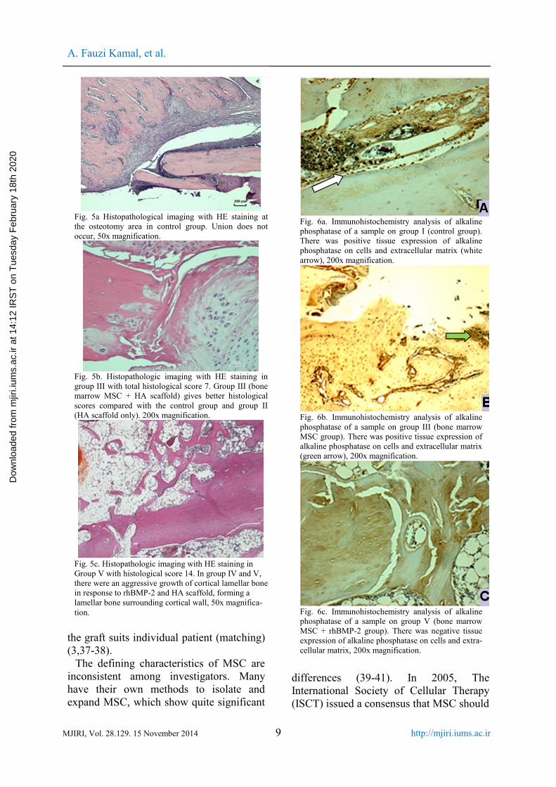
Histological images in each treatmentgroup were shown in Fig. 5(a, b, and c).Union and healing by extensive connectivetissue (score 0) appeared in some controlgroups (Fig. 5a). Most other control groupsexperienced union with fibrocartilagetissue. The majority of group III hasreached union with mature bone or partialunion with mineralized cartilage (figure5b). Unlike the other groups, in general,group IV and V were generally healed withlamellar bone. In group IV and V lamellarbone aggressively grew surroundingcortical wall (Fig. 5c).
Table 1. Mean histological score, serum alkaline phosphatase and osteocalcin level, and biomechanical study.Group Mean Histo-
logicalScore#
Mean SerumAlkaline Phos-phatase level
(ng/µL)
Mean SerumOsteocalcineLevel (pg/µL)
Biomechanical Study at 8th Week
PO DO 2nd
week4th
week2nd
week4th
weekMean Cal-lus Diame-ter (mm)
Mean CallusCrossectionalArea (mm2)
MeanUltimateStrength(Newton)
I (control) 4 3.8 13.18 9.09 189.1 270.7 2.98 5.01 111II (HA) 1.4 4 12.93 6.86 227.8 279.3 4.94 9.31 120III (HA+MSC) 9.5 5.75 8.85 8.29 272.9 358.8 3.24 5.57 111IV (HA+rhBMP-2) 12 12.7 10.89 8.04 283.6 322.3 8.37 16.85 232V (HA+MSC +rhBMP-2) 12 13.25 6.8 11.38 270.8 270.7 8.86 17.94 161Contralateral leg 3.04 5.13 159.3
#: Histological score of union according to Salkeld and Marino at 8th week, PO: Proximal osteotomy, DO: Distal osteotomy, HA: Hy-droxyapatite scaffold, MSC: Bone marrow mesenchymal stem cells, rhBMP-2: Recombinant human bone morphogenetic protein-2
Tabel 2. Expression of tissue alkaline phosphatase and osteocalcin.
GroupΣn Alkaline Phosphatase Osteocalcin
- + F - + FI 6 3 2 1 2 4 0II 6 2 4 0 3 3 0III 5 1 4 0 3 2 0IV 5 5 0 0 5 0 0V 4 4 0 0 4 0 0
Description: n: number of samples; (-): negative expression of alkaline phosphatase or osteocalcin, (+): positive expressionof alkaline phosphatase or osteocalcin, F: failure
Dow
nloa
ded
from
mjir
i.ium
s.ac
.ir a
t 14:
12 IR
ST
on
Tue
sday
Feb
ruar
y 18
th 2
020

Enhancement method for extracorporeal irradiation
8 MJIRI, Vol. 28.129. 15 November 2014http://mjiri.iums.ac.ir
Immunohistochemistry of alkaline phos-phosphatase and osteocalcin
Alkaline phosphatase and osteocalcinexpression of tissue showed positive results
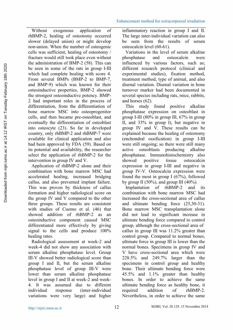
in group I-III and negative in group IV-V.Alkaline phosphatase expression was mostcommonly found in group III (80%),followed by group II (67%), and group I(33%). Osteocalcin expression were foundthe most in group I (67%), followed bygroup II (50%) and III (40%). Theexpression of alkaline phosphatase and thetissue osteocalcin are summarized in Table2.
Tissue expression of alkaline phosphataseand osteocalcin was positive in group I, II,and III. Group IV and V had perfecthealing and remodelling, so that they nolonger express alkaline phosphatase andosteocalcin (Fig. 6a, b, and c).
Mechanical Strength of BoneBone mechanical strength test was used
for different samples. The mean diameterof the bone (callus) was 5.8 ± 2.57 mm, thecross-sectional area of bone (callus) was11.3 ± 5.66 mm2. The mean of ultimateforce on bending test was 151.8 ± 52.66 N.The highest ability to withstand the bendingforce was found in group IV. The averagediameter of the callus, callus cross-sectional area, and ultimate force of eachtreatment group are shown in Table 4.Diameter and cross-sectional area of callusin group IV and V are greater than the othergroups.
DiscussionsIn primary malignant bone tumor with
indication of LSS with ECI technique,fracture could occur due to bonedestruction by osteoclasts, the tumor itself(bone destruction before surgery), or due tocomplication of ECI exposure. In thismodel the various factors mentioned abovewas eliminated, so we evaluated the effectof MSC, rhBMP-2, and their combinationto the union of osteotomy and itsmechanical properties. The use ofautogenous bone as a graft after devitalizedwith ECI exposure as a preferred method ofLSS continued to increase, because thismethod was simple and inexpensive(3,6,11,13) and also the size and shape of
Fig. 4a. Lateral projection radiograph of a sample ingroup I at 8th week. There was nonunion.
Fig. 4b. Lateral projection radiograph of a sample ingroup III at 8th week. There was union with moderateamount of callus.
Fig. 4c. Lateral projection radiograph of a sample ingroup V at 8th week. There was union with enormouscallus formation.
Dow
nloa
ded
from
mjir
i.ium
s.ac
.ir a
t 14:
12 IR
ST
on
Tue
sday
Feb
ruar
y 18
th 2
020
A. Fauzi Kamal, et al.
9MJIRI, Vol. 28.129. 15 November 2014 http://mjiri.iums.ac.ir
the graft suits individual patient (matching)(3,37-38).
The defining characteristics of MSC areinconsistent among investigators. Manyhave their own methods to isolate andexpand MSC, which show quite significant
differences (39-41). In 2005, TheInternational Society of Cellular Therapy(ISCT) issued a consensus that MSC should
Fig. 5a Histopathological imaging with HE staining atthe osteotomy area in control group. Union does notoccur, 50x magnification.
Fig. 5b. Histopathologic imaging with HE staining ingroup III with total histological score 7. Group III (bonemarrow MSC + HA scaffold) gives better histologicalscores compared with the control group and group II(HA scaffold only). 200x magnification.
Fig. 5c. Histopathologic imaging with HE staining inGroup V with histological score 14. In group IV and V,there were an aggressive growth of cortical lamellar bonein response to rhBMP-2 and HA scaffold, forming alamellar bone surrounding cortical wall, 50x magnifica-tion.
Fig. 6a. Immunohistochemistry analysis of alkalinephosphatase of a sample on group I (control group).There was positive tissue expression of alkalinephosphatase on cells and extracellular matrix (whitearrow), 200x magnification.
Fig. 6b. Immunohistochemistry analysis of alkalinephosphatase of a sample on group III (bone marrowMSC group). There was positive tissue expression ofalkaline phosphatase on cells and extracellular matrix(green arrow), 200x magnification.
Fig. 6c. Immunohistochemistry analysis of alkalinephosphatase of a sample on group V (bone marrowMSC + rhBMP-2 group). There was negative tissueexpression of alkaline phosphatase on cells and extra-cellular matrix, 200x magnification.
Dow
nloa
ded
from
mjir
i.ium
s.ac
.ir a
t 14:
12 IR
ST
on
Tue
sday
Feb
ruar
y 18
th 2
020

Enhancement method for extracorporeal irradiation
10 MJIRI, Vol. 28.129. 15 November 2014http://mjiri.iums.ac.ir
have the following three criteria (42). First,MSC must be plastic-adherent when main-maintained in standard culture conditions.Second, MSC must express CD105, CD73and CD90, and lack expression of CD45,CD34, CD14 or CD11b, CD79a or CD19,and HLA-DR surface molecules. Third,MSC could differentiate to osteoblasts,adipocytes, and chondroblasts in vitro. Inorder to prove that, we performedcharacterization with RT-PCR andimmunocytochemistry assays. RT-PCRshowed that cultured cells had positivegene for surface markers CD73, CD90,CD105, and STRO-1, and negative forCD34 and CD45. Those results werefurther supported by immunocytochemistryassay that showed positive expression ofCD73 and CD105. It was the limitation ofthis study that we could only confirm twoof three ISCT criteria. Considering theirfibroblast-like morphology, plastic adherentproperties, ability of cells to grow andproliferate on appropriate medium formesenchymal stem cells, and the result ofRT-PCR and immunocytochemistry, weconcluded that those mononuclear cellswith fibroblast like morphology are verylikely MSC (33).
In this study, we used allogenic bonemarrow MSC from the previous cultureresults (our preliminary study) due toseveral considerations: (i) autogenic MSCdonor is not possible, because it is obtainedfrom the femur and tibia of rats via surgicalhip joint disarticulation, (ii) surgery on therest of the contralateral femur would causeinjury and pain that violate the animalwelfare, (iii) the use of allogenic MSCrelatively did not cause rejection due tolack of expression of HLA class II antigenson the bone marrow MSC (25). The lowrate of immunorejection of the allogenicMSC was reported by Koc et al (43). Thestudy showed no T cell reaction or rejectionon the use of allogenic MSC in patientswith Hurler's syndrome. Horwitz et al. (44)reported allogenic MSC transplantation inthree children with osteogenesis imperfectadid not result in immunorejection. That
result was also supported by Bartholomewet al. (45) and Ryan et al. (26) whichshowed allogenic MSC administration onwound healing gave no rejection reaction.
Our study showed that on the secondweek of observation, the radiological resultof proximal and distal osteotomy in group Iand II only showed signs of new callusformation (score 1), whereas group III, IVand V more than 60% of rats reached thebeginning of healing by bone tissue (score2). The osteotomy line was almostdisappeared (score 3) in more than 60% ofsamples in group III, IV and V (score 3) onthe fourth week, however only 30% ofsamples on group I and II had reachedscore 3. It means that transplantation ofbone marrow MSC, application of rhBMP-2, and its combination improvedradiological score and accelerated union. Itwas supported then, on the sixth week,more than 50% rats on group IV and V hadachieved complete healing (score 4).
Radiological result was supported byhistopathological examinations. Healing ingroup I-III varied from fibrous to maturebone tissue. In group I and II, there wasfibrous tissue between graft and host boneexpanding into medullary canal of femur. Afew osteotomy in group I and II healed withcartilage or osteoid. In general, it could beconcluded that group III had betterhistological score compared to control orgroup II. It was due to the effect of MSCtransplantation on osteogenesis andendochondral ossification at the osteotomysite. This result was supported by researchdone by Cuomo et al. (46) which showedthat tissue healing on control group andMSC transplantation group varied fromfibrous tissue to mature bone.
In group IV and V (rhBMP-2 and bonemarrow MSC + rhBMP-2), there wereextensive bridging callus, contained manytrabecular bone, which bridge the host bonefrom proximal to distal covering thefemoral autograft. Asymmetric callus onone side of the femur was mainly theinfluence of the rhBMP-2 implantationarea. Both bony osteotomized area
Dow
nloa
ded
from
mjir
i.ium
s.ac
.ir a
t 14:
12 IR
ST
on
Tue
sday
Feb
ruar
y 18
th 2
020

A. Fauzi Kamal, et al.
11MJIRI, Vol. 28.129. 15 November 2014 http://mjiri.iums.ac.ir
underwent union with mature bone, byforming lamellar bone that surround thecortical wall. The combination of bonemarrow MSC and rhBMP-2 resulted inbetter histological score compared to thegroups which received bone marrow MSCor rhBMP-2 alone.
Hydroxyapatite administration alonewhich functioned as an osteoconductivescaffold did not improve radiological scoreor accelerate the healing process. In fact,radiological mean score of group II at thesixth week was lower compared withcontrol group. The results of post hocstatistical analysis also showed nosignificant difference between groups thatreceived hydroxyapatite and the controlgroup. These results are similar to studiesconducted by Ozturk et al. (47) whichshowed that the addition of hydroxyapatitedoes not improve fracture healing bothradiologically and histopatologically.According to Finkemeier (21),hydroxyapatite administration would givegood results when administered on a gap orbone defects in areas that have good bloodsupply such as in metaphysis.
Theoretically, hydroxyapatite has thesame chemical composition with bonemineral fraction. It may fill gap betweengraft and host bone and also stimulate bonegrowth. Hydroxyapatite is a nonresorbablescaffold, it is a good substrate for adhesion,proliferation, and differentiation of MSCand osteoblasts. Furthermore, differentiatedcells would produce extracellular matrixand integrated with host tissue (24,48,49).However, according to some researcher,hydroxyapatite may cause osteolysis if itwas exposed to bone marrow and softtissue. Hydroxyapatite debris was allegedlycaused implant failure by stimulatingphagocytosis (by macrophage) and releaseof various cytokines (TNF-α, IL-ß, IL-6,dan prostaglandin E2 (PGE2)). After that,there would be inflammatory process,triggering differentiation of osteoclastprecursor into mature osteoclast, and itmight cause impairment in boneremodeling and result in osteolysis (50).
The healing of osteotomy (union betweengraft and host bone) was influenced by;biologic activity (MSC, viable osteoblastand its product), osteoinductive potential(growth factors such as BMP-2) (31,51),and osteoconductive potential (graft andscaffold) (51-52), and also local hostcondition (53).
Bone marrow MSC transplantation alone(group III) had increased radiologicalscores, with radiologic mean score higherthan group I and II. At week-8, it gavesignificant difference in mean radiologicalscores of distal osteotomy compared togroup I. Theoretically bone marrow MSCmight provide response to biological andmechanical environment and coulddifferentiate into cells which providecomponent required for fracture healing(54). But bone marrow MSC cannot workalone, and very dependent on theextracellular matrix, intercellular signal(54), and also neovascularization to sustainits viability (55).
The limitation of bone marrow MSC inthis study is thought to be caused by in vivoenvironment which does not support theirproliferation and differentiation. Bonemarrow MSC which had been previouslygrowth in artificial medium must adapt tothe new environment after surgicalresection and re-implantation procedure,which was poor in oxygen and growthfactors. LSS may cause extensive tissuedamage (after fracture, oxygen level atfracture site would decrease into 0-2%)(56). Transplanted stem cell was avascularon the state of extensive tissue damage andinadequate nutrition, until there wasvascular invasion from the surroundingtissue (57). Decrease of O2 concentration atthe osteotomy site, extensive tissuedamage, and loss of hematome (54) werenot an ideal environment for theproliferation and differentiation of MSC. Itwas also explained by Cancedda et al. (57)which stated the results of stem celltransplantation cannot be predicted andwould fail when transplanted intounsuitable microenvironment.
Dow
nloa
ded
from
mjir
i.ium
s.ac
.ir a
t 14:
12 IR
ST
on
Tue
sday
Feb
ruar
y 18
th 2
020
Enhancement method for extracorporeal irradiation
12 MJIRI, Vol. 28.129. 15 November 2014http://mjiri.iums.ac.ir
Without exogenous application ofrhBMP-2, healing of osteotomy occurredslower (delayed union) or might developnon-union. When the number of osteogeniccells was sufficient, healing of osteotomy /fracture would still took place even withoutthe administration of BMP-2 (58). This canbe seen in some of the rats in group I-IIIwhich had complete healing with score 4.From several BMPs (BMP-2 to BMP-7,and BMP-9) which was known for theirosteoinductive properties, BMP-2 showedthe strongest osteoinductive potency. BMP-2 had important roles in the process ofdifferentiation, from the differentiation ofbone marrow MSC into osteoprogenitorcells, and then became pre-osteoblast, andeventually the differentiation of osteoblastinto osteocyte (23). So far in developedcountry, only rhBMP-2 and rhBMP-7 wereavailable for clinical application and alsohad been approved by FDA (59). Based onits potential and availability, the researcherselect the application of rhBMP-2 for theintervention in group IV and V.
Application of rhBMP-2 alone and theircombination with bone marrow MSC hadaccelerated healing, increased bridgingcallus, and also prevented implant failure.This was proven by thickness of callusformation and higher radiological score onthe group IV and V compared to the otherthree groups. These results are consistentwith studies of Cuomo et al. (46) thatshowed addition of rhBMP-2 as anosteoinductive component caused MSCdifferentiated more effectively by givingsignal to the cells and produce 100%healing rates.
Radiological assessment at week-2 andweek-4 did not show any association withserum alkaline phosphatase level. GroupIII-V showed better radiological score thangroup I and II, but the serum alkalinephosphatase level of group III-V werelower than serum alkaline phosphataselevel in group I and II at week-2 and week-4. It was assumed due to differentindividual response (inter-individualvariations were very large) and higher
inflammatory reaction in group I and II.The large inter-individual variation can alsobe seen from the results of serumosteocalcin level (60-61).
Variations in the level of serum alkalinephosphatase and osteocalcin wereinfluenced by various factors, such as;different research protocol (clinical andexperimental studies), fixation method,treatment method, type of animal, and alsodiurnal variation. Diurnal variation in boneturnover marker had been documented inseveral species including rats, mice, rabbits,and horses (62).
This study found positive alkalinephosphatase expression on osteoblast ingroup I-III (80% in group III, 67% in groupII, and 33% in group I), but negative ingroup IV and V. These results can beexplained because the healing of osteotomy(enchondral ossification) in group I-IIIwere still ongoing; so there were still manyactive osteoblasts producing alkalinephosphatase. Immunohistochemistry alsoshowed positive tissue osteocalcinexpression in group I-III and negative ingroup IV-V. Osteocalcin expression werefound the most in group I (67%), followedby group II (50%), and group III (40%).
Implantation of rhBMP-2 and itscombination with bone marrow MSC hadincreased the cross-sectional area of callusand ultimate bending force (25,30-31).Bone marrow MSC transplantation alonedid not lead to significant increase inultimate bending force compared to controlgroup, although the cross-sectional area ofcallus in group III was 11.2% greater thancontrol group. Compared to normal bones,ultimate force in group III is lower than thenormal bones. Specimens in group IV andV have cross-sectional area which were228.5% and 249.7% larger than thespecimens in control group and healthybone. Their ultimate bending force were45.5% and 1.1% greater than healthybones. In order to achieve the sameultimate bending force as healthy bone, itrequired addition of rhBMP-2.Nevertheless, in order to achieve the same
Dow
nloa
ded
from
mjir
i.ium
s.ac
.ir a
t 14:
12 IR
ST
on
Tue
sday
Feb
ruar
y 18
th 2
020

A. Fauzi Kamal, et al.
13MJIRI, Vol. 28.129. 15 November 2014 http://mjiri.iums.ac.ir
ultimate bending force as healthy bone, itrequired 228% greater cross-sectional area.
ConclusionThe conclusions of this study are: (1)
transplantation of bone marrow MSC alonewas proven to accelerate union ofosteotomy but inadequate to improve themechanical strength of ECI autograft, (2)combination of bone marrow MSC andrhBMP-2 was proven to accelerate union ofosteotomy and improve the mechanicalstrength of ECI autograft.
AcknowledgementsWe would like to express our deep
gratitude to Professor Sarwono Waspadjifor advice and suggestion inmethodological aspect of this research,Ms. Silmy Mariya for assistance inlaboratory works, Kurniadi for recordingand typing research data, and DaewoongPharmaceutical for the donation ofrhBMP-2. I hereby affirm that there is noconflict of interest in this research.
References1. Futani H, Minamizaki T, Nishimoto Y, Abe S,
Yabe H, Ueda T. Long-term follow-up after limbsalvage in skeletally immature children with aprimary malignant tumor of the distal end of thefemur. J Bone Joint Surg (Am) 2007;88-A:596-603.
2. Aksnes LH, Bauer HC, Jebsen NL, FollerasG, Haugen GS, Hall KS. Limb-sparing surgerypreserves more function than amputation: ascandinavian sarcoma group study of 118 patients.J Bone Joint Surg (Br) 2008;90-B:786-94.
3. Fuchs B, Ossendorf C, Leerapun T, Sim FH.Intercalary segmental reconstruction after bonetumor resection. Eur J Surg Oncol 2008;34:1271-6.
4. Bielack S, Carle D, Jost L. Osteosarcoma:ESMO clinical recommendations for diagnosis,treatment and follow-up. Ann Oncol 2008;19Suppl 2:94-6.
5. Chen WM, Chen TW, Huang CK, Chiang CC,Lo WH. Treatment of malignant bone tumours byextracorporeally irradiated autograft-prostheticcomposite arthroplasty. J Bone Joint Surg (Br)2002;84-B:1156-61.
6. Sewell MD, Spilberg BGI, Hanna SA,Meswania JM, Blunn GW, Henry C. Non-invasive
extendible endoprostheses for limb reconstructionin skeletally-mature patients. J Bone Joint Surg(Br) 2009;91-B:1360-5.
7. Muscolo DL, Ayerza MA, Aponte-Tinao LA,Ranalletta M. Use of distal femoral osteoarticularallografts in limb salvage surgery. J Bone JointSurg (Am) 2005;87:2449-55.
8. Takahashi S, Kotoura Y, Yamamuro T, OkaM, Shibamoto Y, Takahashi M. Incorporation ofcortical bone autografts following intraoperativeextracorporeal irradiation in rabbits. Int J RadiatOncol Biol Phys 1991;21:1221-30.
9. Kamal AF, Ismail, Mi’raj F, Hutagalung EU.Outcome of stage IIB osteosarcoma treated bylimb salvage surgery using extracorporeallyirradiated (ECI) autograft. Med J Indones 2011;20(2): 131-7.
10. Bohm P, Fritz J, Thiede S, Budach W.Reimplantation of extracorporeal irradiated bonesegments in musculoskeletal tumor surgery:clinical experience in eight patients and review ofthe literature. Langenbecks Arch Surg2003;387:355-65.
11. Davidson AW, Hong A, McCarthy SW,Stalley PD. En-bloc resection, extracorporealirradiation, and reimplantation in limb salvage forbony malignancies. J Bone Joint Surg (Br) 2005;87-B:851-6.
12. Krieg AH, Mani M, Speth BM, Stalley PD.Extracorporeal irradiation for pelvicreconstruction in ewing's sarcoma. J Bone JointSurg (Br) 2009;91-B:395-9.
13. Takahashi S, Okudaira S, Sasai K, KotouraY. En bloc resection, extracorporeal irradiation,and reimplantation of an entire tibia. J Orthop Sci2006;11:298-302.
14. Boston SE, Duerr F, Bacon N, Larue S,Ehrhart E, Withrow S. Intraoperative radiation forlimb sparing of the distal aspect of the radiuswithout transcarpal plating in five dogs. Vet Surg2007;36:314-23.
15. Chen TH, Chen WM, Huang CK.Reconstruction after intercalary resection ofmalignant bone tumours: comparison betweensegmental allograft and extracorporeally irradiatedautograft. J Bone Joint Surg (Br) 2005;87-B:704-9.
16. Liptak JM, Dernell WS, Duncan B, LascellesX, Larue SM, Jameson VJ. Intraoperativeextracorporeal irradiation for limb sparing in 13dogs. Vet Surg 2004;33:446-56.
17. Yamamoto T, Akisue T, Marui T, Nagira K,Kurosaka M. Osteosarcoma of the distal radiustreated by intraoperative extracorporealirradiation. J Hand Surg 2002;27-A:160-4.
18. Singh VA, Nagalingam J, Saad M, Pailoor J.Which is the best method of sterilization oftumour bone for reimplantation? a biomechanicaland histopathological study. Biomed Eng 2010;9:48-50.
Dow
nloa
ded
from
mjir
i.ium
s.ac
.ir a
t 14:
12 IR
ST
on
Tue
sday
Feb
ruar
y 18
th 2
020

Enhancement method for extracorporeal irradiation
14 MJIRI, Vol. 28.129. 15 November 2014http://mjiri.iums.ac.ir
19. Santoni BG, Turner AS, Wheeler DL, Nicho-las RW, Anchordoquy TJ, Ehrhart N. Gene thera-py to enhance allograft incorporation after hosttissue irradiation. Clin Orthop Relat Res 2008;466: 921–9.
20. Dong J, Li LX, Mu WD, Wang YH, ZhouDS, Hao W, et al. Bone regeneration with BMP-2gene-modified mesenchymal stem cells seeded onnano-hydroxyapatite/collagen/poly(l-lactic Acid)scaffolds. J Bioactive Compatible Polymers 201025:547-65.
21. Finkemeier CG. Current concepts review:bone grafting and bone graft substitutes. J BoneJoint Surg (Am) 2002;84-A:454-63.
22. Salkeld SL, Patron LP, Barrack RL, CookSD. The effect of osteogenic protein-1 on thehealing of segmental bone defects treated withautograft or allograft bone. J Bone Joint Surg(Am) 2001;83:803-16.
23. James D, Heckman WE, Brooks BP,Aufdemorte TB, Lohmann CH, Morgan T, et al.Bone morphogenetic protein but not transforminggrowth factor-β enhances bone formation incanine diaphyseal nonunions implanted with abiodegradable composite polymer. J Bone JointSurg (Am) 1999;81:1717-29.
24. Vats A, Tolley NS, Buttery LDK, Polak JM.The stem cell in ortopaedic surgery. J Bone JointSurg (Br) 2004;86-B:159-64.
25. Hee HT, Dilogo IH, Lim CT, Goh JC, WongHK. Effects of implantation of bone marrowmesenchymal stem cells, disc distraction, andcombined therapy on reversing degeneration of theintervertebral disc. J Bone Joint Surg (Br)2010;92: 726-36.
26. Ryan JM, Barry FP, Murphy JM, Mahon BP.Mesenchymal stem cells avoid allogeneicrejection. J Inflam 2005; 2:1-11.
27. Goldberg VM, Stevenson S. Biology of boneand cartilage allografts. In: Czitrom AA, GrossAE, editors. Allografts in orthopaedic pratice.Baltimore: Williams & Wilkins; 1992. pp. 1-14.
28. Yamaguchi A, Katagiri T, Ikeda T.Recombinant human bone morphogenetic protein-2 stimulates osteoblastic maturation and inhibitsmyogenic differentiation in vitro. J Cell Biol 1991;133: 681-7.
29. Sasso RC, Williams JI, Dimasi N.Postoperative drains at the donor sites of iliac-crest bone grafts: a prospective, randomized studyof morbidity at donor site in patients who had atraumatic injury of the spine. J Bone Joint Surg(Am) 1998; 80:631-5.
30. Reddi A. Bone morphogenetic proteins: frombasic science to clinical applications. J Bone JointSurg (Am) 2001;83-A:S1-6.
31. Cheng H, Jiang W, Phillips FM, HaydonRC, Peng Y, Zhou L, et al. Osteogenic activity ofthe fourteen types of human bone morphogeneticproteins (BMPs). J Bone Joint Surg (Am) 2003;
85:1544-52.32. Lane JM, Sandhu HS. Current approaches to
experimental bone grafting. Orthop Clin NorthAm 1987;18(2):213-25.
33. Kamal AF, Iskandriati D, Dilogo IH, SiregarNC, Hutagalung EU, Yusuf AA, Mariya S,Husodo K. Comparison of cultured mesenchymalstem cells derived from bone marrow or peripheralblood of rats. J Exp Integr Med 2014;4(1):17-22.
34. Tsuchida H, Hashimoto J, Crawford E,Manske P, Low J. Engineered allogeneicmesenchymal stem cells repair femoral segmentaldefect in rats. J Orthop Res 2002;21:44-53.
35. Yue B, Lu B, Dai KR, Zhang XL, Yu CF,Lou JR, et al. BMP-2 gene therapy on the repair ofbone defects of aged rats. Calcif Tissue Int 2005;77:395-403.
36. Yoshiki S, Umeda T, Kurahashi Y. Aneffective reactivation of alkaline phosphatase inhard tissue completely decalcified for light andelectron microscopy. Histochemie1972;29(4):296-304.
37. Abed YY, Beltrami G, Campanacci DA,Innocenti M, Capanna R. Biologicalreconstruction after resection of bone tumoursaround the knee. Long-term follow-up. J BoneJoint Surg (Br) 2009;91-B:1366-72.
38. Ahlmann ER, Menenzes IR. Intercalaryendoprosthetic reconstruction for diaphysealtumours. J Bone Joint Surg (Br) 2006;88-B:1487-91.
39. Cheng MT, Yang HW, Chne TH, Lee OKS.Isolation and characterization of multipotent stemcells from human cruciate ligaments. Cell Prolif2009;42:448-60.
40. Villaron EM, Almeida J, Holgado NL,Alcoceba M, Sanchez-Abarca LI, Sanchez-GuijoFM, et al. Mesenchymal stem cells are present inperipheral blood and can engraft after allogenichematopoietic stem cell transplantation.Haematologica 2004;89:1421-7.
41. Bernacki SH, Wall ME, Loboa EG. Isolationof human mesenchymal stem cells from bone andadipose tissue. In: Mather JP (ed) Methods in CellBiology, Amsterdam: Elsevier; 2008. pp.257-277.
42. Dominici M, Le Blanc K, Mueller I, Slaper-Cortenbach I, Marini FC, Krause DS, et al.Minimal criteria for definingmultipotentmesenchymal stromal cells. TheInternational Society for Cellular Therapy positionstatement. Cytotherapy 2006; 8:315-7.
43. Koc O, Day J, Nieder M, Gerson SL,Lazarus HM, Krivit W. Allogeneic mesenchymalstem cell infusion for treatment of metachromaticleukodystrphy (MLD) and Hurler's syndrome(MPS-IH). Bone Marrow Transplant 2002;30:215-22.
44. Horwitz EM, Prockop DJ, Gordon PL, et al.Clinical responses to bone marrow transplantationin children with severe osteogenesis imperfecta.
Dow
nloa
ded
from
mjir
i.ium
s.ac
.ir a
t 14:
12 IR
ST
on
Tue
sday
Feb
ruar
y 18
th 2
020

A. Fauzi Kamal, et al.
15MJIRI, Vol. 28.129. 15 November 2014 http://mjiri.iums.ac.ir
Blood 2001;97:12227-31.45. Bartholomew A, Stuegeon C, Siatskas M,
Ferrer K, McIntosh K, Patil S. Mesenchymal stemcells suppress lymphocyte proliferation in vitroand prolong skin graft survival in vitro. ExpHematol 2002;30:42-8.
46. Cuomo AV, Virk M, Petrigliano F, MorganEF, Lieberman JR. Mesenchymal stem cellconcentration and bone repair: potential pitfallsfrom bench to bedside. J Bone Joint Surg (Am)2009; 91:1073-83.
47. Ozturk A, Yetkin H, Memis L, Cila E,Bolukbasi S, Gemalmaz C. Demineralized bonematrix and hydroxyapatite/tri-calcium phosphatemixture for bone healing in rat. Int Orthop 2006;30:147-52.
48. Khan Y, Yaszemski MJ, Mikos AG,Laurencin CT. Tissue engineering of bone:material and matrix considerations. J Bone JointSurg (Am) 2008;90:36-42.
49. Kakar S, Einhorn T. Tissue engineering ofbone. In: Hollinger JO, Einhorn TA, Doll BA,Sfeir C, editors. Bone tissue engineering. 1stedition. Washington (DC): CRC Press; 2005.pp.277-302.
50. Nuss KRM, Rechenberg V. Biocompatibilityissues with modern Implants in bone - a review forclinical orthopedics. Open Orthop J 2008;2:66-78.
51. Nather A, Han YY. Biology of healing ofbone allograft. In: Nather A, Yusof N, Hilmy N,editors. Allograft procurement, processing, andtransplantation acomprehensive guide for tissuebanks. Singapore: World Scientific; 2010. pp.175-93.
52. Lieberman JR, Stevenson S. Bone grafts. In:Pellici PM, Tria AJ, Garvin KL, editors.Orthopaedic knowledge update hip and kneereconstruction 2. Illinois: AAOS; 2000. pp.35-42.
53. Beaman FD, Peterson JJ, Kransdorf J. Bonegraft materials and synthetic substitutes. RadiolClin N Am 2006;44:451–61.
54. Kraus KH, Kirker-Head C. Mesenchymalstem cells and bone regeneration. Vet Surg 2006;35: 232-42.
55. Tomford WW, Bloem RM. The biology ofautograft and allograft. In: Conrad EU, Eckardt JJ,Finn HA, Genhardt MC, Gitelis S, Malawer MM,et al, editors. Surgery for bone and soft tissuetumors. Philadelphia: Lippincott-Raven; 1998.pp.481-6.
56. Lepperdinger G, Singh S, Kloss F. Reponsesof MSC to varying oxygen availability in vitro andin vivo. In: Artmann GM, Hescheler J, Minger S,editors. Stem cell engineering, principles &applications. Berlin: Springer; 2010. pp.199-211.
57. Cancedda R, Bianchi G, Derubeis A, QuartoR. Cell therapy for bone disease: a review ofcurrent status. Stem Cells 2003;21:610-19.
58. Seeherman H, Wozney J. Delivery of bonemorphogenetic proteins for orthopaedic tissueregeneration. Cytokine Growth Factor Rev 2005;16: 329-45.
59. Jansen TB, Overgaard S, Lind M, Rahbek O,Bunger C, Soballe K. Osteogenic protein-1increases the fixation of implants grafted withmorcellised bone allograft and ProOsteon bonesubstitute: an experimental study in dogs. J BoneJoint Surg (Br) 2007;89(1):121-6.
60. Allen MJ. Biochemical markers of bonemetabolism in animals: uses and limitations. VetClin Pathol 2003;32:101-13.
61. Stoffel K, Engler H, Kuster M, Riesen W.Changes in biochemical markers after lower limbfractures. Clin Chem 2007;53(1):131-4.
62. Paskalev M, Krastev S, Sotirov L. Variation ofserum bone alkaline phosphatase activities andosteocalcin concentrations in dogs with experimentalosteotomy fixed by two different osteosynthesistechniques. Revue Med Vet 2008;159:444-9.D
ownl
oade
d fr
om m
jiri.i
ums.
ac.ir
at 1
4:12
IRS
T o
n T
uesd
ay F
ebru
ary
18th
202
0