trap procedure – isolation by immunoprecipitation of...
TRANSCRIPT

TRAP Protocol (8-2013 JBS) 1
TRAP Procedure – Isolation by immunoprecipitation of polysomes from transgenic plants expressing FLAG-tagged RPL18 Bailey-Serres Lab
Center for Plant Cell Biology University of California, Riverside Overview: Polyribosomes (polysomes) are multiple ribosomes engaged in translation on a single mRNA. To evaluate the translation state of an mRNA, ribosomal subunits, ribosomes, and polysomes can be isolated from detergent-treated cell extracts. Traditionally this is done by high-speed differential centrifugation to obtain a ribosome pellet. The ribonucleoprotein particles of the pellet can be further purified by centrifugation through a sucrose density gradient. By fractionation of the gradient the amount of an individual mRNA in a sub-population of polysomes can be quantitatively determined. In this method we outline polysome isolation using transgenic Arabidopsis thaliana that express an epitope tagged version of ribosomal protein L18 (RPL18). The addition of a FLAG epitope to the amino terminus to RPL18 allows for the rapid immunoprecipitation of ribosomes from crude cell extracts (Zanetti et al., 2005 Plant Physiol. 138(2), 624-635). We call this method “TRAP” – Translating Ribosomes Affinity Purification Method references: 1. Zanetti, M.E., Chang I.-.F, Gong, F.-C., Galbraith D.W., Bailey-Serres, J. (2005) Immuno-
affinity purification of polyribosomal complexes of Arabidopsis for global analysis of gene expression. Plant Physiology. 138:624-635.
2. Mustroph, A., Juntawong, P. and Bailey-Serres, J. (2009) Isolation of plant polysomal mRNA by differential centrifugation and ribosome immunopurification methods. Methods Mol Biol. Plant Systems Biology (D. Belostotsky, ed) 553:109-26.
3. Mustroph A, Zanetti ME, Girke T, Bailey-Serres J. (2013) Methods Mol Biol. 959:277-302. doi: 10.1007/978-1-62703-221-6_19.
Method objectives:
(1) Perform polysome immunopurifications (TRAP) to obtain subpopulations of cellular mRNAs. Isolate total mRNA from the same samples.
(2) Examine levels of specific mRNAs by use of cDNA synthesis and quantitative real time polymerase chain reaction (qRT-PCR).
(3) Analyze the proteins obtained by TRAP using SDS-polyacrylamide gel electrophoresis. In this protocol we use rosette tissue from two genotypes of transgenic Arabidopsis thaliana (Col-0): 35S:HF-RPL18 - near constitutive expression of a FLAG-epitope tagged RPL18 CER5:HF-RPL18 – expression of FLAG-epitope tagged RPL18 in the epidermal cell layer

TRAP Protocol (8-2013 JBS) 2
Experiment Overview
Tissue sample (5 mL packed volume)
Trizol RNA extraction and DNAse treatment
isolation
Immunopurification with anti-FLAG Protein A agarose beads
50% RNA analyses
50% Protein analysis
FLAG peptide used to release IP
from FLAG-agarose in
presence of RNAsin
Quantitate and quality control
q-RT-PCR
90% polysome isolation
Strataclean concentration
cDNA synthesis with Reverse transcriptase and oligo dT
10% total RNA isolation
Evaluate Total Proteins and
supernatant of the immunopurified
fraction (Unbound fraction)
90% polysome isolation
SDS-Polyacrylamide gel
electrophoresis
Coomassie Blue or Silver staining

TRAP Protocol (8-2013 JBS) 3
Materials for the TRAP
- All solutions and equipment used in this protocol need to be free of RNAse. Glassware, pipette tips, tubes and solutions must be sterilized by autoclaving for 15 min.
- All steps are carried out on ice or at 4 oC. - Unless otherwise stated, all solutions are prepared with sterile deionized water. - The plant material must be harvested directly into liquid nitrogen, ground to a fine powder
using sufficient liquid nitrogen to maintain a frozen state. Pulverization can be accomplished with a porcelain mortar and pestle. The pulverized tissue is stored at –80 °C until use.
Solutions and chemicals
- Sucrose (Ultracentrifuge grade, Fisher, Catalog # BP220-212) - Miracloth for filtering, autoclaved - α-FLAG agarose beads (Sigma, EZview FLAG M2 Agarose beads, F 2426) - FLAG3 peptide (Sigma, product number F 4799) - RNAse Out (Invitrogen) - TriZOL (Invitrogen Catalog# 15596) - Chloroform, Isopropanol - Qiagen RNeasy kit (Catalog # 74904), Qiagen RNase free DNAse kit (#79254) - 99 % (v/v) ethanol
The following stock solutions are autoclaved and stored at room temperature
2 M Tris, adjust to pH 9.0 with HCl 2 M KCl 0.5 M ethylene glycol-bis(2-aminoethylether)-N,N,N´,N´-tetraacetic acid
(EGTA), adjust to pH 8.0 with 10 M NaOH. Note, EGTA only dissolves after the pH has been adjusted.
1 M MgCl2 20 % (v/v) Polyoxyethylene 10 tridecyl ether (PTE). Note, shake before use. 10 % Sodium Deoxycholate (DOC), Use lung protection while weighing DOC. 50 mM NH4CO3 (for the proteomics sample)
20 % Detergent mix (Dissolve while heating to about 60 °C)
20 % (w/v) polyoxyethylene(23)lauryl ether (Brij-35) 20 % (v/v) Triton X-100 20 % (v/v) Octylphenyl-polyethylene glycol (Igepal CA 630) 20 % (v/v) polyoxyethylene sorbitan monolaurate 20 (Tween 20)
Solutions NOT to be autoclaved, stored at -20 °C in aliquots
0.5 M Dithiothreitol (DTT) 50 mg/mL Cycloheximide, dissolved in ethanol 50 mg/mL Chloramphenicol, dissolved in ethanol 0.5 M Phenylmethylsulfonyl fluoride (PMSF), dissolved in isopropanol
TRAP Protocol (8-2013 JBS) 4
Equipment
- This technique is based on the usage of transgenic Arabidopsis thaliana or other plants expressing a FLAG-tagged ribosomal protein. These stable transgenic lines are essential for this protocol.
- Preparative centrifuge with fixed angle or swinging bucket rotor accommodating 30 mL tubes (i.e. Beckman J2-21 highspeed centrifuge and JA-20 rotor, fitted with rubber inserts to accommodate 15 or 30 mL Corex tubes)
- Low speed benchtop centrifuge with swinging buckets for 15 or 50 mL Falcon tubes (required speed 8,200 g)
- Rocking shaker, capable of shaking at about 60 rpm/min Day 1: Isolation of epitope-tagged polysomes by immunopurification Prepare Buffers (estimated time, 20 minutes – this will be done for students in workshop) Polysome Extraction buffer (PEB): prepared on the day of each experiment and kept on ice Final concentration Amount of stock solution for 50 mL 0.2 M Tris, pH 9.0 5 mL 0.2 M KCl 5 mL 0.025 M EGTA 2.5 mL 0.035 M MgCl2 1.75 mL 1 % Detergent mix1 2.5 mL 1 % DOC 5 mL 1 % PTE 2.5 mL 5 mM DTT 0.5 mL 1 mM PMSF 0.1 mL 50 µg/mL Cycloheximide 50 µL 50 µg/mL Chloramphenicol 50 µL 1 Warm solution at 42 °C before use; pipette with a 1000 µL tip enlarged by cutting 0.5 cm from the end. It takes time for the detergents to go into solution. Wash buffer: prepared on the day of each experiment and kept on ice* Final concentration Amount of stock solution for 100 mL 0.2 M Tris, pH 9.0 10 mL 0.2 M KCl 10 mL 0.025 M EGTA 5 mL 0.035 M MgCl2 3.5 mL 5 mM DTT 1 mL 1 mM PMSF 0.2 mL 50 µg/mL Cycloheximide 100 µL 50 µg/mL Chloramphenicol 100 µL * This wash buffer will be used in the final elution step. To the amount needed for the elution of the sample for RNA, add RNAsin or RNAse OUT to a final concentration of 20 U/mL. Do not include this inhibitor for the elution of the sample for protein analyses.
TRAP Protocol (8-2013 JBS) 5
Tissue extraction (Estimated time, 60 min)
- The pulverized tissue is placed into a 50 mL Falcon tube. Keep sample on ice. - Estimate volume of pulverized tissue powder (5 mL rosette tissue) and add two times (10
mL) the volume of freshly prepared Polysome Extraction buffer (PEB). Use a plastic transfer pipette. For preparative immunoprecipitation use at least 5 mL of packed leaf tissue. We will perform one immunoprecipitation and then split the sample for RNA and protein analyses.
- Let the mixture thaw on ice. During thawing, stir gently with a glass rod. - Transfer tissue to glass homogenizer with a plastic transfer pipette. - Homogenize the mixture by use of a glass homogenizer; five strokes; keep on ice. - Let the mixture stand on ice for 10 min (or until all samples are prepared) - Pour into 15 mL Corex tube (tubes will need to be balanced to within 0.5 g) - Centrifuge the samples at 4°C, 16,000 g, for 15 min in a preparative centrifuge; use a
fixed angle or swinging bucket rotor with rubber Corex tube adapters. - [Perform the Preparation of the α-FLAG M2 agarose beads during this c’fuge run, if
possible] - Put a fresh Corex tube on ice. Place a piece of Miracloth in the opening to form a small
funnel. - Using a transfer pipette, filter the supernatant into the new tube, avoiding the pellet. If any
of the pellet has been transferred, then repeat the centrifugation step to ensure removal of material that pellets at 16,000 g. This is the clarified extract.
- Label a 1.5 mL microfuge tube (i.e. 35S total RNA) and put 0.75 mL of the extract into the tube; this is to be used to save 5 % of the clarified extract to isolate total RNA as a control. Label another 1.5 mL microfuge tube (i.e. 35S T protein) and pipette 30 µl of the extract into the tube; this is to be the “total” protein sample. Keep these reserves on ice.
Preparation of the α-FLAG M2 agarose beads (Estimated time, 15 min; can do during sample centrifugations)
- Remove α-FLAG M2 / Protein A affinity agarose gel from -20 freezer and thaw on ice. - Thoroughly suspend the α-FLAG M2 / Protein A affinity agarose gel in the reagent vial
to make a uniform suspension of the resin. - You will prepare two sets of beads, one for the RNA extraction and one for the
protein extraction (label tubes accordingly). Transfer 100 µL of the beads to a new 15 mL Falcon tube (sterile). Use cut pipette tips for easier transfer. (These will be aliquoted for you in the workshop).
- Add 1.5 mL of wash buffer, and resuspend beads gently by shaking/flicking tube. - Centrifuge at 8,200 g for 60 sec (we use a benchtop clinical c’fuge on speed 3; can be in
cold room or deli) - Keep on ice until the plant extract is ready. - Remove the supernatant with a pipette and wash one more time with 1.5 mL of wash
buffer before continuing with the immunoprecipitation. These are the washed α−FLAG-M2 agarose beads. You will add the clarified extract (supernatant) to the beads (remember to reserve the sample for total RNA).

TRAP Protocol (8-2013 JBS) 6
Immunoprecipitation of polysomes (Estimated time 4 hours, including 2 hour pause)
- Add half of the clarified extract to each of the two tubes with washed α-FLAG M2 agarose beads (label one for “Protein” and the other “RNA”; i.e. 35S RNA, 35S Protein).
- To bind the epitope tagged ribosomes to the affinity matrix, incubate for 2 h at 4 °C with gentle back-and-forth shaking on a rocking platform. If done at room temperature, tubes should be lying on their sides in an ice bucket with maximal movement of the liquid.
2 h incubation
- Label a microfuge tubes (i.e. 35S UB protein) - Centrifuge the 15 mL Falcon tubes containing the sample for 60 sec at 8,200 g at 4 °C - Transfer 30 µl of the supernatant from the “protein” sample (unbound fraction
supernatant) into the labeled microfuge tube. This is the reserve for the protein gel of the “unbound” protein fraction.
- Carefully, remove the remaining supernatant from the beads (IP) using a 1 mL disposable pipette. This should be done without disturbing the bead pellet. If necessary, re-centrifuge to get all but about 100 µl of the supernatant off. The beads should have absorbed the epitope tagged ribosomes from the solution. You can discard the supernatant.
- Add 6 mL of Wash Buffer to the beads, mix by gently inverting the tube, incubate at 4 °C for 5 min with gentle shaking on a rocking platform on ice; centrifuge for 60 sec at 8,200 g at 4 oC (First wash).
- Remove the supernatant with a pipette, add 6 mL of Wash Buffer to the beads. Incubate at 4 °C for 5 min with gentle shaking (Second wash)
- Centrifuge for 60 sec at 8,200 g at 4 °C - Repeat the washing steps one more time to remove any contaminating RNA or protein. - Centrifuge for 60 sec at 8,200 g at 4 °C - Remove the supernatant, add 1 mL Wash Buffer. Carefully transfer the sample from the
Falcon tube to a pre-labelled microfuge tube (you should have two microfuge tubes (i.e. 35S protein IP; 35S RNA IP)). Use an additional 0.5 mL to remove all of the affinity matrix from the tube.
- Centrifuge for 60 sec at 8,200 g at 4 °C in a microfuge - Use a fine tipped pipette to remove as much of the supernatant as possible from the beads.
For the RNA sample:
- Prepare Elution Buffer (this can be done during the washing steps): 300 uL Wash Buffer, 12 uL of 5 mg/mL FLAG3 peptide, 0.5 uL of 2 U/mL RNAsin (final concentrations 200 ng/µL of FLAG3; 20 U/mL RNAsin).
- Add 300 µL Elution Buffer to the beads. Incubate for 30 min at 4 °C with shaking on a rocking platform.
- Centrifuge for 60 sec at 8,200 g at 4 oC. Transfer the supernatant to a new tube (contains the RNA and proteins). If the supernatant still contains the beads (white or red particles), centrifuge again at 13,000 g for 2 min at 4°C, and transfer to a new tube. It is extremely important to remove all beads from the supernatant. Use a very small bore pipette tip.
- The resulting solution is the eluate of the immunoprecipitation that contains released FLAG-tagged polysomes including the associated proteins and RNAs.
- Add 800 uL TriZOL (Invitrogen) and vortex for 30 sec.
TRAP Protocol (8-2013 JBS) 7
- At this time, you will also want to perform the extraction with the aliquot of supernatant that you reserved for total RNA. (30 µl total extract and 800 µl Triazol is fine)
- Samples can be further processed immediately or stored at -80 or -20 °C -
For the Protein sample:
- Centrifuge to remove all of the supernatant from the beads; then add 300 µL of Elution Buffer containing 200 ng/µL of FLAG3 peptide (no RNAsin)
- Incubate for 30 min at 4 °C with shaking on a rocking platform. - Centrifuge for 60 sec at 8,200 g at 4 oC. Transfer the supernatant to a new tube (contains
the RNA and proteins). If the supernatant still contains the beads (white or red particles), centrifuge again at 13,000 g for 2 min at 4 °C, and transfer to a new tube. It is extremely important to remove all beads from the supernatant. Use a very small bore pipette tip.
- The resulting solution is the eluate of the immunoprecipitation that contains released FLAG-tagged polysomes including the associated proteins and RNAs.
- The sample can be stored at -80 °C or further processed as described below.
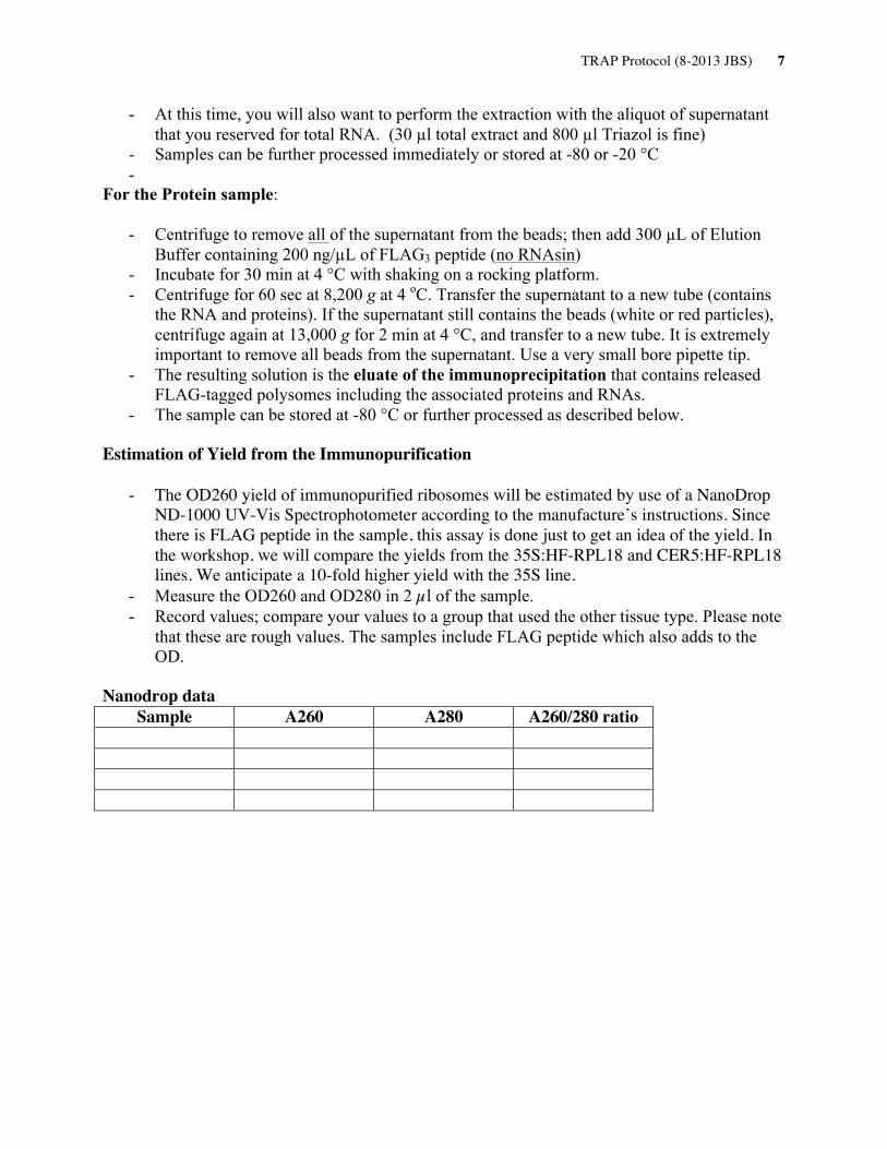
Estimation of Yield from the Immunopurification
- The OD260 yield of immunopurified ribosomes will be estimated by use of a NanoDrop ND-1000 UV-Vis Spectrophotometer according to the manufacture’s instructions. Since there is FLAG peptide in the sample, this assay is done just to get an idea of the yield. In the workshop, we will compare the yields from the 35S:HF-RPL18 and CER5:HF-RPL18 lines. We anticipate a 10-fold higher yield with the 35S line.
- Measure the OD260 and OD280 in 2 µl of the sample. - Record values; compare your values to a group that used the other tissue type. Please note
that these are rough values. The samples include FLAG peptide which also adds to the OD.
Nanodrop data
Sample A260 A280 A260/280 ratio
TRAP Protocol (8-2013 JBS) 8
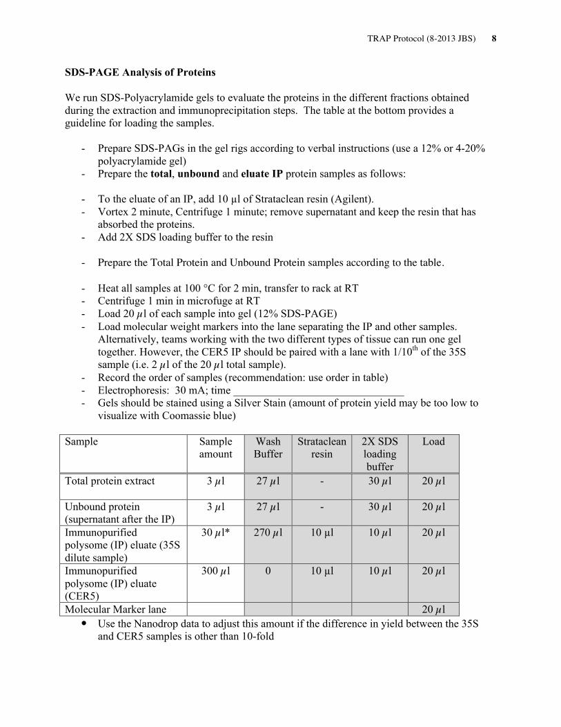
SDS-PAGE Analysis of Proteins We run SDS-Polyacrylamide gels to evaluate the proteins in the different fractions obtained during the extraction and immunoprecipitation steps. The table at the bottom provides a guideline for loading the samples.
- Prepare SDS-PAGs in the gel rigs according to verbal instructions (use a 12% or 4-20% polyacrylamide gel)
- Prepare the total, unbound and eluate IP protein samples as follows:
- To the eluate of an IP, add 10 µl of Strataclean resin (Agilent). - Vortex 2 minute, Centrifuge 1 minute; remove supernatant and keep the resin that has
absorbed the proteins. - Add 2X SDS loading buffer to the resin - Prepare the Total Protein and Unbound Protein samples according to the table.
- Heat all samples at 100 °C for 2 min, transfer to rack at RT - Centrifuge 1 min in microfuge at RT - Load 20 µl of each sample into gel (12% SDS-PAGE) - Load molecular weight markers into the lane separating the IP and other samples.
Alternatively, teams working with the two different types of tissue can run one gel together. However, the CER5 IP should be paired with a lane with 1/10th of the 35S sample (i.e. 2 µl of the 20 µl total sample).
- Record the order of samples (recommendation: use order in table) - Electrophoresis: 30 mA; time _______________________________ - Gels should be stained using a Silver Stain (amount of protein yield may be too low to
visualize with Coomassie blue) Sample Sample
amount Wash Buffer
Strataclean resin
2X SDS loading buffer
Load
Total protein extract
3 µl 27 µl - 30 µl 20 µl
Unbound protein (supernatant after the IP)
3 µl 27 µl - 30 µl 20 µl
Immunopurified polysome (IP) eluate (35S dilute sample)
30 µl* 270 µl 10 µl 10 µl 20 µl
Immunopurified polysome (IP) eluate (CER5)
300 µl 0 10 µl 10 µl 20 µl
Molecular Marker lane 20 µl • Use the Nanodrop data to adjust this amount if the difference in yield between the 35S
and CER5 samples is other than 10-fold
TRAP Protocol (8-2013 JBS) 9
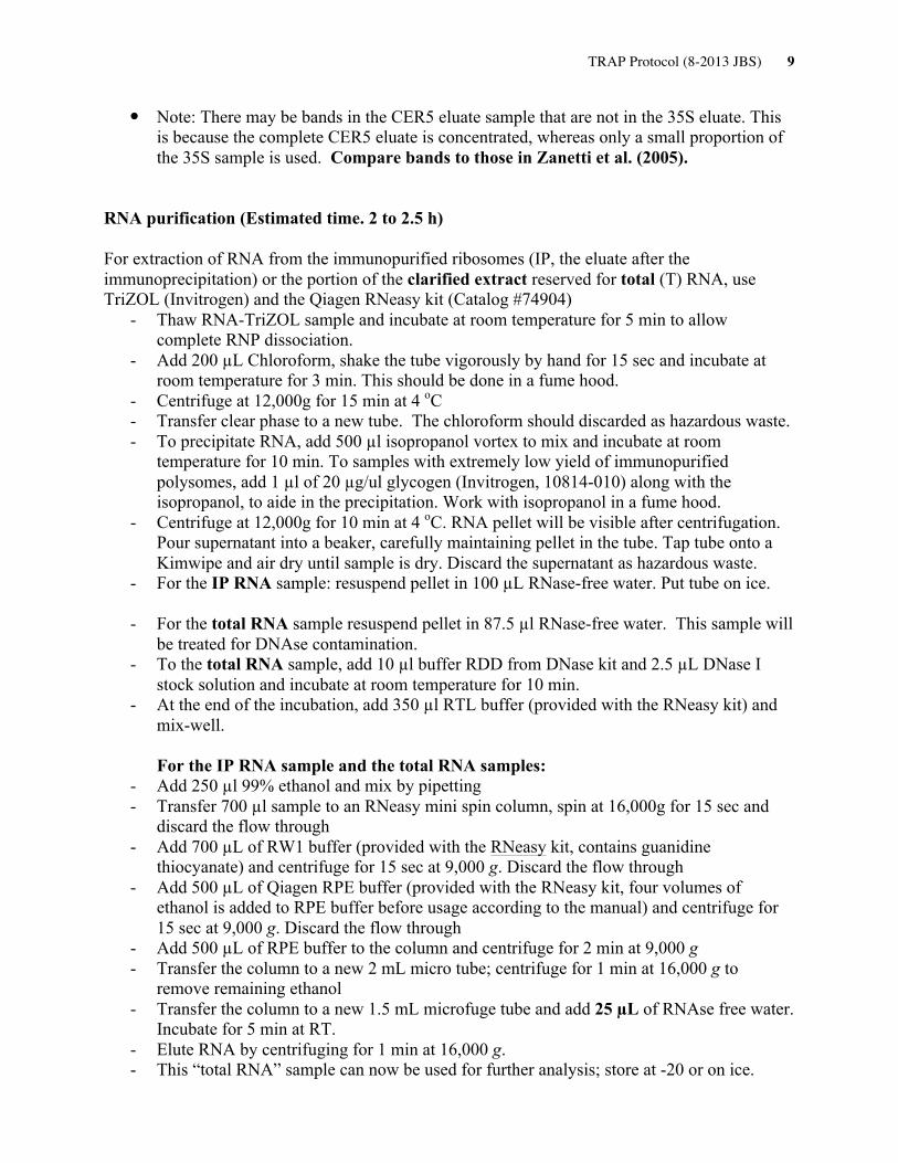
• Note: There may be bands in the CER5 eluate sample that are not in the 35S eluate. This is because the complete CER5 eluate is concentrated, whereas only a small proportion of the 35S sample is used. Compare bands to those in Zanetti et al. (2005).
RNA purification (Estimated time. 2 to 2.5 h) For extraction of RNA from the immunopurified ribosomes (IP, the eluate after the immunoprecipitation) or the portion of the clarified extract reserved for total (T) RNA, use TriZOL (Invitrogen) and the Qiagen RNeasy kit (Catalog #74904)
- Thaw RNA-TriZOL sample and incubate at room temperature for 5 min to allow complete RNP dissociation.
- Add 200 µL Chloroform, shake the tube vigorously by hand for 15 sec and incubate at room temperature for 3 min. This should be done in a fume hood.
- Centrifuge at 12,000g for 15 min at 4 oC - Transfer clear phase to a new tube. The chloroform should discarded as hazardous waste. - To precipitate RNA, add 500 µl isopropanol vortex to mix and incubate at room
temperature for 10 min. To samples with extremely low yield of immunopurified polysomes, add 1 µl of 20 µg/ul glycogen (Invitrogen, 10814-010) along with the isopropanol, to aide in the precipitation. Work with isopropanol in a fume hood.
- Centrifuge at 12,000g for 10 min at 4 oC. RNA pellet will be visible after centrifugation. Pour supernatant into a beaker, carefully maintaining pellet in the tube. Tap tube onto a Kimwipe and air dry until sample is dry. Discard the supernatant as hazardous waste.
- For the IP RNA sample: resuspend pellet in 100 µL RNase-free water. Put tube on ice. - For the total RNA sample resuspend pellet in 87.5 µl RNase-free water. This sample will
be treated for DNAse contamination. - To the total RNA sample, add 10 µl buffer RDD from DNase kit and 2.5 µL DNase I
stock solution and incubate at room temperature for 10 min. - At the end of the incubation, add 350 µl RTL buffer (provided with the RNeasy kit) and
mix-well. For the IP RNA sample and the total RNA samples:
- Add 250 µl 99% ethanol and mix by pipetting - Transfer 700 µl sample to an RNeasy mini spin column, spin at 16,000g for 15 sec and
discard the flow through - Add 700 µL of RW1 buffer (provided with the RNeasy kit, contains guanidine
thiocyanate) and centrifuge for 15 sec at 9,000 g. Discard the flow through - Add 500 µL of Qiagen RPE buffer (provided with the RNeasy kit, four volumes of
ethanol is added to RPE buffer before usage according to the manual) and centrifuge for 15 sec at 9,000 g. Discard the flow through
- Add 500 µL of RPE buffer to the column and centrifuge for 2 min at 9,000 g - Transfer the column to a new 2 mL micro tube; centrifuge for 1 min at 16,000 g to
remove remaining ethanol - Transfer the column to a new 1.5 mL microfuge tube and add 25 µL of RNAse free water.
Incubate for 5 min at RT. - Elute RNA by centrifuging for 1 min at 16,000 g. - This “total RNA” sample can now be used for further analysis; store at -20 or on ice.

TRAP Protocol (8-2013 JBS) 10
Estimation of RNA Yield The yields of RNA (rRNA and mRNA) in each sample will be estimated by use of a NanoDrop ND-1000 UV-Vis Spectrophotometer according to the manufacture’s instructions. 1. Determine total amount of RNA per volume of tissue used. 2. Determine the µg RNA/µL sample Analysis of RNA Quality The sample quality can be evaluated by use of Agilent 2100 Bioanalyzer with either RNA 6000 Nano or Pico Assay reagent kits (Agilent Technology). Sucrose gradient fractionated polysomes and Agilent 2100 analysis of RNA samples. Panel A: Leaf polysomes fractionated over a sucrose density gradient (Method in Mustroph et al., 2009). Panel B: RNA gel of total RNA and IP’d polysomal RNA from leaves of Arabidopsis seedlings expressing p35S:HF-RPL18. N; nuclear rRNAs, P; plastid rRNAs (23S, 16S) and their degradation products (23S*).
Sucrose concentration
Abs
orba
nce
at 2
54 n
m
40S60S
80S polysomes small large
Siz
e m
arke
r
Tota
l RN
A (l
eaf)
IP’ d
pol
ysom
alR
NA
(lea
f)
A B
N25S
18S
5.8S5S
P
23S23S*
16S23S*23S*

TRAP Protocol (8-2013 JBS) 11
Quantitative Real-Time Reverse Transcriptase PCR (qRT-PCR) cDNA synthesis (estimated time, 2 hours)
1) Start with immunopurified polysomal or total RNA. The total RNA sample needs to have been DNAse treated, as described earlier in this protocol.
Nanodrop data
Sample A260 A280 A260/280 µg/µl
2) Add the following components to a 0.5 ml microcentrifuge tube on ice:
Oligo(dT) 15 (500 μg/ml) 1 μl dNTP Mix (10 mM each) 1 μl RNase-free water to 12 μl (the total reaction volume is 12 μl) 0.01 to 2 μg RNA _ μl
3) Heat mixture to 65°C for 5 min and quick chill on ice. Collect the contents of the tube by brief centrifugation and add:
5X First-Strand Buffer (Invitrogen) 4 μl 0.1 M DTT 2 μl RNasin (Promega) 1 μl (or RNaseOUT (Invitrogen))
4) Mix contents of the tube gently. Incubate at 42°C for 2 min.
5) Add 1 μl (200 units) of SuperScript II Reverse transcriptase (Invitrogen) and mix by pipetting gently up and down.
6) Incubate at 42°C for 50 min.
7) Inactivate the reaction by heating at 70°C for 15 min.

TRAP Protocol (8-2013 JBS) 12
Quantitative Real Time RT-PCR (qRT-PCR) (estimated time, 2 hours) This is a modified reverse transcriptase PCR reaction that allows monitoring of the amplification of a DNA product in Real Time. The incorporation of SYBR Green, a fluorescent dye that is detected when in double stranded DNA but not single stranded provides a means to monitor change in concentration of the product over time. The output of a qRT-PCR experiment is a plot of the PCR cycles versus fluorescence. Multiple qRT-PCR reactions are compared by considering the number of cycles required for the fluorescence to cross an arbitrary threshold that is above background in the linear range of amplification (Ct). For example, if one sample reaches the threshold value after 25 cycles and the other after 30 cycles, then the one that took only 25 cycles has 32-fold (25) more of the gene being monitored. In our assays we will monitor levels of (a control gene) versus levels of genes that may be enriched in the different polysomal mRNA populations.
1) Dilution of cDNA if necessary.
2) Add the following components to a 0.5 ml qPCR tube (total volume: 20 μl): cDNA 2 μl Forward primer (10 μM) 1 μl Reverse primer (10 μM) 1 μl iQ SYBR Green supermix (Bio-Rad) 10 μl Sterile, Nanopure water to 6 μl (or 8 μl; negative control)
2) Spin down mixture by centrifugation and place in MyiQ real-time PCR detection system (Bio-Rad)
PCR program 1. 95 oC for 3 min.
2. 95 oC for 15 sec. 3. 60 oC for 30 sec. (annealing and extension together) 4. 72 oC for 30 sec. (detect fluorescence signal here) à Return to step 2 and repeat 45 times 5. 95 oC for 1 min. (Steps 5-7 are for determining the melting curve of the amplicon) 6. 60 oC for 1 min. 7. 60 to 95 oC (detect fluorescence signal here) à Increase the temperature by 0.5 oC /10 sec.
TRAP Protocol (8-2013 JBS) 13
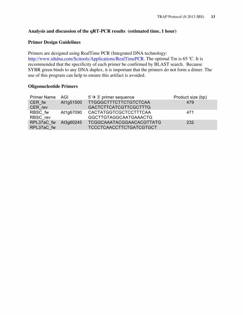
Analysis and discussion of the qRT-PCR results (estimated time, 1 hour)
Primer Design Guidelines Primers are designed using RealTime PCR (Integrated DNA technology: http://www.idtdna.com/Scitools/Applications/RealTimePCR. The optimal Tm is 65 oC. It is recommended that the specificity of each primer be confirmed by BLAST search. Because SYBR green binds to any DNA duplex, it is important that the primers do not form a dimer. The use of this program can help to ensure this artifact is avoided. Oligonucleotide Primers Primer Name AGI 5’à 3’ primer sequence Product size (bp) CER_fw At1g51500 TTGGGCTTTCTTCTGTCTCAA 479 CER_rev GACTCTTCATCGTTCGCTTTG RBSC_fw At1g67090 CACTATGGTCGCTCCTTTCAA 471 RBSC_rev GGCTTGTAGGCAATGAAACTG RPL37aC_fw At3g60245 TCGGCAAATACGGAACACGTTATG 232 RPL37aC_fw TCCCTCAACCTTCTGATCGTGCT