trauma imaging of the acute cervical spine - thieme · trauma imaging of the acute cervical spine...
TRANSCRIPT
Trauma Imaging of the Acute Cervical SpineDaniela Berritto, MD1 Antonio Pinto, MD2 Paul Michelin, MD, MSc3 Xavier Demondion, MD, PhD4
Sammy Badr, MD, MSc4
1Department of Radiology, Private Hospital “Villa dei Fiori,” Acerra,Naples, Italy
2Department of Radiology, Cardarelli Hospital, Naples, Italy3Department of Radiology, University Hospital of Rouen,Rouen, France
4Department of Musculoskeletal Radiology, CHU Lille, Lille, France
Semin Musculoskelet Radiol 2017;21:184–198.
Address for correspondence Sammy Badr, MD, MSc, CHU Lille,Service d’imagerie musculosquelettique, rue Émile Laine, 59037 Lille,France (e-mail: [email protected]).
Cervical spine trauma is a frequent condition encountered inemergency departments. Road traffic accidents, falls, andsports injuries (mainly equestrian events followed by rugby)are the most common causes of admission.1 Patients withhead trauma are more likely to have cervical spine injuries, asevere neurologic outcome, and an early mortality rate.2–4
However, an intervention (surgical stabilization, halo, orcervical-thoracic orthosis) is required in < 5% of adultpatients who did not match the National EmergencyX-Radiography Utilization Study (NEXUS) low-risk criteria.5
Therefore, imaging plays a key role in screening for unstablelesions and preventing devastating undiagnosed spinalinjuries.
Imaging Strategy
Choosing the right initial imagingmodality is challenging forclinicians and has been widely discussed. Clinical decision-making tools have been developed to help physicians(►Table 1).
For alert and asymptomatic patients in stable condition,the Canadian C-Spine Rule and the NEXUS low-risk criteriamay be used to reduce the number of unnecessary radio-graphs.6–8 Caution must be exercised, however, especiallywith the NEXUS low-risk criteria that do not have an age
limitation. Medical history must also be taken into consid-eration, particularly if the patient has an ankylosing spinaldisorder like spondyloarthritis (ankylosing spondylitis)because fractures can occur even from minor trauma.9
If cervical spine imaging is needed, the minimal radio-graphic assessment must include a three-view cervical spineseries (odontoid, or “open-mouth,” anteroposterior, andlateral), supplemented with computed tomography (CT)in case of inadequate radiographs, suspicious areas, orill-defined structures.
Moreover, current literature provides more evidenceto use CT first if available, especially for unconscious, inter-mediate-risk, and high-risk patients.10 Its superior diagnos-tic performance, as well as time and cost-effectivenessanalyses, have supported this assessment.11–13 Recommen-dations from the American College of Radiology and theAmerican Association of Neurological Surgeons approvedthese data and suggested CT as the primary modality forpatients not meeting low-risk criteria,10,14 supplementedwith magnetic resonance imaging (MRI) if spinal cord com-pression or contusion is suspected or discoligamentousevaluation is required.10
In patients < 9 years of age, radiography is recommendedas the first imaging modality, however, and use of cervicalCT must be limited to selected cases.15 MRI should also be
Keywords
► cervical spine► trauma► diagnostic imaging► emergency radiology
Abstract Among patients admitted in emergency departments for acute cervical spine trauma,only a few have a confirmed and significant injury requiring surgical intervention.Imaging plays a key role in screening for unstable lesions that can be responsible fordevastating neurologic complications or death. The complex anatomy of the cervicalspine is associated with a wide spectrum of osseous and ligamentous pathologies. Thisreview focuses on the imaging features of the main cervical spine fractures that can beencountered in emergency radiology.
Issue Theme Emergency and Trauma inMSK Radiology; Guest Editor, GiuseppeGuglielmi, MD
Copyright © 2017 by Thieme MedicalPublishers, Inc., 333 Seventh Avenue,New York, NY 10001, USA.Tel: +1(212) 584-4662.
DOI https://doi.org/10.1055/s-0037-1602713.ISSN 1089-7860.
184
Thi
s do
cum
ent w
as d
ownl
oade
d fo
r pe
rson
al u
se o
nly.
Una
utho
rized
dis
trib
utio
n is
str
ictly
pro
hibi
ted.
considered because ligamentous and spinal cord injurieswithout radiologic abnormality are more common in thepediatric population.15
Radiographic Assessment
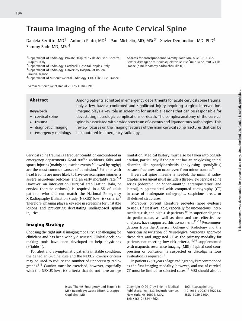
Lateral, anteroposterior, and odontoid views (►Fig. 1)must be appraised methodically after assessing theirquality (ABC’s method, proposed by Daffner and Harris16)as follows:
– Quality: All seven cervical vertebrae must be demon-strated, the cervicothoracic junction (apophyseal jointsof C7–T1) visualized, and the mandible and articularfacets superimposed (head and neck in a neutral position,with no rotation).
– Alignment: Bony alignment can be evaluated by drawingfour lines on the lateral view (the anterior and posteriorvertebral lines, and the spinolaminar and interspinouslines). On the anteroposterior view, spinous processesshould be in midline and regularly spaced, with no focaldisplacement. On the odontoid view, lateral massesshould also be aligned.
– Bone integrity: Bony landmarks must be identified, withno loss of vertebral body height and no fracture line.
– Cartilage (joints): No space abnormalities. In addition tospecific indices, Daffner and Harris simplified radio-graphic interpretation by using the “rule of twos.” Theinterlaminar (interspinous) space, interpedicle distance(transverse or vertical), unilateral or bilateral atlantoaxialoffset, and the interfacetal joint width must not differby > 2 mm.16 The interlaminar distance is more reliable
and accurate than the interspinous distance to explorehyperflexion injuries.
– Soft tissue: Retropharyngeal thickness should not exceed7 mm in children and adults at the C2 level, and 14 mm (inchildren) or 22 mm (in adults) at the C6 level.16
The lateral view is the most important radiograph toacquire. Because nearly half of all cervical spine injuriesaffect C6 and C7,2,3 the cervicothoracic junction must beseen, supplemented by additional views (swimmer’s oroblique views) or by gently pulling down the shoulders. Ifplain radiographs are still inadequate or a lesion is suspected,CT imaging must be done.
Upper Cervical Spine Injuries (C0–C2)
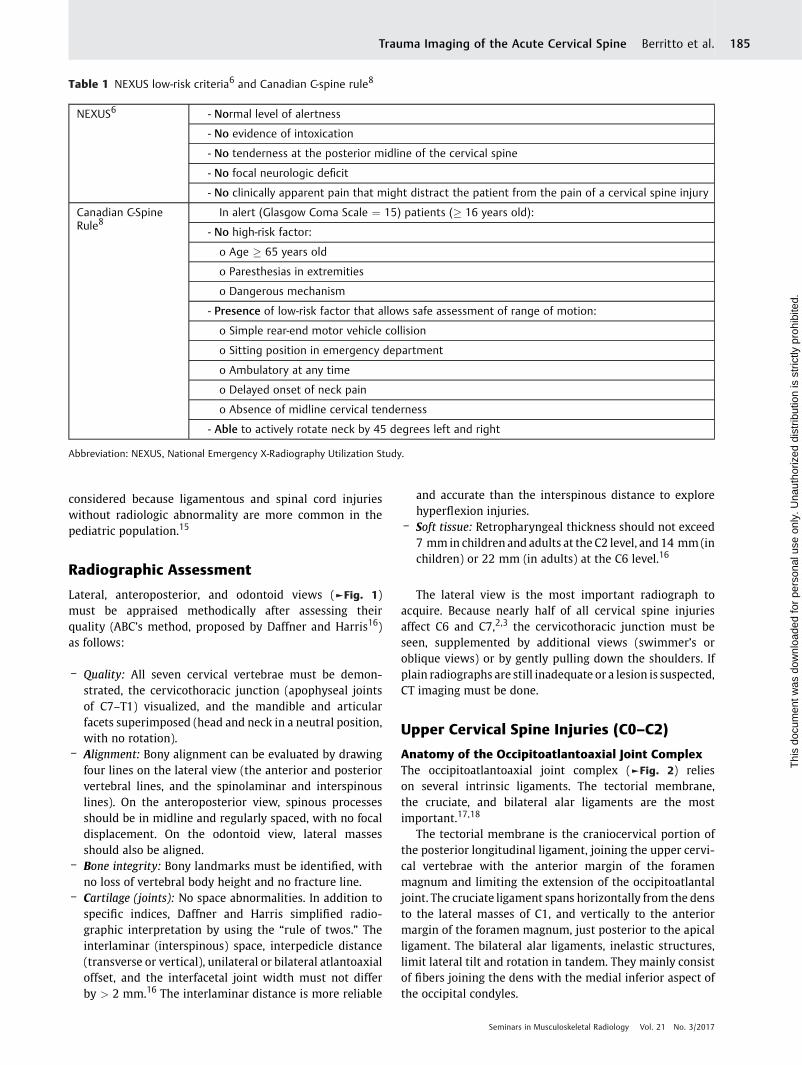
Anatomy of the Occipitoatlantoaxial Joint ComplexThe occipitoatlantoaxial joint complex (►Fig. 2) relieson several intrinsic ligaments. The tectorial membrane,the cruciate, and bilateral alar ligaments are the mostimportant.17,18
The tectorial membrane is the craniocervical portion ofthe posterior longitudinal ligament, joining the upper cervi-cal vertebrae with the anterior margin of the foramenmagnum and limiting the extension of the occipitoatlantaljoint. The cruciate ligament spans horizontally from the densto the lateral masses of C1, and vertically to the anteriormargin of the foramen magnum, just posterior to the apicalligament. The bilateral alar ligaments, inelastic structures,limit lateral tilt and rotation in tandem. They mainly consistof fibers joining the dens with the medial inferior aspect ofthe occipital condyles.
Table 1 NEXUS low-risk criteria6 and Canadian C-spine rule8
NEXUS6 - Normal level of alertness
- No evidence of intoxication
- No tenderness at the posterior midline of the cervical spine
- No focal neurologic deficit
- No clinically apparent pain that might distract the patient from the pain of a cervical spine injury
Canadian C-SpineRule8
In alert (Glasgow Coma Scale ¼ 15) patients (� 16 years old):
- No high-risk factor:
o Age � 65 years old
o Paresthesias in extremities
o Dangerous mechanism
- Presence of low-risk factor that allows safe assessment of range of motion:
o Simple rear-end motor vehicle collision
o Sitting position in emergency department
o Ambulatory at any time
o Delayed onset of neck pain
o Absence of midline cervical tenderness
- Able to actively rotate neck by 45 degrees left and right
Abbreviation: NEXUS, National Emergency X-Radiography Utilization Study.
Seminars in Musculoskeletal Radiology Vol. 21 No. 3/2017
Trauma Imaging of the Acute Cervical Spine Berritto et al. 185
Thi
s do
cum
ent w
as d
ownl
oade
d fo
r pe
rson
al u
se o
nly.
Una
utho
rized
dis
trib
utio
n is
str
ictly
pro
hibi
ted.
In addition to these structures, extrinsic ligaments (thearticular capsule ligaments and the anterior, posterior, andlateral atlantooccipital membranes) strengthen the cranio-cervical junction by connecting the ring of C1 to the base ofthe skull.19 Another distinctive feature of this region is theabsence of intervertebral discs: The load of the skull isdirectly transferred through the lateral masses of C1 andC2.20 Through this unique joint complex, many upper cervi-cal vertebrae injuries can be associated because of the closerelationship between the occipital condyles (C0), atlas (C1),and axis (C2) vertebrae.
Normal anatomical variants and congenital defects mayappear as pseudo fractures, so knowledge of the normaldevelopment of the upper cervical spine is critical.20,21 Thedescription of these variations is beyond the scope of thisarticle.
Occipital Condyle Fractures (C0)Occipital condyle fractures are rare injuries, now increas-ingly diagnosed in survivors of high-energy blunt traumabecause of the widespread use of CT.17 Clinical presentationmay vary, from only upper cervical pain without neurologic
Fig. 1 Normal standard three-view series (plain radiographs). (a) On lateral views, the anterior (solid line), posterior (dotted line), spinolaminar(dashed line), and interspinous (dash-dotted line) must be smoothly drawn. Soft tissue swelling can be assessed at C2 and C6 levels (asterisks). Aspheno-occipital synchondrosis may be seen in young adults andmust not be mistaken as a fracture (black arrows). (b) On anteroposterior views,spinous processes are in midline and regularly spaced (dashed line). No vertebrae misalignment should be observed. (c) On odontoid views,lateral masses of C1 (m) are equally distant (double-headed arrows) from the dens of the odontoid (d) and aligned with the articular facets of C2(solid lines). Occipitoatlantal joints may be identified (asterisks).
Seminars in Musculoskeletal Radiology Vol. 21 No. 3/2017
Trauma Imaging of the Acute Cervical Spine Berritto et al.186
Thi
s do
cum
ent w
as d
ownl
oade
d fo
r pe
rson
al u
se o
nly.
Una
utho
rized
dis
trib
utio
n is
str
ictly
pro
hibi
ted.
deficit to palsy. Because they may be associated with cra-niocervical instability, these fractures are important toscreen.
Lateral cervical spine radiography has a limited role in thedetection of occipital condyle fractures, especially becauseendotracheal intubation and pooling of pharyngeal secre-tions are responsible for inadequate lateral cervical spineradiographs.When available, CT imaging is the best modalityto explore occipital condyle fractures.
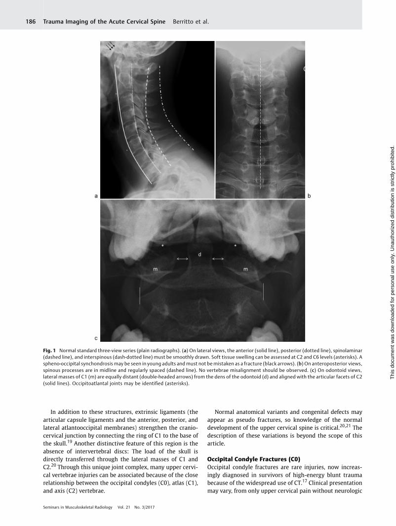
If only plain radiographs are available, alignment of thecraniocervical junction should be evaluated from a cross-table lateral view using the basion-dental interval (BDI) andbasion-axial interval (BAI) methods, described by Harriset al.22,23 (►Fig. 3):
– BDI: The distance between the basion and the dens shouldnot be > 12 mm in adults or children > than 13 years ofage.24
– BAI: The distance between the basion and the posterioraxial line should be between þ 12 mm and � 4 mm.
The most widely used radiologic classification of occipitalcondyle fractures was described by Anderson and Monte-sano25 as follows:
– Type I: Comminuted impaction-fracture due to axial load-ing that may compromise the ipsilateral alar ligament.Stability is maintained by the contralateral alar ligamentand the tectorial membrane. This type is unusual and notfrequently observed.17
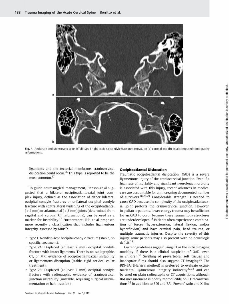
– Type II: Skull base fracture that extends from the occipitalbone to the condyle before reaching the foramen mag-num. Stability is maintained by intact alar ligaments andthe tectorial membrane (►Fig. 4).
– Type III: Avulsion fracture mediated through tension in analar ligament. When associated with a disruption of alar
Fig. 3 Computed tomography (CT) assessment of the occipitoatlantoaxial joint complex (Harris’s method). (a) The basion-dental intervalshould not be more than 12 mm in adults or children > 13 years.22,23 (b) The basion-axial interval should be between þ 12 mm and � 4 mm.
Fig. 2 Normal T2-weighted MRI anatomy. (a) Sagittal plane demonstrating the hypointense anterior atlanto-occipital membrane (thin arrow),the apical ligament (white star), and the tectorial membrane (plain arrow). (b) Axial plane demonstrating the transverse ligament (thin arrows).(c) Coronal plane demonstrating the alar ligaments (thin arrows).
Seminars in Musculoskeletal Radiology Vol. 21 No. 3/2017
Trauma Imaging of the Acute Cervical Spine Berritto et al. 187
Thi
s do
cum
ent w
as d
ownl
oade
d fo
r pe
rson
al u
se o
nly.
Una
utho
rized
dis
trib
utio
n is
str
ictly
pro
hibi
ted.
ligaments and the tectorial membrane, craniocervicaldislocation could occur.26 This type is reported to be themost common.17
To guide neurosurgical management, Hanson et al sug-gested that a bilateral occipitoatlantoaxial joint com-plex injury, defined as the association of either bilateraloccipital condyle fractures or unilateral occipital condylefracture with contralateral widening of the occipitoatlantal(> 2 mm) or atlantoaxial (> 3 mm) joints (determined fromsagittal and coronal CT reformations), can be used as amarker for instability.17 Furthermore, Tuli et al proposedmore recently a classification that includes ligamentousintegrity, assessed by MRI27:
– Type 1:Nondisplaced occipital condyle fracture (stable, nospecific treatment).
– Type 2A: Displaced (at least 2 mm) occipital condylefracture with intact ligaments. There is no radiographic,CT, or MRI evidence of occipitoatlantoaxial instabilityor ligamentous disruption (stable, rigid cervical collartreatment).
– Type 2B: Displaced (at least 2 mm) occipital condylefracture with radiographic evidence of craniocervicaljunction instability (unstable, requiring surgical instru-mentation or halo traction).
Occipitoatlantal DislocationTraumatic occipitoatlantal dislocation (OAD) is a severeligamentous injury of the craniocervical junction. Even if ahigh rate of mortality and significant neurologic morbidityis associated with this injury, recent advances in medicalcare are accountable for an increasing documented numberof survivors.18,28,29 Considerable strength is needed tocause OAD because the complexity of the occipitoatlantoax-ial joint protects the craniocervical junction. However,in pediatric patients, lower energy trauma may be sufficientfor an OAD to occur because these ligamentous structuresare underdeveloped.18 Patients often experience a combina-tion of forces (hyperextension, lateral flexion, and/orhyperflexion) and have cervical pain, head trauma, ormultiple traumatic injuries. Despite the severity of thisinjury, some patients may also present with no neurologicdeficit.28
Current guidelines suggest using CT as the initial imagingmodality if there is a clinical suspicion of OAD, evenin children.30 Swelling of prevertebral soft tissues andinadequate films should also suggest CT imaging.30 TheBDI-BAI (Harris’s method) is preferred to evaluate occipi-toatlantal ligamentous integrity indirectly22,31 and canbe used on plain radiographs or CT acquisitions, althoughBAI measurement is poorly reproducible on CT reconstruc-tions.32 In addition to BDI and BAI, Powers’ ratio and X-line
Fig. 4 Anderson and Montosano type II/Tuli type I right occipital condyle fracture (arrow), on (a) coronal and (b) axial computed tomographyreformations.
Seminars in Musculoskeletal Radiology Vol. 21 No. 3/2017
Trauma Imaging of the Acute Cervical Spine Berritto et al.188
Thi
s do
cum
ent w
as d
ownl
oade
d fo
r pe
rson
al u
se o
nly.
Una
utho
rized
dis
trib
utio
n is
str
ictly
pro
hibi
ted.
methods can be used to strengthen the diagnostic accuracyof OAD:
– Powers’ ratio is the ratio of the basion-posterior atlas archdistance to the opisthion-anterior arch distance. Values> 1 are pathologic.33
– X-line method is considered pathologic if the line from thebasion to the axis spinolaminar junction does not intersectC2 or if a line from the opisthion to the posteroinferiorcorner of the body of the axis does not intersect C1.34
An initial classificationwas proposed in 1986 by Trayneliset al, in accordance with OAD orientation (type 1: anteriordisplacement; type 2: vertical distraction; type 3: posteriordisplacement).35However, this gradation did not sufficientlymatch with injury severity. To better guide neurosurgicaltreatment, Horn et al classified OAD fractures as follows28:
– Grade I: Normal CT findings in relation to establishedmethods of diagnosis (Powers’ ratio, X-line, BDI and BAImethods) and equivocal MRI findings consisting of highposterior ligamentous or occipitoatlantal signal and mildto no signal change at the occipitoatlantal joint. Nono-perative treatment,with halo or cervical collar, in patientswith grade I injuries, is supported.
– Grade II: At least one abnormal finding on CT-basedcriteria or grossly abnormal MRI findings in the occipi-toatlantal joints, tectorial membrane, alar ligaments, orcruciate ligaments. For these patients, surgical fixation isrequired.
Atlas Fractures (C1)Atlas fractures account for 3 to 13% of acute cervical spineinjuries and 1 to 3% of all spinal injuries.36–40 A bimodaldistribution is seen, with individuals in their mid-20s andbetween 80 and 84 years most at risk for C1 fractures. How-ever, the mean age of diagnosis is 64 years, and nearly threequartersofatlas fracturesoccur inpatients > 50yearsofage.38
Patients with C1 fractures classically have cervical painand neck stiffness, with a history of indirect trauma throughaxial compression (fall on the head) or violent hyperexten-sion (automobile collision).37 These fractures are not usuallyassociated with a neurologic deficit because the fragmentstend to spread away from the spinal cord.40 However, thevertebral arteries are at high risk of injury (dissection and/orthrombosis) or spasm due to local inflammation that canresult in neurologic deficits.
Because the atlas is a ring, fractures can affect in a combinedor isolatedwayeacharch,a lateralmass,ora transverseprocess.Adescriptive classificationwas initiallygivenbyGehweiler et alin 1976,41 updated a decade later by Landells et al42:
– Type I: Fractures confined to a single arch (anterior orposterior). A “plow fracture” affects one anterior archfracture and is caused by forces driving the odontoidanteriorly.
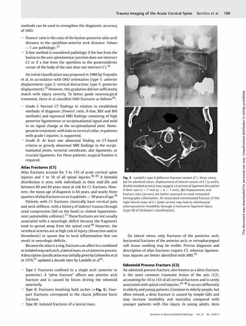
– Type II: Fractures involving both arches (►Fig. 5). Four-part fractures correspond to the classic Jefferson burstfracture.
– Type III: Isolated fractures of a lateral mass.
On lateral views, only fractures of the posterior arch,horizontal fractures of the anterior arch, or retropharyngealsoft tissue swelling may be visible. Precise diagnosis anddescription of atlas fractures require CT, whereas ligamen-tous injuries are better identified with MRI.43
Odontoid Process Fracture (C2)An odontoid process fracture, also known as a dens fracture,is the most common traumatic lesion of the axis (C2),accounting for 10 to 15% of all cervical fractures and is rarelyassociatedwith spinal cord injuries.44–46 It occurs differentlyin elderly and young patients. Common in elderly people, butoften missed, a dens fracture is caused by simple falls andmay increase morbidity and mortality compared withyounger patients with this injury. In young adults, dens
Fig. 5 Landell’s type II Jefferson fracture variant (C1, three sites).(a) On odontoid views, displacement of lateral masses of C1 (a and b,double-headed arrows) may suggest a transversal ligament disruptionif their sum is > 7 mm (a þ b > 7 mm). (b) Displacement andfracture sites (arrows) are better assessed on axial computedtomography reformation. An associated comminuted fracture of theright lateral mass of C1 (plain arrow) may lead to atlantoaxialanteroposterior instability through a transverse ligament injury(type IIB of Dickman’s classification).
Seminars in Musculoskeletal Radiology Vol. 21 No. 3/2017
Trauma Imaging of the Acute Cervical Spine Berritto et al. 189
Thi
s do
cum
ent w
as d
ownl
oade
d fo
r pe
rson
al u
se o
nly.
Una
utho
rized
dis
trib
utio
n is
str
ictly
pro
hibi
ted.
fractures result from head blunt trauma through cervicalhyperflexion or hyperextension.
Patients with a dens fracture usually present with neckpain, worsening with motion. Dysphagia may be presentwhen associated with a large retropharyngeal hematoma.Myelopathy is very rare due to the large cross-sectional areaof the spinal canal at this level.
Dens fractures can be classified according to Andersonand D’Alonzo’s classification as follow47,48:
– Type I:Oblique avulsion fracture of the tip of the odontoid,above the transverse ligament.
– Type II: Fracture occurring at the base of the odontoid,between the level of the transverse ligament and the bodyof C2, associated with a high nonunion rate due to inter-ruption of blood supply. This type is the most common.49
– Type III: Fracture extending into the vertebral body.
To better predict the degree of instability, Roy-Camille’sclassification described four subtypes of type II Anderson’sand D’Alonzo’s injuries, depending on the direction of thefracture line. Anterior oblique fractures are more stable thanthe posterior oblique ones50:
– Oblique fracture line sloping forward,with dens displacedanteriorly.
– Oblique fracture line sloping backward, with dens dis-placed posteriorly (►Fig. 6).
– Horizontal fracture line, with dens displaced anteriorly orposteriorly.
– Fracture with rotation or “English policeman’s hat.”
Flexion-extension radiographs are important optionalviews to diagnose atlantoaxial instability in type I fracturesand variants (os odontoideum). Anterior instability is de-fined as an atlantodens interval (ADI) > 10 mm and a spaceavailable for the spinal cord < 13 mm.51
CT is the imaging modality of choice for fracture delinea-tion and assessing its stability. CT angiogram is required todetermine the location of the vertebral artery prior toposterior instrumentation procedures. MRI is indicated ifneurologic symptoms are present.
Nondisplaced fractures can be initially managed withcervical orthosis. Comminution and displaced fractures(> 5 mm) require surgical stabilization.52
Traumatic Spondylolisthesis of C2 (Hangman’sFracture)Traumatic spondylolisthesis is considered one of themost frequent upper cervical spine injury and correspondsto the fracture of both “pars interarticularis” (isthmus)of C2.2 It usually results from sudden cervical spinehyperextension and distraction, tearing apart thelongitudinal ligaments and the intervertebral disc,which can lead to vertebral subluxation. Overall, 30% ofconcomitant cervical spine fractures are associated withthis injury.53
Patients with hangman’s fracture usually present withneck pain and no neurologic deficit.54 Most authors affirmthat hangman’s fractures have a good prognosis.53,55 Thetreatment of this fracture is predominantly nonoperativeaccording to the literature, although surgery is preferred forneurologic symptoms, severe ligamentous injuries, or dis-ruption of the C2–C3 disc space.52
The classification of these fractures, initially defined byEffendi et al,56 then modified by Levine and Edwards whoincluded the traumamechanism,57 is divided into threemaintypes48:
– Type I: Fractures with minimal displacement (< 3 mm),secondary to an axial compression with cervical exten-sion (stable) (►Fig. 7).
–
Fig. 6 Roy-Camille “oblique odontoid fracture sloping backward,” on (a) lateral view and (b) sagittal computed tomography reformation.
Seminars in Musculoskeletal Radiology Vol. 21 No. 3/2017
Trauma Imaging of the Acute Cervical Spine Berritto et al.190
Thi
s do
cum
ent w
as d
ownl
oade
d fo
r pe
rson
al u
se o
nly.
Una
utho
rized
dis
trib
utio
n is
str
ictly
pro
hibi
ted.

Type II: Displaced fractures (> 3 mm), or angulated (> 11degrees), secondary to axial compression with cervicalextension, followed by a rebound flexion (unstable).
– Type IIa: Fractures with high angulation (often > 14degrees) and minimal translation (< 3 mm), secondaryto a flexion-distraction mechanism (unstable).
– Type III (rare): Translation and accentuated angulation arepresent, with dislocation of one or two C2–C3 articularfacets and ligamentous disruptions (unstable).
On plain radiographs, anomalies could be subtle, and CT isthe study of choice to delineate fracture pattern.MRImust be
considered to evaluate discoligamentous structures and ifthere are neurologic symptoms and/or a suspicion of avertebral artery injury.
Atlantoaxial Anteroposterior DislocationThe complexity of the atlantoaxial joint provides robusthead stabilization while the cervical spine is involved incomplex movements. Atlantoaxial anteroposterior disloca-tion (AAD) is associated with a loss of stability. Unlikethe lower cervical spine, the C1–C2 facets are not physicallylimited in their horizontal movements. The transverseligament is the sole element preventing C1 from being
Fig. 7 Levine and Edwards type I (nondisplaced) hangman’s fracture (arrows), on (a) axial and (b) parasagittal computed tomography (CT)reformations. The anterior C2–C3 intersomatic space is initially widened (star) on the sagittal CT reformation (c), suggesting a disc injury andpotential instability, confirmed by an anterior subluxation of C2 (double-headed arrow) demonstrated 3 months later (d).
Seminars in Musculoskeletal Radiology Vol. 21 No. 3/2017
Trauma Imaging of the Acute Cervical Spine Berritto et al. 191
Thi
s do
cum
ent w
as d
ownl
oade
d fo
r pe
rson
al u
se o
nly.
Una
utho
rized
dis
trib
utio
n is
str
ictly
pro
hibi
ted.

dislocated anteriorly and the dens from being displacedposteriorly.
Clinical presentation of atlantoaxial dislocation variesfrom minor neck pain to death. Approximately 50% ofpatients have neck pain and/or neck movement restriction,70% with weakness and/or numbness, and 90% with pyra-midal signs.58–60
Dickman et al proposed a classification to describe liga-mentous injuries following AAD48,61:
– Type I: Pure transverse ligament injuries, requiringsurgery (IA: midportion disruption; IB: disruption at itsinsertion without fracture of the tubercle).
– Type II: Injuries involving a bone fracture, requiring sur-gery if nonoperative management fails (IIA: avulsionfracture of a tubercle; IIB: comminuted lateral massfracture).
On open-mouth views, if the sum of each lateral massdisplacement is > 7 mm, the transverse ligament is likely tobe disrupted (“rule of Spence”). An avulsion fragment of themedial aspect of a lateral mass of C1 could also be visible. Onlateral views, ligamentous instability can be assessed by theADI. Normal ADI is < 3 mm in adults and 5 mm in children,and a larger distance reflects a high probability of ligamen-tous injury.62 Available space for the spinal cord can beestimated by measuring the distance from the posterioraspect of the dens to the anterior aspect of the posterioratlantal ring.
Moreover, because plain radiographs acquired in neutralposition may be normal, dynamic cervical radiographs canbe helpful to evaluate atlantoaxial stability with sponta-neous reducible dislocation. MRI must be used to assesssoft tissues, joints, and the spinal cord.
According to Wang’s classification, and to guide treat-ment, AAD can be categorized into four types after flexionand extension radiographs63:
– Type I: Dynamic reduction on radiographs (instability).– Type II: Reducible dislocation after skeletal traction under
general anesthesia.– Type III: Irreducible dislocation after skeletal traction
under general anesthesia.– Type IV: Bony dislocation (C1–C2 bony fusion).
Atlantoaxial Rotary SubluxationAtlantoaxial rotary subluxation (ARS) is more common inchildren due to their discoligamentous laxity, and it occursafter a trauma with sudden head rotation. This pathologicpattern may also be caused by inflammatory processes, as inGrisel’s syndrome, although rarely encountered. Becauserotation of C1 over C2 is fixed, the head is blocked in lateralrotation and muscle contracture is responsible for cervicalpain.
Whereas the transverse ligament and C1–C2 joint capsuleskeep the atlantoaxial joint from shifting in a sagittal plane, thealar ligaments prevent excessive rotation. The whole atlan-toaxial ligamentous complex is involved in this injury.
CT is essential for ARS diagnosis. Evaluation of the ADI andanatomical relationship between C1 and C2 in an axial plane
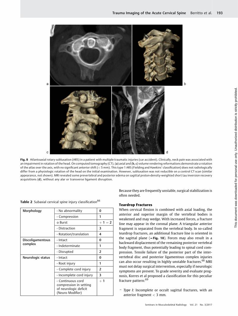
are required. Fielding and Hawkins in 1977 described fourtypes of rotary subluxations64:
– Type 1: Rotary subluxation without anterior shift(►Fig. 8).
– Type 2: Rotary fixation with 3- to 5-mm anterior shift.– Type 3: Rotary fixation with > 5 mm anterior shift.– Type 4: Rotary fixation with posterior shift (rare).
Because the atlantoaxial joint can rotate up to 40 degrees,physiologic rotation due to torticollis and type 1 ARS can bedifficult to distinguish on a single CT scan. Some authorssuggest scanning patients with maximum voluntary con-tralateral head rotation if symptoms do not resolve 3 to4 weeks after the initial diagnosis. In patients with ARS,subluxation was not reducible on functional scans.65
Lower Cervical Region Injuries (C3–C7)
Due to its considerable mobility and proximity with themorerigid thoracic spine, the subaxial cervical spine is particularlyvulnerable to traumatic injuries. More than two thirds ofcervical fractures and spinal dislocations take place in thisregion,mostly fromC5 to C7.2,3 In addition to being frequentlyassociated with neurologic symptoms, numerous patterns ofosseous and ligamentous lesions can be observed in the lowercervical spine, and described according to the injury mechan-ism (compression, distraction, or rotation).
Subaxial Cervical Spine Injury Classification SystemSeveral descriptive classifications of lower cervical vertebraefractures have been proposed. However, in 2013 the Con-gress of Neurosurgeons recommended the use of the Sub-axial Cervical Spine Injury Classification System (SLIC).66,67
This severity scale is based on three main components: (1)injury morphology (compression, distraction, rotation/translation), (2) integrity of the discoligamentous complex,and (3) the patient’s neurologic status. This gradation com-bines all imaging modalities available, especially MRI fordiscoligamentous complex assessment, and takes clinicalevaluation into account (►Table 2). Even if only partiallyvalidated, this system is less complicated than other scales(the Cervical Spine Injury Severity Score is the main andmore complex alternative) and may guide treatment.
Compression InjuriesVertical compression injuries occur when sufficient axialstrength is exerted through the spinal column.Minor verticalcompression results in central cupping of vertebral endplates and/or sagittal/coronal split fractures. Most fracturesaffect only the vertebral body. With high-energy compres-sion mechanisms, typical burst fractures show severe ver-tebral body height loss and may extend to the posteriorcolumn (►Fig. 9). Minimally displaced articular pillars andfacet fractures may occur with lateral compression mechan-isms. Fragments reaching the spinal canal can result in severeneural damages.
Of all cervical traumas, compressive hyperflexion injuriesare responsible for the highest rate of neurologic outcomes.
Seminars in Musculoskeletal Radiology Vol. 21 No. 3/2017
Trauma Imaging of the Acute Cervical Spine Berritto et al.192
Thi
s do
cum
ent w
as d
ownl
oade
d fo
r pe
rson
al u
se o
nly.
Una
utho
rized
dis
trib
utio
n is
str
ictly
pro
hibi
ted.
Because they are frequently unstable, surgical stabilization isoften needed.
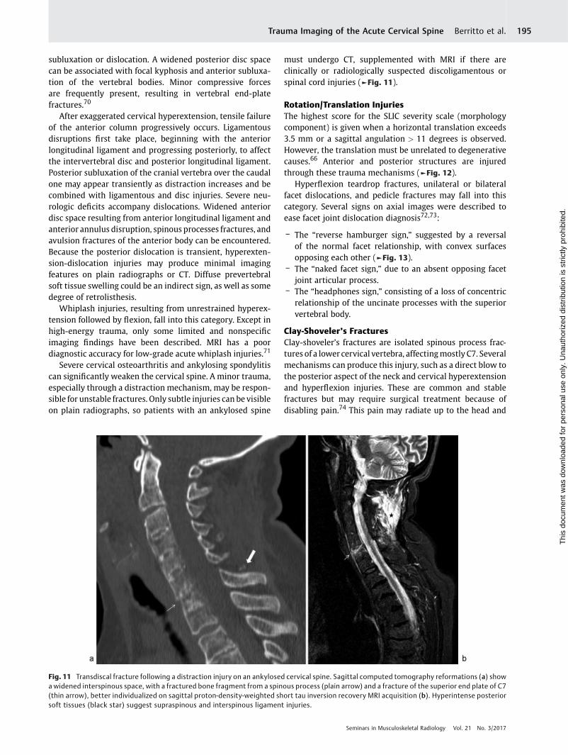
Teardrop FracturesWhen cervical flexion is combined with axial loading, theanterior and superior margin of the vertebral bodies isweakened and may wedge. With increased forces, a fractureline may appear in the coronal plane. A triangular anteriorfragment is separated from the vertebral body. In so-calledteardrop fractures, an additional fracture line is oriented inthe sagittal plane (►Fig. 10). Forces may also result in abackward displacement of the remaining posterior vertebralbody fragment, thus potentially leading to spinal cord com-pression. Tensile failure of the posterior part of the inter-vertebral disc and posterior ligamentous complex injuriescan also occur resulting in highly unstable fractures.68 MRImust not delay surgical intervention, especially if neurologicsymptoms are present. To grade severity and evaluate prog-nosis, Korres et al proposed a classification for this peculiarfracture pattern:69
– Type I: Incomplete or occult sagittal fractures, with ananterior fragment < 3 mm.
Table 2 Subaxial cervical spine injury classification66
Morphology - No abnormality 0
- Compression 1
o Burst þ 1 ¼ 2
- Distraction 3
- Rotation/translation 4
Discoligamentouscomplex
- Intact 0
- Indeterminate 1
- Disrupted 2
Neurologic status - Intact 0
- Root injury 1
- Complete cord injury 2
- Incomplete cord injury 3
- Continuous cordcompression in settingof neurologic deficit(Neuro Modifier)
þ 1
Fig. 8 Atlantoaxial rotary subluxation (ARS) in a patient with multiple traumatic injuries (car accident). Clinically, neck pain was associated withan impairment in rotation of the head. On computed tomography (CT), (a) axial and (b, c) volume rendering reformations demonstrate a rotationof the atlas over the axis, with no significant anterior shift (< 5mm). This type 1 ARS (Fielding and Hawkins’ classification) does not radiologicallydiffer from a physiologic rotation of the head on the initial examination. However, subluxation was not reducible on a control CT scan (similarappearance, not shown). MRI revealed some prevertebral and posterior edema on sagittal proton-density-weighted short tau inversion recoveryacquisitions (d), without any alar or transverse ligament disruption.
Seminars in Musculoskeletal Radiology Vol. 21 No. 3/2017
Trauma Imaging of the Acute Cervical Spine Berritto et al. 193
Thi
s do
cum
ent w
as d
ownl
oade
d fo
r pe
rson
al u
se o
nly.
Una
utho
rized
dis
trib
utio
n is
str
ictly
pro
hibi
ted.
– Type II: Complete sagittal fractures, without posteriordisplacement.
– Type III: Type II with posterior displacement (IIIa: < 4mm; IIIb: > 4 mm).
– Type IV: Type IIIb with locked facet (the inferior articularfacet is dislocated and locked over the superior articularprocess of the subjacent vertebra) and anterior dislocationof the above vertebra.
Distraction InjuriesThe main feature of distraction injuries is the anatomicaldissociation in the vertical axis, with severe discoligamen-tous lesions. In flexion, the posterior column is affected first.The supraspinous ligament begins to disrupt, followed bythe interspinous ligament, facet capsular ligaments, and theligamentum flava, thus leading to interspinous and inter-laminar widening and unilateral or bilateral facet joint
Fig. 10 Teardrop fractures. (a) A triangular fragment is seen at the anterior and inferior corner of C3–C5 vertebral bodies (arrows) on sagittalcomputed tomography (CT) reformations. (b) The sagittal fracture line (arrowhead) is best seen on axial CT reformations.
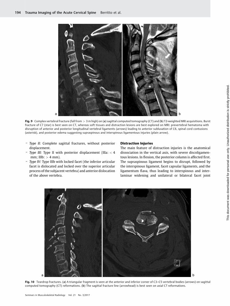
Fig. 9 Complex vertebral fracture (fall from > 3 m high) on (a) sagittal computed tomography (CT) and (b) T2-weighted MRI acquisitions. Burstfracture of C7 (star) is best seen on CT, whereas soft tissues and distraction lesions are best explored on MRI: prevertebral hematoma withdisruption of anterior and posterior longitudinal vertebral ligaments (arrows) leading to anterior subluxation of C6, spinal cord contusions(asterisk), and posterior edema suggesting supraspinous and interspinous ligamentous injuries (plain arrow).
Seminars in Musculoskeletal Radiology Vol. 21 No. 3/2017
Trauma Imaging of the Acute Cervical Spine Berritto et al.194
Thi
s do
cum
ent w
as d
ownl
oade
d fo
r pe
rson
al u
se o
nly.
Una
utho
rized
dis
trib
utio
n is
str
ictly
pro
hibi
ted.
subluxation or dislocation. A widened posterior disc spacecan be associated with focal kyphosis and anterior subluxa-tion of the vertebral bodies. Minor compressive forcesare frequently present, resulting in vertebral end-platefractures.70
After exaggerated cervical hyperextension, tensile failureof the anterior column progressively occurs. Ligamentousdisruptions first take place, beginning with the anteriorlongitudinal ligament and progressing posteriorly, to affectthe intervertebral disc and posterior longitudinal ligament.Posterior subluxation of the cranial vertebra over the caudalone may appear transiently as distraction increases and becombined with ligamentous and disc injuries. Severe neu-rologic deficits accompany dislocations. Widened anteriordisc space resulting from anterior longitudinal ligament andanterior annulus disruption, spinous processes fractures, andavulsion fractures of the anterior body can be encountered.Because the posterior dislocation is transient, hyperexten-sion-dislocation injuries may produce minimal imagingfeatures on plain radiographs or CT. Diffuse prevertebralsoft tissue swelling could be an indirect sign, as well as somedegree of retrolisthesis.
Whiplash injuries, resulting from unrestrained hyperex-tension followed by flexion, fall into this category. Except inhigh-energy trauma, only some limited and nonspecificimaging findings have been described. MRI has a poordiagnostic accuracy for low-grade acute whiplash injuries.71
Severe cervical osteoarthritis and ankylosing spondylitiscan significantly weaken the cervical spine. A minor trauma,especially through a distraction mechanism, may be respon-sible for unstable fractures. Only subtle injuries can be visibleon plain radiographs, so patients with an ankylosed spine
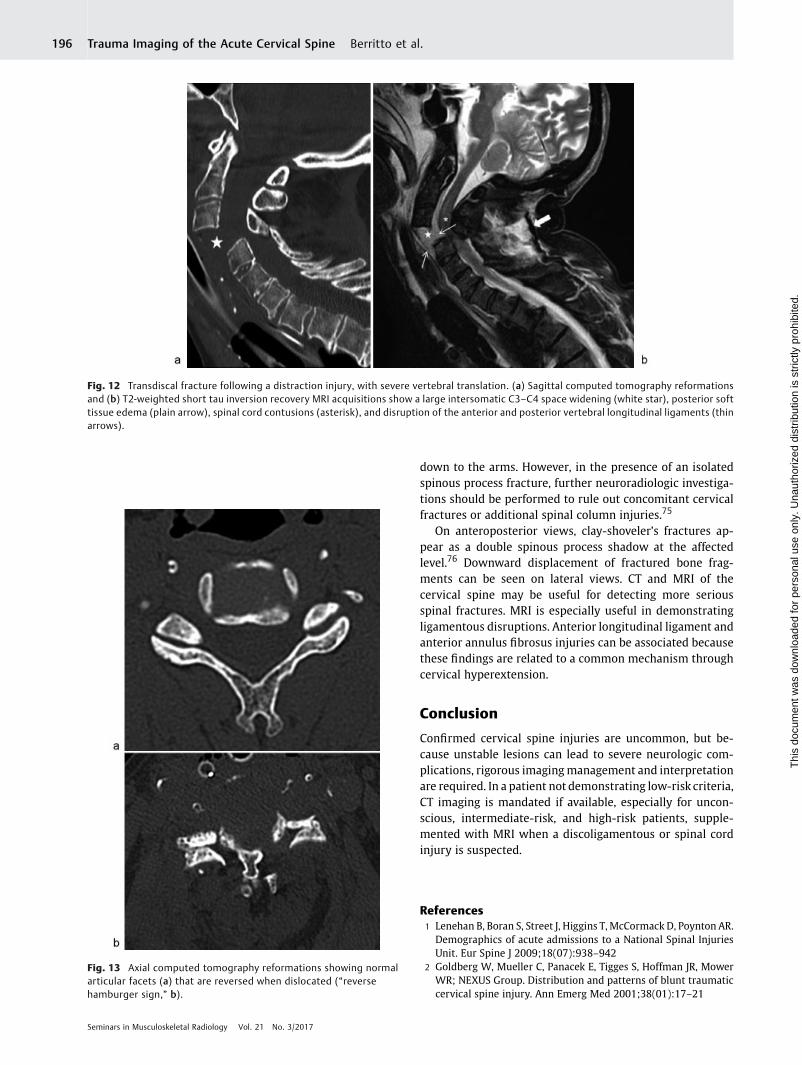
must undergo CT, supplemented with MRI if there areclinically or radiologically suspected discoligamentous orspinal cord injuries (►Fig. 11).
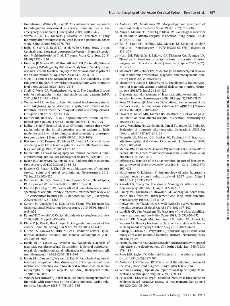
Rotation/Translation InjuriesThe highest score for the SLIC severity scale (morphologycomponent) is given when a horizontal translation exceeds3.5 mm or a sagittal angulation > 11 degrees is observed.However, the translation must be unrelated to degenerativecauses.66 Anterior and posterior structures are injuredthrough these trauma mechanisms (►Fig. 12).
Hyperflexion teardrop fractures, unilateral or bilateralfacet dislocations, and pedicle fractures may fall into thiscategory. Several signs on axial images were described toease facet joint dislocation diagnosis72,73:
– The “reverse hamburger sign,” suggested by a reversalof the normal facet relationship, with convex surfacesopposing each other (►Fig. 13).
– The “naked facet sign,” due to an absent opposing facetjoint articular process.
– The “headphones sign,” consisting of a loss of concentricrelationship of the uncinate processes with the superiorvertebral body.
Clay-Shoveler’s FracturesClay-shoveler’s fractures are isolated spinous process frac-tures of a lower cervical vertebra, affectingmostly C7. Severalmechanisms can produce this injury, such as a direct blow tothe posterior aspect of the neck and cervical hyperextensionand hyperflexion injuries. These are common and stablefractures but may require surgical treatment because ofdisabling pain.74 This pain may radiate up to the head and
Fig. 11 Transdiscal fracture following a distraction injury on an ankylosed cervical spine. Sagittal computed tomography reformations (a) showa widened interspinous space, with a fractured bone fragment from a spinous process (plain arrow) and a fracture of the superior end plate of C7(thin arrow), better individualized on sagittal proton-density-weighted short tau inversion recovery MRI acquisition (b). Hyperintense posteriorsoft tissues (black star) suggest supraspinous and interspinous ligament injuries.
Seminars in Musculoskeletal Radiology Vol. 21 No. 3/2017
Trauma Imaging of the Acute Cervical Spine Berritto et al. 195
Thi
s do
cum
ent w
as d
ownl
oade
d fo
r pe
rson
al u
se o
nly.
Una
utho
rized
dis
trib
utio
n is
str
ictly
pro
hibi
ted.
down to the arms. However, in the presence of an isolatedspinous process fracture, further neuroradiologic investiga-tions should be performed to rule out concomitant cervicalfractures or additional spinal column injuries.75
On anteroposterior views, clay-shoveler’s fractures ap-pear as a double spinous process shadow at the affectedlevel.76 Downward displacement of fractured bone frag-ments can be seen on lateral views. CT and MRI of thecervical spine may be useful for detecting more seriousspinal fractures. MRI is especially useful in demonstratingligamentous disruptions. Anterior longitudinal ligament andanterior annulus fibrosus injuries can be associated becausethese findings are related to a common mechanism throughcervical hyperextension.
Conclusion
Confirmed cervical spine injuries are uncommon, but be-cause unstable lesions can lead to severe neurologic com-plications, rigorous imagingmanagement and interpretationare required. In a patient not demonstrating low-risk criteria,CT imaging is mandated if available, especially for uncon-scious, intermediate-risk, and high-risk patients, supple-mented with MRI when a discoligamentous or spinal cordinjury is suspected.
References1 Lenehan B, Boran S, Street J, Higgins T, McCormack D, Poynton AR.
Demographics of acute admissions to a National Spinal InjuriesUnit. Eur Spine J 2009;18(07):938–942
2 Goldberg W, Mueller C, Panacek E, Tigges S, Hoffman JR, MowerWR; NEXUS Group. Distribution and patterns of blunt traumaticcervical spine injury. Ann Emerg Med 2001;38(01):17–21
Fig. 13 Axial computed tomography reformations showing normalarticular facets (a) that are reversed when dislocated (“reversehamburger sign,” b).
Fig. 12 Transdiscal fracture following a distraction injury, with severe vertebral translation. (a) Sagittal computed tomography reformationsand (b) T2-weighted short tau inversion recovery MRI acquisitions show a large intersomatic C3–C4 space widening (white star), posterior softtissue edema (plain arrow), spinal cord contusions (asterisk), and disruption of the anterior and posterior vertebral longitudinal ligaments (thinarrows).
Seminars in Musculoskeletal Radiology Vol. 21 No. 3/2017
Trauma Imaging of the Acute Cervical Spine Berritto et al.196
Thi
s do
cum
ent w
as d
ownl
oade
d fo
r pe
rson
al u
se o
nly.
Una
utho
rized
dis
trib
utio
n is
str
ictly
pro
hibi
ted.
3 Greenbaum J, Walters N, Levy PD. An evidenced-based approachto radiographic assessment of cervical spine injuries in theemergency department. J Emerg Med 2009;36(01):64–71
4 Varma A, Hill EG, Nicholas J, Selassie A. Predictors of earlymortality after traumatic spinal cord injury: a population-basedstudy. Spine 2010;35(07):778–783
5 Inaba K, Byerly S, Bush LD, et al; WTA C-Spine Study Group.Cervical spinal clearance: a prospectiveWestern Trauma Associa-tion Multi-Institutional Trial. J Trauma Acute Care Surg 2016;81(06):1122–1130
6 Hoffman JR, MowerWR,Wolfson AB, Todd KH, ZuckerMI; NationalEmergency X-RadiographyUtilization StudyGroup. Validity of a setof clinical criteria to rule out injury to the cervical spine in patientswith blunt trauma. N Engl J Med 2000;343(02):94–99
7 Stiell IG, Clement CM, McKnight RD, et al. The Canadian C-spinerule versus the NEXUS low-risk criteria in patients with trauma. NEngl J Med 2003;349(26):2510–2518
8 Stiell IG, Wells GA, Vandemheen KL, et al. The Canadian C-spinerule for radiography in alert and stable trauma patients. JAMA2001;286(15):1841–1848
9 Westerveld LA, Verlaan JJ, Oner FC. Spinal fractures in patientswith ankylosing spinal disorders: a systematic review of theliterature on treatment, neurological status and complications.Eur Spine J 2009;18(02):145–156
10 Daffner RH, Hackney DB. ACR Appropriateness Criteria on sus-pected spine trauma. J Am Coll Radiol 2007;4(11):762–775
11 Bailitz J, Starr F, Beecroft M, et al. CT should replace three-viewradiographs as the initial screening test in patients at high,moderate, and low risk for blunt cervical spine injury: a prospec-tive comparison. J Trauma 2009;66(06):1605–1609
12 Blackmore CC, Ramsey SD, Mann FA, Deyo RA. Cervical spinescreening with CT in trauma patients: a cost-effectiveness ana-lysis. Radiology 1999;212(01):117–125
13 Daffner RH. Cervical radiography for trauma patients: a time-effective technique?AJR AmJRoentgenol 2000;175(05):1309–1311
14 Ryken TC, Hadley MN,Walters BC, et al. Radiographic assessment.Neurosurgery 2013;72(Suppl 2):54–72
15 Rozzelle CJ, Aarabi B, Dhall SS, et al. Management of pediatriccervical spine and spinal cord injuries. Neurosurgery 2013;72(Suppl 2):205–226
16 Daffner RH, Harris JH. Cervical Spine Injuries. 5th ed. Philadelphia,PA: Lippincott Williams & Wilkins; 2013:139–260
17 Hanson JA, Deliganis AV, Baxter AB, et al. Radiologic and clinicalspectrum of occipital condyle fractures: retrospective review of107 consecutive fractures in 95 patients. AJR Am J Roentgenol2002;178(05):1261–1268
18 Garrett M, Consiglieri G, Kakarla UK, Chang SW, Dickman CA.Occipitoatlantal Dislocation. Neurosurgery 2010;66(03, Suppl 3):A48–A55
19 KaramYR, Traynelis VC. Occipital condyle fractures. Neurosurgery2010;66(03, Suppl 3):A56–A59
20 Klimo P Jr, Rao G, Brockmeyer D. Congenital anomalies of thecervical spine. Neurosurg Clin N Am 2007;18(03):463–478
21 Lustrin ES, Karakas SP, Ortiz AO, et al. Pediatric cervical spine:normal anatomy, variants, and trauma. Radiographics 2003;23(03):539–560
22 Harris JH Jr, Carson GC, Wagner LK. Radiologic diagnosis oftraumatic occipitovertebral dissociation: 1. Normal occipitover-tebral relationships on lateral radiographs of supine subjects. AJRAm J Roentgenol 1994;162(04):881–886
23 Harris JH Jr, Carson GC,Wagner LK, Kerr N. Radiologic diagnosis oftraumatic occipitovertebral dissociation: 2. Comparison of threemethods of detecting occipitovertebral relationships on lateralradiographs of supine subjects. AJR Am J Roentgenol 1994;162(04):887–892
24 Wholey MH, Bruwer AJ, Baker HL Jr. The lateral roentgenogram ofthe neck; with comments on the atlanto-odontoid-basion rela-tionship. Radiology 1958;71(03):350–356
25 Anderson PA, Montesano PX. Morphology and treatment ofoccipital condyle fractures. Spine 1988;13(07):731–736
26 Ahuja A, Glasauer FE, Alker GJ Jr, Klein DM. Radiology in survivorsof traumatic atlanto-occipital dislocation. Surg Neurol 1994;41(02):112–118
27 Tuli S, Tator CH, Fehlings MG, Mackay M. Occipital condylefractures. Neurosurgery 1997;41(02):368–376; discussion376–377
28 Horn EM, Feiz-Erfan I, Lekovic GP, Dickman CA, Sonntag VK,Theodore N. Survivors of occipitoatlantal dislocation injuries:imaging and clinical correlates. J Neurosurg Spine 2007;6(02):113–120
29 Steinmetz MP, Lechner RM, Anderson JS. Atlantooccipital disloca-tion in children: presentation, diagnosis, and management. Neu-rosurg Focus 2003;14(02):ecp1
30 Theodore N, Aarabi B, Dhall SS, et al. The diagnosis and manage-ment of traumatic atlanto-occipital dislocation injuries. Neuro-surgery 2013;72(Suppl 2):114–126
31 Diagnosis and Management of Traumatic Atlanto-occipital Dis-location Injuries. Neurosurgery 2002;50(Suppl 3):S105–S113
32 Rojas CA, Bertozzi JC,Martinez CR,Whitlow J. Reassessment of thecraniocervical junction: normal values on CT. AJNR Am J Neuror-adiol 2007;28(09):1819–1823
33 Powers B, Miller MD, Kramer RS, Martinez S, Gehweiler JA Jr.Traumatic anterior atlanto-occipital dislocation. Neurosurgery1979;4(01):12–17
34 Lee C, Woodring JH, Goldstein SJ, Daniel TL, Young AB, Tibbs PA.Evaluation of traumatic atlantooccipital dislocations. AJNR AmJ Neuroradiol 1987;8(01):19–26
35 Traynelis VC, Marano GD, Dunker RO, Kaufman HH. Traumaticatlanto-occipital dislocation. Case report. J Neurosurg 1986;65(06):863–870
36 Marcon RM, Cristante AF, TeixeiraWJ, Narasaki DK, Oliveira RP, deBarros FilhoTE. Fractures of the cervical spine. Clinics (Sao Paulo)2013;68(11):1455–1461
37 Jefferson G. Fracture of the atlas vertebra. Report of four cases,and a review of those previously recorded. Br J Surg 1919;7(27):407–422
38 Matthiessen C, Robinson Y. Epidemiology of atlas fractures–anational registry-based cohort study of 1,537 cases. Spine J2015;15(11):2332–2337
39 Kakarla UK, Chang SW, Theodore N, Sonntag VK. Atlas fractures.Neurosurgery 2010;66(03, Suppl 3):A60–A67
40 Hadley MN, Dickman CA, Browner CM, Sonntag VK. Acute trau-matic atlas fractures: management and long term outcome.Neurosurgery 1988;23(01):31–35
41 Gehweiler J, Duff D,Martinez S,MillerMD, ClarkWM. Fractures ofthe atlas vertebra. Skeletal Radiol 1976;1(02):97–102
42 Landells CD, Van Peteghem PK. Fractures of the atlas: classifica-tion, treatment and morbidity. Spine 1988;13(05):450–452
43 Radcliff KE, Sonagli MA, Rodrigues LM, Sidhu GS, Albert TJ,Vaccaro AR. Does C1 fracture displacement correlate with trans-verse ligament integrity? Orthop Surg 2013;5(02):94–99
44 Harrop JS, Sharan AD, Przybylski GJ. Epidemiology of spinal cordinjury after acute odontoid fractures [abstract]. Neurosurg Focus2000;8(06):e4
45 Pepin JW, Bourne RB, Hawkins RJ. Odontoid fractures,with specialreference to the elderly patient. Clin Orthop Relat Res 1985;(193):178–183
46 Ryan MD, Taylor TK. Odontoid fractures in the elderly. J SpinalDisord 1993;6(05):397–401
47 Anderson LD, D’Alonzo RT. Fractures of the odontoid process ofthe axis. J Bone Joint Surg Am 1974;56(08):1663–1674
48 Wilson J, Harrop J. Update on upper cervical spine injury classi-fications. Semin Spine Surg 2017;29(01):9–13
49 Pal D, Sell P, Grevitt M. Type II odontoid fractures in the elderly: anevidence-based narrative review of management. Eur Spine J2011;20(02):195–204
Seminars in Musculoskeletal Radiology Vol. 21 No. 3/2017
Trauma Imaging of the Acute Cervical Spine Berritto et al. 197
Thi
s do
cum
ent w
as d
ownl
oade
d fo
r pe
rson
al u
se o
nly.
Una
utho
rized
dis
trib
utio
n is
str
ictly
pro
hibi
ted.
50 Roy-Camille R, Saillant G, Judet T, de Botton G,Michel G. Factors ofseverity in the fractures of the odontoid process [in French]. RevChir Orthop Repar Appar Mot 1980;66(03):183–186
51 Rozzelle CJ, Aarabi B, Dhall SS, et al. Os odontoideum. Neurosur-gery 2013;72(Suppl 2):159–169
52 Ryken TC, Hadley MN, Aarabi B, et al. Management of isolatedfractures of the axis in adults. Neurosurgery 2013;72(03, Suppl 2):132–150
53 Gleizes V, Jacquot FP, Signoret F, Féron JM. Combined injuries inthe upper cervical spine: clinical and epidemiological data over a14-year period. Eur Spine J 2000;9(05):386–392
54 Garber JN. Abnormalities of the atlas and axis vertebrae—congenital and traumatic. J Bone Joint Surg Am 1964;46(08):1782–1791
55 Barros TE, Bohlman HH, Capen DA, et al. Traumatic spondylo-listhesis of the axis: analysis of management. Spinal Cord 1999;37(03):166–171
56 Effendi B, Roy D, Cornish B, Dussault RG, Laurin CA. Fractures ofthe ring of the axis. A classification based on the analysis of 131cases. J Bone Joint Surg Br 1981;63-B(03):319–327
57 Levine AM, Edwards CC. The management of traumatic spondy-lolisthesis of the axis. J Bone Joint Surg Am 1985;67(02):217–226
58 Passias PG, Wang S, Kozanek M, Wang S, Wang C. Relationshipbetween the alignment of the occipitoaxial and subaxial cervicalspine in patients with congenital atlantoxial dislocations. J SpinalDisord Tech 2013;26(01):15–21
59 Yin Y-H, Qiao G-Y, Yu X-G, Tong H-Y, Zhang Y-Z. Posteriorrealignment of irreducible atlantoaxial dislocation with C1-C2screw and rod system: a technique of direct reduction andfixation. Spine J 2013;13(12):1864–1871
60 Sobolewski BA, Mittiga MR, Reed JL. Atlantoaxial rotary subluxa-tion after minor trauma. Pediatr Emerg Care 2008;24(12):852–856
61 Dickman CA, Greene KA, Sonntag VK. Injuries involving thetransverse atlantal ligament: classification and treatment guide-lines based upon experience with 39 injuries [abstract]. Neuro-surgery 1996;38(01):44–50
62 Jackson RS, Banit DM, Rhyne AL III, Darden BV II. Upper cervicalspine injuries. J Am Acad Orthop Surg 2002;10(04):271–280
63 Wang S, Wang C, YanM, Zhou H, Dang G. Novel surgical classifica-tion and treatment strategy for atlantoaxial dislocations. Spine2013;38(21):E1348–E1356
64 Fielding JW, Hawkins RJ. Atlanto-axial rotatory fixation. (Fixedrotatory subluxation of the atlanto-axial joint) [abstract]. J BoneJoint Surg Am 1977;59(01):37–44
65 Kowalski HM, Cohen WA, Cooper P, Wisoff JH. Pitfalls in the CTdiagnosis of atlantoaxial rotary subluxation. AJR Am J Roentgenol1987;149(03):595–600
66 Vaccaro AR, Hulbert RJ, Patel AA, et al; Spine Trauma Study Group.The subaxial cervical spine injury classification system: a novelapproach to recognize the importance of morphology, neurology,and integrity of the disco-ligamentous complex. Spine 2007;32(21):2365–2374
67 Aarabi B, Walters BC, Dhall SS, et al. Subaxial cervical spine injuryclassification systems. Neurosurgery 2013;72(Suppl 2):170–186
68 Favero KJ, Van Peteghem PK. The quadrangular fragment fracture.Roentgenographic features and treatment protocol. Clin OrthopRelat Res 1989;239(239):40–46
69 Korres D, Benetos I, Evangelopoulos D, et al. Tear-drop fractures ofthe lower cervical spine: classification and analysis of 54 cases.Eur J Orthop Surg Traumatol 2007;17(06):521–526
70 Allen BL Jr, Ferguson RL, Lehmann TR, O’Brien RP. A mechanisticclassification of closed, indirect fractures and dislocations of thelower cervical spine. Spine 1982;7(01):1–27
71 Anderson SE, Boesch C, Zimmermann H, et al. Are there cervicalspine findings at MR imaging that are specific to acute sympto-matic whiplash injury? A prospective controlled study with fourexperienced blinded readers. Radiology 2012;262(02):567–575
72 Palmieri F, Cassar-Pullicino VN, Dell’Atti C, et al. Uncovertebraljoint injury in cervical facet dislocation: the headphones sign. EurRadiol 2006;16(06):1312–1315
73 Shanmuganathan K, Mirvis SE, Levine AM. Rotational injury ofcervical facets: CT analysis of fracture patterns with implicationsfor management and neurologic outcome. AJR Am J Roentgenol1994;163(05):1165–1169
74 Hirsh LF, Duarte LE, Wolfson EH, Gerhard W. Isolated sympto-matic cervical spinous process fracture requiring surgery. Casereport. J Neurosurg 1991;75(01):131–133
75 Meyer PG, Hartman JT, Leo JS. Sentinel spinous process fractures.Surg Neurol 1982;18(03):174–178
76 Solaroğlu I, Kaptanoğlu E, Okutan O, Beşkonakli E. Multipleisolated spinous process fracture (Clay-shoveler’s fracture) ofcervical spine: a case report. Ulus Travma Acil Cerrahi Derg2007;13(02):162–164
Seminars in Musculoskeletal Radiology Vol. 21 No. 3/2017
Trauma Imaging of the Acute Cervical Spine Berritto et al.198
Thi
s do
cum
ent w
as d
ownl
oade
d fo
r pe
rson
al u
se o
nly.
Una
utho
rized
dis
trib
utio
n is
str
ictly
pro
hibi
ted.