traumatic pneumothorax - derangedphysiology.comderangedphysiology.com/files/traumatic...
TRANSCRIPT
Traumatic Pneumothorax Traumatic Pneumothorax Traumatic Pneumothorax Traumatic Pneumothorax Detailed History of Presenting Illness (HPI) As relevant to a motor vehicle accident: - Bruised chest - Painful area of ribs - Shortness of breath - Lacerations consistent with road trauma (eg. “gravel rash”) - Pt uncooperative, confused or unconscious
- OF PARTICULAR IMPORTANCE is the time from ACCIDENT UNTIL ARRIVAL
Differential Diagnoses (DDx) in emergency situation Focus on Shortness Of Breath which should be the first priority
- C SPINE INJURY (always assumed in MVA trauma) - Head injury (will get in trouble for not suspecting this one) - Pneumothorax - Hemothorax - Blunt cardiac injury - Cardiac tamponade - Ruptured diaphragm - Ruptured oesophagus - Ruptured tracheobronchial tree - Faciomaxillary injury - Neck / larynx / chest trauma - Shock - Foreign body obstruction Differential diagnoses for any presenting pneumothorax:Absence of air entry
• Pneumothorax
• Blocked endotracheal tube (ETT)
• Accidental extubation
• Space occupying lesion
Unequal breath sounds or air entry
• Atelectasis
• Pneumothorax
• Intubation of right main stem
Asymmetric chest excursion
• Pneumothorax
• Intubation of right main stem
Increased chest excursion
• Change in compliance resulting in over ventilation
Decreased chest excursion
• Under ventilation
• Blocked ETT
• Accidental extubation
• Airleak
Pertinent Findings on History (Hx)- what ER physicians are interested in
Past History
• allergies
• current medications.
• Last tetanus injection
• Smoking, drinking, drugs
• Past hospital stay
Personal History
• Next of kin (important for consent)
MECHANISM OF INJURY: All-important clues, which may have to be extracted
from bystanders or ambulance officers. If you have
some idea of what they ploughed into with their
chest, you can guess better whether the ribs are
fractured and the lungs collapsed.
put together by Alex Yartsev: Sorry if i used your imagesor data and forgot to reference you. Tell me who you are.
Examination and Immediate Management PRIMARY SURVEY: ABCDE (on arrival to ER)
Airway obstruction? Breathing support Circulation control- IV fluids, control bleeding Disability eg. nerve lesion or spine injury Exposure and environment control
VITALS (!!of vital importance!!)
In Pneumothorax, primary survey is likely to find - The Pt. alert but uncooperative - Multiple superficial grazes
- Pulse rate 120/min, - BP 140/85, - oximetry 99%, - RR 20/min.
- Crackles over right chest with bony crepitus. - Decreased breath sounds evident over right hemi thorax. - Pelvis stable, abdomen not tender.
- Neck hard collar in place.
IMMEDIATE MANAGEMENT: give O2 at rate 6L/min via face mask SECONDARY SURVEY: Comprehensive head-to-toe systems review
approx. 5 minutes later Respiratory:
• Some distress & tachypnoeic
• Some decreased movement in the area of bruising
• Not cyanosed
• In pain and anxious
• Respiration rate increased to 30 per minute
• Trachea is mid-line
• Chest not percussed due to pain being experienced
• Auscultation - breath sounds reduced on right Cardiovascular:
• Pulse, BP and heart sounds normal
• Apex beat not displaced
• Peripheral pulses normal Cervical Spine Assessment:
• Head immobilised, neck collar carefully removed
• Palpation of neck revealed no tenderness, swelling or distortion
• Collar replaced pending X-ray Other System View:
• Abdomen, musculoskeletal (all bones and joints) and neurological assessments all showed no abnormality
10 minutes later - Mental state - decreased responsiveness. ( ! ) - Pulse rate 160/min, - BP 60/- ( ! ) - RR 35 per minute. ( ! ) - Oximetry 89%. - Trachea deviated to left (diagnostic for Tension Pneumothorax) - Hyperresonant right hemithorax. - No breath sounds right hemithorax.
- Breath sounds on left - normal.
Tests and Investigations
FBC and "group and screen"- 2 specimens
(in case transfusion becomes necessary or the police wish to test for alcohol content)
Biochemistry - arterial blood gases taken (on 30% oxygen).
Chest X-ray - to confirm pneumothorax
neck X-ray – to confirm C-spine injury
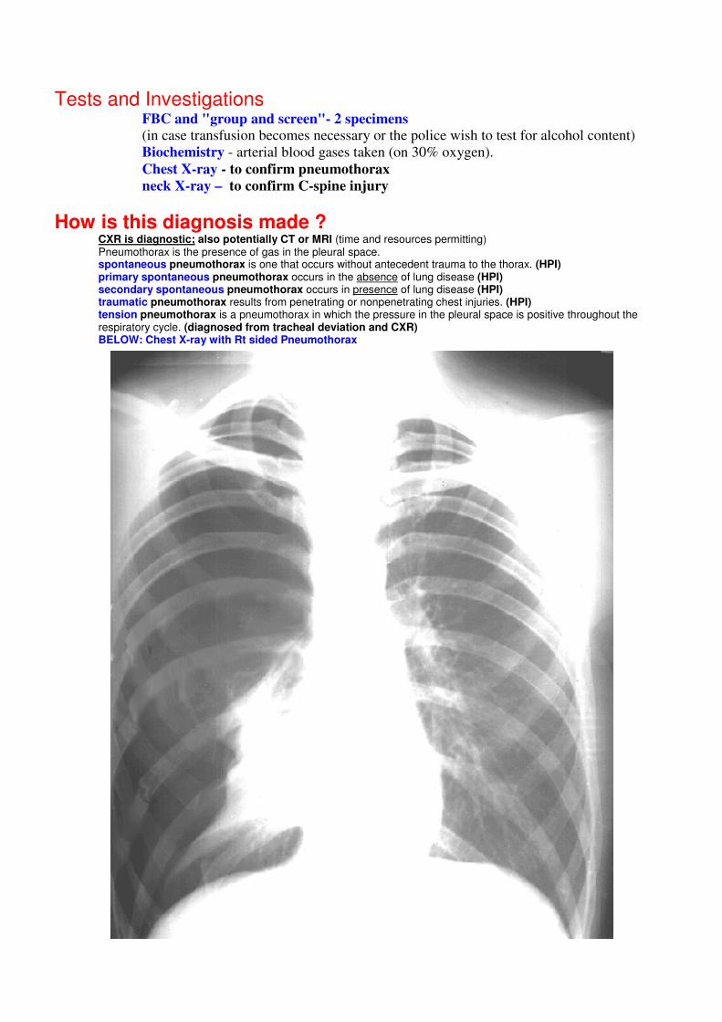
How is this diagnosis made ? CXR is diagnostic; also potentially CT or MRI (time and resources permitting) Pneumothorax is the presence of gas in the pleural space. spontaneous pneumothorax is one that occurs without antecedent trauma to the thorax. (HPI) primary spontaneous pneumothorax occurs in the absence of lung disease (HPI) secondary spontaneous pneumothorax occurs in presence of lung disease (HPI) traumatic pneumothorax results from penetrating or nonpenetrating chest injuries. (HPI) tension pneumothorax is a pneumothorax in which the pressure in the pleural space is positive throughout the respiratory cycle. (diagnosed from tracheal deviation and CXR) BELOW: Chest X-ray with Rt sided Pneumothorax
There is no one correct way to interpret the chest X-Ray. One suggestion of a systematic approach is: • Basic Identification (Name and Date particularly)
• Correct Orientation (?Side Marker)
• Adequate Technical Quality (particularly as regards factors which are going to modify your assessment of abnormalities. ?Supine, for example)
• Search for Abnormalities (both in an organised search and as a review of areas where you often fail to see the abnormalities - such as apices; hila; behind the heart; below the diaphragm)
• Abnormalities which are alterations of normal appearances
• Overall density (?R=L)
• Soft Tissues
• Bones
• Diaphragm
• Root of neck & Trachea
• Mediastinum & Heart
• Hila
• Fissures
• Vessels (peripheral vessels, as in left heart failure) • Abnormalities which are abnormal opacities
• Pleural
• Parenchymal
• Airspace
• Interstitial
• Nodules Some Rules of Thumb for CXR interpretation: The normal CXR is taken, on full inspiration, with the beam passing from the back to the front (PA frontal film, as opposed to AP frontal film) in a very short exposure (approximately 40 msec).
The convention for viewing plain X-Rays is as though you are facing the person who is turned towards you,
with the patient’s right to your left.
Common CXR signs: No border between heart and lung: means that the lung or a mass in the lung abutting the heart border is filled with fluid
(or pus as in pneumonia or blood as in pulmonary contusion or tumour cells as in a cancer) A.K.A. the “Silhouette sign”
A large tension pneumothorax is usually clinically obvious and the lack of lung markings and mediastinal shift away from the air collection makes it an obvious CXR diagnosis.
The usual pneumothorax, however, is smaller and more subtle: an area lacking lung markings with the lung bordered by a fine white line (the visceral pleural edge). This, the definitive sign of a pneumothorax, needs to be differentiated from two common false signs: - Skin folds, as in the sick patient who has lost weight and is having his film AP (with the
film cassette catching a roll of loose skin on his back), show a gradual increase in density to the skin fold edge when the density suddenly drops to the black of the air caught at the edge of the fold — a border not a line;
- Starch in the patient’s gown can look just like a pleural edge, but it usually extends off
the chest The usual pleural effusion is seen as a white area with a concave up “meniscus” (the fluid creeping up one of the walls of the
pleural cavity Pleural masses usually have a distinctive appearance with a sharp border on the inside and a fading-out border approaching
the chest wall. If a pleural mass is abutting the chest wall there tends to be an obtuse angle between the mass and the chest wall (like a cardinal’s hat a British WW1 soldier’s hat), whereas a parenchymal mass close to the chest wall forms an acute angle at the chest wall.
If there is volume loss or collapse the structures shift towards that side; if there is a mass or large collection of fluid the structures shift away from that side.
The commonest type of collapse is that of the left lower lobe. When this lobe collapses it collapses towards the mediastinum, behind the heart. This creates an area of extra whiteness behind the heart, adjacent to the left hemidiaphragm. Notice that in this situation the border (or silhouette) of the left hemidiaphragm is lost.
DEFINITIVE MANAGEMENT
Intravenous infusion started - 0.9% saline.
• Oxygen.
• Analgesia. (titratng 5mg morphine is OK apparently)
• Emergency needle thoracostomy (14 gauge intravenous cannula placed in second intercostal space in mid-clavicular line)
based on clinical diagnosis of tension pneumothorax.
• This followed by tube thoracostomy after X-ray confirms right pneumothorax.
• Fractured ribs evident on CXR - right 8, 9, 10.
• Wounds cleaned and dressed.
• Tetanus toxoid booster.
Progress
• Chest tube removed when pneumothorax resolution is confirmed by CXR
• Outpatient follow-up at 2 months reveals difficulty readjusting to work, often wakening through
the night in a cold sweat and spending the day as if in "cotton wool".
• Referred to counsellor for follow-up.
Disease Definition Blunt trauma to right chest causes rib fracture and damage to pleura. Visceral pleura is breached
(direct injury or shearing) with alveoli leaking air into the pleural space leading to equalisation of
pressure in lung pleural space decreasing blood oxygenation. Uncooperative behaviour due
combination of hypoxia and reaction to trauma +/- broken ribs (simple pneumothorax)
Epidemiology & Prevention …of Road Trauma:
- 1759 deaths per year in Australia; ~ 10/100,000 - Risk of crashing = 90% due to human factors - Risk of injury/death from crash = 70% due to vehicle factors
In association with SPEEDING: …speeding is associated with 30% of road fatalities
- Risk of Injury and Death rises EXPONENTIALLY with speed - - Factors associated with speeding:
- Young driver age - Single or 2+ occupants - Business - Behind schedule - Late model vehicle - Not owner of vehicle - Long weekly distances
- High frequency accident history In association with ALCOHOL:
- Alcohol alone - 35% - Alcohol and other drug - 11% - Other drug (single) - 5%
- Other drugs (multiple) - 2% RISK OF DEATH and INJURY ALSO RISES EXPONENTIALLY WITH DOSE
Most commonly young males at night
?? Marijuana seems to have NO EFFECT on risk of crash / injury (users compensate?) EPIDEMIOLOGY of Chest Trauma:
- 10% mortality; 25% of all trauma deaths
Prevention of Road Trauma: Traffic Law Enforcement - Education, Deterrence and Punishment - Driver-specific intervention:
- Random breath tests (limit = 0.05): - 19 - 24% reduction in fatal crashes (1982-1987) - 13 - 15% reduction in serious injury - attitudinal change: now drink driving viewed as illegal by 86% of people
- Speed cameras - Must be covert and intensive for good results; effective in reducing speeding
- Halo Effect (visible police) - Taxes the human resources but effective in reducing speed by 25% within 2km of cop.
( ~ 20 km if regularly deployed) - Vehicle-specific interventions:
- Assessed by Australasian NCAP (New Car Assessment Program) since 1992 - Seat belts - Air bag (roughly halves the chance of severe head trauma)
Medical Practitioner’s Responsibilities:
- ADVICE about prescription drugs that may affect driving - EXAMINATIONS for fitness to drive - REPORT to RTA review process
Aetiology of Pneumothorax: - TRAUMA results in broken ribs - Broken Ribs pierce the pleural lining of the lung, allowing air to escape from the alveoli into the pleural space - Air collects in the pleural space, occupying space and preventing the lung from inflating fully - The Air cannot escape because the pierced deflated alveoli create a “flap-valve” - Continuing accumulation of air causes the lung to collapse completely as air comes to occupy the hemithorax - Increasing Air Pressure within the Hemithorax pushes the other structures to the contralateral side: - Thus, the trachea deviates - Increasing Air Pressure within the Hemithorax
- compresses the heart (thus cardiac output decreases) - compresses the other lung, reducing its ability to inflate - compresses the great vessels (particularly the Vena Cava), thus reducing venous return
Prognosis - An uncomplicated, untreated pneumothorax will resolve slowly--approximately 1.25 percent per day. This rate of resorption can be increased by using supplemental oxygen, which increases the nitrogen gradient from the lung to the pleura. Basic Sciences and Clinical Procedures
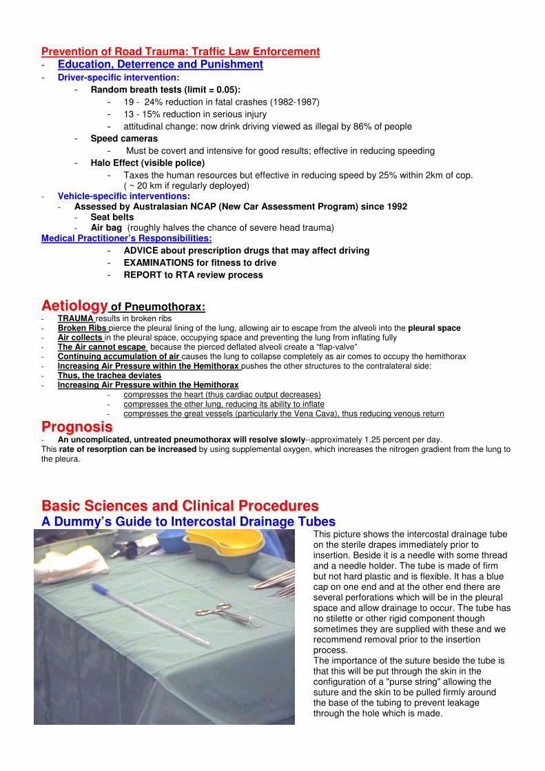
A Dummy’s Guide to Intercostal Drainage Tubes This picture shows the intercostal drainage tube on the sterile drapes immediately prior to insertion. Beside it is a needle with some thread and a needle holder. The tube is made of firm but not hard plastic and is flexible. It has a blue cap on one end and at the other end there are several perforations which will be in the pleural space and allow drainage to occur. The tube has no stilette or other rigid component though sometimes they are supplied with these and we recommend removal prior to the insertion process. The importance of the suture beside the tube is that this will be put through the skin in the configuration of a "purse string" allowing the suture and the skin to be pulled firmly around the base of the tubing to prevent leakage through the hole which is made.
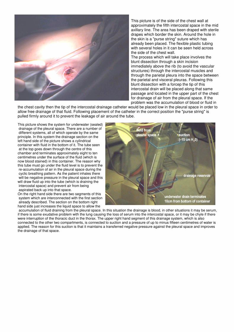
This picture is of the side of the chest wall at approximately the fifth intercostal space in the mid axillary line. The area has been draped with sterile drapes which border the skin. Around the hole in the skin is a "purse string" suture which has already been placed. The flexible plastic tubing with several holes in it can be seen held across the side of the chest wall. The process which will take place involves the blunt dissection through a skin incision immediately above the rib (to avoid the vascular structures) through the intercostal muscles and through the parietal pleura into the space between the parietal and visceral pleuras. Following this blunt dissection with a forcep the tip of this intercostal drain will be placed along that same passage and located in the upper part of the chest for drainage of air from the pleural space. If the problem was the accumulation of blood or fluid in
the chest cavity then the tip of the intercostal drainage catheter would be placed low in the pleural space in order to allow free drainage of that fluid. Following placement of the catheter in the correct position the "purse string" is pulled firmly around it to prevent the leakage of air around the tube. This picture shows the system for underwater (sealed) drainage of the pleural space. There are a number of different systems, all of which operate by the same principle. In this system the drainage section on the left hand side of the picture shows a cylindrical container with fluid in the bottom of it. The tube seen at the top goes down through the centre of this chamber and terminates approximately eight to ten centimetres under the surface of the fluid (which is now blood stained) in this container. The reason why this tube must go under the fluid level is to prevent the re-accumulation of air in the pleural space during the cyclic breathing pattern. As the patient inhales there will be negative pressure in the pleural space and this will draw fluid up into the tube (which is draining the intercostal space) and prevent air from being aspirated back up into that space. On the right hand side there are two segments of this system which are interconnected with the first section already described. The section on the bottom right hand side just increases the liquid space to allow the accumulation of fluid draining from the pleural space. In this situation the drainage is blood, in other situations it may be serum, if there is some exudative problem with the lung causing the loss of serum into the intercostal space, or it may be chyle if there were interruption of the thoracic duct in the thorax. The upper right hand segment of this drainage system, which is also connected to the other two compartments, is connected to suction and a pressure of up to minus fifteen centimetres of water is applied. The reason for this suction is that it maintains a transferred negative pressure against the pleural space and improves the drainage of that space.
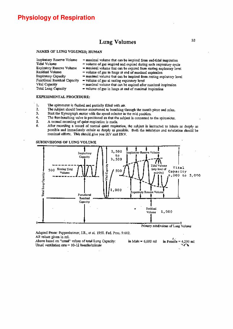
Physiology of Respiration
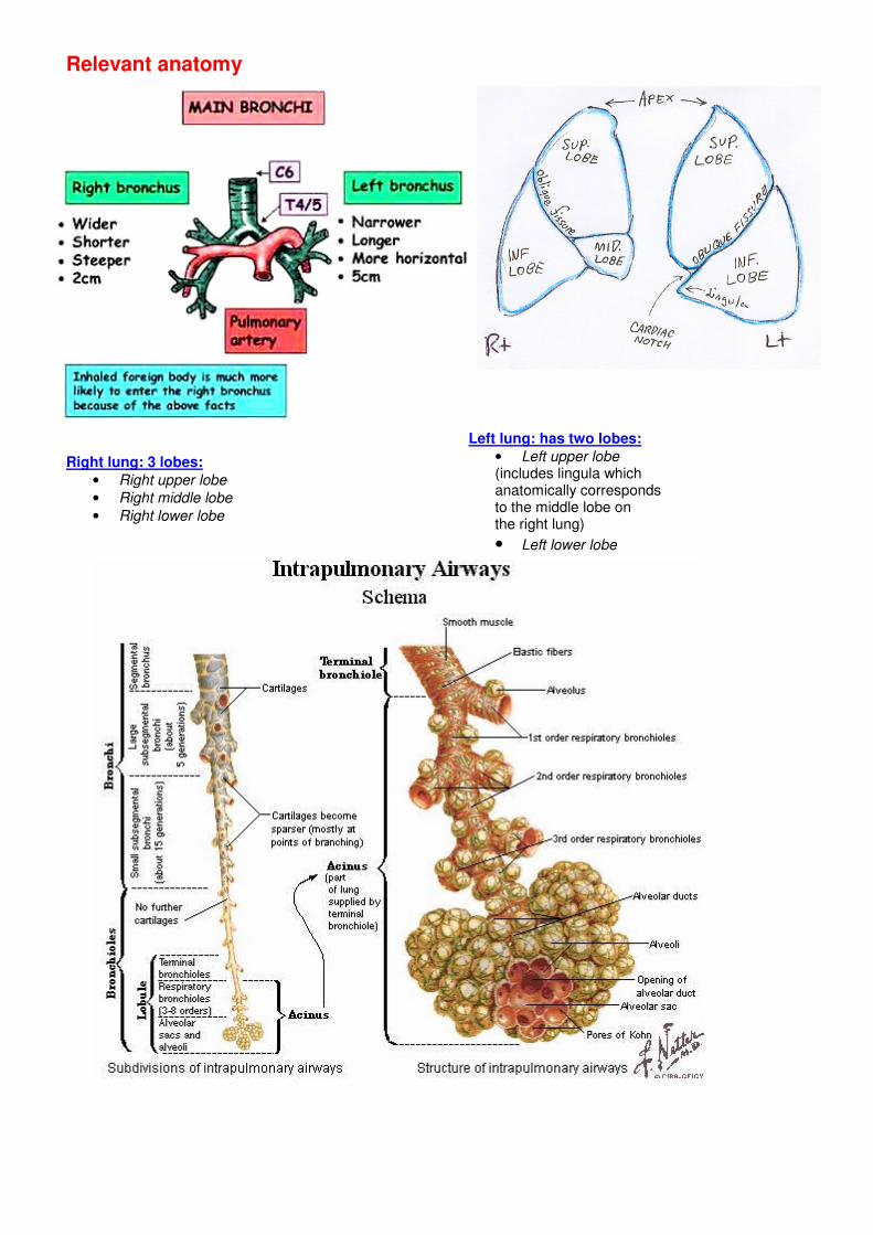
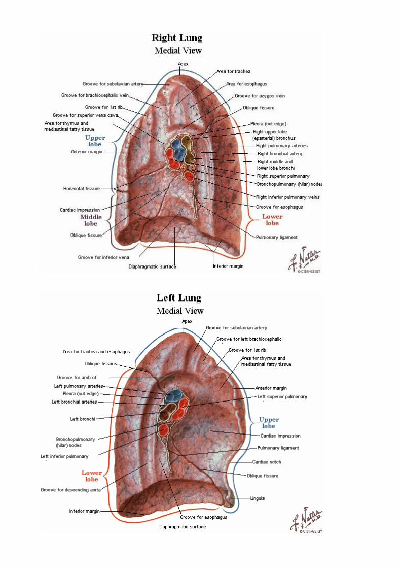
Relevant anatomy Right lung: 3 lobes:
• Right upper lobe
• Right middle lobe
• Right lower lobe
Left lung: has two lobes:
• Left upper lobe (includes lingula which anatomically corresponds to the middle lobe on the right lung)
• Left lower lobe
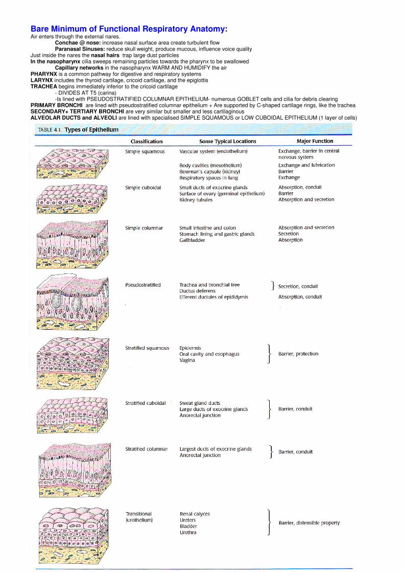
Bare Minimum of Functional Respiratory Anatomy: Air enters through the external nares.
Conchae @ nose: increase nasal surface area create turbulent flow Paranasal Sinuses: reduce skull weight, produce mucous, influence voice quality
Just inside the nares the nasal hairs trap large dust particles In the nasopharynx cilia sweeps remaining particles towards the pharynx to be swallowed Capillary networks in the nasopharynx WARM AND HUMIDIFY the air PHARYNX is a common pathway for digestive and respiratory systems LARYNX includes the thyroid cartilage, cricoid cartilage, and the epiglottis TRACHEA begins immediately inferior to the cricoid cartilage - DIVIDES AT T5 (carina) -Is lined with PSEUDOSTRATIFIED COLUMNAR EPITHELIUM- numerous GOBLET cells and cilia for debris clearing PRIMARY BRONCHI are lined with pseudostratified columnar epithelium + Are supported by C-shaped cartilage rings, like the trachea SECONDARY+ TERTIARY BRONCHI are very similar but smaller and less cartilaginous ALVEOLAR DUCTS and ALVEOLI are lined with specialised SIMPLE SQUAMOUS or LOW CUBOIDAL EPITHELIUM (1 layer of cells)
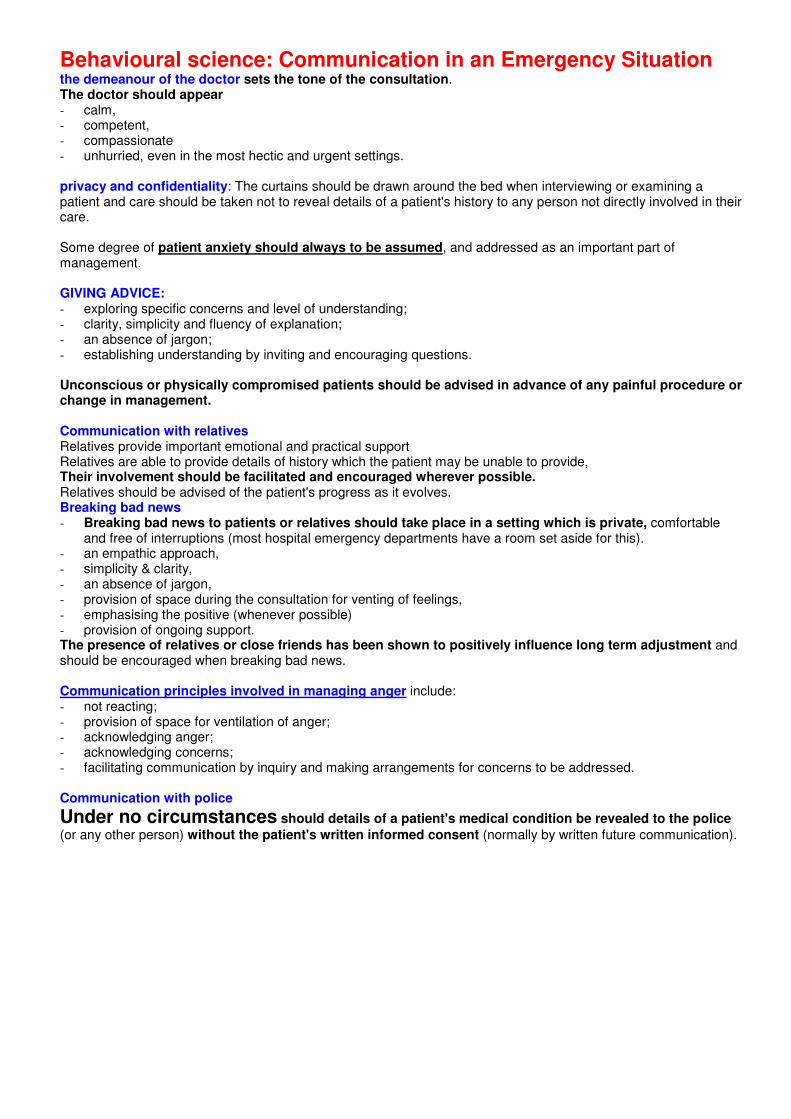
Behavioural science: Communication in an Emergency Situation the demeanour of the doctor sets the tone of the consultation. The doctor should appear - calm, - competent, - compassionate - unhurried, even in the most hectic and urgent settings. privacy and confidentiality: The curtains should be drawn around the bed when interviewing or examining a patient and care should be taken not to reveal details of a patient's history to any person not directly involved in their care. Some degree of patient anxiety should always to be assumed, and addressed as an important part of management. GIVING ADVICE: - exploring specific concerns and level of understanding; - clarity, simplicity and fluency of explanation; - an absence of jargon; - establishing understanding by inviting and encouraging questions. Unconscious or physically compromised patients should be advised in advance of any painful procedure or change in management. Communication with relatives Relatives provide important emotional and practical support Relatives are able to provide details of history which the patient may be unable to provide, Their involvement should be facilitated and encouraged wherever possible. Relatives should be advised of the patient's progress as it evolves. Breaking bad news - Breaking bad news to patients or relatives should take place in a setting which is private, comfortable
and free of interruptions (most hospital emergency departments have a room set aside for this). - an empathic approach, - simplicity & clarity, - an absence of jargon, - provision of space during the consultation for venting of feelings, - emphasising the positive (whenever possible) - provision of ongoing support. The presence of relatives or close friends has been shown to positively influence long term adjustment and should be encouraged when breaking bad news. Communication principles involved in managing anger include: - not reacting; - provision of space for ventilation of anger; - acknowledging anger; - acknowledging concerns; - facilitating communication by inquiry and making arrangements for concerns to be addressed. Communication with police
Under no circumstances should details of a patient's medical condition be revealed to the police
(or any other person) without the patient's written informed consent (normally by written future communication).
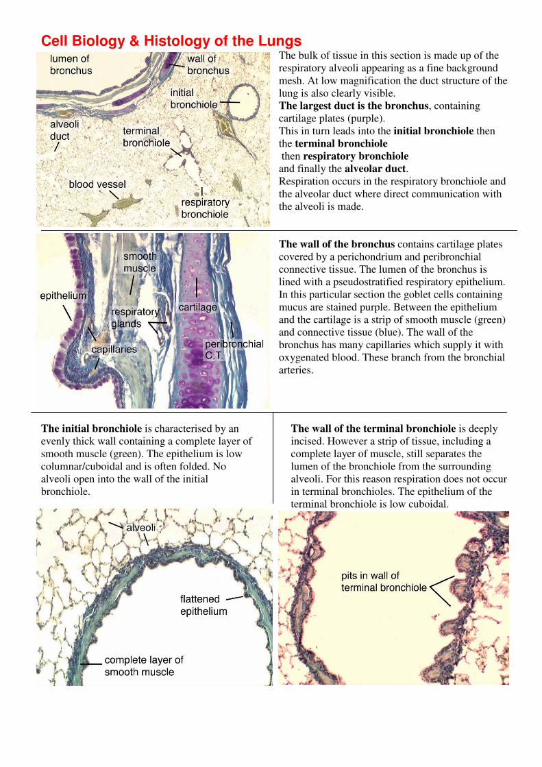
Cell Biology & Histology of the Lungs The bulk of tissue in this section is made up of the
respiratory alveoli appearing as a fine background
mesh. At low magnification the duct structure of the
lung is also clearly visible.
The largest duct is the bronchus, containing
cartilage plates (purple).
This in turn leads into the initial bronchiole then
the terminal bronchiole
then respiratory bronchiole
and finally the alveolar duct.
Respiration occurs in the respiratory bronchiole and
the alveolar duct where direct communication with
the alveoli is made.
The wall of the bronchus contains cartilage plates
covered by a perichondrium and peribronchial
connective tissue. The lumen of the bronchus is
lined with a pseudostratified respiratory epithelium.
In this particular section the goblet cells containing
mucus are stained purple. Between the epithelium
and the cartilage is a strip of smooth muscle (green)
and connective tissue (blue). The wall of the
bronchus has many capillaries which supply it with
oxygenated blood. These branch from the bronchial
arteries.
The initial bronchiole is characterised by an
evenly thick wall containing a complete layer of
smooth muscle (green). The epithelium is low
columnar/cuboidal and is often folded. No
alveoli open into the wall of the initial
bronchiole.
The wall of the terminal bronchiole is deeply
incised. However a strip of tissue, including a
complete layer of muscle, still separates the
lumen of the bronchiole from the surrounding
alveoli. For this reason respiration does not occur
in terminal bronchioles. The epithelium of the
terminal bronchiole is low cuboidal.
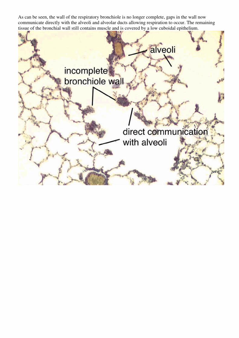
As can be seen, the wall of the respiratory bronchiole is no longer complete, gaps in the wall now
communicate directly with the alveoli and alveolar ducts allowing respiration to occur. The remaining
tissue of the bronchial wall still contains muscle and is covered by a low cuboidal epithelium.