trends developments in bioprocess technolog … vs nbcs.pdf · trends & developments in...
TRANSCRIPT

TRENDS & DEVELOPMENTS IN BIOPROCESS TECHNOLOGY
OPEN ACCESS ARTICLE www.bioprocessingjournal.com1
Abstract
The continued use of animal serum as an important component in biotechnol-ogy manufacturing processes has raised questions regarding both the reliability of
geographic origin and possible adulteration of prod-uct. The International Serum Industry Association (ISIA) has implemented a traceability certification program designed to demonstrate traceability from slaughterhouse or abattoir to the end-user. This is based on an audit performed by an indepen-dent, approved third-party auditor according to an approved audit plan, using a detailed audit check-list. Recent advances have led to the development of a complementary testing program to determine geographic origin of material. The methodology described in this paper differentiates fetal bovine serum from newborn calf serum on the basis of biochemical composition.
A Method for Differentiating Fetal Bovine Serum from Newborn Calf Serum
By Michelle Cheever, Alyssa Master, and Rosemary Versteegendisease, both human and veterinary. One of the most varied factors in culture systems is the growth medium, which provides the nutrients and/or the growth factors required for cell growth and product yield. Growth media are most often supplemented at concentrations of 5–20 % serum by volume.
The choice of which serum is most appropriate for use in any application can be a difficult one, and the selection of the serum supplier can be important to ensure quality product is available. FBS provides the proper cell growth environment for many applications, but is not yet fully characterized. NBCS presents a less expensive alternative to FBS but is also appropriate for use in some applications. This difference in value, which is based on availability, could provide an incentive for misrepresentation. The ability to differentiate between these serum types would therefore be important, and would, among other aspects, help to ensure reproducibility of results. This capability has not been available in the past. ISIA has promulgated an indus-try-approved list of serum definitions.ISIA Definitions of FBS and NBCS:
• Fetal Bovine Serum is obtained from the blood of fetuses of healthy, pre-partum bovine dams that have been deemed fit for human consumption through ante- and/or post-mortem veterinary inspection. It is collected in abattoirs inspected by the competent authority in the country of origin. There are no dele-tions or additions (including preservatives) allowed.
• Newborn Calf Serum is the liquid fraction of clotted blood derived from healthy, slaughtered bovine calves aged less than 20 days, deemed fit for human consump-tion through ante- and/or post-mortem inspection. It is collected in abattoirs inspected by the competent authority of the country of origin. There are no dele-tions or additions (including preservatives) allowed.
One of the goals of this study was to determine a biochemical fingerprint that could be used to individually identify these two types of bovine serum. A major differ-ence between FBS and NBCS is the levels of circulating antibodies. This is due to the fact that colostrum, the first feed from mother to calf, is rich in antibodies which are essential for the establishment of the immune system of the calf. It has long been known that higher levels of immuno-globulin (IgG) are found in NBCS.[1] It is also known in other species that the birthing process can result in increased levels of other marker molecules.[2, 3, 4]
One of the simplest methods for assessing animal health
Serum and its Role in Cell Culture Despite significant efforts to avoid the use of animal-
source products, serum and other animal-derived products continue to play critical roles as culture medium supple-ments in the biomedical and biopharmaceutical arenas. Animal blood is a by-product of the meat industry and, as such, is sourced from animals fit for human consump-tion. Bovine-sourced products are the most commonly used. Serum, plasma, and other animal-derived materials from a large variety of animal sources are also used by the biopharmaceutical industry in daily applications including:
• Biomedical research• Cell culture-based safety testing of pharmaceuticals
and cosmetics• Cell culture-based production of human and veteri-
nary vaccines as well as therapeutic proteins• Cell therapies
Serum UseSerum has been used extensively as a growth medium
supplement in cell culture for many years, and its use for this purpose has greatly contributed to the battle against
2OPEN ACCESS ARTICLE www.bioprocessingjournal.com
status is to run a general veterinary panel of tests on a serum sample. This panel will give an indication of the general health and function of various organs. A list of the
tests usually performed is shown in Table 1. Such a panel is of particular interest because it provides useful markers for distinguishing between FBS and NBCS.
A Method for Differentiating Fetal Bovine Serum from Newborn Calf Serum
Methods
Biochemical Profile of FBS and NBCS
Each unique FBS or NBCS sample was the result of the standard, multi- animal pool manufacturing process. Where indicated, samples were either filtered, finished p r o d u c t o r u n f i l te r e d , un pro cessed raw serum, as processing did not appear to affect the results obtained. Statistical analysis was per-formed using GraphPad I nSt a t Ve r s i o n 3 .1 f o r Microsoft Windows. A P-value less than 0.05 was considered statistically significant.
Thirty-nine FBS samples and 32 NBCS sample were obtained from f ive ISIA-certified traceable suppliers. Of these, 55 samples were submitted to a third party to be aliquoted, blinded, and directly submitted to the testing facility while
TABLE 1. Veterinary panel tests and focus.
Group Measurements Function
Electrolyte, Fluid, pH Balance
• Sodium • BiCarb (HCO3)• Potassium
Indicators of dehydration, electrolyte, and acid/base balance. They are the main intracellular ions, and are responsible for the maintenance of osmotic pressure.
Vitamins and Minerals
• Phosphorus• Iron
Important for synthesis of hemoglobin and activation of numerous enzymes and hormones.
Renal and Pancreas Function, Injury
• Creatinine• Glucose
Abnormal values indicate skeletal muscle damage. Includes assessment of kidney function.
Lipid • Cholesterol• Triglycerides
Critical role in the function of the cell membrane. Lipids are precursors to hormones and aid in the absorption of nutrients from food.
Liver
• Aspartate Aminotransferase (AST)• Alkaline Phosphatase (ALP)• Gamma-Glutamyl Transferase (GGT)• Bilirubin, Total
Enzymes and protein that indicate soft tissue, liver, and bile duct damage.
Proteins
• Protein, Total• Albumin• Globulin• Immunoglobulin G
Used to identify serum protein disorders. Albumin provides 80 % of osmotic activity in plasma and serves as a transport protein in metabolic processes. Changes in alpha globulin indicate acute phase of inflammation. Beta fractions of globulin are part of complement, transferrin, c-reactive protein, and some immunoglobulin. IgG is a type of antibody that is produced by the immune system.[5]
• Anion Gap• Chloride
• Calcium • Magnesium
• Creatine Kinase (CK)
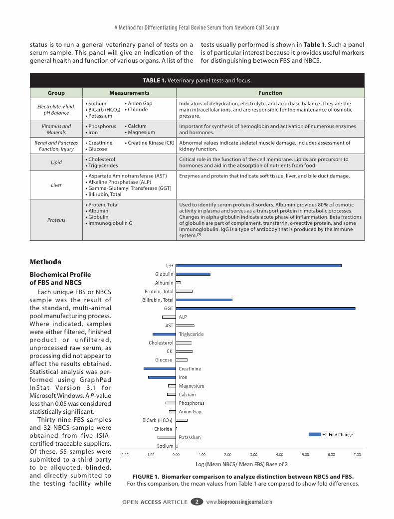
FIGURE 1. Biomarker comparison to analyze distinction between NBCS and FBS. For this comparison, the mean values from Table 1 are compared to show fold differences.

3OPEN ACCESS ARTICLEwww.bioprocessingjournal.com www.bioprocessingjournal.com
A Method for Differentiating Fetal Bovine Serum from Newborn Calf Serum
TABLE 2. Comparison of veterinary panel results for FBS and NBCS. “N” indicates the number of samples tested.
Function Analyte Serum N Mean SD P-Value
Electrolyte, Fluid,
pH Balance
Sodium mEQ/LFBS 39 132.03 14.68
0.1353NBCS 32 139.19 24.25
Potassium mEQ/LFBS 39 11.84 1.81
< 0.0001NBCS 32 7.26 2.50
Chloride mEQ/LFBS 39 93.37 11.75
0.3010NBCS 32 95.88 18.97
BiCarb (HCO3) mEQ/LFBS 39 16.84 6.21
0.0004NBCS 32 22.70 6.25
Anion Gap mmol/LFBS 39 33.72 6.26
0.0164NBCS 32 28.03 10.53
Vitamins and Minerals
Phosphorus mg/dLFBS 39 10.19 2.97
< 0.0001NBCS 32 7.74 2.06
Calcium mg/dLFBS 39 13.22 1.76
< 0.0001NBCS 32 10.45 3.12
Magnesium mg/dLFBS 39 3.09 0.44
0.0006NBCS 32 2.51 0.96
Iron µg/dLFBS 39 179.77 33.59
< 0.0001NBCS 32 85.91 14.32
Renal and Pancreas Function,
Injury
Creatinine mg/dLFBS 39 2.49 0.46
< 0.0001NBCS 32 1.05 0.34
Glucose mg/dLFBS 39 102.77 29.35
0.9080NBCS 32 139.43 111.56
CK IU/LFBS 39 178.74 75.32
0.0006NBCS 32 277.34 138.47
Lipid
Cholesterol mg/dLFBS 39 28.33 7.07
< 0.0001NBCS 32 42.91 7.66
Triglyceride mg/dLFBS 39 66.85 11.35
< 0.0001NBCS 32 36.06 16.17
Liver
AST IU/LFBS 39 32.10 14.21
< 0.0001NBCS 32 52.06 23.31
ALP IU/LFBS 31 213.65 65.17
< 0.0001NBCS 24 148.04 34.35
GGT IU/LFBS 39 3.64 1.14
< 0.0001NBCS 32 441.56 188.57
Bilirubin, Total mg/dLFBS 39 0.14 0.06
< 0.0001NBCS 32 0.62 0.28
Proteins
Protein, Total g/dLFBS 39 3.52 0.45
< 0.0001NBCS 32 5.42 0.80
Albumin g/dLFBS 39 2.47 0.33
< 0.0001NBCS 32 2.78 0.44
Globulin g/dLFBS 39 1.05 0.16
< 0.0001NBCS 32 2.63 0.44
IgG µg/mLFBS 39 183.86 135.65
< 0.0001NBCS 32 15425.81 8854.20
16 samples were directly submitted (with-out blinding) for testing. The geographic origin of source material included United States (US), Mexico, Australia, New Zealand, and Costa Rica. Unless otherwise indicated, serum biochemical determination was performed by Colorado State University (CSU) Diagnostic Laboratory (Fort Collins, Colorado USA) using the Roche cobas c 501 Analyzer. Serum IgG determination by enzyme-linked immunosorbent assay (ELISA) was performed by ZeptoMetrix Corporation (Buffalo, New York USA). The panel of 22 analytes included electrolytes, pH, vitamins, minerals, indicators of renal and pancreatic function, lipids, liver func-tion, and proteins, as outlined in Table 1. Mean, standard deviation (SD), and P-value (as calculated by the Mann-Whitney test) for each analyte are summarized in Table 2.
In order to identify which of the bio -chemical properties of NBCS were distinct from FBS, a comparison of the difference in the mean value of the results was performed. To determine the increase or decrease in level of analytes, the mean value for NBCS was divided by the mean value for FBS for each analyte, and trans-formed data (logarithm to base 2) were plotted as shown in Figure 1 (previous page).
Analytes exhibiting a fold change ≥ 2 are indicated in blue. Differences to the right of zero and ≥ 2 indicate that the NBCS value was greater than that for FBS (i.e., GGT, IgG, total bilirubin, and globulin). Changes to the left of zero and ≤ 2 indi-cate that the NBCS result was less than that for FBS (i.e., triglyceride, creatinine, and iron). The fold difference was statistically significant for GGT at 121.3 and IgG at 83.9. The maximum GGT value of the sample population for FBS was 5 IU/L. In contrast, the minimum GGT value of the sample population for NBCS was 157 IU/L. GGT was the analyte with the highest differ-ence seen and became the focus of further experiments.
High GGT and IgG were found to have statistically significant associations with NBCS. A GGT of > 100 was associated with NBCS while that for FBS was < 10. FBS is characterized as having a significantly lower IgG when compared to NBCS but showed a wider range of values when compared to GGT ranges. Although a

4OPEN ACCESS ARTICLE www.bioprocessingjournal.com
A Method for Differentiating Fetal Bovine Serum from Newborn Calf Serum
specification level for IgG has not yet been set by ISIA, it is more commonly reported on manufacturer’s FBS certif icates of analysis (COA). This analysis shows that in instances where IgG is > 320 μg/mL (+ 1 SD from the mean), then an evaluation of GGT would be a determining factor for charac-terizing serum.
VariationFive FBS and three NBCS samples of US
origin were submitted to nine separate facilities for GGT analysis. Samples were transported on dry ice and stored frozen until the initial analysis was performed. Samples were then refrigerated until a re-evaluation could be performed 24 hours later.
In Figure 2, N was adjusted for instances where analysis was not performed 24 hours after initial evaluation. Analytical facilities included: CSU Diagnostic Lab, Cornell University Animal Health Diagnostic Center (Ithaca, New York USA), University of Illinois at Urbana-Champaign College of Veterinary Medicine (Urbana, Illinois USA), University of Missouri College of Veterinary Medicine (Columbia, Missouri USA), Oregon State University Diagnostic Laboratory (Corvallis, Oregon USA), Rocky Mountain Biologicals (Missoula, Montana USA), University of California Davis (UC Davis) Veterinary Medical Teaching Hospital (Davis, California USA), Utah State University Veterinary Diagnostic Laborator y (Logan, Utah USA), and Virginia Tech Animal Laboratory Services (Blacksburg, Virginia USA). Chemical anal-ysis was performed using six analyzers: Beckman AU400, AU480, and AU680; Roche Modular P and cobas c 501; and Siemens Dimension Xpand.
GGT results obtained by the different analyzers are summarized in Figure 2A for FBS and Figure 2B for NBCS. A statisti-cally significant difference was observed for FBS using the Siemens Dimension Xpand when compared to the Beckman and Roche analyzers. Maximum FBS GGT for the Beckman and Roche analyzers was 7 IU/L. No statistical significance with NBCS was observed for all the analyzers. Analysis was performed using Kruskal-Wallis test (nonparametric one-way analysis of vari-ance [ANOVA]). There was a statistically higher amount of FBS GGT, as determined
FIGURE 2. Comparison of GGT results from six different analyzers for: (A) FBS; and (B) NBCS.
B
A

5OPEN ACCESS ARTICLEwww.bioprocessingjournal.com www.bioprocessingjournal.com
A Method for Differentiating Fetal Bovine Serum from Newborn Calf Serum
by the Xpand analyzer. Because this observable significant difference could not be attributed to the method or time, results from the Xpand were excluded from future experiments.
Variation in ResultsFBS GGT data obtained using the Roche
cobas c 501 analyzer are summarized in Table 3. The data set includes five samples analyzed at two locations, CSU and UC Davis, over a period of 24 hours. There was no significant difference in the mean results for GGT tested at these diagnostic facilities (P > 0.05) when analyzed using nonparametric ANOVA and Dunn’s multiple comparison. No significant difference was observed in GGT (P > 0.999, unpaired t -test) when samples were re-analyzed within a 24-hour period. There was no significant variation in GGT results based on testing facility, time of analysis, or the Beckman and Roche analyzers.
The Siemens Dimension Xpand analyzer gave statistically significant differences in measurements of GGT in FBS, but not for NBCS. Variations between Beckman and Roche analyzers were not statistically significant for FBS or NBCS. GGT values were reproducible regardless of the testing facility and not susceptible to variation, suggesting that this might be the most useful marker for differentiating NBCS from FBS. It should be stressed that if FBS GGT is analyzed using the Siemens Dimension Xpand, values over 10 IU/L will be reported.
Detecting NBCS in FBSThree NBCS samples were diluted into
FBS from 5–20 % (in increments of 5 %) by volume. Diluted samples were submitted to the CSU diagnostic facility for GGT determination by the cobas c 501 analyzer.
Figure 3A shows the results of the mixing experiments. The average GGT level detected was plotted against the percentage of NBCS dilution. This suggested a strong linear relationship between the percent dilution of NBCS and the corresponding GGT measurement (R2 = 0.9994). In order to further analyze statistical variance, the sum of the squares of the deviations from the mean (SDM) was calculated to be 9.113, indicating little variance. The predicted GGT value of the various levels of mixtures was determined
B
A
FIGURE 3. Determination of GGT levels in FBS containing up to 20 % NCBS: (A) Observable GGT; and (B) Predicted GGT.
TABLE 3. Summary of GGT results obtained by two facilities over 24 hours using the Roche cobas c 501 analyzer.
c 501 Analyzer
Initial 24 hrMean
CSU UC Davis CSU UC Davis
Sample 1 4.00 4.00 5.00 4.00 4.25
Sample 2 5.00 5.00 5.00 5.00 5.00
Sample 3 5.00 5.00 5.00 5.00 5.00
Sample 4 5.00 4.00 4.00 4.00 4.25
Sample 5 4.00 5.00 5.00 4.00 4.50
CSU vs. UC Davis P > 0.05 P > 0.05 P > 0.05 P > 0.05 Dunn’s*
Initial vs. 24 hr P > 0.999 P > 0.999 t-test**
*Non-parametric ANOVA with Dunn’s post-test. **Unpaired t -test.

6OPEN ACCESS ARTICLE www.bioprocessingjournal.com
A Method for Differentiating Fetal Bovine Serum from Newborn Calf Serum
by adding the mean GGT value of 100 % NBCS (814.33 IU/L) and FBS (5 IU/L) and multiplying by the dilution factors. Analysis confirmed that there was excellent agreement between the predicted and the observed GGT values. The maximum observed value of all the FBS samples analyzed was 7 IU/L, and was calculated excluding data generated by the Siemens Dimension Xpand. In these experiments, 7 IU/L would equate to a NBCS concentration of 0.4092 %.
To determine if the percentage of NBCS in a mixture could be calculated from reported GGT values, GGT results at various percent-ages were calculated based on the average sample results for NBCS (441.56 IU/L) and FBS (3.64 IU/L) (Table 2). The slope was 4.342, the Y intercept was 2.145, and there was an R2 value of 0.9993. The percent of NBCS diluted in FBS can be calculated using reported values of GGT, and the slope and Y intercept from the average sample results. For example, a GGT result of 50 IU/L would intercept this standard curve at 11.02 %. A reasonable assumption would therefore seem to be that a GGT result of 50 IU/L could contain 8–20 % NBCS (+/– 1 SD). In Figure 3B (previous page), the maximum observed value for GGT of 7 IU/L is shown for reference and intercepts at 1.15 %.
In this study, we categorized FBS as having GGT values
of < 10 IU/L. Reported GGT values over 10 IU/L in FBS would strongly suggest that the material is not entirely derived from fetal origin.
ConclusionsFor many years, low levels of IgG were thought to be
a suitable marker for FBS.[3, 6] This has been questionable, based on the wide range of levels observed, and
also the potential for removal of IgG from serum using protein A chromatography. In fact,
commercially available ultra-low IgG FBS can be obtained from several suppliers.
Earlier work[3, 4] indicated the presence of GGT in bovine serum. GGT was a potential indicator for passive transfer of immunity
and might be an indicator of neonatal vs. fetal origin. The work discussed here strongly indi-
cates that GGT can be used to distinguish NBCS from FBS. The presence of NBCS in FBS may be detected using this method, based on our analysis showing higher than expected GGT levels. Therefore, the method may be useful for detecting the adulteration of FBS by dilution in NBCS. ISIA will be working in the very near future to recommend specifications for IgG and GGT levels for FBS based on the results of this analytical study.
Michelle Cheever* is Director of Quality Assurance at Atlas Biologicals, 2649 Mulberry St., Suite A-3, Fort Collins, Colorado 80524 USA.
Alyssa Master, PhD, is Senior Manager, Science and Applications, Nucleus Biologics, San Diego, California USA.
Rosemary J. Versteegen, PhD, is Chief Executive Officer, International Serum Industry Association (ISIA), McHenry, Maryland USA.
*Corresponding Author: Email: [email protected], Website: www.atlasbio.com, Phone: 970-494-2051
About the Authors
Acknowledgement
The authors would like to acknowledge Linda Vap, DVM, of Colorado State Universit y.
References
[1] Weaver DM, Ty ler JW, VanMe t re D C, Hos te t ler DE, Barrington GM. Passive transfer of colostral immunoglob-ulins in calves. J Vet Intern Med, 2000; 14(6): 569–77. PMid: 11110376[2] Carski TR, Smith JL, Rehak MJ. Chemical characterization of pooled animal sera. Appl Microbiol, 1967; 15(6): 1502–4. PMid: 16349778; PMCid: PMC547253[3] Festen R. Understanding animal sera: considerations for use in the production of biological therapeutics. In: Medicines from animal cell culture. Stacey GN & Davis J eds; John Wiley & Sons, Inc; 2007; pp 45–58. ISBN: 978- 0 -470 -85094-7[4] K l i n ko n M, J e že k J. Va lu e s o f b l o o d v a r ia b l e s i n
c a l v e s . I n: A b i rd ’s- e y e v i e w o f ve te r i n a r y m e d i c i n e . Perez-Marin CC ed; InTech; 2012; pp 301–21. ISBN: 978-953-51- 0 031-7. Av a i l a b l e f r o m: h t t p s : // w w w. i n t e c h o p e n .com/ b o ok s/a - bir d -s- eye -v iew- of-ve ter inar y- me dic ine/values-of-blood-variables-in-calves[5] Mazzaferro EM, Rudlof f E, Kirby R. The role of albumin replacement in the cr it ically i l l veter inar y patient. J Vet Em e rg Cr i t Ca re, 20 02; 12(2): 113–24. h t t p s : //d x .d o i .org/10.1046/j.1435- 6935.2002.00025.x [6] Ellis WA, Logan EF, O’Brien JJ. Serum immunoglobulins in aborted and non-aborted bovine foetuses. Clin Exp Immunol, 1978; 33(1): 136–41. PMid: 101325; PMCid: PMC1537509