trf2-tethered tin2 can mediate telomere protection...
TRANSCRIPT
Published Ahead of Print 27 January 2014. 10.1128/MCB.01052-13.
2014, 34(7):1349. DOI:Mol. Cell. Biol. David Frescas and Titia de Lange Protection by TPP1/POT1TRF2-Tethered TIN2 Can Mediate Telomere
http://mcb.asm.org/content/34/7/1349Updated information and services can be found at:
These include:REFERENCES
http://mcb.asm.org/content/34/7/1349#ref-list-1at: This article cites 29 articles, 12 of which can be accessed free
CONTENT ALERTS more»articles cite this article),
Receive: RSS Feeds, eTOCs, free email alerts (when new
http://journals.asm.org/site/misc/reprints.xhtmlInformation about commercial reprint orders: http://journals.asm.org/site/subscriptions/To subscribe to to another ASM Journal go to:
on March 14, 2014 by Rockefeller University
http://mcb.asm
.org/Downloaded from
on M
arch 14, 2014 by Rockefeller Universityhttp://m
cb.asm.org/
Downloaded from
TRF2-Tethered TIN2 Can Mediate Telomere Protection byTPP1/POT1
David Frescas, Titia de LangeLaboratory for Cell Biology and Genetics, The Rockefeller University, New York, New York, USA
The shelterin protein TIN2 is required for the telomeric accumulation of TPP1/POT1 heterodimers and for the protection oftelomeres by the POT1 proteins (POT1a and POT1b in the mouse). TIN2 also binds to TRF1 and TRF2, improving the telomericlocalization of TRF2 and its function. Here, we ask whether TIN2 needs to interact with both TRF1 and TRF2 to mediate the telo-mere protection afforded by TRF2 and POT1a/b. Using a TIN2 allele deficient in TRF1 binding (TIN2-L247E), we demonstratethat TRF1 is required for optimal recruitment of TIN2 to telomeres and document phenotypes associated with the TIN2-L247Eallele that are explained by insufficient TIN2 loading onto telomeres. To bypass the requirement for TRF1-dependent recruit-ment, we fused TIN2-L247E to the TRF2-interacting (RCT) domain of Rap1. The RCT-TIN2-L247E fusion showed improved te-lomeric localization and was fully functional in terms of chromosome end protection by TRF2, TPP1/POT1a, and TPP1/POT1b.These data indicate that when sufficient TIN2 is loaded onto telomeres, its interaction with TRF1 is not required to mediate thefunction of TRF2 and the TPP1/POT1 heterodimers. We therefore conclude that shelterin can protect chromosome ends as aTRF2-tethered TIN2/TPP1/POT1 complex that lacks a physical connection to TRF1.
Shelterin represses DNA damage signaling by the ATM andATR kinases and prevents double-stranded break (DSB) re-
pair at chromosome ends (reviewed in reference 1). Conditionaldeletion of individual shelterin proteins has revealed considerablefunctional compartmentalization within shelterin. TRF2 is re-quired for the repression of ATM-dependent signaling and classi-cal nonhomologous end joining (c-NHEJ), whereas the closelyrelated TRF1 protein prevents replication fork stalling in telo-meric DNA and represses a fragile site phenotype at telomeres.POT1a prevents ATR kinase signaling at telomeres by blocking thebinding of RPA to the single-stranded TTAGGG repeats. POT1b,on the other hand, is required for the correct formation of the 3=telomeric overhang.
TIN2, the central component of shelterin, is critical for thefunction of POT1a and POT1b because it links TPP1/POT1 het-erodimers to TRF1 and TRF2 (2–6). Consistent with this role,TIN2 deletion phenocopies the loss of TPP1 and/or POT1a/b,including the activation of ATR signaling, the deregulation of theamount of single-stranded telomeric DNA, and a low frequency of(postreplicative) sister telomere fusions (7–10). In addition, dele-tion of TIN2 elicits ATM kinase signaling and a moderate level ofchromosome-type telomere fusions that are hallmarks of TRF2loss (7, 10–12). The chromosome-type fusions associated withTIN2 deletion are due to decreased telomeric accumulation ofTRF2, as overexpression of TRF2 in TIN2-null cells alleviates thisphenotype (10). However, overexpression of TRF2 does not fullyrepress ATM kinase signaling in TIN2-knockout (KO) cells, and aTRF2 mutant that lacks the TIN2 binding domain is partially de-fective in preventing ATM activation, suggesting that TIN2 di-rectly contributes to this TRF2 function. There is no indication fora role of TIN2 in the function of TRF1 at telomeres since TIN2-deleted cells do not display the fragile telomere phenotype typicalof TRF1 loss (10, 13, 14).
Here, we ask whether TIN2 requires its interaction with bothTRF1 and TRF2 in order to function as a tether for the TPP1/POT1 heterodimers. Deletion of TRF2 does not give rise to any ofthe phenotypes associated with POT1a/b deficiency (7, 11). Sim-
ilarly, deletion of TRF1 does not appear to elicit the phenotypestypical of the loss of POT1a/b from telomeres (14). However, de-letion of either TRF1 or TRF2 induces a prominent telomericDNA damage response, which could mask (some of) the pheno-types of POT1a/b loss. Thus, to further elucidate the role of theTIN2-TRF1 and TIN2-TRF2 interactions in the protective func-tion of shelterin, we generated a TIN2 allele defective in TRF1association. We show that TRF1 is principally responsible for therecruitment of TIN2 to telomeres and describe a distinct set oftelomere dysfunction phenotypes that arise from the severing ofthis interaction. By artificially tethering TIN2 to TRF2 via thefusion of the Rap1 TRF2-interacting domain (RCT) to TIN2, weprovide direct evidence that TRF2-bound TIN2 is in principlesufficient to fulfill the TPP1/POT1 tethering function required toprotect chromosome ends.
MATERIALS AND METHODSCell lines and expression plasmids. SV40-LT-immortalized TIN2F/F,TIN2F/F ATM!/!, TIN2F/F ATRF/F (10), TRF2F/F, and TRF1F/F mouseembryonic fibroblasts (MEFs) (14) were grown in Dulbecco modifiedEagle medium (DMEM) supplemented with L-glutamine, penicillin-streptomycin, nonessential amino acids, and 10% fetal bovine serum (In-vitrogen). HT1080 and 293T cells were maintained in DMEM supple-mented with L-glutamine, penicillin-streptomycin, nonessential aminoacids, and 10% bovine calf serum (BCS) (HyClone). TIN2F/F, TIN2F/F
ATM!/!, and TIN2F/F ATRF/F MEFs were infected with pWZL-hygromy-cin retroviruses expressing FLAG-hemagglutinin (HA)-HA (FLAG-HA2)-tagged mouse TIN2 alleles or an empty vector and selected withhygromycin. In some experiments, the hygromycin-selected MEFs were
Received 13 August 2013 Returned for modification 9 September 2013Accepted 21 January 2014
Published ahead of print 27 January 2014
Address correspondence to Titia de Lange, [email protected].
Copyright © 2014, American Society for Microbiology. All Rights Reserved.
doi:10.1128/MCB.01052-13
April 2014 Volume 34 Number 7 Molecular and Cellular Biology p. 1349 –1362 mcb.asm.org 1349
on March 14, 2014 by Rockefeller University
http://mcb.asm
.org/Downloaded from
subsequently infected with pLPC-puromycin retroviruses expressingMYC-tagged TRF2, TPP1, or POT1b. In all cases, following three roundsof retroviral infection, MEFs were selected with hygromycin or puromy-cin for 2 to 4 days. The human HT1080 fibrosarcoma cells were firstinfected with pWZL-hygromycin retroviruses expressing short hairpinRNA (shRNA)-insensitive FH2-TIN2 alleles or an empty vector. TIN2iconstructs possess silent mutations to generate shRNA resistance (AGACAATATGGTGTGGACAT to AGGCAGTACGGCGTGGACAT; under-lining identifies which bases were changed to introduce silent mutationsto confer shRNA resistance). Following selection with hygromycin,HT1080 cells were infected with a previously characterized shRNA (pu-romycin-resistant lentiviral vector) against human TIN2 and selectedwith puromycin (10). For immunoprecipitation studies, 293T cells weretransiently cotransfected with MYC-tagged TRF1 or TRF2 plasmids(pLPC-based) and FLAG-HA2-tagged mouse TIN2 alleles (pLPC-based)using the calcium phosphate coprecipitation method. All introduced mu-tations and deletions in TIN2 were made by using a QuikChange II site-directed mutagenesis kit (Agilent Technologies) and confirmed by DNAsequencing. Deletion of floxed alleles in all MEF lines was induced by twoinfections with Hit&Run Cre retrovirus at 12-h intervals. Time points aredefined in hours or days after the second infection.
Immunoblotting. Cell extracts were made from MEFs using 450 mMNaCl lysis buffer (50 mM Tris-HCl [pH 7.4], 1% Triton X-100, 0.1% SDS,450 mM NaCl, 1 mM EDTA, 1 mM dithiothreitol [DTT]) and proteaseinhibitors. Lysates were centrifuged at 13,200 rpm for 10 min. Proteinconcentrations from supernatants were determined using the Bradfordprotein assay (Bio-Rad). Samples were suspended in 2" sample buffer (75mM Tris-HCl [pH 6.8], 10% glycerol, 2% SDS, 0.05% bromophenol blue,2.5% #-mercaptoethanol) prior to being loaded on 8% or 10% SDS-PAGE gels and then transferred to nitrocellulose membranes in transferbuffer (25 mM Tris, 0.192 M glycine, 20% methanol). After being blockedwith 5% nonfat dry milk in phosphate-buffered saline (PBS)– 0.1%Tween 20 (PBST) at room temperature for 20 min, membranes wereincubated in PBST for 1 h with the following primary antibodies: rabbitpolyclonal anti-mTRF1 antibody (1449), rabbit polyclonal anti-mTRF2antibody (1254), rabbit polyclonal anti-mRap1 antibody (1252), rabbitpolyclonal anti-hTRF1 antibody (370), mouse monoclonal anti-$-tubu-lin (Sigma-Aldrich), mouse monoclonal anti-MYC antibody (9E10 [Cal-biochem]) and mouse monoclonal anti-HA antibody (12CA5 [Roche]).Membranes were developed with enhanced chemiluminescence (ECL;Amersham).
Coimmunoprecipitation. Cell extracts were made from MEFs or293T cells using 150 mM NaCl lysis buffer (50 mM Tris-HCl [pH 7.4], 1%Triton X-100, 0.1% SDS, 150 mM NaCl, 1 mM EDTA, 1 mM DTT) andprotease inhibitors. Lysates were centrifuged at 13,200 rpm for 10 min.One percent of the supernatant was taken to serve as the input. Followingpreclearing with protein G beads (GE Healthcare) for 30 min at 4°C,supernatants were incubated with anti-HA affinity matrix (Roche) over-night at 4°C on a rotator wheel. Following three 10-min washes with lysisbuffer, beads were pelleted by centrifugation and resuspended in 2" sam-ple buffer (75 mM Tris-HCl [pH 6.8], 10% glycerol, 2% SDS, 0.05%bromophenol blue, 2.5% #-mercaptoethanol) prior to being loaded on8% or 10% SDS-PAGE gels and then transferred to nitrocellulose mem-branes in transfer buffer (25 mM Tris, 0.192 M glycine, 20% methanol).Membranes were immunoblotted as described above.
Indirect immunofluorescence. Cells were grown on coverslips andfixed with 2% paraformaldehyde (PFA) in PBS for 10 min. TelomericDNA fluorescence in situ hybridization (FISH) was combined with im-munofluorescence (IF) as described previously (7) by using the followingantibodies: polyclonal rabbit anti-mTIN2 (1447), polyclonal rabbit anti-mRap1 (1252), polyclonal rabbit anti-human 53BP1 antibody NB 100-304 (Novus), mouse monoclonal anti-MYC antibody 9E10 (Sigma), andanti-HA antibody (12CA5 [Roche]). Secondary antibodies were AlexaFluor 555 goat anti-rabbit IgG (Molecular Probe) or RRX-conjugateddonkey anti-mouse IgG (Jackson Laboratory). A fluorescein isothiocya-
nate (FITC)-TelC (FITC-OO-CCCTAACCCTAACCCTAA; Applied Bio-systems) probe was used to detect telomeric DNA using the protocoldeveloped by Herbig and colleagues (15). DNA was stained with 4,6-diamino-2-phenylindole (DAPI). Where noted, nucleoplasmic proteinswere removed using Triton X-100 buffer (0.5% Triton X-100, 20 mMHEPES-KOH [pH 7.9], 50 mM NaCl, 3 mM MgCl2, 300 mM sucrose) onice for 2 min prior to fixation with 2% paraformaldehyde (PFA) in PBS.Digital images were captured with a Zeiss Axioplan II microscope with aHamamatsu C4742-95 camera using Improvision OpenLab software.
ChIP. Chromatin immunoprecipitation (ChIP) was performed as de-scribed previously (16) with minor modifications. Cells were fixed in me-dium with 1% paraformaldehyde for 60 min at room temperature. Gly-cine was added to 0.2 M to stop the cross-linking. Cells were pelleted bycentrifugation, washed once with cold PBS, followed by a final wash inPBS-1 mM phenylmethylsulfonyl fluoride (PMSF). The cells were resus-pended in cell lysis buffer [5 mM piperazine-N,N=-bis(2-ethanesulfonicacid) (PIPES) at pH 8.0, 85 mM KCl, 0.5% NP-40, 1 mM PMSF, Completeprotease inhibitor cocktail (Roche)] and incubated on ice for 15 min.After sonication, the lysates were centrifuged at 13,200 rpm for 10 min.The supernatants were incubated with crude rabbit antibody serum raisedagainst mTRF1 (1448), mTRF2 (1254), mRap1 (1252), or mTIN2 (1447)or with commercial purified antibodies against MYC (9B11 Cell Signal-ing) or HA (Abcam). Samples were incubated at 4°C overnight and for 45min with ChIP-grade protein G magnetic beads (Invitrogen Dynal). Theimmunoprecipitated DNA was collected and washed using a 16-tubemagnetic separation rack (Cell Signaling) and precipitated with ethanolafter reversal of the cross-links. The DNA samples were dissolved in Tris-EDTA (TE), blotted using a slot blotter, and hybridized with an 800-bpprobe labeled with Klenow and a primer for the C-rich telomeric repeatstrand. The signal intensity measured by ImageQuant software was nor-malized to the signals of the input DNA on the same blot.
Telomere analysis. Telomeric overhang signals and telomeric restric-tion fragment patterns were analyzed by in-gel analysis as previously de-scribed (11). Briefly, a (CCCTAA)4 oligonucleotide was hybridized tonative MboI-digested genomic DNA fractionated on contour-clampedhomogeneous electric field (CHEF) gels to determine the overhang signal.After capture of the signal, the DNA was denatured in situ, neutralized,and then rehybridized with the same probe to determine the total telo-meric DNA signals. The overhang signal in each lane is normalized to theduplex telomeric signal so that comparisons of these ratios reveal changesin the overhang status. The procedures for telomeric FISH on metaphasespreads were as described previously (14). Briefly, cells at the indicatedtime points and treatments were incubated for 2 h with 0.2 %g/ml Colce-mid. The cells were harvested, swollen in 75 mM KCl, fixed in methanol-acetic acid (3:1), and dropped onto glass slides. After aging overnight, theslides were washed in 1" PBS for 5 min followed by consecutive incuba-tion with 75%, 95%, and 100% ethanol. The slides were allowed to air drybefore applying hybridizing solution (70% formamide, 1 mg/ml blockingreagent [Roche], 10 mM Tris-HCl [pH 7.2]) containing FITC-OO-(CCCTAA)3 PNA probe (Applied Biosystems). The spreads were dena-tured for 3 min at 80°C on a heat block and hybridized at room temper-ature for 2 h. The slides were washed twice for 15 min with 70%formamide-10 mM Tris-HCl (pH 7.0), followed by three 5-min washes in0.1 M Tris-HCl (pH 7.0)-0.15 M NaCl-0.08% Tween 20. The chromo-somal DNA was counterstained with DAPI added to the second wash.Slides were mounted in antifade reagent (ProLong Gold; Invitrogen), anddigital images were captured with a Zeiss Axioplan II microscope with aHamamatsu C4742-95 camera using Improvision OpenLab software.
RESULTSTIN2 is dependent on TRF1 for optimal telomeric association.Human TIN2 uses an FXLXP motif around position 260 to inter-act with the TRFH domain of TRF1, and substitution of the leu-cine at position 260 of TIN2 with glutamate (L260E) abolishes itsbinding to TRF1 but not to TRF2 (17). A mouse TIN2 allele har-
Frescas and de Lange
1350 mcb.asm.org Molecular and Cellular Biology
on March 14, 2014 by Rockefeller University
http://mcb.asm
.org/Downloaded from
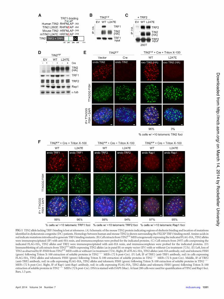
FIG 1 TIN2 allele lacking TRF1 binding is lost at telomeres. (A) Schematic of the mouse TIN2 protein indicating regions of shelterin binding and location of mutationsidentified in dyskeratosis congenita (DC) patients. Homology between human and mouse TIN2 is shown surrounding the FXLXP TRF1 binding motif. Amino acids inred indicate mutations introduced to generate TRF1 binding mutants. (B) Cell extracts from TIN2F/F MEFs exogenously expressing the indicated FLAG-HA2 TIN2 alleleswere immunoprecipitated (IP) with anti-HA resin, and immunocomplexes were probed for the indicated proteins. (C) Cell extracts from 293T cells coexpressing theindicated FLAG-HA2 TIN2 alleles and TRF2 were immunoprecipitated with anti-HA resin, and immunocomplexes were probed for the indicated proteins. (D)Immunoblotting of cell extracts from TIN2F/F MEFs expressing TIN2 alleles (as in panel B) or empty vector (EV) with or without Cre treatment (72 h). (E) Left, loss ofTIN2 as observed by IF-FISH from TIN2F/F MEFs with or without Cre treatment (72 h). Right, IF of FLAG-HA2 TIN2 alleles (anti-HA antibody, red) and telomeric FISH(green) following Triton X-100 extraction of soluble proteins in TIN2!/! MEFs (72 h post-Cre). (F) Left, IF of TRF2 (anti-TRF1 antibody, red) in cells expressingFLAG-HA2 TIN2 alleles and telomeric FISH (green) following Triton X-100 extraction of soluble proteins in TIN2!/! MEFs (72 h post-Cre). Middle, IF of TRF2(anti-TRF2 antibody, red) in cells expressing FLAG-HA2 TIN2 alleles and telomeric FISH (green) following Triton X-100 extraction of soluble proteins in TIN2!/!
MEFs (72 h post-Cre). Right, IF of Rap1 (anti-Rap1 antibody, red) in cells expressing FLAG-HA2 TIN2 alleles and telomeric FISH (green) following Triton X-100extraction of soluble proteins in TIN2!/! MEFs (72 h post-Cre). DNA is stained with DAPI (blue). At least 200 cells were used for quantification of TIN2 and Rap1 foci.Bars, 1.5 %m.
April 2014 Volume 34 Number 7 mcb.asm.org 1351
on March 14, 2014 by Rockefeller University
http://mcb.asm
.org/Downloaded from
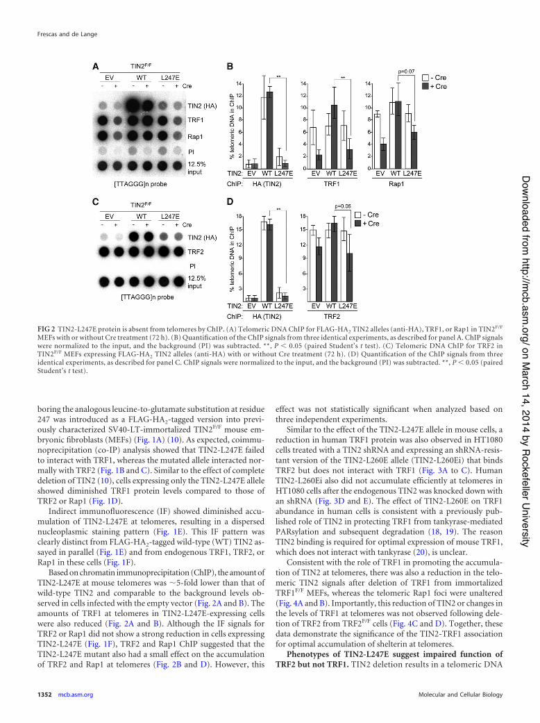
boring the analogous leucine-to-glutamate substitution at residue247 was introduced as a FLAG-HA2-tagged version into previ-ously characterized SV40-LT-immortalized TIN2F/F mouse em-bryonic fibroblasts (MEFs) (Fig. 1A) (10). As expected, coimmu-noprecipitation (co-IP) analysis showed that TIN2-L247E failedto interact with TRF1, whereas the mutated allele interacted nor-mally with TRF2 (Fig. 1B and C). Similar to the effect of completedeletion of TIN2 (10), cells expressing only the TIN2-L247E alleleshowed diminished TRF1 protein levels compared to those ofTRF2 or Rap1 (Fig. 1D).
Indirect immunofluorescence (IF) showed diminished accu-mulation of TIN2-L247E at telomeres, resulting in a dispersednucleoplasmic staining pattern (Fig. 1E). This IF pattern wasclearly distinct from FLAG-HA2-tagged wild-type (WT) TIN2 as-sayed in parallel (Fig. 1E) and from endogenous TRF1, TRF2, orRap1 in these cells (Fig. 1F).
Based on chromatin immunoprecipitation (ChIP), the amount ofTIN2-L247E at mouse telomeres was &5-fold lower than that ofwild-type TIN2 and comparable to the background levels ob-served in cells infected with the empty vector (Fig. 2A and B). Theamounts of TRF1 at telomeres in TIN2-L247E-expressing cellswere also reduced (Fig. 2A and B). Although the IF signals forTRF2 or Rap1 did not show a strong reduction in cells expressingTIN2-L247E (Fig. 1F), TRF2 and Rap1 ChIP suggested that theTIN2-L247E mutant also had a small effect on the accumulationof TRF2 and Rap1 at telomeres (Fig. 2B and D). However, this
effect was not statistically significant when analyzed based onthree independent experiments.
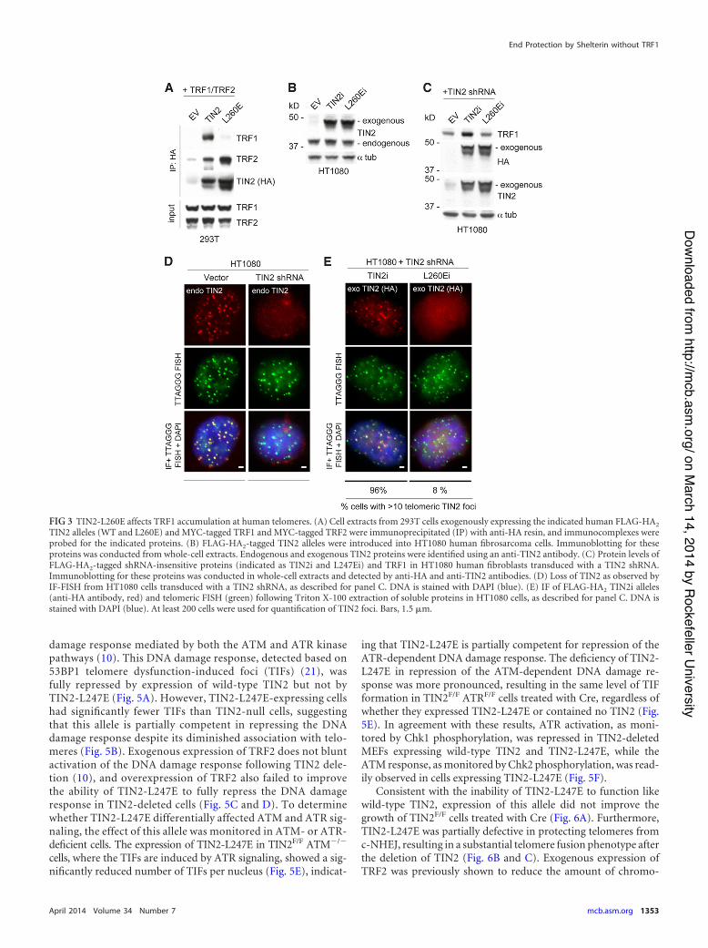
Similar to the effect of the TIN2-L247E allele in mouse cells, areduction in human TRF1 protein was also observed in HT1080cells treated with a TIN2 shRNA and expressing an shRNA-resis-tant version of the TIN2-L260E allele (TIN2-L260Ei) that bindsTRF2 but does not interact with TRF1 (Fig. 3A to C). HumanTIN2-L260Ei also did not accumulate efficiently at telomeres inHT1080 cells after the endogenous TIN2 was knocked down withan shRNA (Fig. 3D and E). The effect of TIN2-L260E on TRF1abundance in human cells is consistent with a previously pub-lished role of TIN2 in protecting TRF1 from tankyrase-mediatedPARsylation and subsequent degradation (18, 19). The reasonTIN2 binding is required for optimal expression of mouse TRF1,which does not interact with tankyrase (20), is unclear.
Consistent with the role of TRF1 in promoting the accumula-tion of TIN2 at telomeres, there was also a reduction in the telo-meric TIN2 signals after deletion of TRF1 from immortalizedTRF1F/F MEFs, whereas the telomeric Rap1 foci were unaltered(Fig. 4A and B). Importantly, this reduction of TIN2 or changes inthe levels of TRF1 at telomeres was not observed following dele-tion of TRF2 from TRF2F/F cells (Fig. 4C and D). Together, thesedata demonstrate the significance of the TIN2-TRF1 associationfor optimal accumulation of shelterin at telomeres.
Phenotypes of TIN2-L247E suggest impaired function ofTRF2 but not TRF1. TIN2 deletion results in a telomeric DNA
FIG 2 TIN2-L247E protein is absent from telomeres by ChIP. (A) Telomeric DNA ChIP for FLAG-HA2 TIN2 alleles (anti-HA), TRF1, or Rap1 in TIN2F/F
MEFs with or without Cre treatment (72 h). (B) Quantification of the ChIP signals from three identical experiments, as described for panel A. ChIP signalswere normalized to the input, and the background (PI) was subtracted. **, P ' 0.05 (paired Student’s t test). (C) Telomeric DNA ChIP for TRF2 inTIN2F/F MEFs expressing FLAG-HA2 TIN2 alleles (anti-HA) with or without Cre treatment (72 h). (D) Quantification of the ChIP signals from threeidentical experiments, as described for panel C. ChIP signals were normalized to the input, and the background (PI) was subtracted. **, P ' 0.05 (pairedStudent’s t test).
Frescas and de Lange
1352 mcb.asm.org Molecular and Cellular Biology
on March 14, 2014 by Rockefeller University
http://mcb.asm
.org/Downloaded from
damage response mediated by both the ATM and ATR kinasepathways (10). This DNA damage response, detected based on53BP1 telomere dysfunction-induced foci (TIFs) (21), wasfully repressed by expression of wild-type TIN2 but not byTIN2-L247E (Fig. 5A). However, TIN2-L247E-expressing cellshad significantly fewer TIFs than TIN2-null cells, suggestingthat this allele is partially competent in repressing the DNAdamage response despite its diminished association with telo-meres (Fig. 5B). Exogenous expression of TRF2 does not bluntactivation of the DNA damage response following TIN2 dele-tion (10), and overexpression of TRF2 also failed to improvethe ability of TIN2-L247E to fully repress the DNA damageresponse in TIN2-deleted cells (Fig. 5C and D). To determinewhether TIN2-L247E differentially affected ATM and ATR sig-naling, the effect of this allele was monitored in ATM- or ATR-deficient cells. The expression of TIN2-L247E in TIN2F/F ATM!/!
cells, where the TIFs are induced by ATR signaling, showed a sig-nificantly reduced number of TIFs per nucleus (Fig. 5E), indicat-
ing that TIN2-L247E is partially competent for repression of theATR-dependent DNA damage response. The deficiency of TIN2-L247E in repression of the ATM-dependent DNA damage re-sponse was more pronounced, resulting in the same level of TIFformation in TIN2F/F ATRF/F cells treated with Cre, regardless ofwhether they expressed TIN2-L247E or contained no TIN2 (Fig.5E). In agreement with these results, ATR activation, as moni-tored by Chk1 phosphorylation, was repressed in TIN2-deletedMEFs expressing wild-type TIN2 and TIN2-L247E, while theATM response, as monitored by Chk2 phosphorylation, was read-ily observed in cells expressing TIN2-L247E (Fig. 5F).
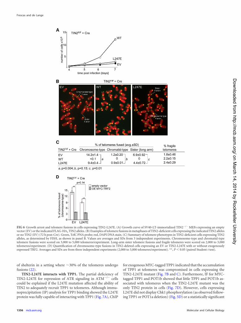
Consistent with the inability of TIN2-L247E to function likewild-type TIN2, expression of this allele did not improve thegrowth of TIN2F/F cells treated with Cre (Fig. 6A). Furthermore,TIN2-L247E was partially defective in protecting telomeres fromc-NHEJ, resulting in a substantial telomere fusion phenotype afterthe deletion of TIN2 (Fig. 6B and C). Exogenous expression ofTRF2 was previously shown to reduce the amount of chromo-
FIG 3 TIN2-L260E affects TRF1 accumulation at human telomeres. (A) Cell extracts from 293T cells exogenously expressing the indicated human FLAG-HA2
TIN2 alleles (WT and L260E) and MYC-tagged TRF1 and MYC-tagged TRF2 were immunoprecipitated (IP) with anti-HA resin, and immunocomplexes wereprobed for the indicated proteins. (B) FLAG-HA2-tagged TIN2 alleles were introduced into HT1080 human fibrosarcoma cells. Immunoblotting for theseproteins was conducted from whole-cell extracts. Endogenous and exogenous TIN2 proteins were identified using an anti-TIN2 antibody. (C) Protein levels ofFLAG-HA2-tagged shRNA-insensitive proteins (indicated as TIN2i and L247Ei) and TRF1 in HT1080 human fibroblasts transduced with a TIN2 shRNA.Immunoblotting for these proteins was conducted in whole-cell extracts and detected by anti-HA and anti-TIN2 antibodies. (D) Loss of TIN2 as observed byIF-FISH from HT1080 cells transduced with a TIN2 shRNA, as described for panel C. DNA is stained with DAPI (blue). (E) IF of FLAG-HA2 TIN2i alleles(anti-HA antibody, red) and telomeric FISH (green) following Triton X-100 extraction of soluble proteins in HT1080 cells, as described for panel C. DNA isstained with DAPI (blue). At least 200 cells were used for quantification of TIN2 foci. Bars, 1.5 %m.
End Protection by Shelterin without TRF1
April 2014 Volume 34 Number 7 mcb.asm.org 1353
on March 14, 2014 by Rockefeller University
http://mcb.asm
.org/Downloaded from

some-type fusions resulting from TIN2 deletion, suggesting thatTIN2 is not required for the ability of TRF2 to repress c-NHEJ(10), and exogenous expression of TRF2 in TIN2-deleted cellsexpressing TIN2-L247E similarly reduced the amount of chromo-some-type fusions (Fig. 6D). Thus, the L247E allele of TIN2, de-ficient in TRF1 binding, acts as if it has a diminished capacity tosupport TRF2 function at telomeres.
On the other hand, TIN2-L247E-expressing cells did not show
one of the hallmarks of TRF1 deficiency, the formation of fragiletelomeres (Fig. 6B and C), arguing that the TIN2-TRF1 interac-tion is not essential for the replication function of TRF1, despitethe fact that the overall expression level of TRF1 is lowered in thesecells. This conclusion is consistent with the absence of a fragiletelomere phenotype in TIN2-KO cells (10). The occurrence oftelomere fusions does not mask our ability to detect fragile telo-meres since they were readily detected after the complete removal
FIG 4 Accumulation of TIN2 at telomeres is reduced in TRF1-deficient MEFs. (A) An anti-TRF1 antibody was used to detect TRF1 protein in immunoblots inTRF1F/F MEFs with or without Cre treatment (72 h). (B) Endogenous TIN2 (left) and endogenous Rap1 (right) were detected by IF in combination withtelomeric FISH (green) with or without Cre treatment (72 h). Triton X-100 was used to extract soluble proteins. DNA is stained with DAPI (blue). Bars, 1.5 %m.(C) An anti-TRF2 antibody was used to detect TRF2 protein in immunoblots in TRF2F/F MEFs with or without Cre treatment (72 h). *, unspecific bands. (D)Endogenous TIN2 (left) and endogenous TRF1 (right) were detected by IF in combination with telomeric FISH (green) with or without Cre treatment (72 h).Triton X-100 was used to extract soluble proteins. DNA is stained with DAPI (blue). Bars, 1.5 %m.
Frescas and de Lange
1354 mcb.asm.org Molecular and Cellular Biology
on March 14, 2014 by Rockefeller University
http://mcb.asm
.org/Downloaded from
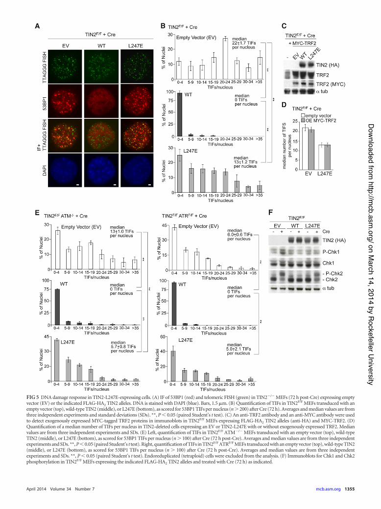
FIG 5 DNA damage response in TIN2-L247E-expressing cells. (A) IF of 53BP1 (red) and telomeric FISH (green) in TIN2!/! MEFs (72 h post-Cre) expressing emptyvector (EV) or the indicated FLAG-HA2 TIN2 alleles. DNA is stained with DAPI (blue). Bars, 1.5 %m. (B) Quantification of TIFs in TIN2F/F MEFs transduced with anempty vector (top), wild-type TIN2 (middle), or L247E (bottom), as scored for 53BP1 TIFs per nucleus (n ( 200) after Cre (72 h). Averages and median values are fromthree independent experiments and standard deviations (SDs). **, P ' 0.05 (paired Student’s t test). (C) An anti-TRF2 antibody and an anti-MYC antibody were usedto detect exogenously expressed MYC-tagged TRF2 proteins in immunoblots in TIN2F/F MEFs expressing FLAG-HA2 TIN2 alleles (anti-HA) and MYC-TRF2. (D)Quantification of a median number of TIFs per nucleus in TIN2-deleted cells expressing an EV or TIN2-L247E with or without exogenously expressed TRF2. Medianvalues are from three independent experiments and SDs. (E) Left, quantification of TIFs in TIN2F/F ATM!/! MEFs transduced with an empty vector (top), wild-typeTIN2 (middle), or L247E (bottom), as scored for 53BP1 TIFs per nucleus (n ( 100) after Cre (72 h post-Cre). Averages and median values are from three independentexperiments and SDs. **, P ' 0.05 (paired Student’s t test). Right, quantification of TIFs in TIN2F/F ATRF/F MEFs transduced with an empty vector (top), wild-type TIN2(middle), or L247E (bottom), as scored for 53BP1 TIFs per nucleus (n ( 100) after Cre (72 h post-Cre). Averages and median values are from three independentexperiments and SDs. **, P ' 0.05 (paired Student’s t test). Endoreduplicated (tetraploid) cells were excluded from the analysis. (F) Immunoblots for Chk1 and Chk2phosphorylation in TIN2F/F MEFs expressing the indicated FLAG-HA2 TIN2 alleles and treated with Cre (72 h) as indicated.
April 2014 Volume 34 Number 7 mcb.asm.org 1355
on March 14, 2014 by Rockefeller University
http://mcb.asm
.org/Downloaded from
of shelterin in a setting where &30% of the telomeres undergofusions (22).
TIN2-L247E interacts with TPP1. The partial deficiency ofTIN2-L247E for repression of ATR signaling in ATM!/! cellscould be explained if the L247E mutation affected the ability ofTIN2 to adequately recruit TPP1 to telomeres. Although immu-noprecipitation (IP) analysis for TPP1 binding showed the L247Eprotein was fully capable of interacting with TPP1 (Fig. 7A), ChIP
for exogenous MYC-tagged TPP1 indicated that the accumulationof TPP1 at telomeres was compromised in cells expressing theTIN2-L247E mutant (Fig. 7B and C). Furthermore, IF for MYC-tagged TPP1 and POT1b showed that little TPP1 and POT1b as-sociated with telomeres when the TIN2-L247E mutant was theonly TIN2 protein in cells (Fig. 7D). However, cells expressingL247E did not display Chk1 phosphorylation (as observed follow-ing TPP1 or POT1a deletion) (Fig. 5D) or a statistically significant
FIG 6 Growth arrest and telomere fusions in cells expressing TIN2-L247E. (A) Growth curve of SV40-LT-immortalized TIN2!/! MEFs expressing an emptyvector (EV) or the indicated FLAG-HA2 TIN2 alleles. (B) Examples of telomere fusions in metaphases of TIN2-deficient cells expressing the indicated TIN2 allelesor no TIN2 (EV) (72 h post-Cre). Green, TelC PNA probe; red, DAPI DNA stain. (C) Summary of telomere phenotypes in TIN2-deficient cells expressing TIN2alleles, as determined by FISH, as shown in panel B. Values are averages and SDs from 3 independent experiments. Chromosome-type and chromatid-typetelomere fusions were scored on 3,000 to 5,000 telomeres/experiment. Long-arm sister telomere fusions and fragile telomeres were scored on 2,000 to 3,000telomeres/experiment. (D) Quantification of chromosome-type fusions in TIN2-deleted cells expressing an EV or TIN2-L247E with or without exogenouslyexpressed TRF2. Averages and SDs are from three independent experiments (2,000 to 3,000 telomeres/experiment). **, P ' 0.05 (paired Student t test).
Frescas and de Lange
1356 mcb.asm.org Molecular and Cellular Biology
on March 14, 2014 by Rockefeller University
http://mcb.asm
.org/Downloaded from
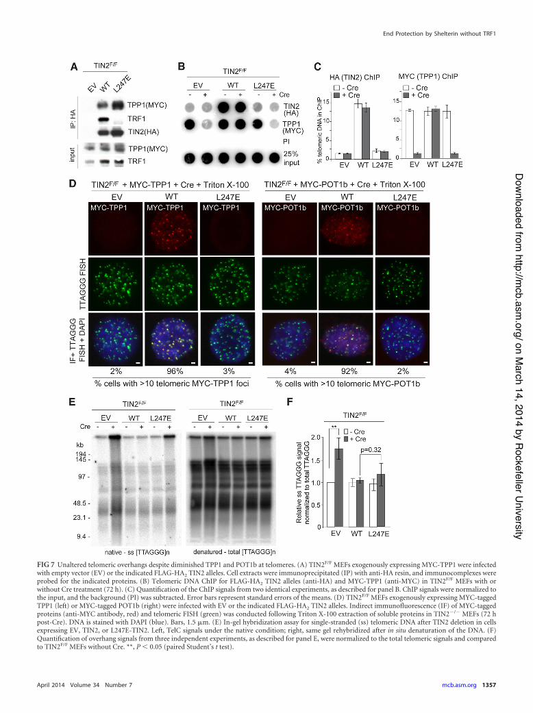
FIG 7 Unaltered telomeric overhangs despite diminished TPP1 and POT1b at telomeres. (A) TIN2F/F MEFs exogenously expressing MYC-TPP1 were infectedwith empty vector (EV) or the indicated FLAG-HA2 TIN2 alleles. Cell extracts were immunoprecipitated (IP) with anti-HA resin, and immunocomplexes wereprobed for the indicated proteins. (B) Telomeric DNA ChIP for FLAG-HA2 TIN2 alleles (anti-HA) and MYC-TPP1 (anti-MYC) in TIN2F/F MEFs with orwithout Cre treatment (72 h). (C) Quantification of the ChIP signals from two identical experiments, as described for panel B. ChIP signals were normalized tothe input, and the background (PI) was subtracted. Error bars represent standard errors of the means. (D) TIN2F/F MEFs exogenously expressing MYC-taggedTPP1 (left) or MYC-tagged POT1b (right) were infected with EV or the indicated FLAG-HA2 TIN2 alleles. Indirect immunofluorescence (IF) of MYC-taggedproteins (anti-MYC antibody, red) and telomeric FISH (green) was conducted following Triton X-100 extraction of soluble proteins in TIN2!/! MEFs (72 hpost-Cre). DNA is stained with DAPI (blue). Bars, 1.5 %m. (E) In-gel hybridization assay for single-stranded (ss) telomeric DNA after TIN2 deletion in cellsexpressing EV, TIN2, or L247E-TIN2. Left, TelC signals under the native condition; right, same gel rehybridized after in situ denaturation of the DNA. (F)Quantification of overhang signals from three independent experiments, as described for panel E, were normalized to the total telomeric signals and comparedto TIN2F/F MEFs without Cre. **, P ' 0.05 (paired Student’s t test).
End Protection by Shelterin without TRF1
April 2014 Volume 34 Number 7 mcb.asm.org 1357
on March 14, 2014 by Rockefeller University
http://mcb.asm
.org/Downloaded from
increase in the single-stranded TTAGGG repeats compared toTIN2-deficient MEFs (Fig. 7E and F), as would have been expectedif TPP1 or the POT1 proteins were completely absent. This resultwould suggest that despite the obvious reduction in telomericTPP1/POT1, there is sufficient TPP1/POT1a and TPP1/POT1bheterodimers at telomeres in the TIN2-L247E cells for the POT1proteins to fulfill most of their functions. These results are consis-tent with a prior analysis of MEFs bearing the TPP1 ACD allele,which also show severely reduced TPP1 accumulation at telo-meres yet do not have an increase in the telomeric overhang or ahigh level of TIFs (23). Moreover, despite a substantial reductionof endogenous TIN2 at telomeres (Fig. 4B), TRF1-deficient MEFsalso do not display an increase in the single-stranded telomericoverhang (14). Nevertheless, given that TIN2-L247E does not ac-cumulate at telomeres efficiently, our results are difficult to inter-pret; thus, we sought to improve the loading of the TIN2-L247Emutant on telomeres.
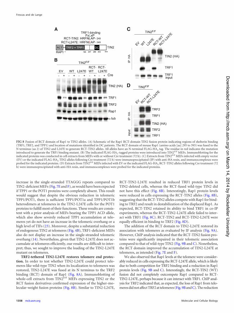
TRF2-tethered TIN2-L247E restores telomere end protec-tion. In order to test whether TIN2-L247E could protect telo-meres like wild-type TIN2 when its localization to telomeres wasrestored, TIN2-L247E was fused at its N terminus to the TRF2binding (RCT) domain of Rap1 (Fig. 8A). Immunoblotting ofwhole-cell extracts from TIN2F/F MEFs expressing TIN2 or theRCT fusion derivatives confirmed expression of the higher-mo-lecular-weight fusion proteins (Fig. 8B). Similar to TIN2-L247E,
RCT-TIN2-L247E resulted in reduced TRF1 protein levels inTIN2-deleted cells, whereas the RCT-fused wild-type TIN2 didnot have this effect (Fig. 8B). Interestingly, Rap1 protein levelswere reduced in cells expressing the RCT-TIN2 alleles (Fig. 8B),suggesting that the RCT-TIN2 alleles compete with Rap1 for bind-ing to TRF2 and result in destabilization of the displaced Rap1. Asexpected, RCT-TIN2 retained its ability to bind TRF1 in co-IPexperiments, whereas the RCT-TIN2-L247E allele failed to inter-act with TRF1 (Fig. 8C). RCT-TIN2 and RCT-TIN2-L247E wereequally efficient in binding to TRF2 (Fig. 8D).
The addition of the RCT domain to TIN2-L247E restored itsassociation with telomeres as evaluated by IF analysis (Fig. 9A).However, ChIP analysis indicated that the RCT-TIN2 fusion pro-teins were significantly impaired in their telomeric associationcompared to that of wild-type TIN2 (Fig. 9B and C). Nonetheless,the RCT domain improved the accumulation of TIN2-L247E attelomeres, as intended (Fig. 7E and F).
We also observed that Rap1 levels at the telomere were consider-ably reduced in cells expressing the RCT-L247E allele, which is likelydue to both competition for TRF2 binding and a reduction in Rap1protein levels (Fig. 9B and C). Interestingly, the RCT-TIN2 (WT)fusion did not completely outcompete Rap1 compared to RCT-TIN2-L247E, perhaps because it can interact with TRF1. ChIP anal-ysis for TRF2 indicated that, as expected, the loss of Rap1 from telo-meres did not affect TRF2 at telomeres (Fig. 9B and C). The reduction
FIG 8 Fusion of RCT domain of Rap1 to TIN2 alleles. (A) Schematic of the Rap1 RCT-domain TIN2 fusion protein indicating regions of shelterin binding(TRF1, TRF2, and TPP1) and location of mutations identified in DC patients. The RCT domain of mouse Rap1 (amino acids [aa] 295 to 393) was fused to theN terminus (aa 2) of TIN2 and L247E to generate RCT-TIN2 alleles. All alleles have an N-terminal FLAG-HA2 tag. The residue in red indicates the mutationintroduced to generate the TRF1 binding mutant. (B) The indicated FLAG-HA2-tagged proteins were introduced into TIN2F/F MEFs. Immunoblotting for theindicated proteins was conducted in cell extracts from MEFs with or without Cre treatment (72 h). (C) Extracts from TIN2F/F MEFs infected with empty vector(EV) or the indicated FLAG-HA2 TIN2 alleles following Cre treatment (72 h) were immunoprecipitated (IP) with anti-HA resin, and immunocomplexes wereprobed for the indicated proteins. (D) Extracts from TIN2F/F MEFs infected with EV or the indicated FLAG-HA2 RCT-TIN2 alleles following Cre treatment (72h) were immunoprecipitated with anti-HA resin, and immunocomplexes were probed for the indicated proteins.
Frescas and de Lange
1358 mcb.asm.org Molecular and Cellular Biology
on March 14, 2014 by Rockefeller University
http://mcb.asm
.org/Downloaded from
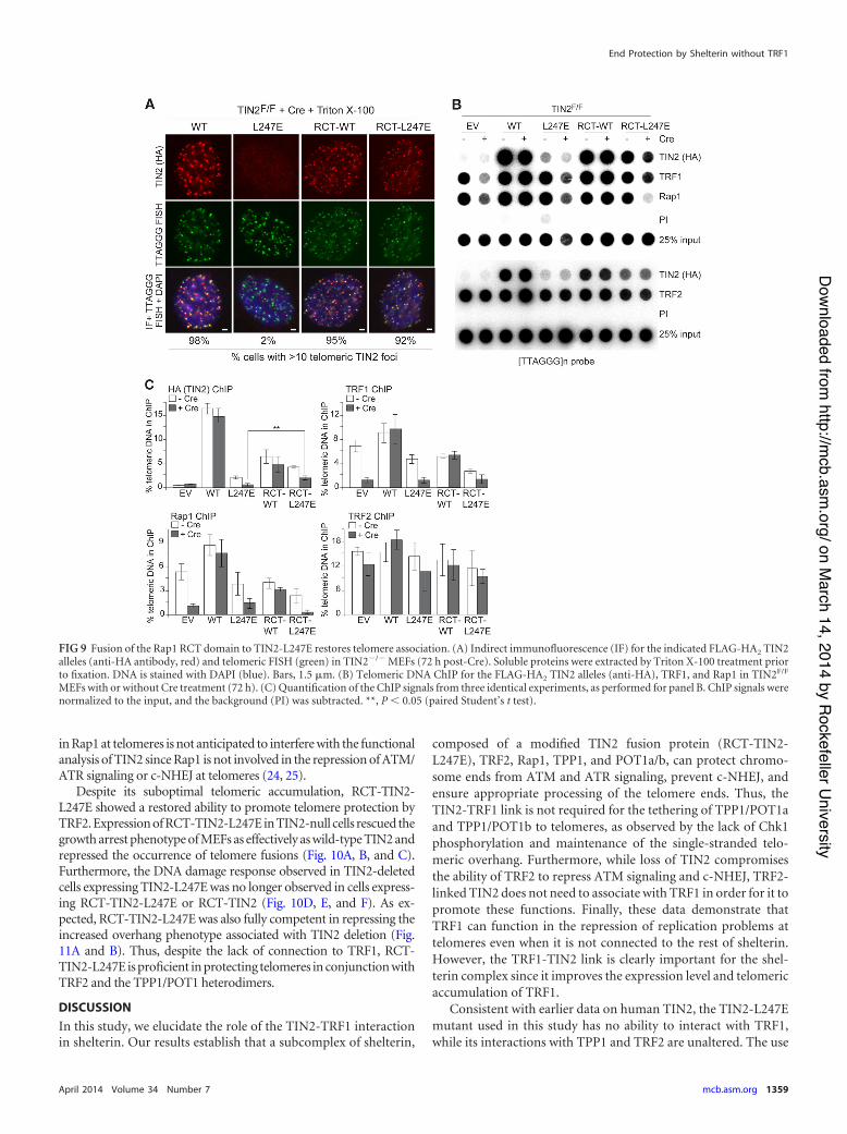
in Rap1 at telomeres is not anticipated to interfere with the functionalanalysis of TIN2 since Rap1 is not involved in the repression of ATM/ATR signaling or c-NHEJ at telomeres (24, 25).
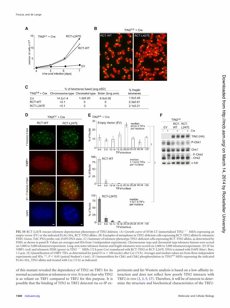
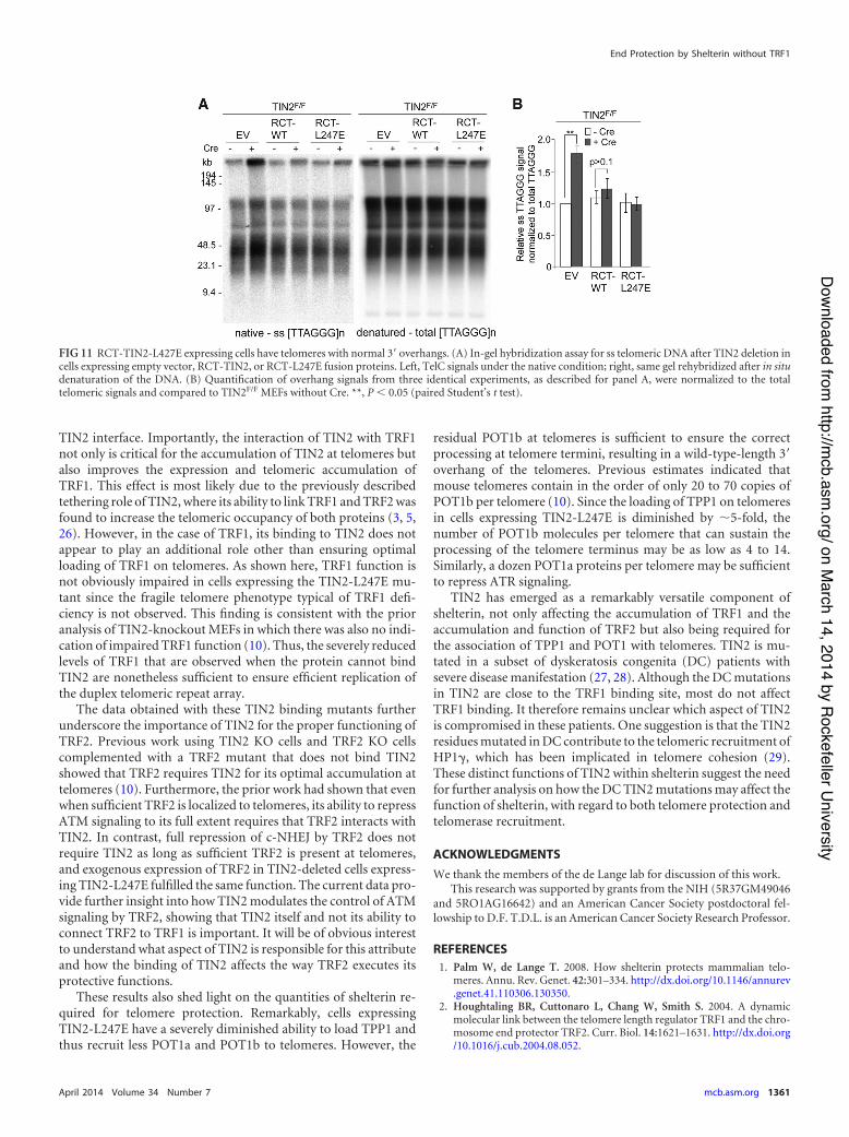
Despite its suboptimal telomeric accumulation, RCT-TIN2-L247E showed a restored ability to promote telomere protection byTRF2. Expression of RCT-TIN2-L247E in TIN2-null cells rescued thegrowth arrest phenotype of MEFs as effectively as wild-type TIN2 andrepressed the occurrence of telomere fusions (Fig. 10A, B, and C).Furthermore, the DNA damage response observed in TIN2-deletedcells expressing TIN2-L247E was no longer observed in cells express-ing RCT-TIN2-L247E or RCT-TIN2 (Fig. 10D, E, and F). As ex-pected, RCT-TIN2-L247E was also fully competent in repressing theincreased overhang phenotype associated with TIN2 deletion (Fig.11A and B). Thus, despite the lack of connection to TRF1, RCT-TIN2-L247E is proficient in protecting telomeres in conjunction withTRF2 and the TPP1/POT1 heterodimers.
DISCUSSIONIn this study, we elucidate the role of the TIN2-TRF1 interactionin shelterin. Our results establish that a subcomplex of shelterin,
composed of a modified TIN2 fusion protein (RCT-TIN2-L247E), TRF2, Rap1, TPP1, and POT1a/b, can protect chromo-some ends from ATM and ATR signaling, prevent c-NHEJ, andensure appropriate processing of the telomere ends. Thus, theTIN2-TRF1 link is not required for the tethering of TPP1/POT1aand TPP1/POT1b to telomeres, as observed by the lack of Chk1phosphorylation and maintenance of the single-stranded telo-meric overhang. Furthermore, while loss of TIN2 compromisesthe ability of TRF2 to repress ATM signaling and c-NHEJ, TRF2-linked TIN2 does not need to associate with TRF1 in order for it topromote these functions. Finally, these data demonstrate thatTRF1 can function in the repression of replication problems attelomeres even when it is not connected to the rest of shelterin.However, the TRF1-TIN2 link is clearly important for the shel-terin complex since it improves the expression level and telomericaccumulation of TRF1.
Consistent with earlier data on human TIN2, the TIN2-L247Emutant used in this study has no ability to interact with TRF1,while its interactions with TPP1 and TRF2 are unaltered. The use
FIG 9 Fusion of the Rap1 RCT domain to TIN2-L247E restores telomere association. (A) Indirect immunofluorescence (IF) for the indicated FLAG-HA2 TIN2alleles (anti-HA antibody, red) and telomeric FISH (green) in TIN2!/! MEFs (72 h post-Cre). Soluble proteins were extracted by Triton X-100 treatment priorto fixation. DNA is stained with DAPI (blue). Bars, 1.5 %m. (B) Telomeric DNA ChIP for the FLAG-HA2 TIN2 alleles (anti-HA), TRF1, and Rap1 in TIN2F/F
MEFs with or without Cre treatment (72 h). (C) Quantification of the ChIP signals from three identical experiments, as performed for panel B. ChIP signals werenormalized to the input, and the background (PI) was subtracted. **, P ' 0.05 (paired Student’s t test).
End Protection by Shelterin without TRF1
April 2014 Volume 34 Number 7 mcb.asm.org 1359
on March 14, 2014 by Rockefeller University
http://mcb.asm
.org/Downloaded from
of this mutant revealed the dependency of TIN2 on TRF1 for itsnormal accumulation at telomeres in vivo. It is not clear why TIN2is so reliant on TRF1 compared to TRF2 for this purpose. It ispossible that the binding of TIN2 to TRF2 detected via co-IP ex-
periments and far-Western analysis is based on a low-affinity in-teraction and does not reflect how poorly TIN2 interacts withTRF2 in vivo (2, 3, 5, 17). Therefore, it will be of interest to deter-mine the structure and biochemical characteristics of the TRF2-
FIG 10 RCT-L247E rescues telomere deprotection phenotypes of TIN2 deletion. (A) Growth curve of SV40-LT-immortalized TIN2!/! MEFs expressing anempty vector (EV) or the indicated FLAG-HA2 RCT-TIN2 alleles. (B) Examples of metaphases in TIN2-deficient cells expressing RCT-TIN2 alleles by telomericFISH. Green, TelC PNA probe; red, DAPI DNA stain. (C) Summary of telomere phenotype TIN2-deficient cells expressing RCT-TIN2 alleles, as determined byFISH, as shown in panel B. Values are averages and SDs from 3 independent experiments. Chromosome-type and chromatid-type telomere fusions were scoredon 3,000 to 5,000 telomeres/experiment. Long-arm sister telomere fusions and fragile telomeres were scored on 2,000 to 3,000 telomeres/experiment. (D) IF for53BP1 (red) and telomeric FISH (green) in TIN2!/! MEFs (72 h post-Cre) transduced with RCT-TIN2 or RCT-L247E. DNA is stained with DAPI (blue). Bars,1.5 %m. (E) Quantification of 53BP1 TIFs, as determined for panel D (n ( 100 nuclei) after Cre (72 h). Averages and median values are from three independentexperiments and SDs. **, P ' 0.05 (paired Student’s t test). (F) Immunoblots for Chk1 and Chk2 phosphorylation in TIN2F/F MEFs expressing the indicatedFLAG-HA2 TIN2 alleles and treated with Cre (72 h) as indicated.
Frescas and de Lange
1360 mcb.asm.org Molecular and Cellular Biology
on March 14, 2014 by Rockefeller University
http://mcb.asm
.org/Downloaded from
TIN2 interface. Importantly, the interaction of TIN2 with TRF1not only is critical for the accumulation of TIN2 at telomeres butalso improves the expression and telomeric accumulation ofTRF1. This effect is most likely due to the previously describedtethering role of TIN2, where its ability to link TRF1 and TRF2 wasfound to increase the telomeric occupancy of both proteins (3, 5,26). However, in the case of TRF1, its binding to TIN2 does notappear to play an additional role other than ensuring optimalloading of TRF1 on telomeres. As shown here, TRF1 function isnot obviously impaired in cells expressing the TIN2-L247E mu-tant since the fragile telomere phenotype typical of TRF1 defi-ciency is not observed. This finding is consistent with the prioranalysis of TIN2-knockout MEFs in which there was also no indi-cation of impaired TRF1 function (10). Thus, the severely reducedlevels of TRF1 that are observed when the protein cannot bindTIN2 are nonetheless sufficient to ensure efficient replication ofthe duplex telomeric repeat array.
The data obtained with these TIN2 binding mutants furtherunderscore the importance of TIN2 for the proper functioning ofTRF2. Previous work using TIN2 KO cells and TRF2 KO cellscomplemented with a TRF2 mutant that does not bind TIN2showed that TRF2 requires TIN2 for its optimal accumulation attelomeres (10). Furthermore, the prior work had shown that evenwhen sufficient TRF2 is localized to telomeres, its ability to repressATM signaling to its full extent requires that TRF2 interacts withTIN2. In contrast, full repression of c-NHEJ by TRF2 does notrequire TIN2 as long as sufficient TRF2 is present at telomeres,and exogenous expression of TRF2 in TIN2-deleted cells express-ing TIN2-L247E fulfilled the same function. The current data pro-vide further insight into how TIN2 modulates the control of ATMsignaling by TRF2, showing that TIN2 itself and not its ability toconnect TRF2 to TRF1 is important. It will be of obvious interestto understand what aspect of TIN2 is responsible for this attributeand how the binding of TIN2 affects the way TRF2 executes itsprotective functions.
These results also shed light on the quantities of shelterin re-quired for telomere protection. Remarkably, cells expressingTIN2-L247E have a severely diminished ability to load TPP1 andthus recruit less POT1a and POT1b to telomeres. However, the
residual POT1b at telomeres is sufficient to ensure the correctprocessing at telomere termini, resulting in a wild-type-length 3=overhang of the telomeres. Previous estimates indicated thatmouse telomeres contain in the order of only 20 to 70 copies ofPOT1b per telomere (10). Since the loading of TPP1 on telomeresin cells expressing TIN2-L247E is diminished by &5-fold, thenumber of POT1b molecules per telomere that can sustain theprocessing of the telomere terminus may be as low as 4 to 14.Similarly, a dozen POT1a proteins per telomere may be sufficientto repress ATR signaling.
TIN2 has emerged as a remarkably versatile component ofshelterin, not only affecting the accumulation of TRF1 and theaccumulation and function of TRF2 but also being required forthe association of TPP1 and POT1 with telomeres. TIN2 is mu-tated in a subset of dyskeratosis congenita (DC) patients withsevere disease manifestation (27, 28). Although the DC mutationsin TIN2 are close to the TRF1 binding site, most do not affectTRF1 binding. It therefore remains unclear which aspect of TIN2is compromised in these patients. One suggestion is that the TIN2residues mutated in DC contribute to the telomeric recruitment ofHP1), which has been implicated in telomere cohesion (29).These distinct functions of TIN2 within shelterin suggest the needfor further analysis on how the DC TIN2 mutations may affect thefunction of shelterin, with regard to both telomere protection andtelomerase recruitment.
ACKNOWLEDGMENTSWe thank the members of the de Lange lab for discussion of this work.
This research was supported by grants from the NIH (5R37GM49046and 5RO1AG16642) and an American Cancer Society postdoctoral fel-lowship to D.F. T.D.L. is an American Cancer Society Research Professor.
REFERENCES1. Palm W, de Lange T. 2008. How shelterin protects mammalian telo-
meres. Annu. Rev. Genet. 42:301–334. http://dx.doi.org/10.1146/annurev.genet.41.110306.130350.
2. Houghtaling BR, Cuttonaro L, Chang W, Smith S. 2004. A dynamicmolecular link between the telomere length regulator TRF1 and the chro-mosome end protector TRF2. Curr. Biol. 14:1621–1631. http://dx.doi.org/10.1016/j.cub.2004.08.052.
FIG 11 RCT-TIN2-L427E expressing cells have telomeres with normal 3= overhangs. (A) In-gel hybridization assay for ss telomeric DNA after TIN2 deletion incells expressing empty vector, RCT-TIN2, or RCT-L247E fusion proteins. Left, TelC signals under the native condition; right, same gel rehybridized after in situdenaturation of the DNA. (B) Quantification of overhang signals from three identical experiments, as described for panel A, were normalized to the totaltelomeric signals and compared to TIN2F/F MEFs without Cre. **, P ' 0.05 (paired Student’s t test).
End Protection by Shelterin without TRF1
April 2014 Volume 34 Number 7 mcb.asm.org 1361
on March 14, 2014 by Rockefeller University
http://mcb.asm
.org/Downloaded from
3. Liu D, O’Connor MS, Qin J, Songyang Z. 2004. Telosome, a mammaliantelomere-associated complex formed by multiple telomeric proteins. J. Biol.Chem. 279:51338–51342. http://dx.doi.org/10.1074/jbc.M409293200.
4. Liu D, Safari A, O’Connor MS, Chan DW, Laegeler A, Qin J, SongyangZ. 2004. PTOP interacts with POT1 and regulates its localization to telo-meres. Nat. Cell Biol. 6:673– 680. http://dx.doi.org/10.1038/ncb1142.
5. Ye JZ, Donigian JR, Van Overbeek M, Loayza D, Luo Y, KrutchinskyAN, Chait BT, de Lange T. 2004. TIN2 binds TRF1 and TRF2 simulta-neously and stabilizes the TRF2 complex on telomeres. J. Biol. Chem.279:47264 – 47271. http://dx.doi.org/10.1074/jbc.M409047200.
6. Ye JZ, Hockemeyer D, Krutchinsky AN, Loayza D, Hooper SM, ChaitBT, de Lange T. 2004. POT1-interacting protein PIP1: a telomere lengthregulator that recruits POT1 to the TIN2/TRF1 complex. Genes Dev. 18:1649 –1654. http://dx.doi.org/10.1101/gad.1215404.
7. Denchi EL, de Lange T. 2007. Protection of telomeres through indepen-dent control of ATM and ATR by TRF2 and POT1. Nature 448:1068 –1071. http://dx.doi.org/10.1038/nature06065.
8. Hockemeyer D, Daniels JP, Takai H, de Lange T. 2006. Recent expan-sion of the telomeric complex in rodents: two distinct POT1 proteinsprotect mouse telomeres. Cell 126:63–77. http://dx.doi.org/10.1016/j.cell.2006.04.044.
9. Kibe T, Osawa GA, Keegan CE, de Lange T. 2010. Telomere protectionby TPP1 is mediated by POT1a and POT1b. Mol. Cell. Biol. 30:1059 –1066. http://dx.doi.org/10.1128/MCB.01498-09.
10. Takai KK, Kibe T, Donigian JR, Frescas D, de Lange T. 2011. Telomereprotection by TPP1/POT1 requires tethering to TIN2. Mol. Cell 44:647–659. http://dx.doi.org/10.1016/j.molcel.2011.08.043.
11. Celli GB, de Lange T. 2005. DNA processing is not required for ATM-mediated telomere damage response after TRF2 deletion. Nat. Cell Biol.7:712–718. http://dx.doi.org/10.1038/ncb1275.
12. van Steensel B, Smogorzewska A, de Lange T. 1998. TRF2 protectshuman telomeres from end-to-end fusions. Cell 92:401– 413. http://dx.doi.org/10.1016/S0092-8674(00)80932-0.
13. Martinez P, Thanasoula M, Munoz P, Liao C, Tejera A, McNees C,Flores JM, Fernandez-Capetillo O, Tarsounas M, Blasco MA. 2009.Increased telomere fragility and fusions resulting from TRF1 deficiencylead to degenerative pathologies and increased cancer in mice. Genes Dev.23:2060 –2075. http://dx.doi.org/10.1101/gad.543509.
14. Sfeir A, Kosiyatrakul ST, Hockemeyer D, MacRae SL, Karlseder J,Schildkraut CL, de Lange T. 2009. Mammalian telomeres resemble frag-ile sites and require TRF1 for efficient replication. Cell 138:90 –103. http://dx.doi.org/10.1016/j.cell.2009.06.021.
15. Herbig U, Jobling WA, Chen BP, Chen DJ, Sedivy JM. 2004. Telomereshortening triggers senescence of human cells through a pathway involv-ing ATM, p53, and p21(CIP1), but not p16(INK4a). Mol. Cell 14:501–513. http://dx.doi.org/10.1016/S1097-2765(04)00256-4.
16. Loayza D, de Lange T. 2003. POT1 as a terminal transducer of TRF1telomere length control. Nature 424:1013–1018. http://dx.doi.org/10.1038/4241013a.
17. Chen Y, Yang Y, van Overbeek M, Donigian JR, Baciu P, de Lange T,Lei M. 2008. A shared docking motif in TRF1 and TRF2 used for differ-ential recruitment of telomeric proteins. Science 319:1092–1096. http://dx.doi.org/10.1126/science.1151804.
18. Chang W, Dynek JN, Smith S. 2003. TRF1 is degraded by ubiquitin-mediated proteolysis after release from telomeres. Genes Dev. 17:1328 –1333. http://dx.doi.org/10.1101/gad.1077103.
19. Ye JZ, de Lange T. 2004. TIN2 is a tankyrase 1 PARP modulator in theTRF1 telomere length control complex. Nat. Genet. 36:618 – 623. http://dx.doi.org/10.1038/ng1360.
20. Donigian JR, de Lange T. 2007. The role of the poly(ADP-ribose) poly-merase tankyrase1 in telomere length control by the TRF1 component ofthe shelterin complex. J. Biol. Chem. 282:22662–22667. http://dx.doi.org/10.1074/jbc.M702620200.
21. Takai H, Smogorzewska A, de Lange T. 2003. DNA damage foci atdysfunctional telomeres. Curr. Biol. 13:1549 –1556. http://dx.doi.org/10.1016/S0960-9822(03)00542-6.
22. Sfeir A, de Lange T. 2012. Removal of shelterin reveals the telomereend-protection problem. Science 336:593–597. http://dx.doi.org/10.1126/science.1218498.
23. Hockemeyer D, Palm W, Else T, Daniels JP, Takai KK, Ye JZ, KeeganCE, de Lange T, Hammer GD. 2007. Telomere protection by mammalianPOT1 requires interaction with TPP1. Nat. Struct. Mol. Biol. 14:754 –761.http://dx.doi.org/10.1038/nsmb1270.
24. Martinez P, Thanasoula M, Carlos AR, Gomez-Lopez G, Tejera AM,Schoeftner S, Dominguez O, Pisano DG, Tarsounas M, Blasco MA.2010. Mammalian Rap1 controls telomere function and gene expressionthrough binding to telomeric and extratelomeric sites. Nat. Cell Biol. 12:768 –780. http://dx.doi.org/10.1038/ncb2081.
25. Sfeir A, Kabir S, van Overbeek M, Celli GB, de Lange T. 2010. Loss ofRap1 induces telomere recombination in the absence of NHEJ or a DNAdamage signal. Science 327:1657–1661. http://dx.doi.org/10.1126/science.1185100.
26. Kim SH, Beausejour C, Davalos AR, Kaminker P, Heo SJ, Campisi J.2004. TIN2 mediates functions of TRF2 at human telomeres. J. Biol.Chem. 279:43799 – 43804. http://dx.doi.org/10.1074/jbc.M408650200.
27. Savage SA, Bertuch AA. 2010. The genetics and clinical manifestations oftelomere biology disorders. Genet. Med. 12:753–764. http://dx.doi.org/10.1097/GIM.0b013e3181f415b5.
28. Savage SA, Giri N, Baerlocher GM, Orr N, Lansdorp PM, Alter BP.2008. TINF2, a component of the shelterin telomere protection complex,is mutated in dyskeratosis congenita. Am. J. Hum. Genet. 82:501–509.http://dx.doi.org/10.1016/j.ajhg.2007.10.004.
29. Canudas S, Houghtaling BR, Bhanot M, Sasa G, Savage SA, BertuchAA, Smith S. 2011. A role for heterochromatin protein 1gamma athuman telomeres. Genes Dev. 25:1807–1819. http://dx.doi.org/10.1101/gad.17325211.
Frescas and de Lange
1362 mcb.asm.org Molecular and Cellular Biology
on March 14, 2014 by Rockefeller University
http://mcb.asm
.org/Downloaded from