triangles of the neck
TRANSCRIPT

TRIANGLES OF THE
NECKDr. Deepak K Gupta
www.facebook.com/notesdental

Introduction
• The side of the neck presents a somewhat
quadrilateral outline .
• It is limited above by the lower border of the
body of the mandible ,and an imaginary line
drawn from the angle of the mandible to the
mastoid process.
• Below, it is limited by the upper border of the
clavicle.
• Medially, by the midline of the neck.
• Posteriorly, by the anterior border of the
Trapezius muscle .www.facebook.com/notesdental

Quadrilateral outline in the neck
www.facebook.com/notesdental

Sternocleidomastoid Muscle
www.facebook.com/notesdental

Sternocleidomastoid Muscles
• This quadrilateral space is divided by the
Sternocleidomastoid muscle into two main
triangles .
• It passes obliquely upwards and backwards from
its site of origin at the clavicle and sternum to
its point of insertion on the mastoid process
and the occipital bone .
• The triangle in front of this muscle is the
anterior triangle and the one behind it is the
posterior triangle .
www.facebook.com/notesdental

www.facebook.com/notesdental

Anterior Triangle
www.facebook.com/notesdental

Posterior triangle
www.facebook.com/notesdental

Posterior triangle
• Boundaries
– SCM anteriorly
– Trapezius muscle, posteriorly.
– Clavicle, inferiorly.
• The apex of the triangle is formed by the
occipital bone.
• The ROOF of the posterior triangle is formed by:
– Skin
– Superficial fascia
– Platysma muscle
– Investing layer of the deep cervical fasciawww.facebook.com/notesdental

FLOOR
Formed by the following muscles from
above downwards:
– Splenius Capitis
– Levator scapulae
– Posterior scalene
– Middle scalene
– Anterior scalene
www.facebook.com/notesdental

Subdivisions of the posterior
triangle
• The posterior triangle is further divided into two smaller triangles by the Inferior belly of the Omohyoid muscle.
• Supraclavicular triangle
– Inferior belly of the Omohyoid
– the Clavicle,
– Sternocleidomastoid muscle
• Occipital triangle– Inferior belly of the Omohyoid
– Trapezius muscle
– Sternocleidomastoid muscle
www.facebook.com/notesdental

www.facebook.com/notesdental

CONTENTS: NERVES
• Spinal acessory nerve.
• Branches of Cervical plexus
– Lesser occipital
– Transverse cervical
– Great auricular
– Supraclavicular
• Roots and trunks of brachial plexus.
• Dorsal scapular
• Long thoracic
• Phrenicwww.facebook.com/notesdental

www.facebook.com/notesdental

VESSELS
• Arteries
– Subclavian artery
– Transverse Cervical artery
– Suprascapular artery
• Vein
– External jugular vein (terminal part)
• Lymph Nodes
– Occipital
– Supraclavicularwww.facebook.com/notesdental

www.facebook.com/notesdental

CLINICAL SIGNIFICANCE OF
THE POSTERIOR TRIANGLE
• The Accessory Nerve may be damaged,
while taking lymph node biopsy.
• The External Jugular Vein is present in a
superficial location here and this makes it
vulnerable to injury.
www.facebook.com/notesdental

THE ANTERIOR
TRIANGLE
www.facebook.com/notesdental

ANTERIOR TRIANGLE
• BOUNDARIES
– Anterior border of the SCM muscle
– midline of the neck
– inferior border of the mandible
• ROOF
– Skin
– Superfacial fascia and platysma muscle
– Investing layer of deep cervical fascia
www.facebook.com/notesdental

SUBDIVISIONS OF THE
ANTERIOR TRIANGLE
• The anterior triangle is divided into four
smaller triangles:
1. SUBMENTAL TRIANGLE
2. SUBMANDIBULAR TRIANGLE
3. CAROTID TRIANGLE
4. MUSCULAR TRIANGLE
www.facebook.com/notesdental

SUBMENTAL TRIANGLE
• Borders
– Body of hyoid
– Anterior digastric on right
– Anterior digastric on left
• Floor : Mylohyoid
• Roof is made of the:
– Skin
– Superficial fascia with platysma
– Deep cervical fascia
• Submental triangle is unpaired
• Content: Anterior Jugular vein and sub-mental lymph
nodes www.facebook.com/notesdental

www.facebook.com/notesdental

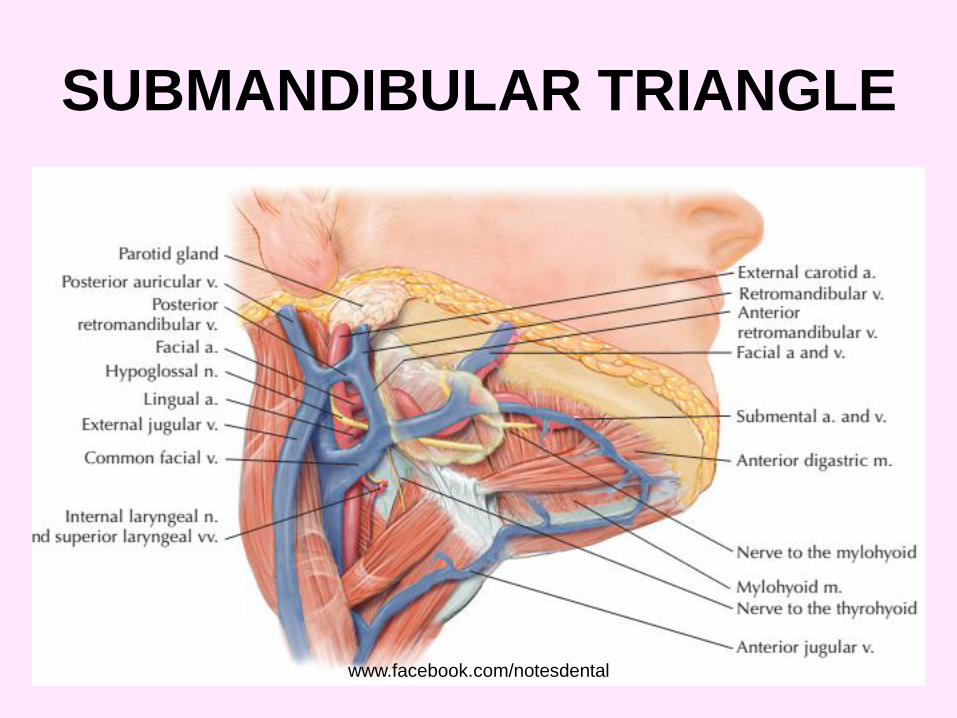
SUBMANDIBULAR TRIANGLE• Borders
– Inferior border of the mandible
– Posterior digastric
– Anterior digastric
• Floor of the triangle– Hyoglossus
– Mylohyoid
– Middle constrictor
• Roof is made of the:– Skin
– Superficial fascia with platysma
– Deep cervical fascia
• Submandibular triangle is paired
www.facebook.com/notesdental

www.facebook.com/notesdental

SUBMANDIBULAR TRIANGLE
• Lesser’s triangle is a small subdivision of
the submandibular triangle, which aids in
identifying the lingual artery (especially
for ligation)
• Boundaries of Lesser’s triangle:
– Hypoglossal nerve
– Anterior digastric
– Posterior digastric
www.facebook.com/notesdental
SUBMANDIBULAR TRIANGLE
www.facebook.com/notesdental

SUBMANDIBULAR
TRIANGLE: Content• Arteries
– Facial
– Sublingual
– Submental
• Veins: same as arteries
• Nerves : Mylohyoid and hypoglossal
• Structures
– Submandibular gland
– Inferior portion of parotid gland
– Submandibular lymph nodewww.facebook.com/notesdental

CAROTID TRIANGLE• Named because parts of all three carotid arteries are located within it
• Borders of the carotid triangle:
– Anterior border of the sternocleidomastoid
– Posterior digastric
– Superior omohyoid
• Floor of the triangle is composed of the:
– Hyoglossus
– Thyrohyoid
– Middle constrictorInferior constrictor
• Roof
– Skin
– Superficial fascia with platysma
– Deep cervical fascia
• Carotid triangle is paired
www.facebook.com/notesdental

CAROTID TRIANGLE
www.facebook.com/notesdental

CAROTID TRIANGLE
• Arteries
– Common carotid (with carotid
body)
– Internal carotid (with carotid
sinus)
• Superior thyroid (with superior
Superior thyroid
• Lingual
• Facial
• Ascending pharyngeal
• Occipital
• Vein
– Internal Jugalar vein
– Common facial vein
– Lingual Vein
– Superior Thyroid vein
– Middle thyroid vein
• Nerves
– Vagus• External laryngeal
• Internal laryngeal
– Spinal Acessory
– Hypoglossal
– Ansa cervicalos
– Sympathetic trunk
• Structures• Larynx
• Thyroid
www.facebook.com/notesdental

www.facebook.com/notesdental

MUSCULAR TRIANGLE• Borders
– Anterior border of the sternocleidomastoid
– Superior omohyoid
– Midline
• Floor
– Sternohyoid
– Sternothyroid
• Roof
– Skin
– Superficial fascia with platysma
– Deep cervical fascia
• Muscular triangle is paired
www.facebook.com/notesdental

MUSCULAR TRIANGLE
www.facebook.com/notesdental

MUSCULAR TRIANGLE:
Content• Arteries
– Superior thyroid
• Veins
– Inferior thyroid
– Anterior jugular
• Nerves: Ansa cervicalis
• Structures
– Strap muscles: Sternohyoid, Sternothyroid, Thyrohyoid
– Thyroid gland
– Parathyroid gland
– Larynx
– Trachea
– Esophagus www.facebook.com/notesdental

MUSCULAR TRIANGLE:
Content
www.facebook.com/notesdental

References
• Grays Anatomy for Students 2nd Edition
• Head and Neck Anatomy for Dental
Medicine
• Head, Neck and Dental Anatomy, 4th
Edition
• Netter’s Head and Neck Anatomy for
Dentistry, 2nd Edition Neil S norton
www.facebook.com/notesdental