trichomycterus dali : a new highly troglomorphic catfish ... · 478 trichomycterus dali: a new...
TRANSCRIPT

477
Neotropical Ichthyology, 9(3): 477-491, 2011Copyright © 2011 Sociedade Brasileira de Ictiologia
Trichomycterus dali: a new highly troglomorphic catfish
(Silurifomes: Trichomycteridae) from Serra da Bodoquena,
Mato Grosso do Sul State, Central Brazil
Pedro Pereira Rizzato1, Edmundo P. D. Costa-Jr.2, Eleonora Trajano3 and
Maria Elina Bichuette1
Trichomycterus dali, new species, is described from flooded limestone caves in Serra da Bodoquena karst area, State of MatoGrosso do Sul, Central Brazil. The new species is diagnosed by a unique character in the genus, the presence of conspicuous,ridge-like adipose folds lining dorsally throughout the body. Trichomycterus dali can be further distinguished readily fromepigean congeners by the reduction of eyes and skin pigmentation (except for T. gorgona), and from remaining congeners(i.e., all hypogean plus T. gorgona) by the total loss of eyes, not visible externally (except for T. sandovali and T. spelaeus).Other diagnostic features includes very long barbels, especially the nasal (99.3-143.5% HL) and the maxillary (97.0-131.3%HL), pectoral-fin ray count reaching I,9 and a unique cranial fontanel with a conspicuous constriction on the meeting point ofsupraoccipital and the two frontal bones. The troglobitic status of the species is suggested by the presence of troglomorphismson an advanced degree, especially the reduction of skin pigmentation, the total loss of eyes and the enlarged barbels. Inaddition, the presence of a well developed adipose fold in adults may indicate a distinctive adaptation acquired by neoteny towithstand the food scarce conditions of its hypogean habitat.
Trichomycterus dali, espécie nova, é descrita de cavernas calcárias alagadas na área cárstica da Serra da Bodoquena, estadodo Mato Grosso do Sul, Brasil central. A nova espécie é diagnosticada por um caráter único no gênero, a presença de dobrasadiposas conspícuas, em forma de crista, passando por toda a região dorsal do corpo. Trichomycterus dali pode ser aindadiferenciada facilmente de suas congêneres epígeas pela redução dos olhos e da pigmentação cutânea (exceto para T. gorgona),e das congêneres restantes (isto é, todas as hipógeas mais T. gorgona) pela perda total dos olhos, não visíveis externamente(exceto para T. sandovali e T. spelaeus). Outras características diagnósticas incluem barbilhões muito longos, especialmenteo nasal (99,3-143,5% do comprimento da cabeça) e o maxilar (97,0-131,3% do comprimento da cabeça), contagem de raios danadadeira peitoral atingindo I,9 e uma fontanela cranial única com uma constrição conspícua no ponto de encontro dosupraoccipital e dos dois ossos frontais. O status troglóbio da espécie é sugerido pela presença de troglomorfismos emavançado grau, especialmente a redução da pigmentação cutânea, a perda total dos olhos e os barbilhões alongados. Alémdisso, a presença de uma dobra adiposa bem desenvolvida nos adultos pode indicar uma adaptação distintiva, adquirida porneotenia, para enfrentar as condições alimentares escassas do seu habitat hipógeo.
Key words. New cave fish, rio Paraguai basin, Subterranean ichthyofauna, Taxonomy, Troglobite.
1Universidade Federal de São Carlos, Departamento de Ecologia e Biologia Evolutiva, Via Washington Luis, km 235, Caixa Postal 676,13565-905 São Carlos, SP, Brazil.2Prefeitura Municipal de Bonito-MS, Secretaria Municipal de Meio Ambiente, 79290-000 Bonito, MS, Brazil.3Universidade de São Paulo, Departamento de Zoologia, Instituto de Biociências, Caixa Postal 11461, 05422-970 São Paulo, SP, Brazil.
Introduction
Brazil has one of the most remarkable subterraneanichthyofaunas in the world, comparable to few other countriesor geographically comparable karst areas, such as Mexico,China and southeastern Asia (Bichuette & Trajano, 2008;Trajano et al., 2009; Trajano & Bichuette, 2010). Brazilian cavefishes are of worldwide relevance not only in terms of species
richness, but also in view of the high diversity of ecologicand evolutionary patterns (Trajano & Bichuette, 2010). Up tonow, 25 troglomorphic species were reported, but many ofthem are still waiting for description (Trajano & Bichuette,2010), a process that may take several years. As the discoveryrate of new possible troglobitic species still high (Trajano &Bichuette, 2010), it will take time until the number of nominalspecies corresponds to a figure approaching the actual

Trichomycterus dali: a new highly troglomorphic catfish478
subterranean fish diversity of Brazil. Despite this, the numberof described species increased significantly in recent years(Fernández & Bichuette, 2002; Bichuette & Trajano, 2004,2005, 2008; Bichuette et al. 2008; Moreira et al., 2010,Bockmann & Castro, 2010), and the rate of new descriptionswill surely maintain high as the already discovered speciesare being described.
All but two species of Brazilian troglomorphic fishes aresiluriforms which belong to four families: Callichthyidae,Loricariidae, Heptapteridae and Trichomycteridae, the twolatter being the most species-rich (Trajano & Bichuette, 2010).The family Trichomycteridae is the third largest family ofneotropical siluriforms, with more than 240 species of widelydistributed freshwater fishes, all endemic of Central and SouthAmericas (Nelson, 2006; Castellanos-Morales, 2008;Eschmeyer & Fong, 2010; Datovo & Bockmann, 2010). Itsmonophyly is well-corroborated and the most conspicuoussynapomorphies are based on its highly specialized opercular-interopercular apparatus (de Pinna, 1998; Datovo & Bockmann,2010). Trichomycterids are among the most successful fishescolonizing subterranean habitats (Castellanos-Morales, 2008),and up to now there are 13 nominal species in the generaSilvinichthys, Glaphyropoma, Ituglanis and Trichomycterus(Proudlove, 2010). The latter includes the majority of describedtroglomorphic species: T. chaberti Durand, from Bolivia, T.spelaeus DoNascimiento, Villarreal e Provenzano, fromVenezuela, T. sandovali Ardila-Rodriguez, T. santanderensisCastellanos-Morales and T. uisae Castellanos-Morales, fromColombia, and T. itacarambiensis Trajano e de Pinna, fromeastern Brazil.
Trichomycterus is the most diverse trichomycterid genus,with more than 130 species already described and many otherswaiting for description (Alencar & Costa, 2004; Wosiacki,2005; Wosiacki & de Pinna, 2008a; Fernández & Vari, 2009).Many authors, however, point out to its non-monophyleticcharacter (de Pinna, 1998; Wosiacki, 2002; Datovo &Bockmann, 2010). The species-level identification iscomplicated by many factors, such as the scarce informationavailable for most species (da Silva et al., 2010), and in generalonly those species presenting very distinctive features havebeen described (de Pinna, 1998; Lima & Costa, 2004; Wosiacki& de Pinna, 2008a).
Trichomycterus catfishes occur in a great variety ofhabitats in South America, from Amazonian lowlands toAndean cordilleras (Fernández & Miranda, 2007), and arefrequently endemic to a single river basin (Alencar & Costa,2004; Lima & Costa, 2004). These fish show a great potentialto colonize extreme habitats, including subterranean habitats(Fernández & Miranda, 2007). We describe herein a newsubterranean species of the genus, which exhibits anadvanced degree of troglomorphism. The species was firstsampled in 1998 by Edmundo P. da Costa Jr., in limestoneflooded caves in Serra da Bodoquena karst area, Paraguairiver basin, Central Brazil, and since then it has been cited inliterature as Trichomycterus sp. 2 (Trajano, 1997, 2003) and,later on, as Trichomycterus sp. 1 (Trajano & Bichuette, 2004;
Mattox et al., 2008; Trajano et al., 2009; Proudlove 2010;Trajano & Bichuette 2010).
Study area
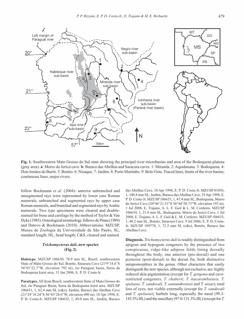
Serra da Bodoquena is located in the State of Mato Grossodo Sul, southwestern Brazil, within the limits of the rioParaguay basin (Fig. 1) and consists of Neoproterozoiccarbonate rocks of the Corumbá Group (Almeida, 1965;Boggiani et al., 1993). This highland feature is elongated informat, oriented in a north-south direction, with a length of200 km and a width varying between 10 and 70 km.
It is possible to identify two main geomorphologicalcompartments in the Serra da Bodoquena area (Alvarenga etal., 1982): a high calcareous massif, the Bodoquena Plateau,or the Serra da Bodoquena (sensu Almeida, 1965); and the rioMiranda lowlands, a region of lower topographic elevation,lying to the east. The Bodoquena Plateau consists of a plateauthat slopes to the east, with a 200-meter escarpment at itswestern border, facing towards the Pantanal region. In thisrocky massif, calcareous rocks outcrop at the surface and, inthose portions covered with soil, a dense forest hasdeveloped, still preserved due to difficulty of access, whichhas justified the creation of the “Parque Nacional da Serra daBodoquena”. The rio Miranda lowlands (Alvarenga et al.,1982), which includes the “Zona Serrana Oriental” (EasternHighland Zone - Almeida, 1965), constitutes a vast lowlandarea (elevation of 100-350 m), bordered to the east by theMaracaju-Campo Grande Plateau.
In both Bodoquena Plateau and rio Miranda lowlands,the landscape is influenced by the presence of CorumbáGroup carbonate rocks, which give rise to karstic surface reliefwith many caves, dolines and other typical features. Theclimate is classified as Aw (Justo, 2000), tropical with wetsummer and dry winter. Monthly average temperatures areabove 18°C, with the hottest months between October andApril. Annual precipitation ranges between 1000 and 2000mm. Various types of vegetation occur in the region, dependingon topography and soil type, such as tropical forests, Cerrado(Brazilian savannah), grasslands, and the complex of thePantanal (Galati et al., 2003). Generally, the vegetation can becharacterized as a remnant of Atlantic forest in transition toCerrado.
Material and Methods
The study specimens were hand-netted in thepermanently dark zones of three flooded caves, anesthetizedin benzocaine solution until death, preserved in formalin andthen transferred to alcohol 70%. All measurements werestraight-line, taken under stereomicroscope with a dial caliper,0.1 mm precision, on the left side of specimens. Measurementsfollow Tchernavin (1944) and de Pinna (1992). Vertebrae countsinclude only free vertebrae (those related to weberian complexnot included) and the compound pre-ural centrum (PU1 + U1)was considered a single element. Ray counts and symbology
P. P. Rizzato, E. P. D. Costa-Jr., E. Trajano & M. E. Bichuette 479
follow Bockmann et al. (2004): anterior unbranched andunsegmented rays were represented by lower case Romannumerals, unbranched and segmented rays by upper caseRoman numerals, and branched and segmented rays by Arabicnumerals. Two type specimens were cleared and double-stained for bone and cartilage by the method of Taylor & VanDyke (1985). Osteological terminology follows de Pinna (1989)and Datovo & Bockmann (2010). Abbreviations: MZUSP,Museu de Zoologia da Universidade de São Paulo; SL,standard length; HL, head length; C&S, cleared and stained.
Trichomycterus dali, new species(Fig. 2)
Holotype. MZUSP 106630, 78.9 mm SL, Brazil, southwesternState of Mato Grosso do Sul, Bonito, Saracura Cave (21º9’14.6”S56º43’22.7”W, elevation 792 m), rio Paraguai basin, Serra daBodoquena karst area, 15 Jan 2006, E. P. D. Costa-Jr.
Paratypes. All from Brazil, southwestern State of Mato Grosso doSul, rio Paraguai Basin, Serra da Bodoquena karst area. MZUSP106631, 1, 62.4 mm SL (c&s), Jardim, Buraco das Abelhas Cave(21°29’18.24”S 56°43’28.6”W, elevation 490 m), 18 Apr 1998, E.P. D. Costa-Jr. MZUSP 106632, 1, 49.0 mm SL, Jardim, Buraco
das Abelhas Cave, 18 Apr 1998, E. P. D. Costa-Jr. MZUSP 81056,1, 100.8 mm SL, Jardim, Buraco das Abelhas Cave, 19 Apr 1998, E.P. D. Costa-Jr. MZUSP 106633, 1, 47.4 mm SL, Bodoquena, Morrodo Jericó Cave (20°46’21.51"S 56°44’58.73"W, elevation 193 m),1 Jul 2008, E. Trajano, A. L. F. Guil & L. M. Cordeiro. MZUSP106634, 1, 21.6 mm SL, Bodoquena, Morro do Jericó Cave, 1 Jul2008, E. Trajano, A. L. F. Guil & L. M. Cordeiro. MZUSP 106635,1, 48.2 mm SL, Bonito, Saracura Cave, 9 Jul 2006, E. P. D. Costa-Jr. MZUSP 109770, 1, 72.5 mm SL (c&s), Bonito, Buraco dasAbelhas Cave.
Diagnosis. Trichomycterus dali is readily distinguished fromepigean and hypogean congeners by the presence of twoconspicuous, ridge-like adipose folds lining dorsallythroughout the body, one anterior (pre-dorsal) and oneposterior (post-dorsal) to the dorsal fin, both distinctiveautapomorphies in the genus. Other characters that easilydistinguish the new species, although not exclusive, are: highlyreduced skin pigmentation (except for T. gorgona and cave-restricted congeners, T. chaberti, T. itacarambiensis, T.spelaeus, T. sandovali, T. santanderensis and T. uisae); totalloss of eyes, not visible externally (except for T. sandovaliand T. spelaeus); barbels long, especially the nasal (99.3-143.5% HL) and the maxillary (97.0-131.3% HL) (except for T.
Fig. 1. Southwestern Mato Grosso do Sul state showing the principal river microbasins and area of the Bodoquena plateau(gray area). a: Morro do Jericó cave. b: Buraco das Abelhas and Saracura caves. 1: Miranda. 2: Aquidauana. 3: Bodoquena. 4:Dois Irmãos do Buriti. 5: Bonito. 6: Nioaque. 7: Jardim. 8: Porto Murtinho. 9: Bela Vista. Traced lines, limits of the river basins;continuous lines, major rivers.
Trichomycterus dali: a new highly troglomorphic catfish480
longibarbatus and T. spelaeus); scapulocoracoid with aconspicuous anterior process projected forward (except forT. sandovali, T. spelaeus and T. uisae), with a narrow base, awide apex and a rounded distal margin; and pectoral-fin raycount reaching I,9 (except for T. hualco). Characters possiblyexclusive, but about which many taxa lack information, arelisted as complementary diagnoses: cranial fontanel unique,extending from the posterior half of supraoccipital to theposterior region of the frontal bones, with a conspicuousconstriction on the meeting point of supraoccipital and thetwo frontal bones; supraorbital long and cylindrical, withoutprojections, with a needle appearance; 27-29 interopercularand 16-19 opercular odontodes.
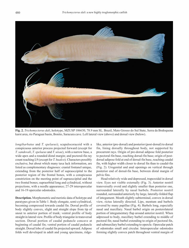
Description. Morphometric and meristic data of holotype andparatypes given in Table 1. Body elongate, semi-cylindrical,becoming compressed towards caudal fin. Dorsal profile ofbody slightly convex, slight and straight slope from tip ofsnout to anterior portion of trunk; ventral profile of bodystraight in lateral view. Profile of body triangular in transversalsection. Dorsal portion of caudal peduncle concave atbeginning of caudal fin; ventral portion of caudal pedunclestraight. Dorsal lobe of caudal fin projected upward. Adiposefolds well developed in adult and young specimens, ridge-
like, anterior (pre-dorsal) and posterior (post-dorsal) to dorsalfin, lining dorsally throughout body, not supported byprocurrent rays. Origin of pre-dorsal adipose fold posteriorto pectoral-fin base, reaching dorsal-fin base; origin of post-dorsal adipose fold at end of dorsal-fin base, reaching caudalfin, with higher width closer to dorsal fin than to caudal-fin(Fig. 2). Urogenital and anal openings on vertical throughposterior end of dorsal-fin base, between distal margin ofpelvic fins.
Head relatively wide and depressed, trapezoidal in dorsalview. Eyes not visible externally (Fig. 3). Anterior nostriltransversally ovoid and slightly smaller than posterior one,surrounded laterally by nasal barbels. Posterior nostrilrounded, surrounded anteriorly by large, laterally-folded flapof integument. Mouth slightly subterminal, convex in dorsalview, rictus laterally directed. Lips, mentum and barbelscovered by many papillae (Fig. 4). Barbels long, especiallynasal and maxillary. Nasal barbel origin on posterolateralportion of integumentary flap around anterior nostril. Whenadpressed to body, maxillary barbel extending to middle ofpectoral fin; nasal barbel extending to origin of pectoral fin,and submaxilary barbel extending to opercle. Opercular patchof odontodes small and circular. Interopercular odontodesforming slightly convex patch throughout ventral margin of
Fig. 2. Trichomycterus dali, holotype, MZUSP 106630, 78.9 mm SL. Brazil, Mato Grosso do Sul State, Serra da Bodoquenakarst area, rio Paraguai basin, Bonito, Saracura cave. Left lateral view (above) and dorsal view (below).
P. P. Rizzato, E. P. D. Costa-Jr., E. Trajano & M. E. Bichuette 481
interopercle. Opercle with two processes, upper sharp andpointed backwards, lower acute, and 27-29 opercularodontodes. Interopercle with 16-19 odontodes.
General morphology of cranium: Cranial fontanel unique,extending from posterior half of supraoccipital to posteriorregion of frontal bones; conspicuous constriction on meetingpoint of supraoccipital and two frontal bones (Fig. 5). Anteriorprocess of sphenotic and posterolateral process of frontal asconspicuous, hollow horn-like structure, inside of whichemerge infraorbital sensory branches. Supraorbital long andcylindrical, without projections, and needle appearance.Palatine with convex anterior margin covered with cartilage,waist-like medial region and long, narrow posterior process,that becomes sharp on distal region and covers almost totallymetapterygoid. Vomer arrow-shaped with long posteriorprocess. Distal profile of mesethmoid straight on dorsal view,main body axis large, cornua reaching 3/4 of premaxillary length.Three to five irregular premaxillary rows of conic teeth curvedbackwards. Rounded proximal margin of maxilla not reachingpremaxilla, covering cartilaginous margin of palatine. Lower
jaw with three rows of conical teeth, curved backwards.Hyomandibular with conspicuous semicircular depression,joined tightly to metapterygoid and quadrate. Metapterygoidand quadrate united together by anterior block of cartilage(Fig. 6).
Branchial skeleton and associated structures:Branchiostegal-rays eight, rays 5, 6 and 7 with enlarged distaltip, ray 8 covered by interopercle and reaching ventral marginof opercular patch of odontodes. Urohyal with long, verynarrow dorsal process, broad convex posterior margin, lateralsurface with distal margins chipped, urohyal-foramen slightlyovoid. Hypohyal with depression to which articulates anterior
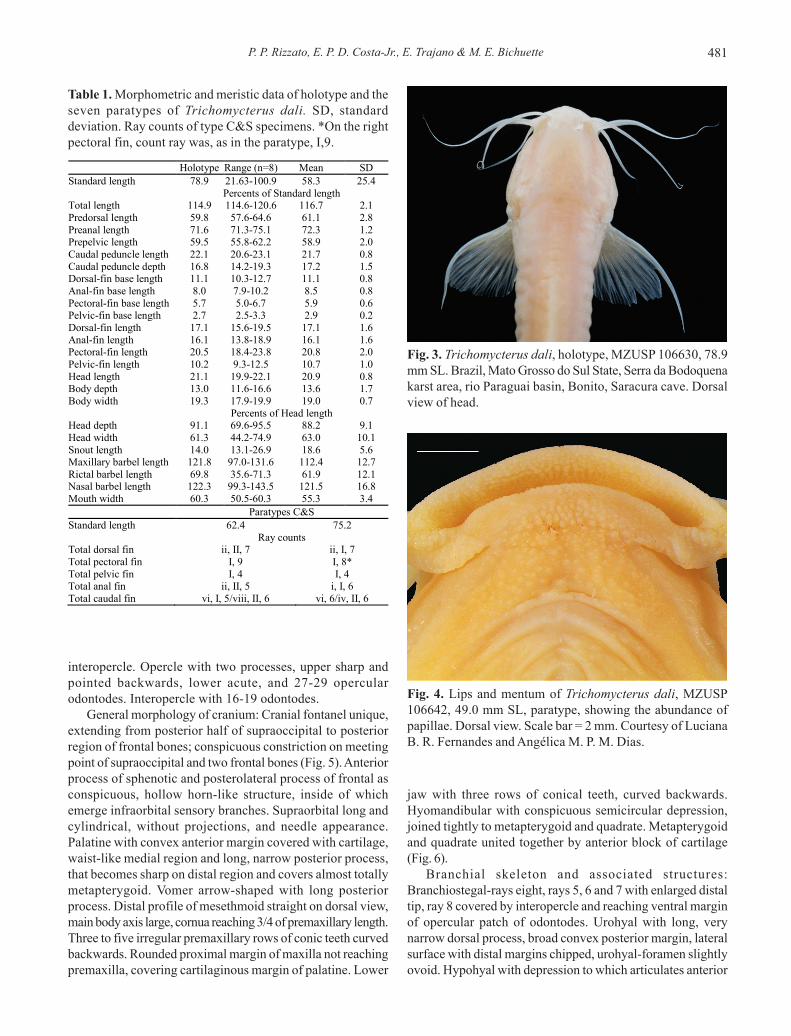
Fig. 3. Trichomycterus dali, holotype, MZUSP 106630, 78.9mm SL. Brazil, Mato Grosso do Sul State, Serra da Bodoquenakarst area, rio Paraguai basin, Bonito, Saracura cave. Dorsalview of head.
Table 1. Morphometric and meristic data of holotype and theseven paratypes of Trichomycterus dali. SD, standarddeviation. Ray counts of type C&S specimens. *On the rightpectoral fin, count ray was, as in the paratype, I,9.
Fig. 4. Lips and mentum of Trichomycterus dali, MZUSP106642, 49.0 mm SL, paratype, showing the abundance ofpapillae. Dorsal view. Scale bar = 2 mm. Courtesy of LucianaB. R. Fernandes and Angélica M. P. M. Dias.
Holotype Range (n=8) Mean SD Standard length 78.9 21.63-100.9 58.3 25.4 Percents of Standard length Total length 114.9 114.6-120.6 116.7 2.1 Predorsal length 59.8 57.6-64.6 61.1 2.8 Preanal length 71.6 71.3-75.1 72.3 1.2 Prepelvic length 59.5 55.8-62.2 58.9 2.0 Caudal peduncle length 22.1 20.6-23.1 21.7 0.8 Caudal peduncle depth 16.8 14.2-19.3 17.2 1.5 Dorsal-fin base length 11.1 10.3-12.7 11.1 0.8 Anal-fin base length 8.0 7.9-10.2 8.5 0.8 Pectoral-fin base length 5.7 5.0-6.7 5.9 0.6 Pelvic-fin base length 2.7 2.5-3.3 2.9 0.2 Dorsal-fin length 17.1 15.6-19.5 17.1 1.6 Anal-fin length 16.1 13.8-18.9 16.1 1.6 Pectoral-fin length 20.5 18.4-23.8 20.8 2.0 Pelvic-fin length 10.2 9.3-12.5 10.7 1.0 Head length 21.1 19.9-22.1 20.9 0.8 Body depth 13.0 11.6-16.6 13.6 1.7 Body width 19.3 17.9-19.9 19.0 0.7 Percents of Head length Head depth 91.1 69.6-95.5 88.2 9.1 Head width 61.3 44.2-74.9 63.0 10.1 Snout length 14.0 13.1-26.9 18.6 5.6 Maxillary barbel length 121.8 97.0-131.6 112.4 12.7 Rictal barbel length 69.8 35.6-71.3 61.9 12.1 Nasal barbel length 122.3 99.3-143.5 121.5 16.8 Mouth width 60.3 50.5-60.3 55.3 3.4 Paratypes C&S Standard length 62.4 75.2 Ray counts Total dorsal fin ii, II, 7 ii, I, 7 Total pectoral fin I, 9 I, 8* Total pelvic fin I, 4 I, 4 Total anal fin ii, II, 5 i, I, 6 Total caudal fin vi, I, 5/viii, II, 6 vi, 6/iv, II, 6

Trichomycterus dali: a new highly troglomorphic catfish482
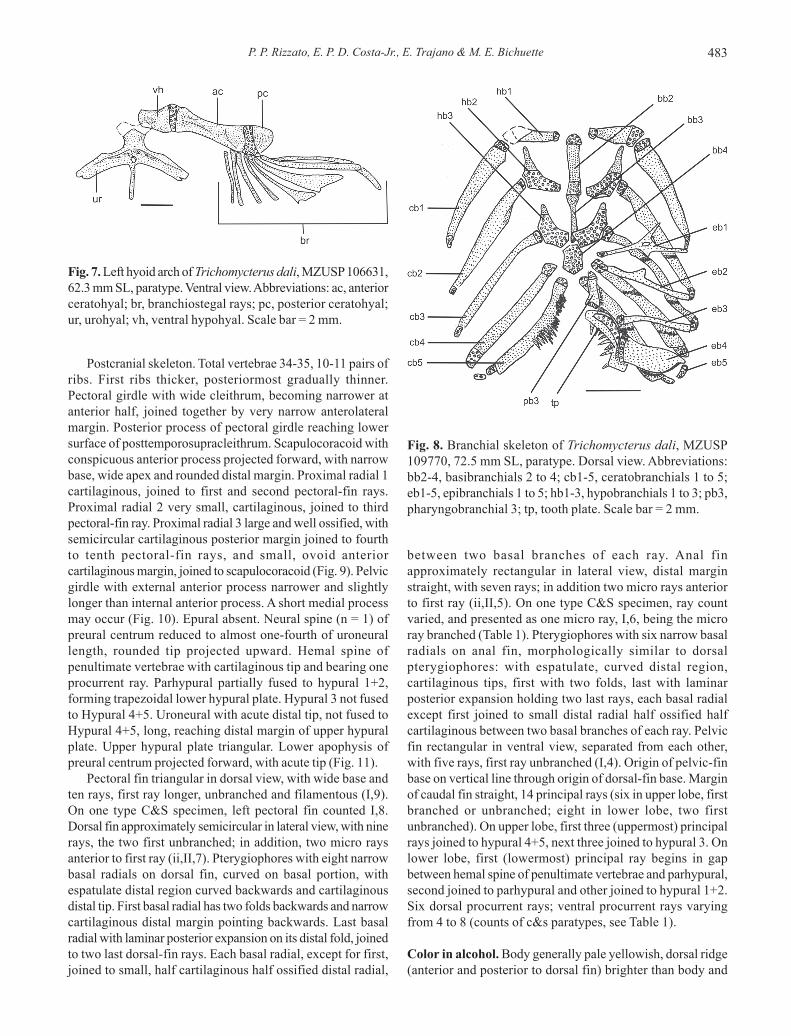
process of urohyal. Posterior ceratohyal rounded triangular(Fig. 7). Basibranchials 3, hypobranchials 3, ceratobranchials5, epibranchials 5, pharyngobranchials 2 (Fig. 8). Basibranchial1 absent. Basibranchial 2 and 3 connected to each other bytheir cartilaginous tips, forming long rod. Basibranchial 3 longand narrow, rectangular on posterior half, almost triangularon anterior half, similar to chalice. Posterior tip of basibranchial3 covered by cartilaginous posterior portion ofhypobranchials 3. Basibranchial 4 completely cartilaginous,approximately hexagonal shaped. Anterior margins ofbasibranchial 4 bordered by cartilaginous posterior portionof hypobranchials 3, lateral and posterior margins borderedby cartilaginous anterior tips of ceratobranchial 4 andceratobranchial 5, respectively. Hypobranchial 1 with externalportion wider than internal portion, with cartilaginous tips,external cartilaginous tip two to three times wider than internal.Hypobranchial 2 boomerang shaped, posterior half
cartilaginous, anterior half ossified, forming long anteriorprocess that almost reaches external posterior margin ofhipobranchial 1. Hypobranchial 3 almost completelycartilaginous, only anterior tip ossified in triangular shape,and closely joined to anterior cartilaginous tip ofceratobranchial 3. Hypobranchial 4 absent. Ceratobranchialsslightly curved, with cartilaginous tips. Ceratobranchial 1 withinternal tip wider than external tip. Ceratobranchial 2 withshallow concavity on its posterior margin, without definedposterior process. Ceratobranchial 3 with pronouncedconcavity on its posterior margin, limited posteriorly by verysmall process. Ceratobranchial 5 slightly enlarged, bearingpatch of small, narrow conical teeth pointed dorsally on itsanterior half, and connected to epibranchial 5 only by upperhalf of posterior margin. Ceratobranchials 3, 4 and 5 bearingone row of conical, very sharp teeth, on posterior margin ofceratobranchial 3, on both margins of ceratobranchial 4, andon anterior margin of ceratobranchial 5. Epibranchials 1, 2and 3 narrow, rod-like, with cartilaginous tips. Epibranchial 1with long, narrow and sharp anterior process, pointedoutwards in acute angle. Epibranchial 2 with small, acuteprocess, not uncinate. Epibranchial 3 with conspicuous,posteriorly directed, large uncinate process. Epibranchial 4large, curved, with wide dorsal margin slightly convex joinedto posterior half of tooth plate, covered with cartilage, andwith ventral margin very narrow, ovoid and cartilaginous,joined to posterior cartilaginous tip of ceratobranchial 4.Epibranchial 5 very small, curved and completely cartilaginous.Pharyngobranchials 1 and 2 absent. Pharyngobranchial 3elongate, rod-like, slightly depressed, cartilaginous tips.Pharyngobranchial 4 ossified, curved, ventral margincartilaginous, joined tightly to anterodorsal half of tooth plate.Tooth plate well developed, curved, one row of long, conic,internally curved teeth, anteriormost of which smaller,posteriormost gradually larger.
Fig. 5. Neurocranium of Trichomycterus dali, MZUSP 106631,62.4 mm SL, paratype. Dorsal view. Abbreviations: ap,autopalatine; ao, antorbital; cf, cranial fontanel; eo, epioccipital;fr, frontal; ip, infraorbital process; le, lateral ethmoid; me,mesethmoid; mx, maxilla; oc, parieto-supraoccipital; pm, pre-maxilla; psc, posttemporosupracleithrum; pt, pterotic; so,supraorbital tendon bone; sp, sphenotic-prootic-pterosphenoid complex bone; wc, weberian complex andcapsule. Scale bar = 2 mm.
Fig. 6. Left suspensorium and opercular series ofTrichomycterus dali, MZUSP 106631, 62.4 mm SL, paratype.Lateral view. Abbreviations: hm, hyomandibular; io,interopercle; mp, metapterygoid; op, opercle; po, preopercle;qu, quadrate. Scale bar = 2 mm.
P. P. Rizzato, E. P. D. Costa-Jr., E. Trajano & M. E. Bichuette 483
Postcranial skeleton. Total vertebrae 34-35, 10-11 pairs ofribs. First ribs thicker, posteriormost gradually thinner.Pectoral girdle with wide cleithrum, becoming narrower atanterior half, joined together by very narrow anterolateralmargin. Posterior process of pectoral girdle reaching lowersurface of posttemporosupracleithrum. Scapulocoracoid withconspicuous anterior process projected forward, with narrowbase, wide apex and rounded distal margin. Proximal radial 1cartilaginous, joined to first and second pectoral-fin rays.Proximal radial 2 very small, cartilaginous, joined to thirdpectoral-fin ray. Proximal radial 3 large and well ossified, withsemicircular cartilaginous posterior margin joined to fourthto tenth pectoral-fin rays, and small, ovoid anteriorcartilaginous margin, joined to scapulocoracoid (Fig. 9). Pelvicgirdle with external anterior process narrower and slightlylonger than internal anterior process. A short medial processmay occur (Fig. 10). Epural absent. Neural spine (n = 1) ofpreural centrum reduced to almost one-fourth of uroneurallength, rounded tip projected upward. Hemal spine ofpenultimate vertebrae with cartilaginous tip and bearing oneprocurrent ray. Parhypural partially fused to hypural 1+2,forming trapezoidal lower hypural plate. Hypural 3 not fusedto Hypural 4+5. Uroneural with acute distal tip, not fused toHypural 4+5, long, reaching distal margin of upper hypuralplate. Upper hypural plate triangular. Lower apophysis ofpreural centrum projected forward, with acute tip (Fig. 11).
Pectoral fin triangular in dorsal view, with wide base andten rays, first ray longer, unbranched and filamentous (I,9).On one type C&S specimen, left pectoral fin counted I,8.Dorsal fin approximately semicircular in lateral view, with ninerays, the two first unbranched; in addition, two micro raysanterior to first ray (ii,II,7). Pterygiophores with eight narrowbasal radials on dorsal fin, curved on basal portion, withespatulate distal region curved backwards and cartilaginousdistal tip. First basal radial has two folds backwards and narrowcartilaginous distal margin pointing backwards. Last basalradial with laminar posterior expansion on its distal fold, joinedto two last dorsal-fin rays. Each basal radial, except for first,joined to small, half cartilaginous half ossified distal radial,
between two basal branches of each ray. Anal finapproximately rectangular in lateral view, distal marginstraight, with seven rays; in addition two micro rays anteriorto first ray (ii,II,5). On one type C&S specimen, ray countvaried, and presented as one micro ray, I,6, being the microray branched (Table 1). Pterygiophores with six narrow basalradials on anal fin, morphologically similar to dorsalpterygiophores: with espatulate, curved distal region,cartilaginous tips, first with two folds, last with laminarposterior expansion holding two last rays, each basal radialexcept first joined to small distal radial half ossified halfcartilaginous between two basal branches of each ray. Pelvicfin rectangular in ventral view, separated from each other,with five rays, first ray unbranched (I,4). Origin of pelvic-finbase on vertical line through origin of dorsal-fin base. Marginof caudal fin straight, 14 principal rays (six in upper lobe, firstbranched or unbranched; eight in lower lobe, two firstunbranched). On upper lobe, first three (uppermost) principalrays joined to hypural 4+5, next three joined to hypural 3. Onlower lobe, first (lowermost) principal ray begins in gapbetween hemal spine of penultimate vertebrae and parhypural,second joined to parhypural and other joined to hypural 1+2.Six dorsal procurrent rays; ventral procurrent rays varyingfrom 4 to 8 (counts of c&s paratypes, see Table 1).
Color in alcohol. Body generally pale yellowish, dorsal ridge(anterior and posterior to dorsal fin) brighter than body and
Fig. 7. Left hyoid arch of Trichomycterus dali, MZUSP 106631,62.3 mm SL, paratype. Ventral view. Abbreviations: ac, anteriorceratohyal; br, branchiostegal rays; pc, posterior ceratohyal;ur, urohyal; vh, ventral hypohyal. Scale bar = 2 mm.
Fig. 8. Branchial skeleton of Trichomycterus dali, MZUSP109770, 72.5 mm SL, paratype. Dorsal view. Abbreviations:bb2-4, basibranchials 2 to 4; cb1-5, ceratobranchials 1 to 5;eb1-5, epibranchials 1 to 5; hb1-3, hypobranchials 1 to 3; pb3,pharyngobranchial 3; tp, tooth plate. Scale bar = 2 mm.

Trichomycterus dali: a new highly troglomorphic catfish484
tending to translucent. Mouth and barbels light yellowishpale to white. Pectoral, dorsal, pelvic, anal and caudal finshyaline (Fig. 2).
Color in life. Body generally white, tending to translucent,dorsal ridge darker than body, with light yellow color. Internalorgans seen by transparency (Fig. 12).
Etymology. The specific name (a noun in apposition) is anallusion to the Spanish artist Salvador Dali, in reference to hisfamously long moustache (or whisker).
Distribution. Trichomycterus dali is known exclusively fromsubterranean waters in at least three caves in Serra daBodoquena karst area: Buraco das Abelhas, Saracura andMorro do Jericó caves.
Notes on habitat, ecology and behavior. The Buraco dasAbelhas Cave is an underwater cave, with more than 2000 mof explored conduits and maximum depths of 55 m, located inthe southern portion of Serra da Bodoquena National Park, inthe rio Miranda lowlands. The only cave entrance so farknown opens into a large room with a lake, whose level
increases during rain seasons, completely flooding the saloonand only allowing access through cave diving from theentrance. The cave continues as a large, 1200 m long floodedconduit, called tunnel A. Two hundred meters from theentrance, on the right side, the tunnel A branches into tunnelB, 700 meters long and depth varying from 40 meters to up to12 meters at the distal explored end of the conduit. Eighthundred meters far from the cave entrance, again on the rightside of tunnel A, opens tunnel C; 200 meters inside tunnel C,on the left, tunnel D starts, going to a depth of up to 12meters. Non-troglomorphic fish, such as the catfish Rhamdiaquelen and tetra characins, Astyanax spp., were observedregularly in the entrance room, and farther in tunnel A until200 m from the cave entrance, reaching a depth of 49 m. At 100meters from the cave entrance, 12 m deep, these fish becomesyntopic with T. dali.
The Saracura Cave small entrance opens into a 230 m longdry conduit with a travertine lake at its distal end accessing alarge underwater cave, not totally explored so far. Theunderwater portion starts with a nearly vertical drop into a 4m diameter tunnel, becoming horizontal at the depth of 60 m.The last exploratory dive took 130 meters of guidelines andended at a depth of 65 m in a huge horizontal tunnel still to beexplored.
The Jericó Cave has three small entrances opening in theSalobra canyon. It is predominantly horizontal, with dryconduits about 15 m high and 2 m wide. Trichomycterus dalispecimens were collected in a large pool at the distal end ofthe lower conduit, which is partially filled by sandy sediments.During the collections (mid-dry season), maximum depth inthe pool was about 1 m, width was 2.5 m and extension, 4 m;
Fig. 9. Left pectoral girdle of Trichomycterus dali, MZUSP106631, 62.4 mm SL, paratype. Ventral view. Abbreviations:ap, anterior process; cl, cleithrum; fr, fin rays; pr1-3, proximalradials 1 to 3; sc, scapulocoracoid. Scale bar = 2 mm.
Fig. 10. Pelvic girdle skeleton of Trichomycterus dali, MZUSP109770, 72.5 mm SL, paratype. Ventral view. Abbreviations: ip,internal anterior process; ep, external anterior process; lp, lateralprocess; mp, medial process; pr, pelvic fin rays. Scale bar = 2mm.
P. P. Rizzato, E. P. D. Costa-Jr., E. Trajano & M. E. Bichuette 485
water flowed slowly transverly to the conduit (L. M. Cordeiro,pers. comm.).
Trichomycterus dali catfishes can be found in thepermanently dark areas zone, calmly swimming in the watercolumn and close to the rocky substrate (bottom and walls).In Buraco das Abelhas, individuals were observed from 12 upto 55 meters deep (maximum depth of the cave system); inSaracura cave, fish swim from near the surface up to a depthof 65 meters, where the main tunnel stops dropping downand become horizontal. The light from halogen underwaterspotlights, 50W, used at the time the first specimens werecollected, as well as from 10 to 20W HID spotlights usedmore recently, did not trigger any visible reaction in thesefishes. Cave divers also seem not to disturb the fishes, but,as one try to catch them, they start to swim very fast towardsthe deeper parts of the caves, against the current, near thebottom and into small crevices. Sometimes they were observeddigging the soft bottom of Buraco das Abelhas.
After being hand netted at depths of up to 65 m, thespecimens brought to the surface did not exhibit any reactionsrelated to pressure reduction. Several specimens were keptalive in small water boxes, aired with battery powered air pumpsduring at least a week and, then, traveled 1,200 km until the
laboratory in São Paulo. Three specimens collected in 2000and 2001 were still alive in 2010.
Discussion
Taxonomy. To date, more than 130 valid species are assignedto the genus Trichomycterus, of which more than 60 occur inBrazil (Eschmeyer & Fong, 2010), in addition to many otherspresently in the process of or awaiting for description (Alencar& Costa, 2004; Wosiacki, 2005; Wosiacki & de Pinna, 2008a;Fernández & Vari, 2009), turning the genus into the mostspecies-rich within the family (de Pinna, 1998; Wosiacki & dePinna, 2008b; Fernández & Vari, 2009; Eschmeyer & Fong,2010). Nevertheless, as mentioned above, the evolutionaryrelationships and phylogenetic history of the genus are stillobscure (Wosiacki & de Pinna, 2008a), and many authorspoint out to its non-monophyletic character (de Pinna, 1998;Wosiacki, 2002), making the identification of new species ofTrichomycterus very complicated (Wosiacki, 2005; da Silvaet al., 2010). For this reason, only species with broadlydistinctive features, which can be easily recognized as newtaxa, have been recently described (de Pinna, 1998; Wosiacki& de Pinna, 2008b). Moreover, due to the lack ofsynapomorphic characters distinguishing the genusTrichomycterus, it has been included those species lackingthe synapomorphies accepted for Bullockia, Eremophilus,Hatcheria, Ituglanis, Rhizosomichthys, Scleronema, andSilvinichthys, as proposed by Arratia (1990, 1998), Costa andBockmann (1993), and Fernández and de Pinna (2005). As aresult, Trichomycterus is nowadays a large assemblage ofspecies not necessarily closely related (Wosiacki & de Pinna,2008b). Because none of the synapomorphies of otherTrichomycterinae genera could be recognized for the newspecies from Serra da Bodoquena, its inclusion inTrichomycterus seems to be the most reasonable alternativefor the moment.
Trichomycterus dali is a very distinctive species,especially due to four easily noticeable characters: (a) highdegree of reduction on melanic skin pigmentation, characterthat differentiates the new species from all epigeancongeners, except for T. gorgona; (b) eyes reduced, notvisible externally, which also distinguishes the new speciesfrom all epigean congeners (including T. gorgona) and furtherdifferentiates it from all cave-restricted congeners exceptfor T. spelaeus and T. sandovali; (c) very long barbels,especially the nasal and the maxillary ones, which maydistinguish the new species from other congeners, exceptfor T. longibarbatus (epigean) and T. spelaeus (hypogean)and represents one of the longest barbels within the genus;and (d) presence of very conspicuous adipose ridges inboth young and adult individuals, lining dorsally throughoutthe body, which differentiates the new species form all of itscongeners, both epigean and hypogean, including T.gorgona, T. longibarbatus, T. spelaeus and T. sandovali.The three first characters are clearly related to itssubterranean habit, because reduction of eyes and
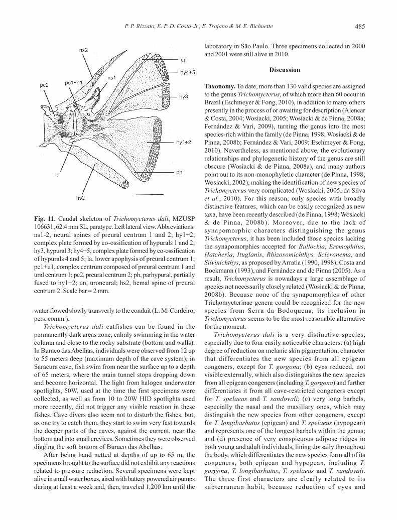
Fig. 11. Caudal skeleton of Trichomycterus dali, MZUSP106631, 62.4 mm SL, paratype. Left lateral view. Abbreviations:ns1-2, neural spines of preural centrum 1 and 2; hy1+2,complex plate formed by co-ossification of hypurals 1 and 2;hy3, hypural 3; hy4+5, complex plate formed by co-ossificationof hypurals 4 and 5; la, lower apophysis of preural centrum 1;pc1+u1, complex centrum composed of preural centrum 1 andural centrum 1; pc2, preural centrum 2; ph, parhypural, partiallyfused to hy1+2; un, uroneural; hs2, hemal spine of preuralcentrum 2. Scale bar = 2 mm.

Trichomycterus dali: a new highly troglomorphic catfish486
pigmentation and enlargement of appendages bearingsensorial structures are broadly recognized as thecommonest traits characterizing troglobites (Hüppop, 2000;Trajano, 2001), and these characters are commonly used indescriptions of exclusively hypogean fishes (Renno et al.,2007). The fourth, although not characteristic of troglobitesand even present in epigean species of the family, may bepossibly related to its subterranean habit, as we discussbelow (see Adipose folds).
Among the six cave-restricted Trichomycterus previouslydescribed, only T. itacarambiensis occurs in Brazil, in theupper rio São Francisco basin, separated from the Paraguaibasin, where T. dali occurs, by the large rio Paraná basin.Trichomycterus dali may be distinguished from T.itacarambiensis by the complete absence of externally visibleeyes (vs. large intraespecific variation in eye development inthe latter), the highly reduced skin pigmentation, with colorpattern pale yellowish to bright white (vs. color patterncomposed of small irregular, roundish black spots), the originof pelvic fin on the vertical line through origin of dorsal finbase (vs. base of pelvic fin slightly anterior to vertical throughorigin of dorsal fin) and by longer barbels (see Table 2).
Among the other troglomorphic species of the genus, T.dali may be distinguished from: (1) T. spelaeus, by the ventralprofile of body straight (vs. slightly convex in the latter), thehead trapezoidal in dorsal view (vs. triangular), the anal finapproximately rectangular in lateral view with distal marginstraight (vs. rounded, distal margin semicircular) and by thesingle cranial fontanel (vs. two cranial fontanels, anterior andposterior, separated by epiphyseal bar); (2) T. chaberti, bythe absence of eyes (vs. presence, in variable degree amongindividuals), the highly reduced skin pigmentation (vs. variable,moderately pigmented, yellowish with brown spots), the originof pelvic fin on the vertical line through origin of dorsal-finbase (vs. vertical line through origin of dorsal fin located atthe end of pelvic fin), the origin of anal fin posterior to theend of dorsal-fin base (vs. origin of anal fin below the dorsal
fin base), and the profile of caudal fin, with a conspicuousconstriction on the origin of rays and a upper lobe projectedupward (vs. dorsal and ventral profile straight); (3) T.sandovali, by the head trapezoidal in dorsal view (vs.triangular), the lateral profile of head straight (vs. semicurve)and the cranial fontanel unique (vs. two cranial fontanels,anterior and posterior, separated by epiphyseal bar); (4) T.santanderensis, by the complete absence of eyes (vs.presence, variable from visible to imperceptible), the highlyreduced skin pigmentation (vs. variable from homogeneouslight-red on adults to pale rose with small, gray, dorsal roundspots on young individuals), the profile of caudal fin, with aconstriction on the origin of rays, dorsal lobe projected upward(vs. dorsal and ventral profile slightly convex), the headtrapezoidal in dorsal view (vs. triangular), and the longer head,ranging from 19.9 to 22.1% SL (vs. 18.2-18.9% SL); (5) T. uisae,by the absence of eyes (vs. presence, reduced but well defined,ranging from 7.4 to 11.1% HL), the highly reduced skinpigmentation (vs. reduced pigmentation, consisting of groundcoloration light brow without spots and a narrow predorsalbluish-gray stripe), origin of the pelvic fin on the vertical linethrough origin of dorsal-fin base (vs. origin of pelvic fin anteriorto vertical through origin of dorsal fin), the profile of caudalfin, with a constriction on the origin of rays, dorsal lobeprojected upward (vs. dorsal and ventral profile slightlyconvex), and the single cranial fontanel (vs. two cranialfontanels, anterior and posterior, separated by a narrowchannel) (see Table 2).
Troglobitic status. According to the classical Schiner-Racovitza system, troglobites are defined as organismsrestricted to subterranean habitats, incapable to formepigean source populations, in opposition to troglophyles,that may form source populations both in epigean andhypogean habitats, and trogloxenos, regularly found insubterranean habitats, but which, to be able to completetheir life cycle, must leave such habitats periodically (Barr,1968; Holsinger & Culver, 1988; Trajano, 2005, 2010). It islogically impossible (see Trajano & Bichuette, 2010), todemonstrate the absence of some biological feature in agiven area, and the only practical solution is a statisticalapproach, based on a surveying effort high enough to testthe sampling sufficiency (such as species accumulationcurves). Unfortunately, this condition is rarely achieved.Therefore, the practice has become to use autapomorphiccharacter states to deduce the troglobitic status, speciallythe so-called classical troglomophisms, with emphasis onreduction of ocular structures and melanic pigmentation,and also hyperdevelopment of sensorial structures, inrelation to closely related epigean species (e.g., Culver &Pipan, 2009).
Extensive fish collections have been made in severalepigean streams of Serra da Bodoquena area, but only oneTrichomycterus specimen was found (L.C. Medeiros & R.Borghezan, pers. comm.), indicating very low densities and/or very localized distributions of epigean populations.
Fig. 12. Trichomycterus dali, adult, live specimenphotographed in aquarium on May, 2011, unpreserved; Brazil,Mato Grosso do Sul State, Serra da Bodoquena karst area, rioParaguai basin, Jardim, Buraco das Abelhas cave. Courtesyof Dante Fenolio.

P. P. Rizzato, E. P. D. Costa-Jr., E. Trajano & M. E. Bichuette 487
Although these collecting efforts are still insufficient for aconclusive statement of restriction to cave habitats, theassociation with advanced troglomorphic traits in T. dali,indicating a long process of differentiation in isolation in thesubterranean habitat, strongly corroborates the hypothesisof a troglobitic status for this species.
Representatives of the family Trichomycteridae have beenvery successful in colonizing subterranean environments(Reis et al., 2006; Castellanos-Morales, 2007), with troglobiticand troglophilic populations (Mattox, 2008). Troglomorphicrepresentatives of the family can be found in five genera, theArgentinian Silvinichthys (one troglobitic species), theBrazilian Copionodon (one species), Glaphyropoma (onespecies) and Ituglanis (six species), and the widespreadTrichomycterus (nine species, including two to be described)(Proudlove, 2010). In all trichomycterids referred to astroglobites, reduction of eyes and pigmentation at some extent,as well as long barbels, have been reported (DoNascimientoet al., 2001; Romero & Paulson, 2001; Bichuette & Trajano,2008; Castellanos-Morales, 2008). However, the total loss ofexternally visible eyes as a fixed character throughout thepopulation was observed for relatively few of these species,namely T. sandovali, T. spelaeus and T. dali. Similarly, not allof these species present an advanced regression of skinpigmentation, with many of them exhibiting melanic pigmentsin variable degrees of regression, mainly at dorsum and flanks(e.g., Glaphyropoma spinosum, Silvinichthys bortayro,Ituglanis bambui, I. ramiroi, T. chaberti, T. itacarambiensis)(Bichuette & Trajano, 2010). Several authors point out thatthere would be a correlation between the reduction degree ofeyes and pigmentation and the time of isolation in the
subterranean habitat, and that species with a high degree ofregression of eyes and melanic pigmentation have beenisolated for a longer time than those with lower or variabledegree of reduction in these characters (Langecker, 2000;Bichuette & Trajano, 2004, 2008).
The elongation of sensorial appendages, basically barbels,has been observed in troglobitic fishes (Hüppop, 2000, amongothers), including heptapterids as Rhamdia (Weber, 1996) andseveral trichomycterids (Fernández & Bichuette, 2002;DoNascimiento et al., 2001; Castellanos-Morales, 2008).Following this trend, T. dali exhibits long barbels, especiallythe nasal and maxillary ones, both surpassing the headlength. Three other species of Trichomycterus exhibits barbelsas long or almost as long as those of T. dali: T. longibarbatus,T. spelaeus and T. sandovali. In the case of T. longibarbatus,no information is given about its habits, but the elongatedbarbels may indicate that this species depends more on thissensorial appendage to acquire information about theirenvironment. The two last, as T. dali, are restricted tosubterranean habitats, where barbels constitute an importantsource of sensory information about the environment in theabsence of light, and its elongation may be involved insensory compensation as an adaptation to the permanentdarkness of the subterranean habitat; however, further studiesare needed to corroborate this hypothesis. Another additionalfeature was proposed by Castellanos-Morales (2008) aspossibly related to a hypogean life, the presence of lateralcutaneous folds encircling the body, present on at least threetroglomorphic species of the genus, T. santanderensis, T.spelaeus and T. uisae. In T. dali, this feature is present inmany, but not all, of the preserved individuals.
Table 2. Comparative data on external morphology of Trichomycterus subterranean species.
T. dali T. itacarambiensis T. spelaeus T. chaberti T. sandovali T. santanderensis T. uisae
Eyes Not visible Presence,
intraespecific variation
Not visible Presence, variable
degree among individuals
Not visible Presence, from visible to
imperceptible Presence, reduced but well defined
Skin pigmentation
Pale yellowish to bright white
Small, irregular roundish black
spots
Bright yellow to light brow
Variable, moderately pigmented,
yellowish with brown spots
Bright yellow
Variable from homogeneous light-red to pale rose with small gray,
dorsal round spots
Reduced, light brown without spots, narrow
predorsal bluish-gray stripe
Origin of pelvic fin
On vertical through origin of dorsal fin
base
Slightly anterior to origin of dorsal fin
On vertical through origin of dorsal fin base
Anterior to origin of dorsal fin
On vertical through origin of dorsal fin
base
On vertical through origin of dorsal fin base
Anterior to origin of dorsal fin
Ventral profile of body
Straight Slightly convex or
nearly straight Slightly convex Slightly concave
Slightly concave
Straight Straight
Dorsal view of head
Trapezoidal Trapezoidal Triangular Triangular Triangular Triangular Trapezoidal
Anal fin in lateral view
Rectangular, distal margin straight
Half-ellipsoidal Rounded, distal
margin semicircular
(unobtained data) Distal margin
rounded Rounded Rounded
Profile of caudal fin
With a constriction on the origin of rays, dorsal lobe projected
upward
Dorsal and ventral profile slightly
convex
Dorsal and ventral profile slightly convex
Dorsal and ventral profile straight
Dorsal and ventral profile slightly convex
Dorsal and ventral profile slightly convex
Dorsal and ventral profile slightly
convex

Trichomycterus dali: a new highly troglomorphic catfish488
Adipose folds. The presence of dorsal adipose folds in adultT. dali is an intriguing character. The presence of a predorsaladipose fold in adults is unique among the Trichomycterinae.Among other trichomycterids, a similar predorsal adipose foldis only found in the highly-derived sarcoglanidineSarcoglanis simplex Myers & Weitzman. However, thatstructure in Sarcoglanis is much less developed than that inT. dali (compare fig. 1 of Myers & Weitzman, 1966, with ourFig. 2). In any event, the predorsal fold of T. dali necessarilyhave evolved independently of the condition of Sarcoglanis(cf. de Pinna, 1989, de Pinna & Starnes, 1990; Costa &Bockmann, 1994; Costa, 1994), being its presence interpretedas an obvious and distinctive diagnostic feature for the newspecies herein described.
The well-developed postdorsal adipose fold notsupported by procurrent rays in adults of T. dali is also uniquewithin Trichomycterus and even among othertrichomycterines. A few other trichomycterines also exhibit asmall postdorsal adipose fold (e.g., T. stramineus, T.nigromaculatus; Eigenmann, 1918), but that structure in thosefishes is never so pronounced as in T. dali. Trichomycterusstawiarski, T. igobi, T. crassicaudatus, and T. lewi exhibit apostdorsal ridge that may resemble the postdorsal adiposefold of T. dali. In these other congeners, however, that ridgeis supported by an unusual large number of procurrent caudalrays distributed along most of its extent. In T. dali, theprocurrent caudal rays are in few number and restricted to theposterior most part of the fold, which is primarily notsupported by procurrent rays.
Other trichomycterid taxa also exhibit a postdorsal adiposefold, such as the sarcoglanidines Sarcoglanis simplex andMalacoglanis gelatinosus Myers & Weitzman, and theCopionodontinae. Given the currently accepted topologiesof the trichomycterid phylogenetic relationships (de Pinna,1992, 1998; Datovo & Bockmann, 2010), those occurrencesare more parsimoniously interpreted as parallelisms.
It is known that, ontogenetically, adipose folds posteriorto dorsal fin of juveniles originates the adipose fin present insome adults. Although juveniles of trichomycterid speciesare not well known because of difficulties in collect thesespecimens, on at least one species, Copionodon lianae, thisontogenetical development is known to occur (Campanario& de Pinna, 2000): juveniles C. lianae exhibit a well-developedadipose fold, both pre- and postdorsal, that gradually becomesmaller as individuals become larger, so that the pre-dorsalfold totally disappears in adults and the post-dorsal foldreaches the appearance of the adipose fin of adults. This isalso true for another species of the genus, Copionodon sp.from northeastern Brazil, analyzed by us. In the trichomycterineItuglanis amazonicus, small juveniles also exhibit pre- andpost-dorsal adipose ridges, lost afterward in adult individuals(Lundberg et al., 2004).
Based on this, we could hypothesize the ontogeneticdevelopment of the adipose fin in trichomycterids as a shiftfrom the ridge-like adipose fold of juveniles to a fin-like adiposefold (the adipose fin), and its further disappearance on the
end of the series. From this hypothetical series, three possibleevolutionary paths are represented by adults of differentspecies: the retention in adults of a ridge-like adipose fold,post-dorsal (M. gelatinosus and C. orthiocarinatus) or bothpre- and postdorsal (S. simplex and T. dali); the modificationto a fin-like adipose fold, i.e., the adipose fin (remainingcopionodontines); and the more advanced step, the total lossof pre- and post-dorsal adipose folds, (most trichomycterids).From this, it become clear that the adipose folds of T. dali, S.simplex, M. gelatinosus and the Copionodontinae are formedby the retention of the adipose folds of juveniles, in differentdegrees in each species, representing then a paedomorphiccharacter.
Although this seems to be a reasonable hypothesis, someessential points need to be emphasized. First, the hypothesisis based solely on external morphologic appearance of theadipose fold, and wasn’t our intention to accomplish a moredetailed analysis of this character, since it would be beyondthe scope of this work. Second, very little information isavailable for juvenile trichomycterids, and as the hypothesisis based on data of only three species (the copionodontinesC. lianae and Copionodon sp. and the trichomycterineItuglanis amazonicus), its weakness must to be considered,as long as new data for other trichomycterid species maycompletely alter or even refute the hypothesis. Third, even iffurther studies corroborate this hypothesis, it is clear that thegroups so defined must not be considered as natural groups,once that many data suggest other phylogenetic relationshipsfor the species within the groups. Therefore, the degree ofretention of adipose folds in adults is better understood as aplastic character that evolved many times within the lineage,and the retention of this fold in adults of different lineages ismore likely to represent parallelism. In conclusion, by nowthis is just a hypothesis that must be further tested before westart to discuss its implications to the understanding oftrichomycterid evolution, and the best way to do so isidentifying and analyzing juvenile specimens oftrichomycterids and building ontogenetical series to trackthe actual development of the adipose fold.
Anyway, as cited above, in T. dali the adipose folds ofadults are very similar morphologically to the adipose foldsdescribed for juvenile C. lianae, and its presence probablybegins early in development, since even in the smallestindividuals analyzed (18.6 mm SL and 19.6 mm SL, non-paratype specimens) it was already present, although poorlydeveloped. This evidence give support to the hypothesis ofpaedomorphic evolution for this character. As all other speciesof the genus do not exibit this character, and as no otherpaedomorphic character state could be recognized in T. dali,the retention of the ridge is more probably related to resultfrom neotenic evolution. In cave vertebrate species, neotenywas already reported for troglobitic salamanders, such as thenotorious European Proteus anguinus and several NorthAmerican plethodontids, and fishes, as the North Americanamblyopsids.
A major ecological feature of subterranean habitats is the
P. P. Rizzato, E. P. D. Costa-Jr., E. Trajano & M. E. Bichuette 489
food scarcity as a limiting factor, and the dependence, ingeneral, on external energy sources, frequently unpredictableand patchy (Langecker, 2000; Culver & Pipan, 2009). Increasedfat layers and large fat deposits have been reported for somehypogean fishes as an adaptation to store energy, in order tocope with periodical starvation (Weber, 1996; Hüppop, 2000;Culver & White, 2005). We then hypothesize that the welldeveloped adipose fold in T. dali, acquired by neoteny, is anadaptation to withstand the energy restrictions andunpredictability of food sources in the subterranean habitat,by accumulating a permanent, reliable storage of energy inthe form of adipose reserve. As similar adipose folds havebeen reported for other, epigean trichomycterids, as discussedabove, where similar food restriction may not exist, alternativehypothesis can be related to its evolution on each group.
Comparative material. Ituglanis bambui, MZUSP 79860,holotype, Brazil: Goiás: São Domingos: Terra Ronca State Park:Angélica Cave. Ituglanis epikarsticus, MZUSP 79869, holotype,Brazil: Goiás: São Domingos: Terra Ronca State Park: São MateusCave. Ituglanis passensis: MZUSP 80097, 3, Brazil: Goiás: SãoDomingos, Passa Três Cave. Ituglanis proops: MZUSP 79576, 15,Brazil: Paraná: Cerro Azul, ribeirão Bonito. Ituglanis ramiroi,MZUSP 79865, holotype, Brazil: Goiás: São Domingos: Terra RoncaState Park: São Bernardo Cave. Ituglanis sp.: MZUSP 53222, 6,Brazil: Goiás: Minaçú, tributary of rio Tocantinzinho.Trichomycetrus bahianus: MZUSP 74655, 10, Brazil: Bahia:Livramento do Brumado, rio Brumado; MZUSP 45887, 7, Brazil:Bahia: Livramento do Brumado, rio Brumado. Trichomycterusitacarambiensis: MZUSP 50548, paratypes, 4, Brazil: MinasGerais: Itacarambi, Olhos D’Água Cave; MZUSP 50549, paratypes,2, Brazil: Minas Gerais: Itacarambi, Olhos D’Água Cave; MZUSP81078, 5, Brazil: Minas Gerais: Itacarambi, Olhos D’Água Cave.Trichomycterus zonatus: MZUSP 68173, 20, Brazil: São Paulo:Cajati, rio do Queimado; MZUSP 23038, 6, Brazil: São Paulo:Caraguatatuba, rio d’Ouro. The comparison with otherTrichomycterus species was based on the literature.
Acknowledgements
Especial thanks are given to A. Datovo da Silva, for hisattention, major contributions and valuable advices thatenriched this work. We are greatly indebted to the colleagueswho joined our speleological expeditions, contributing to thelogistics essential for the highly technical cave diving (carriedout by E. P. D. Costa-Jr.), L. M. Cordeiro and R. Borghezan,responsible by the expedition to Morro do Jericó. We thankD. Fenolio for the picture of Figure 12; L. B. R. Fernandes andA. M. P. M. Dias for the picture of Figure 4; C. Martins for thehelp with other pictures; D. Seripieri of the MZUSP library,for her attention and help with literature. Thanks are due toFAPESP, for a long-term support for our studies (in particular,procs. 97/09274-1, 03/00794-5 and 09/15030-7).
Literature cited
Alencar, A. R. & W. J. E. M. Costa. 2004. Trichomycteruspauciradiatus, a new catfish species from the upper rio Paraná
basin, southeastern Brazil (Siluriformes: Trichomycteridae).Zootaxa, 1269: 43-49.
Almeida, F.F.M. de. 1965. Geologia da Serra da Bodoquena (MatoGrosso), Brasil. Boletim da Divisão de Geologia e Mineralogia,DNPM, 219: 1-96.
Alvarenga, S.M.; A. E. Brasil &D. M. Del’Arco. 1982. Folha SF-21, Campo Grande. 2- Geomorfologia, Projeto RADAM-BRA-SIL, Rio de Janeiro, 28: 125-184.
Arratia, G. 1990. The South American Trichomycterinae (Teleostei:Siluriformes), a problematic group. Pp: 395-403. In: Peters, G.& R. Hutterer (Eds.).Vertebrates in the Tropics. MuseumAlexander Koening, Bonn.
Arratia, G. 1998. Silvinichthys, a new genus of trichomycteridcatfishes from the Argentinian Andes, with redescription ofTrichomycterus nigricans. Ichthyological Explorations ofFreshwaters, 9: 347-370.
Barr, T. C., Jr., 1968. Cave ecology and the evolution of troglobites.Evolutionary Biology, 2: 35-102.
Bichuette, M. E., M. C. C. de Pinna & E. Trajano. 2008. A newspecies of Glaphyropoma: the first subterranean copionodontinecatfish and the first occurrence of opercular odontodes in thesubfamily (Siluriformes: Trichomycteridae). NeotropicalIchthyology, 6:301-306.
Bichuette, M. E. & E. Trajano. 2004. Three new subterranean speciesof Ituglanis from Central Brazil (Siluriformes:Trichomycteridae). Ichthyological Exploration of Freshwaters,15: 243-256.
Bichuette, M. E. & E. Trajano. 2005. A new cave species of RhamdiaBleeker, 1858 (Siluriformes: Heptapteridae) from Serra doRamalho, northeastern Brazil, with notes on ecology andbehavior. Neotropical Ichthyology, 3:587-595.
Bichuette, M. E. & E. Trajano. 2008. Ituglanis mambai, a newsubterranean catfish from a karst area of Central Brazil, rioTocantins basin (Siluriformes: Trichomycteridae). NeotropicalIchthyology, 6: 9-15.
Bockmann, F. A., L. Casatti & M. C. C. de Pinna. 2004. A newspecies of trichomycterid catfish from the Rio Paranapanemabasin, southeastern Brazil (Teleostei: Siluriformes), withcomments on the phylogeny of the family. IchthyologicalExploration of Freshwaters, 15: 225-242.
Bockmann, F. A. & R. M. C. Castro. 2010. The blind catfish fromthe caves of Chapada Diamantina, Bahia, Brazil (Siluriformes:Heptapteridae): description, anatomy, phylogeneticrelationships, natural history, and biogeography. NeotropicalIchthyology, 8(4): 673-706.
Boggiani, P. C.; T. R. Fairchild & A. M. Coimbra.1993. O GrupoCorumbá (Neoproterozóico-Cambriano) na região Central daSerra da Bodoquena, Mato Grosso do Sul (Faixa Paraguai).Revista Brasileira de Geociências, 23: 301-305.
Campanario, C. F. & M. C. C. de Pinna. 2000. A new species of theprimitive trichomycterid subfamily Copionodontinae fromnortheastern Brazil (Teleostei: Trichomycteridae). IchthyologicalExploration of Freshwaters, 11: 369-375.
Castellanos-Morales, C. A. 2007. Trichomycterus santanderensis:A new species of troglomorphic catfish (Siluriformes:Trichomycteridae) from Colombia. Zootaxa, 1541: 49-55.
Castellanos-Morales, C. A. 2008. Trichomycterus uisae: a newspecies of hypogean catfish (Siluriformes: Trichomycteridae)from the northeastern Andean Cordillera of Colombia.Neotropical Ichthyology, 6:307-314.
Costa, W. J. E. M. 1994. A new genus and species of Sarcoglanidinae(Siluriformes: Trichomycteridae) from the Araguaia basin, cen-
Trichomycterus dali: a new highly troglomorphic catfish490
tral Brazil, with notes on subfamilial phylogeny. IchthyologicalExploration of Freshwaters, 5: 207-216.
Costa, W. J. E. M. & F. A. Bockmann. 1993. Un nouveau genrenéotropical de la famille des Trichomycteridae (Siluriformes:Loricarioidei). Revue Française d’Aquariologie et Herpetologie,20: 43-46.
Culver, D. C. 1982. Cave Life: Evolution and Ecology. Cambridge,Harvard University Press, 189p.
Culver, D.C. & T. Pipan.2009. Biology of caves and othersubterranean habitats. Oxford, Oxford University Press, 256 p.
Culver, D. C. & W. B. White. 2005. Encyclopedia of Caves.Amsterdam, Elsevier Academic Press, 654p.
Datovo, A. & F. A. Bockmann. 2010. Dorsolateral head muscles ofthe catfish families Nematogenyidae and Trichomycteridae(Siluriformes: Loricarioidei): comparative anatomy andphylogenetic analysis. Neotropical Ichthyology, 8: 193-246.
DoNascimiento, C., O. Villarreal & F. Provenzano. 2001. Descripciónde una nueva especie de bagre anoftalmo del géneroTrichomycterus (Siluriformes: Trichomycteridae), de una cuevade la Sierra de Perijá, Venezuela. Boletín de la SociedadVenezolana de Espelelología, 35: 20-26.
Eschmeyer, W. N. & J. D. Fong. 2010. Species of Fishes by family/subfamily. Available from:http://research.calacademy.org/redirect?url=http://researcharchive.calacademy.org/research/Ichthyology/catalog/fishcatmain.asp. Accessed May 6, 2010.
Fernández, L. & M. E. Bichuette. 2002. A new cave dwelling speciesof Ituglanis from the São Domingos karst, central Brazil(Siluriformes: Trichomycteridae). Ichthyological Exploration ofFreshwaters, 13: 273-278.
Fernández, L. & G. Miranda. 2007. A catfish of the genusTrichomycterus from a thermal stream in southern South America(Teleostei: Siluriformes: Trichomycteridae), with comments onrelationships within the genus. Journal of Fish Biology, 71:1303-1316.
Fernández, L. & M. C. C. de Pinna. 2005. Phreatic catfish of thegenus Silvinichthys from southern South America (Teleostei,Siluriformes, Trichomycteridae). Copeia, 2005: 100-108.
Fernández, L. & R. P. Vari. 2009. New Species of Trichomycterusfrom the Andean Cordillera of Argentina (Siluriformes:Trichomycteridae). Copeia, 2009: 195-202.
Galati, E. A. B., V. L. B. Nunes, P. C. Boggiani, M. E. C. Dorval, G.Cristaldo, H. C. Rocha, E. T. Oshiro, R. M. Gonçalves-Andrade& G. Naufel. 2003. Phlebotomine (Diptera, Psychodidae) incaves of the Serra da Bodoquena, Mato Grosso do Sul state,Brazil. Revista Brasileira de Entomologia 47: 283-296.
Holsinger, J. R. & D. C. Culver. 1988. The invertebrate cave faunaof Virginia and a part of Eastern Tennessee: Zoogeography andecology. Brimleyana, 14: 1-162.
Hüppop, K. 2000. How do cave animals cope with the food scarcityin caves? Pp. 159–188. In: Wilkens, H., D. C. Culver and W. F.Humphreys (Eds.). Ecosystems of the World, Vol. 30:Subterranean Ecosystems. Amsterdam, Elsevier, 808p.
Justo, L. J. E. C. 2000. Fosfato da Serra a Bodoquena - MatoGrosso do Sul. In: Programa de Avaliação Geológico-econômicade insumos minerais para agricultura no Brasil, Projeto PIMA-GO/TO/MT/MS. Goiânia, CPRM, Relatório Final. 38p.
Langecker, T. G. 2000. The effects of continuous darkness on caveecology and cavernicolous evolution. Pp. 135-137. In: Wilkens,H., D. C. Culver and W. F. Humphreys (Eds.). Ecosystems ofthe World, Vol. 30: Subterranean Ecosystems. Amsterdam,Elsevier, 808p.
Lima, S. M. Q. & W. J. E. M. Costa. 2004. Trichomycterus giganteus(Siluriformes: Loricarioidea: Trichomycteridae): a new catfishfrom the Rio Guandu basin, southeastern Brazil. Zootaxa, 761:1-6.
Lundberg, J. G., T. M. Berra & J. P. Friel. 2004. First description ofsmall juveniles of the primitive catfish Diplomystes (Siluriformes:Diplomystidae). Icthyological Exploration of Freshwaters, 15:71-82.
Mattox, G. M. T., M. E. Bichuette, S. Secutti & E. Trajano. 2008.Surface and subterranean ichthyofauna in the Serra do Ramalhokarst area, northeastern Brazil, with updated lists of Braziliantroglobitic and troglophilic fishes. Biota Neotropica, 8: 145-152.
Moreira, C. R., M. E. Bichuette, O. T. Oyakawa, M. C. C. de Pinna& E. Trajano. 2010. Rediscovery and redescription of theunusual subterranean characiform Stygichthys typhlops, withnotes on its life history. Journal of Fish Biology, 76: 1815–1824.
Myers, G. S. & S. H. Weitzman. 1966. Two remarkable newtrichomycterid catfishes from the Amazon basin in Brazil andColombia. Journal of Zoology, London, 149: 277-287.
Nelson, J. S. 2006. Fishes of the World, 4 ed., New York, Wiley,624p.
de Pinna, M. C. C. 1989. A new sarcoglanidine catfish, phylogenyof its subfamily, and an appraisal of the phyletic status of theTrichomycterinae. American Museum Novitates, 2950: 1-39
de Pinna, M. C. C. 1992. A new subfamily of Trichomycteridae(Teleostei, Siluriformes), lower loricarioid relationships and adiscussion on the impact of additional taxa for phylogeneticanalysis. Zoological Journal of the Linnean Society, 106: 175-229.
de Pinna, M. C. C. 1998. Phylogenetic relationships of NeotropicalSiluriformes (Teleostei: Ostariophysi): historical overview andsynthesis of hypotheses. Pp. 279-330. In: Malabarba, L. R., R.E. Reis, R. P. Vari, Z. M. S. Lucena & C. A. S. Lucena (Eds.).Phylogeny and Classification of Neotropical Fishes. Porto Ale-gre, Edipucrs, 603p.
de Pinna, M. C. C. & W. C. Starnes, 1990. A new genus ofSarcoglanidinae from the Rio Mamore, Amazon basin, withcomments on subfamiIial phylogeny (Teleostei,Trichomycteridae). Journal of Zoology, London, 222: 75-88.
Proudlove, G. S. 2010. Biodiversity and distribution of thesubterranean fishes of the world. Pp. 41-63. In: Trajano, E., M.E. Bichuette & B. G. Kapoor (eds.). Biology of SubterraneanFishes. Enfield, Science Publishers, 480p.
Reis, R. E., E. Trajano & E. Hingst-Zaher. 2006. Shape variation insurface and cave populations of the armoured catfishes Ancistrus(Siluriformes: Loricariidae) from the São Domingos karst area,upper Tocantins River, Brazil. Journal of Fish Biology, 68:414-429.
Renno, J. F., C. Gazel, G. Miranda, M. Pouilly & P. Berrebi.2007.Delimiting species by reproductive isolation: the geneticstructure of epigean and hypogean Trichomycterus spp.(Teleostei: Siluriformes) in the restricted area of Torotoro (UpperAmazon, Bolivia). Genetica, 131: 325-336.
Romero, A. 2001. Scientists prefer them blind: the history ofhypogean fish research. Environmental Biology of Fishes, 62:43-71.
Romero, A. & K. M. Paulson. 2001. It’s a wonderful hypogean life:a guide to the troglomorphic fishes of the world. EnvironmentalBiology of Fishes, 62: 13-41.
P. P. Rizzato, E. P. D. Costa-Jr., E. Trajano & M. E. Bichuette 491
da Silva, C. C. F., S. L. S. F. da Matta, A. W. S. Hilsdorf, F. Langeani& A. P. Marceniuk. 2010. Color pattern variation inTrichomycterus iheringi (Eigenmann, 1917) (Siluriformes:Trichomycteridae) from rio Itatinga and rio Claro, São Paulo,Brazil. Neotropical Ichthyology, 8:49-56.
Taylor, W. R. & G. C. Van Dyke. 1985. Revised procedures forstaining and clearing small fishes and other vertebrates for boneand cartilage study. Cybium, 9: 107-119.
Tchernavin, V. V. 1944. A revision of some Trichomycterinae basedon material preserved in the British Museum (Natural History).Proceedings of the Natural Society of London, 114: 234-275.
Trajano. E. 1986. Alguns problemas envolvidos na classificaçãoecológica dos cavernícolas. Espeleo-tema, 15: 25-27.
Trajano, E. 1997. Synopsis of Brazilian troglomorphic fishes. Seriedocuments - Laboratoire souterrain du C.N.R.S, França, 24:119-126.
Trajano. E. 2001. Ecology of subterranean catfishes: an overview.Environmental Biology of Fishes, 62: 133-160.
Trajano, E. 2003.Ecology and ethology of subterranean catfishes.Pp. 601-635. In: Arratia, G., B. G. Kapoor, M. Chardon& R.Diogo (Org.). Catfishes. Ed. 1, Vol. 2 Enfield: Science Publishers,750p.
Trajano, E. 2010. Source versus sink populations concept appliedto the Schiner-Racovitza classification of subterraneanorganisms, p. 174. In: 20th International Conference onSubterranean Biology, Postojna (book of Abstracts).
Trajano, E. & M. E. Bichuette. 2004. Diversity of subterraneanfishes in Brazil. Pp. 161-163. In: Symposium on WorldSubterranean Biodiversity - Proceedings. Lyon, França: CNRSUniversité Claude Bernard Lyon.
Trajano, E. & M. E. Bichuette. 2010. Subterranean Fishes of Brazil.Pp. 331-355. In: Trajano, E., M. E. Bichuette &B. G. Kapoor(Eds.). Biology of Subterranean Fishes. Enfield, SciencePublishers, 480p.
Trajano, E., S. Secutti & M. E. Bichuette.2009. Natural history andpopulation data of fishes in caves of the Serra do Ramalho karstarea, Middle São Francisco basin, northeastern Brazil. BiotaNeotropica, 9: 129-133.
Weber, A. 1996. Cave dwelling catfish populations of the genusRhamdia (Pimelodidae, Siluroidei, teleostei) in Mexico.Mémoires de Biospéologie, 23: 73-85.
Wosiacki, W. B. 2002. Estudo das relações filogenéticas deTrichomycterinae (Teleostei, Siluriformes, Trichomycteridae)com uma proposta de classificação. Unipublished Ph. D.Dissertation, Universidade de São Paulo, São Paulo. 324p.
Wosiacki, W. B. 2005. A new species of Trichomycterus(Siluriformes: Trichomycteridae) from south Brazil andredescription of T. iheringi (Eigenmann). Zootaxa, 1040: 49-64.
Wosiacki, W. B. & M. C. C. de Pinna. 2008a. Trichomycterus igobi,a new catfish species from the rio Iguaçu drainage: the largesthead in Trichomycteridae (Siluriformes: Trichomycteridae).Neotropical Ichthyology, 6: 17-23.
Wosiacki, W. B. & M. C. C. de Pinna. 2008b. A New Species of theNeotropical Catfish Genus Trichomycterus (Siluriformes:Trichomycteridae) representing a new body shape for the family.Copeia, 2008: 273-278.
Submitted November 8, 2010Accepted August 5, 2011
Published September 16, 2011