trichrome stain for diagnosis of amoebae in · trichrome stain for diagnosis of amoebae in...
TRANSCRIPT

© 2013. Daissy J Vargas Sepúlveda. This is a research/review paper, distributed under the terms of the Creative Commons Attribution-Noncommercial 3.0 Unported License http://creativecommons.org/licenses/by-nc/3.0/), permitting all non-commercial use, distribution, and reproduction inany medium, provided the original work is properly cited.
Global Journal of Medical research Interdisciplinary Volume 13 Issue 7 Version 1.0 Year 2013 Type: Double Blind Peer Reviewed International Research Journal Publisher: Global Journals Inc. (USA)
Trichrome Stain for Diagnosis of Amoebae in Parasitology Laboratory
By Daissy J Vargas Sepúlveda University College of Cundinamarca, Colombia
TrichromeStainforDiagnosisofAmoebaeinParasitologyLaboratory
Strictly as per the compliance and regulations of:
Online ISSN: 2249-4618 & Print ISSN: 0975-5888
Abstract - For many years the trichrome staining technique (TricrómicWheatley) has been considered as the most important technique for the identification ofthe most common intestinal protozoa and popularin parasitology (1).
Currently the mostsensiblidad procedure for detecting and identifying protozoa trophozoites stool sample as it helps to easily highlight the morphology of amoebic cysts and trop-hozoites however, the procedure is complicated and tediousto perform and require at seven different reagents which is probably the most critical especially in laboratories with limited staff, this makes it complicated the routine use of this technique in most of the clinical laboratory, using koplic additionally facilitates reagent contamination by repeated use.(4,5)
Keywords : tricrómicwheatley, diagnosis of amoebae.
GJMR-K Classification : NLMC Code: WF 141

© 2013 Global Journals Inc. (US)© 2013 Global Journals Inc. (US)
Globa
l Jo
urna
l of M
edical R
esea
rch
7
Year
013
2(
)K
Volum
e XIII
Issue
VII
Versio
n I
Trichrome Stain for Diagnosis of Amoebae in Parasitology Laboratory
Coloración Tricromica Para El Diagnostico De Amebas En El Laboratorio De Parasitologiaclinica
DaissyJ Vargas Sepúlveda
Abstract- For many years the trichrome staining technique (TricrómicWheatley) has been considered as the most important technique for the identification ofthe most common intestinal protozoa and popularin parasitology (1).
Currently the mostsensiblidad procedure for detecting and identifying protozoa trophozoites stool sample as it helps to easily highlight the morphology of amoebic cysts and trop-hozoites however, the procedure is complicated and tediousto perform and require at seven different reagents which is probably the most critical especially in laboratories with limited staff, this makes it complicated the routine use of this technique in most of the clinical laboratory, using koplic additionally facilitates reagent contamination by repeated use.(4,5) Keywords: tricrómicwheatley, diagnosis of amoebae.
Resumen- Por muchos años la técnica de coloración tricromía (TricrómicWheatley) ha sido considerada como la técnica más importante para la identificación de protozoos intestinales la más común y popular en parasitología (1).
Actualmente es el procedimiento con mayor sensiblidad para detectar e identificar trofozoitos de protozoarios en muestra de materia fecal ya que ayuda a evidenciar fácilmente la morfología de quistes y trofozoitos de amebas sin embargo, el procedimiento es complicado y tedioso de realizar y requiere siete reactivos diferentes lo cual es probablemente lo mas critico especialmente en laboratorios con personal limitado, esto hace que sea complicado el uso rutinario de esta técnica en la mayor parte de los laboratorio clínicos, adicionalmente el uso de koplic facilita la cont- aminación de los reactivos por el uso repetido. (4, 5) Palabras clave: coloración tricromica, diagnostico de amebas.
I. Introduction
he main purpose of this study was to evaluatea new method for obtaining atrichromic staining faster and effective for it used the samedyesand
proce-ededim plementing two different technical koplicone with and one with direct drops of reagent in the lamina.
Author:
Bacteriologa, Universidad Colegio Mayor de Cundinamarca. Colombia, Afiliation: CMD SIPLAS. e-mail: [email protected]
There were 20 positive smearsall parasites and made several technical modifications in order to simplify and expedite the procedure equally maintaining the excellent staining qualities, the nimplemented the steps mentioned in the original technique and then the technique modified.
Original Technical Steps
Wheatley’s Modification of the Gomori Trichrome stain 1. Schaudinn 30 minutes
2. 70% Ethanol 5 minutes. 3. Place slide in70% ethanol iodado al 1 minute. 4. Place slide in 70% ethanol for 5 minutes 5. Place slidein 70% ethanol for 3 minutes in other
Koplic. 6. Trichrome stain 10 minutes 7. Destain in 90% ethanol y acétic acid por 1 a 3
seconds 8. Rinse several times in 100% ethanol 9. Place in two changes of 100% ethanol for 3 minutes
each 10. Place in two changes of xylene or xylene substitute
for 10 minutes 11. Mount with coverslip using mounting medium (e.g.,
permount). 12. Examine the smear microscopically utilizing the
100× objective. Examine at least 200 to 300 oil immersion fields
Step in the technique modified
1. Perform aslidesheetth in extended Let dryat room temperature
2. Add saturated mercury chloride for 20 minutes 3. Add Trichrome stain + alcohol 96% for 10
minutes 4. Wash and add 96% ethanol 4 minutes 5. Examine the smear microscopically utilizing the
100×objective.
Recommendations for modified technique
1. The most important step for aisgood results is the fixation of the sample, because if not set the samplemay shrink orprotozoamaytakean abnormal color identification difficult.
T

Trichrome Stain for Diagnosis of Amoebae in Parasitology Laboratory
© 2013 Global Journals Inc. (US)
Globa
l Jo
urna
l of M
edical R
esea
rch
© 2013 Global Journals Inc. (US)
8
Year
013
2(
)K
Volum
e XIII
Issue
VII
Versio
n I
2. The smear should not be too thick to facilitate identification of cysts and trophozoites.
3. You must completely remove mercury or Schaudinin the first step of coloring because if left too much, it tends to form crystalsor granules that can prevent the identification of any organism.
4. After adding trichrome bleaching should be performed in a short timeasmay appear washedstaining is likely due to excessive discolouration.
5. May. And the final stage of dehydration with 100% ethanol should be as free of water as possible to avoid both the reactive evaporation of moisture absorption as that can preventeasy identification of the parasite.(2)
Note: formalin fixed Fecal samples are suitable for thisdyeing process
a) Important considerationsThe fund continues to see green and cytoplasm
of protozoa is stained a blue green and purple. Therednuclei with inclusions purple and intracellular structuresare easy to distinguish as glycogen vacuoles are theIodamoeba butschlii. (6)
Experimental development
Validation of the art Stian Modified Trichrome in Cmd Siplas
Table 1 : Compared observer 1 and observer 2 with the modified technique
NUMBER SAMPLE
RESULTOBSERVER 1
RESULTOBSERVER 2
COMMENTS
250529 Cysts Endolimax nana ++ Cysts Endolimax nana ++ Agreement in identifying parasitic forms
252022 Cysts Entamoeba coliCysts Blastocystis Hominis
Cysts Entamoeba coli Cysts Blastocystis Hominis
Agreement in identifying parasitic forms
252029 Yeasts ++ Yeasts ++ Agreement in identifying parasitic forms
253555 Cysts Entamoeba coli + Cysts Entamoeba coli + Agreement in identifying parasitic forms
253912 Cysts Blastocystis Hominis+ Cysts Blastocystis Hom-inis+
Agreement in identifying parasitic forms
255717 Cysts Blastocystis Hominis+ Cysts Blastocystis Hominis Agreement in identifying parasitic forms
256920 Cysts Endolimax nana Cysts Endolimax nana Agreement in identifying parasitic forms
257583 Cysts Endolimax nanaescasos
Cysts Endolimax nana + Agreement in identifying parasitic forms
259110 Cysts Blastocystis Hominis+
Cysts Blastocystis Hominis+
Agreement in identifying parasitic forms
259209 Cysts Blastocystis Hom-inis++
Cysts Blastocystis Hom-inis++
Agreement in identifying parasitic forms
261161 Cysts Blastocystis Hom-inis++
Cysts Blastocystis Hom-inis+
Agreement in identifying parasitic forms
254021 Cysts Entamoeba coli ++ Cysts Entamoeba coli ++ Agreement in identifying parasitic forms
266114 Cysts Iodamoeba Buts-chlii+
Cysts Iodamoeba Buts-chlii+
Agreement in identifying parasitic forms
264223 Cysts Endolimax nana + Cysts Endolimax nana +Cysts de Blastocystis Hom-inis escasos
Agreement in identifying parasitic forms
264532 Parasitic structuresare not observedin the sample
Parasitic structuresare not observed in the sample
Agreement in identifying parasitic forms
Trichrome Stain for Diagnosis of Amoebae in Parasitology Laboratory
© 2013 Global Journals Inc. (US)© 2013 Global Journals Inc. (US)
Globa
l Jo
urna
l of M
edical R
esea
rch
9
Year
013
2(
)K
Volum
e XIII
Issue
VII
Versio
n I
269688 Cysts Entamoeba hysto-litica/dispar ++
Cysts Entamoeba hysto-litica/dispar ++
Agreement in identifying parasitic forms
264746 Cysts Endolimax nana ++ Cysts Endolimax nana + Agreement in identifying parasitic forms
264939 Leukocytes++ Leukocytes ++ Agreement in identifying parasitic forms
p- 03 Cysts Giardiaspp + Cysts Giardiaspp + Agreement in identifying parasitic forms
Observer 1: DAISSY VARGASS, Bacteriologyst CMDSIPLAS.Observer 2: YENY BALLESTEROS,Bacteriologyst CMDSIPLAS.
II. Analysis of Results
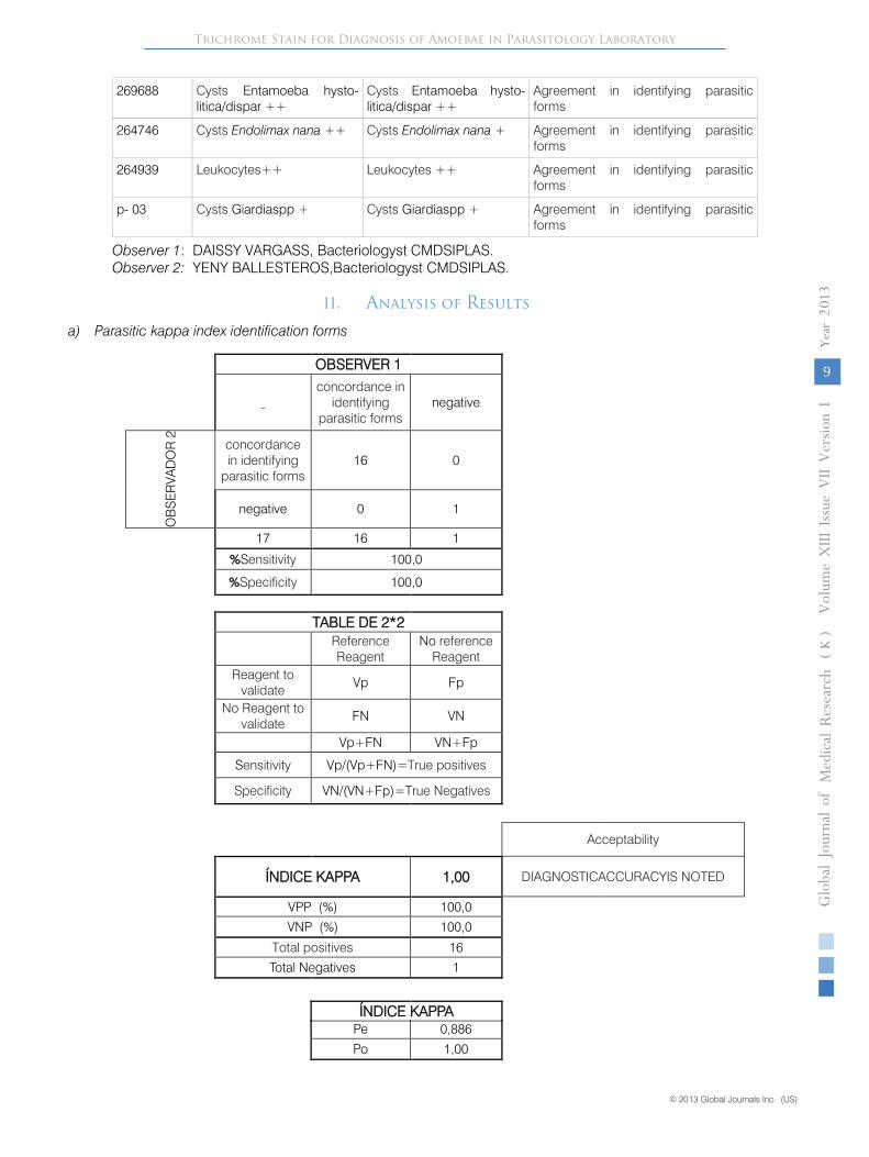
a) Parasitic kappa index identification forms
OBSERVER 1concordance in
identifyingparasitic forms
negative
OB
SE
RVA
DO
R 2 concordance
in identifyingparasitic forms
16 0
negative 0 1
17 16 1
%Sensitivity 100,0
%Specificity 100,0
TABLE DE 2*2 ReferenceReagent
No referenceReagent
Reagent tovalidate
Vp Fp
No Reagent tovalidate
FN VN
Vp+FN VN+Fp
Sensitivity Vp/(Vp+FN)=True positives
Specificity VN/(VN+Fp)=True Negatives
Acceptability
ÍNDICE KAPPA 1,00 DIAGNOSTICACCURACYIS NOTED
VPP (%) 100,0
VNP (%) 100,0
Total positives 16
Total Negatives 1
Pe 0,886
Po 1,00
ÍNDICE KAPPA

Trichrome Stain for Diagnosis of Amoebae in Parasitology Laboratory
© 2013 Global Journals Inc. (US)
Globa
l Jo
urna
l of M
edical R
esea
rch
© 2013 Global Journals Inc. (US)
10
Year
013
2(
)K
Volum
e XIII
Issue
VII
Versio
n I
b) Kappa index leukocytes
OBSERVER 1
concordance in
identificationf l k t
negativeO
BS
ER
VAD
OR
2
concordance inidentifying parasitic
forms1 0
negative 0 16
17 1 16
%Sensitivity 100,0
%Specificity 100,0
TABLA DE 2*2 ReferenceReagent
No referenceReagent
Reagent to validate Vp Fp
No Reagent tovalidate
FN VN
Vp+FN VN+Fp
Sensitivity Vp/(Vp+FN)=True positives
Specificity VN/(VN+Fp)=True Negatives
Aceptability
ÍNDICE KAPPA 1,00 DIAGNOSTICACCURACYIS NOTED
VPP (%) 100,0
VNP (%) 100,0
Total positives 1
Total Negatives 16
ÍNDICE KAPPAPe 0,003
Po 1,00
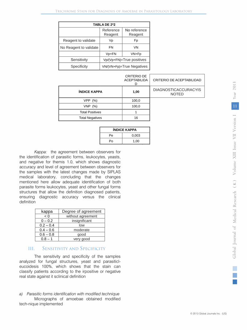
c) Kappa index yeast
OBSERVER 1
concordance inidentification in
yeastnegative
BSER
VAD
OR
2 concordance inidentifying parasitic
forms1 0
negative 0 16
17 1 16
%Sensitivity 100,0
%Specificity 100,0
Trichrome Stain for Diagnosis of Amoebae in Parasitology Laboratory
© 2013 Global Journals Inc. (US)© 2013 Global Journals Inc. (US)
Globa
l Jo
urna
l of M
edical R
esea
rch
11
Year
013
2(
)K
Volum
e XIII
Issue
VII
Versio
n I
TABLA DE 2*2 ReferenceReagent
No referenceReagent
Reagent to validate Vp Fp
No Reagent to validate FN VN
Vp+FN VN+Fp
Sensitivity Vp/(Vp+FN)=True positives
Specificity VN/(VN+Fp)=True Negatives
CRITERIO DE ACEPTABILIDA
D CRITERIO DE ACEPTABILIDAD
ÍNDICE KAPPA 1,00 DIAGNOSTICACCURACYIS NOTED
VPP (%) 100,0VNP (%) 100,0
Total Positives 1Total Negatives 16
ÍNDICE KAPPAPe 0,003Po 1,00
Kappa: the agreement between observers forthe identification of parasitic forms, leukocytes, yeasts, and negative for themis 1.0, which shows diagnostic accuracy and level of agreement between observers forthe samples with the latest changes made by SIPLASmedical laboratory, concluding that the changesmentioned here allow adequate identification of bothparasite forms leukocytes, yeast and other fungal formsstructures that allow the definition diagnosed patients, ensuring diagnostic accuracy versus the clinical definition
kappa Degree of agreement< 0 without agreement
0 – 0.2 insignificant0.2 – 0.4 low0.4 – 0.6 moderate0.6 – 0.8 good0.8 – 1 very good
III. Sensitivity and Specificity
The sensitivity and specificity of the samples analyzed for fungal structures, yeast and parasiticl-eucoidesis 100%, which shows that the stain canclassify patients according to the irpositive or negativereal state against it sclinical definition
a) Parasitic forms identification with modified techniqueMicrographs of amoebae obtained modified
tech-nique implemented

Trichrome Stain for Diagnosis of Amoebae in Parasitology Laboratory
© 2013 Global Journals Inc. (US)
Globa
l Jo
urna
l of M
edical R
esea
rch
© 2013 Global Journals Inc. (US)
12
Year
013
2(
)K
Volum
e XIII
Issue
VII
Versio
n I
Figure 1 : Cysts Blastocystis hominis
Figure 2 : Cysts Entamoeba coli
Figure 3 : Cysts Iodamoeba butschlii
Figure 4 : Cysts Endolimax nana
Figure 5 : Cysts Giardia Duodenalis
Note: The photomicrographs were taken by thebacteriologys Daissy Vargas Sepulveda in CMD SIPLAS
IV. Conclusions
• The quick method is effective and accurate.• It requires less processing time and therefore the
patient must wait less time to get your results• Iteliminates contamination of the reagents
considering that it is not necessary to use Koplic• It saves time and money by having as only three
reagent required to implement this technique
References Références Referencias
1. Diagnóstico Parasitología Médica, 5 ªed., ASM Press, Washington, DC 4. García, LS 2009. PrácticaGuía para el Diagnóstico de Parasitología, 2 ª ed., ASM Press, Washington, DC.
2. Evaluation of Intestinal Protozoan Morphology in Human Fecal Specimens Preserved in Eco Fix: Comparison of Wheatley's Trichrome Stain and EcoStain, Lynne S. Garcia* and Robyn Y. Shimizu ,Journal of Clinical Microbiology, July 1998, p. 1974-1976, Vol. 36, No. 7.
3. Garcia, L.S., and D.A. Bruckner, 1993. Diagnostic Medical Parasitology, 2nd ed. American Society forMicrobiology, Washington D.C.
4. Normas de Laboratorio Clínico Institute, 2005, Procedimientos para la recuperación e identificación de parásitos en el tracto intestinal, Directrices aprobadas, M28-2ª.
5. Normas de Laboratorio Clínico Instituto, Villanova, PA. García, LS (Coordinación Editor), 2003. Selección y Uso de Procedimientos de laboratorio para El diagnóstico de infecciones parasitarias deel tracto gastrointestinal,Cumitech 30A, ASM Press, Washington, DC García, LS 2007.
6. Wheatley, el Banco Mundial de 1951. Un procedimiento de tinción rápida para intestinal amebas y flagelados. Am. J. Clin. Pathol.21:990-991.