trpm3/trpv4 regulates ca -mediated rankl/nfatc1 …
TRANSCRIPT

https://doi.org/10.1530/JME-18-0051https://jme.bioscientifica.com © 2018 Society for Endocrinology
Printed in Great BritainPublished by Bioscientifica Ltd.
Induction of RANKL/NFATc1 via TRPV4/TRPM3
A Son et al.Journal of Molecular Endocrinology
207–21861 4:
RESEARCH
TRPM3/TRPV4 regulates Ca2+-mediated RANKL/NFATc1 expression in osteoblasts
Aran Son1, Namju Kang1,2, Jung Yun Kang1,2, Ki Woo Kim1, Yu-Mi Yang1 and Dong Min Shin1,2
1Department of Oral Biology, Yonsei University College of Dentistry, Seoul, Korea2BK21 PLUS Project, Yonsei University College of Dentistry, Seoul, Korea
Correspondence should be addressed to Y-M Yang or D M Shin: [email protected] or [email protected]
Abstract
Mechanical stress plays an important role in the regulation of bone turnover. However,
the mechanism underlying hypo-osmotic stress-induced cellular response in osteoblasts
remains poorly understood. In this study, we investigated the effect of hypotonic stress
on the expression of bone remodeling factors, including the receptor activator of
nuclear factor-kappa B ligand (RANKL) and the nuclear factor of activated T cells type
c1 (NFATc1) in primary mouse osteoblasts and MC3T3-E1 cells. Hypo-osmotic stress
induced significant increases in RANKL mRNA expression and intracellular Ca2+
concentration ([Ca2+]i) from the extracellular space. Hypo-osmotic stress-induced effects
on [Ca2+]i and RANKL and NFATc1 protein expression were decreased by antagonists
of transient receptor potential melastatin 3 (TRPM3) and vanilloid 4 (TRPV4). Agonists
of TRPM3 and TRPV4 activated [Ca2+]i and RANKL and NFATc1 protein expression.
Furthermore, genetic suppression of Trpm3 and Trpv4 reduced hypo-osmotic stress-induced
effects in mouse osteoblasts. These results suggest that hypo-osmotic stress induces
increases in [Ca2+]i through TRPM3 and TRPV4 to regulate RANKL and NFATc1 expression
in mouse osteoblastic cells and that mechanical stress-activated TRP channels may play a
critical role in bone remodeling.
Introduction
Bone is a highly dynamic tissue that is constantly being renewed throughout the life through a process called remodeling. Interactions between signals regulating bone-forming osteoblasts and bone-resorbing osteoclasts are basically adaptive responses of the skeleton to its environment (Zaidi 2007, Crockett et al. 2011). Although remodeling is initiated by the resorptive action of osteoclasts, it is dependent on signals from osteoblasts. Osteoblasts modulate the expression of the receptor activator of nuclear factor-kappa B ligand (RANKL) and osteoprotegerin (OPG) in response to factors that stimulate resorption. RANKL regulates osteoclast
precursors involved in differentiation into multinuclear osteoclasts, whereas OPG inhibits RANKL action by blocking the interaction between RANK and RANKL (Zaidi 2007, Boyce & Xing 2008, Crockett et al. 2011, Boyce 2013). The relative ratio of RANKL to OPG expression in osteoblasts is believed to be a key determinant of RANKL-mediated bone resorption. Nuclear factor-activated T cells (NFAT) are members of a transcription factor family that is dependent on calcineurin regulation. NFAT proteins are highly phosphorylated in the cytoplasm of resting cells, and NFAT proteins are then dephosphorylated by calcineurin activation. Dephosphorylated NFAT
Journal of Molecular Endocrinology (2018) 61, 207–218
Key Words
f hypo-osmotic stress
f bone remodeling
f RANKL
f osteoblast
f mechanosensitive TRP channels
-18-0051
461
Downloaded from Bioscientifica.com at 03/18/2022 11:08:55AMvia free access

https://doi.org/10.1530/JME-18-0051https://jme.bioscientifica.com © 2018 Society for Endocrinology
Printed in Great BritainPublished by Bioscientifica Ltd.
208A Son et al. Induction of RANKL/NFATc1 via TRPV4/TRPM3
61 4:Journal of Molecular Endocrinology
proteins are translocated into the nucleus to regulate the transcription of NFAT-dependent genes (Hogan et al. 2003). Calcineurin and NFAT are the key factors of osteoclastogenesis and bone resorption (Takayanagi et al. 2002, Hirotani et al. 2004, Ikeda et al. 2004). The Ca2+-calcineurin-NFAT pathway reportedly acts as a positive regulator of osteoblastogenesis and bone formation by maintaining a balanced regulation of osteoclastic and osteoblastic activities (Koga et al. 2005, Sun et al. 2005, Winslow et al. 2006).
Mechanical stress is one of the important regulatory factors for maintaining bone mass and integrity. Mechanical stresses include osmotic stress, shear stress, fluid flow stress and cell membrane stretch (Jin et al. 2011, Yang et al. 2015, Wittkowske et al. 2016). Among the various types of mechanical stress, alteration of osmo-mechanical stresses is often associated with early changes in Ca2+ signaling and downstream Ca2+-dependent processes in osteoblastic cells (Weskamp et al. 2000, Sun et al. 2005, Mehrotra et al. 2006, Mizoguchi et al. 2008, Nakai et al. 2009). There has been much debate regarding the source of the mechanosensitive Ca2+ signals and the downstream effector pathways. Recently, there has been increasing evidence for a key role of Ca2+-permeable transient receptor potential (TRP) channels in this process (Pedersen & Nilius 2007, Mizoguchi et al. 2008, Jin et al. 2011). Mechanosensitive TRP channels (TRPC1, TRPC5, TRPC6; TRPV1, TRPV2, TRPV4; TRPM3, TRPM7; TRPA1 and TRPP2) have been mainly investigated in the cardiovascular system (Christensen & Corey 2007, Pedersen & Nilius 2007). Abed and colleagues have shown that mRNAs of diverse mechanosensitive TRP channels (TRPV2, TRPV4, TRPM3, TRPM4 and TRPM7) are strongly expressed in osteoblastic cells (Abed et al. 2009). TRPV2 acts as a mechanosensor for membrane stretch and osmotic cell swelling in vascular smooth muscle cells (Muraki et al. 2003, Beech et al. 2004). TRPV4 is activated by osmotic cell swelling, excluding potential pathways, such as membrane stretch (Liedtke et al. 2000, Strotmann et al. 2000, Becker et al. 2005, Son et al. 2015). TRPM3 is stimulated by hypo-osmotic cell swelling (Grimm et al. 2003, Son et al. 2015), and TRPM4 has a role as a mechano-/stretch-sensitive channel involved in the control of pressure-induced smooth muscle cell depolarization (Pedersen & Nilius 2007, Guinamard et al. 2010). TRPM7 is activated by cell stretch and hypo-osmotic cell swelling; moreover, shear stress induces the translocation of TRPM7 to the plasma membrane and activation of TRPM7 currents (Oancea et al. 2006, Numata et al. 2007). However, the mechanisms through
which mechanosensitive TRP channels and intracellular Ca2+ are regulated have not been clearly elucidated.
In this study, we investigated the effect of hypo-osmotic stress on basal RANKL expression and the function of mechanosensitive TRP channels in primary mouse osteoblasts and MC3T3-E1 cells. We also demonstrated that hypo-osmotic stress induces large increases in the differentiation markers RANKL and NFATc1 and in intracellular Ca2+ concentration ([Ca2+]i) through the osmo-mechanosensitive TRP channels TRPM3 and TRPV4.
Materials and methods
Reagents
All cell culture media and supplements were purchased from Invitrogen. The acetoxymethyl-ester form of fura2 (fura2/AM) was purchased from Teflabs (Austin, TX, USA). Pluronic F127 was obtained from Molecular Probes. Anti-RANKL and anti-NFATc1 antibodies were obtained from Santa Cruz Biotechnology. Anti-actin antibodies were purchased from Sigma-Aldrich. Ruthenium red (RR), 4a-phorbol didecanoate (4a-PDD), ononetin and HC067047 were obtained from Tocris (Bristol, UK). All other chemicals were purchased from Sigma-Aldrich. Stock solutions of all drugs were made with distilled water, except for 2-aminoethoxydiphenyl borate (2APB) with ethanol and 4a-PDD and fura2/AM with DMSO.
Cell culture
All animal care and experimental procedures complied with institutional guidelines and were approved by the Institutional Animal Care and Use Committee (IACUC) of Yonsei University (IACUC approval no. 2014-0067). Primary osteoblast cell cultures were prepared as described previously (Hogan et al. 2003). In brief, the calvariae of 1-day-old ICR mice were digested with an enzyme solution containing 0.1% collagenase and 0.1% dispase. The isolated osteoblastic cells were cultured for 4 days in alpha-minimum essential medium (a-MEM) supplemented with 10% fetal bovine serum (FBS) and 1% antibiotic-antimycotic solution (100 U/mL penicillin and 100 µg/mL streptomycin) in a humidified incubator containing 5% CO2. MC3T3-E1 cells (Korean Cell Line Bank, Seoul, Korea) were maintained in a-MEM containing 10% FBS and 1% antibiotic-antimycotic solution. Osteoblastic differentiation of MC3T3-E1 cells was induced by 50 μg/mL ascorbic acid and 10 mM beta-glycerophosphate.
Downloaded from Bioscientifica.com at 03/18/2022 11:08:55AMvia free access

https://doi.org/10.1530/JME-18-0051https://jme.bioscientifica.com © 2018 Society for Endocrinology
Printed in Great BritainPublished by Bioscientifica Ltd.
20961 4:A Son et al. Induction of RANKL/NFATc1 via TRPV4/TRPM3
Journal of Molecular Endocrinology
Reverse transcription polymerase chain reaction (RT-PCR)
The expressions of RANKL, OPG, TRPM3, TRPV4 and beta-actin were evaluated by RT-PCR using total RNA isolated from murine osteoblastic cells. Cells were lysed in TRIzol reagent according to the instructions of the manufacturer (Invitrogen). AccuPower RT PreMix (BIONEER, Daejeon, Korea) with total RNA (1 µg) was used for cDNA synthesis. cDNA was amplified by PCR with HiPi Thermostable DNA polymerase (Elpis, Pusan, Korea) using the following primer sets: RANKL (forward): 5′-ATCAGAAGACAGCACTCACT-3′ (reverse): 5′-ATCTAGGACATCCATGCTAATGTTC-3′; OPG (forward): 5′-TGAGTGTGAGGAAGGGCGTT-3′ (reverse): 5′-TTCCTCGTTCTCTCAATCTC-3′; TRPM3 (forward): 5′-CACCTGATGACCAAGGAATG-3′ (reverse): 5′-CTTGTG TTTATCTTCTGGAGTG-3′; TRPV4 (forward): 5′-GCTGAA GGCAAAAGTCTTGG-3′ (reverse): 5′-CTAGGGAACCCCAA CTGTGA-3′; beta-actin (forward): 5′-TGTGATGGTGGG AATGGGTCAG-3′ (reverse): 5′-TTTGATGTCACGCACG ATTTCC-3′. PCR was performed under the following conditions: 94°C for 5 min, 94°C for 30 s, 40 s for annealing step (temperatures varied with primers), 72°C for 30 s, followed by 72°C for 10 min after 38 cycles were finished. The annealing temperature was 60°C for TRPM3, 56.5°C for TRPV4 and RANKL and 57.6°C for OPG and beta-actin. The PCR products were separated on 1.2% agarose gels and visualized with RedSafe nucleic acid staining solution (iNtRon Biotechnology, Gyeonggi-do, Korea).
[Ca2+]i measurement
Osteoblasts were seeded on cover glass in 35 mm dishes (5 × 10⁴ cells/coverslip). The cells were loaded with 5 μM fura2/AM and 0.05% Pluronic F-127 for 30 min in physiological salt solution (PSS) containing (in mM): 140 NaCl, 5 KCl, 1 MgCl2, 10 HEPES, 1 CaCl2 and 10 glucose, adjusted to pH 7.4 and 310 mosmol. For hypo-osmotic stress, PSS was replaced with a hypo-osmotic solution (80 mM NaCl, 215 mosmol). Fura2 fluorescence intensity was measured using excitation wavelengths of 340 and 380 nm, and emitted fluorescence 510 nm (ratio = F340/F380) was collected and monitored at 2-s intervals using a CCD camera (Universal Imaging Co., Downingtown, PA, USA) as described previously (Son et al. 2009). Images were digitized and analyzed by MetaFlour software (Universal Imaging).
Electrophysiology
Whole-cell voltage-clamp recordings were performed at room temperature by the perforated patch-clamp
method. Currents were recorded with a MultiClamp 700B amplifier (Axon Instruments, Foster City, CA, USA), subsequently digitized at a sampling rate of 10 kHz and analyzed with pCLAMP10 software (Axon Instruments). Pipette resistance varied between 3 and 5 MΩ. Whole-cell currents were elicited by voltage ramps from −100 mV to +100 mV (400-ms duration) applied every 10 s from a holding potential of 0 mV. The bath solution contained (in mM) 140 NaCl, 5 KCl, 2 CaCl2, 1 MgCl2, 10 HEPES and 10 glucose, adjusted to pH 7.4 with NaOH. Pipettes for recording TRPM3 currents were filled with an internal solution containing (in mM) 140 CsCl, 5 MgCl2, 10 BAPTA and 10 HEPES, adjusted to pH 7.2 with CsOH and the bath solution was changed to a K+-free external solution (Grimm et al. 2003). The internal solution for recording TRPV4 currents contained (in mM) 140 KCl, 5 EGTA and 10 HEPES, adjusted to pH 7.4 with KOH (Guler et al. 2002).
Western blot
Cell lysates of MC3T3-E1 cells were prepared using RIPA lysis buffer (20 mM Tris, pH 7.4, 250 mM NaCl, 2 mM EDTA, pH 8.0, 0.1% Triton X-100, 0.01 mg/mL aprotinin, 5 μg/mL leupeptin, 0.4 mM PMSF and 4 mM NaVO4) and spun at 12,000 rpm for 10 min to remove insoluble material. Proteins (50–100 μg/well) were subjected to 8–12% SDS-PAGE and separated by size. The proteins were electro-transferred to a nitrocellulose membrane, blocked with 5% skim milk and probed with antibodies against RANKL (1:500), NFATc1 (1:1000) and actin (1:2000). Thereafter, the blots were washed, exposed to horseradish peroxidase-conjugated secondary antibodies for 1 h, and finally detected by chemiluminescence (Amersham Pharmacia Biotech, Arlington Heights, IL, USA).
Transfection of small interfering (si) RNAs
After a cell density of 70% confluence was reached, siRNA duplexes specific for mouse Trpm3, Trpv4 or negative control (BIONEER) were transfected using Lipofectamine2000 (Invitrogen) according to the manufacturer’s instructions.
Statistical analysis
All data were expressed as means ± s.e.m. Statistical significance was determined by a paired Student’s t-test. Statistical significance was set at the P < 0.05 level.
Downloaded from Bioscientifica.com at 03/18/2022 11:08:55AMvia free access

https://doi.org/10.1530/JME-18-0051https://jme.bioscientifica.com © 2018 Society for Endocrinology
Printed in Great BritainPublished by Bioscientifica Ltd.
210A Son et al. Induction of RANKL/NFATc1 via TRPV4/TRPM3
61 4:Journal of Molecular Endocrinology
Results
Hypo-osmotic stress induces increased RANKL expression in osteoblasts
Various mechanical stresses have been identified as regulators of bone formation in the differentiation of bone cells and as inducers of RANKL expression in osteoblasts (Boyce & Xing 2008). In this study, we examined whether hypo-osmotic stress affects RANKL expression after treatment of hypo-osmotic solution (215 mosmol) for 3 h in primary mouse osteoblasts. The expression of RANKL mRNA was significantly increased by hypo-osmotic stress (~1.5-fold) compared with control. This increase was not different from the increase in expression of RANKL mRNA by 1,25(OH)2D3 (~1.6-fold), when the hormonally active form of vitamin D was used as a positive control (Takeda et al. 1999) (Fig. 1A and B). Furthermore, the expression of RANKL mRNA (Supplementary Fig. 1A, see section on supplementary data given at the end of this article) and the ratio of RANKL to OPG expression were significantly increased 3 h after stimulation by hypo-osmotic stress (by ~1.2-fold and 2.3-fold, respectively) (Fig. 1C and D), but the level of OPG mRNA was not altered by osmotic stimulation (Supplementary Fig. 1B). This suggests that increase in RANKL mRNA by hypo-osmotic stress initiates RANKL-mediated bone resorption.
Hypo-osmotic stress-induced increases of [Ca2+]i and RANKL expression are dependent on extracellular Ca2+ entry
Mechanical stress generates the intracellular Ca2+ response in bone cells (Weskamp et al. 2000, Winslow et al. 2006, Huo et al. 2008). Therefore, we examined Ca2+ signaling by osmotic stress in osteoblasts. As shown in Fig. 2A, hypo-osmotic stress increased [Ca2+]i and even the repeated application of hypo-osmotic stress did not significantly change the increase in [Ca2+]i (87.3 ± 0.4% of first application). To identify the Ca2+ influx-related pathways involved, we examined whether the increase in [Ca2+]i promoted by hypo-osmotic stress was dependent on extracellular Ca2+ in osteoblasts. When hypo-osmotic stress was applied in Ca2+-free external solution, the hypo-osmotic stress-induced increase in [Ca2+]i was completely inhibited (Fig. 2B). In addition, 100 μM lanthanum (La3+), as a non-specific blocker of Ca2+-permeable channels, or 10 μM gadolinium (Gd3+), as a non-specific blocker of stretch-activated and some voltage-gated ion channels, also significantly prevented the hypo-osmotic stress-induced increase in [Ca2+]i (Fig. 2C). These results suggest that the major source of hypo-osmotic stress-induced [Ca2+]i increase is Ca2+ influx from the extracellular space. To investigate whether the hypo-osmotic stress-induced increase in [Ca2+]i affected RANKL expression, we examined the changes in RANKL mRNA induced by
Figure 1Effect of hypo-osmotic stress on RANKL mRNA expression in primary mouse osteoblasts. (A) Primary mouse osteoblasts were stimulated with hypo-osmotic solution (215 mosmol) (hypotonic) as mechanical stimulation and active vitamin D, 1,25(OH)2D3 (10 nM), as a positive control for 3 h. RANKL mRNA expression was measured by RT-PCR. (B) The level of RANKL mRNA was quantified after the normalized value to beta-actin (n = 6). (C) Cells were stimulated with the hypo-osmotic solution for the indicated times. Expression of mRNAs was measured. (D) The expression ratio of RANKL/OPG mRNA was quantified after the normalized value to beta-actin (n = 3). Data are expressed as means ± s.e.m. *P < 0.05 compared with control.
Downloaded from Bioscientifica.com at 03/18/2022 11:08:55AMvia free access

https://doi.org/10.1530/JME-18-0051https://jme.bioscientifica.com © 2018 Society for Endocrinology
Printed in Great BritainPublished by Bioscientifica Ltd.
21161 4:A Son et al. Induction of RANKL/NFATc1 via TRPV4/TRPM3
Journal of Molecular Endocrinology
Figure 2Dependence on extracellular Ca2+ of hypo-osmotic stress-induced [Ca2+]i increase and RANKL expression. (A) Treatment with hypo-osmotic solution (hypotonic) induced increases in [Ca2+]i and also showed that [Ca2+]i was increased similarly by repeated application of hypo-osmotic solution in primary mouse osteoblasts using fura2 fluorescence dye. The changes in [Ca2+]i were quantified after the normalized value to the first hypo-osmotic stress (n = 9). (B and C) The effects of increases in [Ca2+]i by hypo-osmotic stress were inhibited by Ca2+-free external solution and 100 μM La3+, which block a wide range of Ca2+-permeable channels. The inhibitory effects on blockers were quantified after the normalized value to hypo-osmotic stress (n = 6) and ionomycin (n = 6). (D) Cells were incubated with Gd3+ or La3+ in the hypo-osmotic solution for 3 h. The increases in RANKL mRNA by hypo-osmotic stress were reduced by 10 μM Gd3+ and 100 μM La3+ and the level of RANKL mRNA was quantified after the normalized value to beta-actin (n = 3). (E) Cells were treated with 1 μM thapsigargin in PSS (a presence of 1 mM CaCl2) for the indicated times. The expression of RANKL mRNA was quantified after the normalized value to beta-actin (n = 6). Data are expressed as means ± s.e.m. *P < 0.05, **P < 0.01, and ***P < 0.001 compared with control.
Downloaded from Bioscientifica.com at 03/18/2022 11:08:55AMvia free access

https://doi.org/10.1530/JME-18-0051https://jme.bioscientifica.com © 2018 Society for Endocrinology
Printed in Great BritainPublished by Bioscientifica Ltd.
212A Son et al. Induction of RANKL/NFATc1 via TRPV4/TRPM3
61 4:Journal of Molecular Endocrinology
hypo-osmotic stress after treatment with 100 μM La3+ or 10 μM Gd3+ for 3 h. The increase of RANKL mRNA by hypo-osmotic stress was significantly decreased by Gd3+ and La3+ (Fig. 2D). This result suggests that the increase in [Ca2+]i from the extracellular space by hypo-osmotic stress leads to the increase in RANKL expression. Thapsigargin, an inhibitor of sarco/endoplasmic reticulum Ca2+ ATPase, can increase [Ca2+]i by depletion of intracellular Ca2+ stores and subsequent influx of Ca2+ into the cytosol via activated plasma membrane Ca2+ channels. Thus, to investigate whether the increase in [Ca2+]i by thapsigargin affected RANKL expression, we examined the level of RANKL mRNA after treatment with 1 μM thapsigargin in PSS (a presence of 1 mM CaCl2). The expression of RANKL mRNA was significantly increased 3 and 6 h (by ~1.8-fold and 1.7-fold, respectively) after treatment with thapsigargin; the increase was not different from the increase in expression of RANKL mRNA by 1,25(OH)2D3 (~1.7-fold) (Fig. 2E). This result suggests that the RANKL expression is related with the increase of [Ca2+]i through extracellular Ca2+ entry as well as thapsigargin-mediated store-operated Ca2+ entry (SOCE).
Activation of TRPM3 and TRPV4 induces increases of [Ca2+]i and the expression of RANKL-mediated NFATc1 in osteoblasts
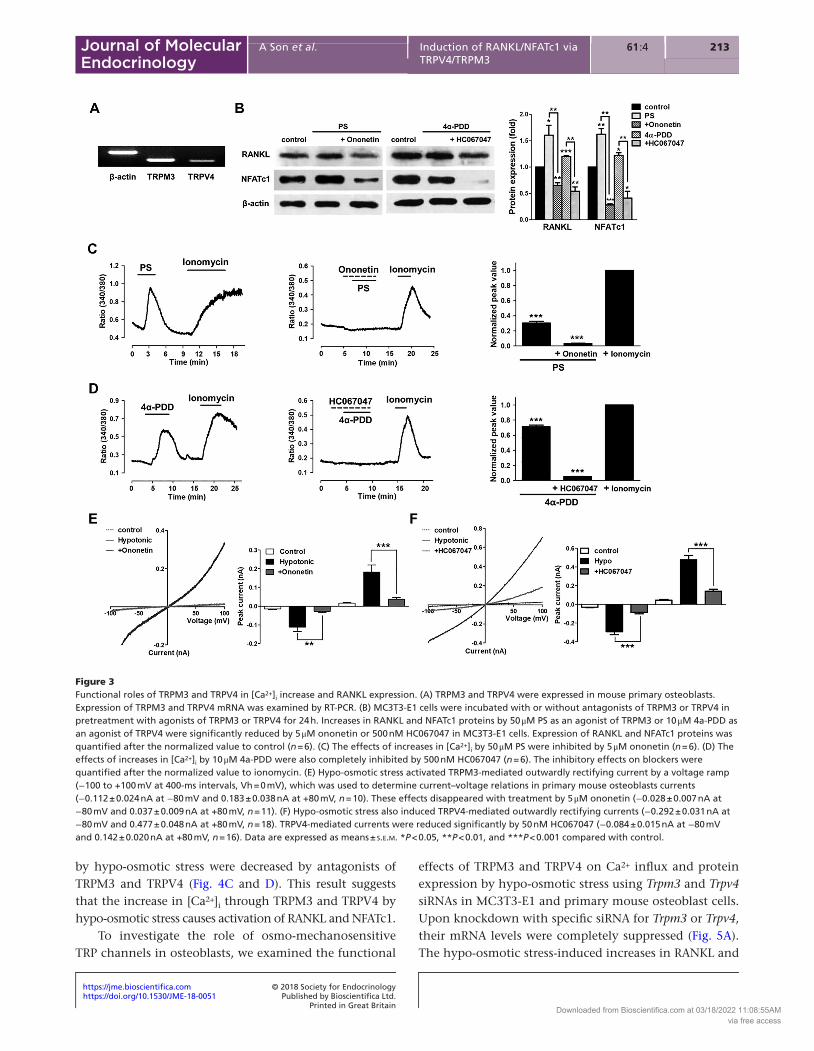
Diverse mechanosensitive TRP channels have been identified in osteoblastic cells (Abed et al. 2009). Osmo-mechanosensitive TRP channels (TRPM2, TRPM3, TRPM7 and TRPV4) have been implicated in controlling the osmosensitive response in osmoregulation organs, such as kidney, sensory neurons, airway epithelia, bone and so on, to changes in osmolarity (Liedtke et al. 2000, Strotmann et al. 2000, Grimm et al. 2003, Koga et al. 2005, Sun et al. 2005, Bessac & Fleig 2007, Christensen & Corey 2007, Numata et al. 2007, 2012, Nakai et al. 2009, Jin et al. 2011). In particular, TRPM3 and TRPV4 are directly activated by hypo-osmotic stress (Strotmann et al. 2000, Alessandri-Haber et al. 2003, Grimm et al. 2003). To determine which osmo-mechanosensitive TRP channels play a role in the hypo-osmotic stress-induced increases in [Ca2+]i, we confirmed the expression and functional activities of osmo-mechanosensitive TRP channels in osteoblasts. As shown in Fig. 3A, both TRPM3 and TRPV4 are present in primary mouse osteoblasts. However, TRPM2 was scarcely expressed, and TRPM7 was not affected by hypo-osmotic stress for 3 h (Supplementary Fig. 2). To directly assess the functional activities of these channels, we examined the expression of RANKL and NFATc1 proteins after treatment
with agonists and antagonists of TRPM3 and TRPV4 for 24 h. The expression of RANKL and NFATc1 protein was markedly increased by pregnenolone sulfate (PS) (50 μM) and 4a-PDD (10 μM), which are agonists of TRPM3 and TRPV4. Both ononetin (5 μM) and HC067047 (500 nM), which are antagonists of TRPM3 and TRPV4, inhibited the agonist-induced increases in protein expression (Fig. 3B). In addition, both PS and 4a-PDD enhanced the increase of [Ca2+]i in osteoblasts, and these effects on increased [Ca2+]i by activation of TRPM3 and TRPV4 were diminished by ononetin and HC067047 (Fig. 3C and D). Taken together, these results show that activation of RANKL and NFATc1 can be induced by increases in [Ca2+]i via TRPM3 and TRPV4. Next, we examined the sensitivity of TRPM3 and TRPV4 to hypotonicity using a whole-cell patch-clamp technique. Hypo-osmotic stress activated TRPM3-mediated outwardly rectifying currents (−0.10 ± 0.01 nA at −80 mV and 0.22 ± 0.05 nA at +80 mV) (Fig. 3E) and TRPV4-mediated outwardly rectifying currents (−0.28 ± 0.07 nA at −80 mV and 0.40 ± 0.05 nA at +80 mV) (Fig. 3F), which reversed near 0 mV. These results suggest that the hypo-osmotic stress-evoked response is mediated by both TRPM3 and TRPV4. Therefore, we confirmed the effects of TRPV4 and TRPM3 on hypo-osmotic stress in subsequent experiments.
Hypo-osmotic stress activates the increases of [Ca2+]i and expression of RANKL-mediated NFATc1 through osmo-mechanosensitive TRP channels (TRPM3 and TRPV4) in osteoblasts
To determine the specific activation of osmo-mechanosensitive TRP channels (TRPM3 and TRPV4) by osmotic stress in osteoblasts, we used blockers of TRPM3 and TRPV4. The hypo-osmotic stress-induced increase in [Ca2+]i was partially inhibited by 100 μM 2APB, a non-specific blocker of the TRPM3 channel, and 5 μM ononetin, a selective inhibitor of the TRPM3 channel (Fig. 4A). Antagonists of TRPV4 (10 μM RR as a non-specific blocker and 500 nM HC067047 as a potent antagonist) also partially inhibited the hypo-osmotic stress-induced increase in [Ca2+]i (Fig. 4B). These results indicate that hypo-osmotic stress is capable of inducing Ca2+ influx from the extracellular space via TRPM3 and TRPV4 on plasma membranes. To investigate whether the expressions of RANKL and NFATc1 was affected by the increase of [Ca2+]i via hypo-osmotic stress, we examined the changes in RANKL and NFATc1 protein levels by hypo-osmotic stress after treatments with antagonists of TRPM3 and TRPV4 for 24 h. The increases in RANKL and NFATc1 protein levels
Downloaded from Bioscientifica.com at 03/18/2022 11:08:55AMvia free access
https://doi.org/10.1530/JME-18-0051https://jme.bioscientifica.com © 2018 Society for Endocrinology
Printed in Great BritainPublished by Bioscientifica Ltd.
21361 4:A Son et al. Induction of RANKL/NFATc1 via TRPV4/TRPM3
Journal of Molecular Endocrinology
by hypo-osmotic stress were decreased by antagonists of TRPM3 and TRPV4 (Fig. 4C and D). This result suggests that the increase in [Ca2+]i through TRPM3 and TRPV4 by hypo-osmotic stress causes activation of RANKL and NFATc1.
To investigate the role of osmo-mechanosensitive TRP channels in osteoblasts, we examined the functional
effects of TRPM3 and TRPV4 on Ca2+ influx and protein expression by hypo-osmotic stress using Trpm3 and Trpv4 siRNAs in MC3T3-E1 and primary mouse osteoblast cells. Upon knockdown with specific siRNA for Trpm3 or Trpv4, their mRNA levels were completely suppressed (Fig. 5A). The hypo-osmotic stress-induced increases in RANKL and
Figure 3Functional roles of TRPM3 and TRPV4 in [Ca2+]i increase and RANKL expression. (A) TRPM3 and TRPV4 were expressed in mouse primary osteoblasts. Expression of TRPM3 and TRPV4 mRNA was examined by RT-PCR. (B) MC3T3-E1 cells were incubated with or without antagonists of TRPM3 or TRPV4 in pretreatment with agonists of TRPM3 or TRPV4 for 24 h. Increases in RANKL and NFATc1 proteins by 50 μM PS as an agonist of TRPM3 or 10 μM 4a-PDD as an agonist of TRPV4 were significantly reduced by 5 μM ononetin or 500 nM HC067047 in MC3T3-E1 cells. Expression of RANKL and NFATc1 proteins was quantified after the normalized value to control (n = 6). (C) The effects of increases in [Ca2+]i by 50 μM PS were inhibited by 5 μM ononetin (n = 6). (D) The effects of increases in [Ca2+]i by 10 μM 4a-PDD were also completely inhibited by 500 nM HC067047 (n = 6). The inhibitory effects on blockers were quantified after the normalized value to ionomycin. (E) Hypo-osmotic stress activated TRPM3-mediated outwardly rectifying current by a voltage ramp (−100 to +100 mV at 400-ms intervals, Vh = 0 mV), which was used to determine current–voltage relations in primary mouse osteoblasts currents (−0.112 ± 0.024 nA at −80 mV and 0.183 ± 0.038 nA at +80 mV, n = 10). These effects disappeared with treatment by 5 μM ononetin (−0.028 ± 0.007 nA at −80 mV and 0.037 ± 0.009 nA at +80 mV, n = 11). (F) Hypo-osmotic stress also induced TRPV4-mediated outwardly rectifying currents (−0.292 ± 0.031 nA at −80 mV and 0.477 ± 0.048 nA at +80 mV, n = 18). TRPV4-mediated currents were reduced significantly by 50 nM HC067047 (−0.084 ± 0.015 nA at −80 mV and 0.142 ± 0.020 nA at +80 mV, n = 16). Data are expressed as means ± s.e.m. *P < 0.05, **P < 0.01, and ***P < 0.001 compared with control.
Downloaded from Bioscientifica.com at 03/18/2022 11:08:55AMvia free access

https://doi.org/10.1530/JME-18-0051https://jme.bioscientifica.com © 2018 Society for Endocrinology
Printed in Great BritainPublished by Bioscientifica Ltd.
214A Son et al. Induction of RANKL/NFATc1 via TRPV4/TRPM3
61 4:Journal of Molecular Endocrinology
NFATc1 expression were diminished in Trpm3- and Trpv4-siRNA-transfected cells (Fig. 5B). Moreover, the amplitude of hypo-osmotic stress-induced increases in [Ca2+]i was reduced by approximately 37 and 35%, respectively, in Trpm3- and Trpv4-siRNA-transfected cells, compared with negative control cells (Fig. 5C and D). These results indicate that activation of TRPM3 and TRPV4 is essential for hypo-osmotic stress-mediated extracellular Ca2+ influx and induction of RANKL and NFATc1 expression in osteoblasts.
Discussion
In this study, we demonstrated a novel regulatory mechanism between osmo-mechanosensitive TRP channels, especially TRPM3 and TRPV4, and differentiation markers of bone cells through entry of extracellular Ca2+ in mouse osteoblast cells (Fig. 6). These effects could be the result of the accelerating activity in [Ca2+]i via TRPM3 and TRPV4 by mechanical stresses during bone remodeling, which is the previously reported physiological function of mechanical stresses such as endogenous RANKL synthesis, maintenance of bone mass and regulation of osteoclast differentiation in osteoblast cells (Tsuzuki et al. 2000, Takayanagi et al. 2002, Becker et al. 2005, Mehrotra et al. 2006, Mizoguchi et al. 2008, Abed et al. 2009, Nakai et al. 2009). However, the major signaling pathway and actions of mechanosensors in osteoblasts remain a mystery. Our results show that hypo-osmotic stress-induced increases
in RANKL mRNA expression depend on the increase of [Ca2+]i in osteoblasts. Although other groups reported that extracellular Ca2+ entry as well as SOCE-elevated [Ca2+]i effected expression levels of RANKL and OPG mRNA in primary osteoblasts (Takami et al. 2000, Hwang et al. 2012), these different results suggest that various stimuli can induce increase of [Ca2+]i through activation of a different mechanism of bone remodeling. Hence, the increase of [Ca2+]i in osteoblasts is necessary for the activation of differentiation markers of osteoblasts and the regulation of osteoclast differentiation (Takami et al. 2000, Weskamp et al. 2000, Romanello et al. 2005, Hwang et al. 2012, Robinson et al. 2012).
In the present study, hypo-osmotic stress-induced increases of RANKL and NFATc1 expressions were also dependent on Ca2+ influx through the extracellular space and osmo-mechanosensitive TRP channels (TRPM3 and TPRV4). In previous reports, various mechanical perturbations, such as mechanical load and fluid shear stress or pressure, played an important role in bone remodeling by activation of Ca2+ signaling and RANKL due to the influx of extracellular Ca2+ through mechanosensitive TRP channels, purinergic receptors, big conductance Ca2+-activated K+ channels in bone (Pavalko et al. 1998, Weskamp et al. 2000, Romanello et al. 2005, Mehrotra et al. 2006, Oancea et al. 2006, Masuyama et al. 2008, Suzuki et al. 2013). TRPV4 was the first mechanosensitive TRP channel shown to be a volume-activated and Ca2+-permeable cation channel by
Figure 4Relationship between osmo-mechanosensitive TRP channels and hypo-osmotic stress. (A) The effects of increases in [Ca2+]i by hypo-osmotic stress (hypotonic stress) were partially inhibited by 100 μM 2APB as a non-specific blocker of TRPM3 and 5 μM ononetin as an antagonist of TRPM3 in osteoblasts (n = 5). (B) The effects of increases in [Ca2+]i by hypo-osmotic stress were also partially inhibited by 10 μM ruthenium red (RR) as a non-specific blocker of TRPV4 and 500 nM HC067047 as an antagonist of TRPV4 in osteoblasts (n = 4). The inhibitory effect on blockers was quantified after the normalized value to hypo-osmotic stress. (C) Cells lysates were collected from MC3T3-E1 cells stimulated with hypo-osmotic solution and blockers of osmo-mechanosensitive TRP channels for 24 h. (D) Expression of RANKL and NFATc1 proteins was quantified after the normalized value to control (n = 4). Data were expressed as means ± s.e.m. *P < 0.05, **P < 0.01, and ***P < 0.001 compared with control.
Downloaded from Bioscientifica.com at 03/18/2022 11:08:55AMvia free access

https://doi.org/10.1530/JME-18-0051https://jme.bioscientifica.com © 2018 Society for Endocrinology
Printed in Great BritainPublished by Bioscientifica Ltd.
21561 4:A Son et al. Induction of RANKL/NFATc1 via TRPV4/TRPM3
Journal of Molecular Endocrinology
hypo-osmotic stimuli (Liedtke et al. 2000, Strotmann et al. 2000). TRPV4 also regulates bone resorption, which is necessary for sustained Ca2+ signaling and NFATc1 activation by the Ca2+/calmodulin signal pathway in osteoclasts, and bone formation, which is essential for flow-induced Ca2+ signaling during differentiation in osteoblasts (Boyce & Xing 2008, Masuyama et al. 2008, Mizoguchi et al. 2008, Suzuki et al. 2013). TRPM3 of osmo-mechanosenstive TRP channels also participates in hypo-osmotic cell swelling (Grimm et al. 2003, Harteneck & Schultz 2007). In our previous study, hypo-osmotic stress induced RANKL expression via TRPM3 and TRPV4 in human periodontal ligament cells (Son et al. 2015). However, the functional mechanism of TRPM3 and TRPV4 in bone remodeling is not fully understood. Our study is the first to show that TRPM3 and TRPV4 are expressed
in osteoblasts and that the activities of differentiation markers are regulated by hypo-osmotic stress. TRPM7 as a mechanosensor is also expressed in osteoblasts and plays an important role in various cell types. TRPM7 has been proposed to be directly activated by volume regulation on mechanical stimuli and magnesium regulation, and the increase in [Ca2+]i via TRPM7 potentiates by hypo-osmotic cell swelling (Oancea et al. 2006, Numata et al. 2007, Abed et al. 2009, Boyce 2013, Kim et al. 2017, Won et al. 2018). According to our previous report, TRPM7 controls RANKL-induced osteoclastogenesis by regulation of Ca2+ oscillation and NFATc1 expression in osteoclast cells (Yang et al. 2013). In the present study, we had ruled out the possibility that TRPM7 was involved in osteoblast differentiation because of the inhibition of TRPM7 activity by regulation of Mg2+ in all buffers
Figure 5Effects of deleted osmo-mechanosensitive TRP channels on [Ca2+]i increases and activation of differentiation factors mediated by hypo-osmotic stress. (A) Osteoblasts were transfected with siTRPM3 and siTRPV4 using Lipofectamine 2000. After 72 h of transfection, whole mRNA was collected using the Trizol method. beta-actin was used as loading control. (B) Transfected MC3T3-E1 cells were treated with the hypotonic solution (Hypotonic) for 24 h. Hypo-osmotic stress-induced increases in RANKL and NFATc1 expressions were decreased in Trpm3- and Trpv4-siRNA-transfected MC3T3-E1 cells. Expression of RANKL and NFATc1 proteins was quantified after the normalized value to negative control (NC) (n = 6). (C and D) Application of hypo-osmotic solution induced increases in [Ca2+]i in negative control osteoblasts (a). However, these effects were reduced in siTRPM3-treated cells (b) (n = 4) and siTRPV4-treated cells (n = 4). We identified the siRNA-transfected cells by GFP fluorescence (upper side) before a test, and increases in [Ca2+]i in single cells were measured using fura2 fluorescence dye (lower side). Data are expressed as means ± s.e.m. *P < 0.05 and ***P < 0.001 compared with control.
Downloaded from Bioscientifica.com at 03/18/2022 11:08:55AMvia free access
https://doi.org/10.1530/JME-18-0051https://jme.bioscientifica.com © 2018 Society for Endocrinology
Printed in Great BritainPublished by Bioscientifica Ltd.
216A Son et al. Induction of RANKL/NFATc1 via TRPV4/TRPM3
61 4:Journal of Molecular Endocrinology
used. We also confirmed that hypo-osmotic stress had no effect on the TRPM7 expression in osteoblast cells (Supplementary Fig. 2). Therefore, activation of TRPM3 and TRPV4 by mechanical stress is essential for control of bone remodeling and osteoblastic differentiation.
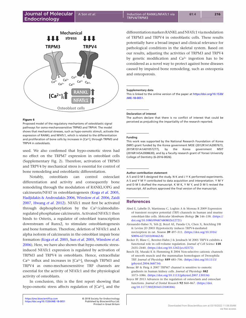
Notably, osteoblasts can control osteoclast differentiation and activity and consequently bone remodeling through the modulation of RANKL/OPG and calcineurin/NFAT in osteoblastogenesis (Koga et al. 2005, Hadjidakis & Androulakis 2006, Winslow et al. 2006, Zaidi 2007, Hwang et al. 2012). NFATc1 must first be activated through dephosphorylation by the Ca2+/calmodulin-regulated phosphatase calcineurin. Activated NFATc1 then binds to Osterix, a regulator of osteoblast transcription downstream of Runx2, to stimulate osteoblastogenesis and bone formation. Therefore, deletion of NFATc1 and A alpha isoform of calcineurin in the osteoblast impair bone formation (Koga et al. 2005, Sun et al. 2005, Winslow et al. 2006). Here, we have also shown that hypo-osmotic stress-induced NFATc1 expression is regulated by activation of TRPM3 and TRPV4 in osteoblasts. Hence, extracellular Ca2+ influx and increases in [Ca2+]i through TRPM3 and TRPV4 as osmo-mechanosensitive TRP channels are essential for the activity of NFATc1 and the physiological activity of osteoblasts.
In conclusion, this is the first report showing that hypo-osmotic stress affects regulation of [Ca2+]i and the
differentiation markers RANKL and NFATc1 via modulation of TRPM3 and TRPV4 in osteoblastic cells. These results potentially have a broad impact and clinical relevance for pathological conditions in the skeletal system. Based on our results, adjusting the activities of TRPM3 and TRPV4 by genetic modification and Ca2+ ingestion has to be considered as a novel way to protect against bone diseases caused by impaired bone remodeling, such as osteopenia and osteoporosis.
Supplementary dataThis is linked to the online version of the paper at https://doi.org/10.1530/JME-18-0051.
Declaration of interestThe authors declare that there is no conflict of interest that could be perceived as prejudicing the impartiality of the research reported.
FundingThis work was supported by the National Research Foundation of Korea (NRF) grant funded by the Korea government MOE (2012R1A1A2007673, 2015R1D1A1A01057277), by the Korea government MSIT (2016R1A5A2008630), and by a faculty research grant of Yonsei University College of Dentistry (6-2016-0026).
Author contribution statementA S and D M S designed the study. N K and J Y K performed experiments. A S and Y M Y contributed to data acquisition and interpretation. Y M Y and D M S drafted the manuscript. K W K, Y M Y, and D M S revised the manuscript. All authors approved the final version of the manuscript.
ReferencesAbed E, Labelle D, Martineau C, Loghin A & Moreau R 2009 Expression
of transient receptor potential (TRP) channels in human and murine osteoblast-like cells. Molecular Membrane Biology 26 146–158. (https://doi.org/10.1080/09687680802612721)
Alessandri-Haber N, Yeh JJ, Boyd AE, Parada CA, Chen X, Reichling DB & Levine JD 2003 Hypotonicity induces TRPV4-mediated nociception in rat. Neuron 39 497–511. (https://doi.org/10.1016/S0896-6273(03)00462-8)
Becker D, Blase C, Bereiter-Hahn J & Jendrach M 2005 TRPV4 exhibits a functional role in cell-volume regulation. Journal of Cell Science 118 2435–2440. (https://doi.org/10.1242/jcs.02372)
Beech DJ, Muraki K & Flemming R 2004 Non-selective cationic channels of smooth muscle and the mammalian homologues of Drosophila TRP. Journal of Physiology 559 685–706. (https://doi.org/10.1113/jphysiol.2004.068734)
Bessac BF & Fleig A 2007 TRPM7 channel is sensitive to osmotic gradients in human kidney cells. Journal of Physiology 582 1073–1086. (https://doi.org/10.1113/jphysiol.2007.130534)
Boyce BF 2013 Advances in the regulation of osteoclasts and osteoclast functions. Journal of Dental Research 92 860–867. (https://doi.org/10.1177/0022034513500306)
Figure 6Proposed model of the regulatory mechanisms of osteoblastic signal pathways for osmo-mechanosensitive TRPM3 and TRPV4. The model shows that mechanical stresses, such as hypo-osmotic stimuli, activate the expression of RANKL and NFATc1, which is related to the differentiation and proliferation of bone cells by increases in [Ca2+]i through TRPM3 and TRPV4 in osteoblasts.
Downloaded from Bioscientifica.com at 03/18/2022 11:08:55AMvia free access

https://doi.org/10.1530/JME-18-0051https://jme.bioscientifica.com © 2018 Society for Endocrinology
Printed in Great BritainPublished by Bioscientifica Ltd.
21761 4:A Son et al. Induction of RANKL/NFATc1 via TRPV4/TRPM3
Journal of Molecular Endocrinology
Boyce BF & Xing L 2008 Functions of RANKL/RANK/OPG in bone modeling and remodeling. Archives of Biochemistry and Biophysics 473 139–146. (https://doi.org/10.1016/j.abb.2008.03.018)
Christensen AP & Corey DP 2007 TRP channels in mechanosensation: direct or indirect activation? Nature Reviews: Neuroscience 8 510–521. (https://doi.org/10.1038/nrn2149)
Crockett JC, Rogers MJ, Coxon FP, Hocking LJ & Helfrich MH 2011 Bone remodelling at a glance. Journal of Cell Science 124 991–998. (https://doi.org/10.1242/jcs.063032)
Grimm C, Kraft R, Sauerbruch S, Schultz G & Harteneck C 2003 Molecular and functional characterization of the melastatin-related cation channel TRPM3. Journal of Biological Chemistry 278 21493–21501. (https://doi.org/10.1074/jbc.M300945200)
Guinamard R, Demion M & Launay P 2010 Physiological roles of the TRPM4 channel extracted from background currents. Physiology 25 155–164. (https://doi.org/10.1152/physiol.00004.2010)
Guler AD, Lee H, Iida T, Shimizu I, Tominaga M & Caterina M 2002 Heat-evoked activation of the ion channel, TRPV4. Journal of Neuroscience 22 6408–6414. (https://doi.org/10.1523/JNEUROSCI.22-15-06408.2002)
Hadjidakis DJ & Androulakis II 2006 Bone remodeling. Annals of the New York Academy of Sciences 1092 385–396. (https://doi.org/10.1196/annals.1365.035)
Harteneck C & Schultz G 2007 TRPV4 and TRPM3 as volume-regulated cation channels. In TRP Ion Channel Function in Sensory Transduction and Cellular Signaling Cascades. Eds WB Liedtke & S Heller. Boca Raton (FL): CRC Press/Taylor & Francis.
Hirotani H, Tuohy NA, Woo JT, Stern PH & Clipstone NA 2004 The calcineurin/nuclear factor of activated T cells signaling pathway regulates osteoclastogenesis in RAW264.7 cells. Journal of Biological Chemistry 279 13984–13992. (https://doi.org/10.1074/jbc.M213067200)
Hogan PG, Chen L, Nardone J & Rao A 2003 Transcriptional regulation by calcium, calcineurin, and NFAT. Genes and Development 17 2205–2232. (https://doi.org/10.1101/gad.1102703)
Huo B, Lu XL, Hung CT, Costa KD, Xu Q, Whitesides GM & Guo XE 2008 Fluid flow induced calcium response in bone cell network. Cellular and Molecular Bioengineering 1 58–66. (https://doi.org/10.1007/s12195-008-0011-0)
Hwang SY, Foley J, Numaga-Tomita T, Petranka JG, Bird GS & Putney JW Jr 2012 Deletion of Orai1 alters expression of multiple genes during osteoclast and osteoblast maturation. Cell Calcium 52 488–500. (https://doi.org/10.1016/j.ceca.2012.10.001)
Ikeda F, Nishimura R, Matsubara T, Tanaka S, Inoue J, Reddy SV, Hata K, Yamashita K, Hiraga T, Watanabe T, et al. 2004 Critical roles of c-Jun signaling in regulation of NFAT family and RANKL-regulated osteoclast differentiation. Journal of Clinical Investigation 114 475–484. (https://doi.org/10.1172/JCI19657)
Jin M, Berrout J & O’Neil RG 2011 Regulation of TRP channels by osmomechanical stress. In TRP Channels. Ed MX Zhu. Boca Raton (FL): CRC Press/Taylor & Francis.
Kim JM, Choi S & Park K 2017 TRPM7 is involved in volume regulation in salivary glands. Journal of Dental Research 96 1044–1050. (https://doi.org/10.1177/0022034517708766)
Koga T, Matsui Y, Asagiri M, Kodama T, de Crombrugghe B, Nakashima K & Takayanagi H 2005 NFAT and Osterix cooperatively regulate bone formation. Nature Medicine 11 880–885. (https://doi.org/10.1038/nm1270)
Liedtke W, Choe Y, Marti-Renom MA, Bell AM, Denis CS, Sali A, Hudspeth AJ, Friedman JM & Heller S 2000 Vanilloid receptor-related osmotically activated channel (VR-OAC), a candidate vertebrate osmoreceptor. Cell 103 525–535. (https://doi.org/10.1016/S0092-8674(00)00143-4)
Masuyama R, Vriens J, Voets T, Karashima Y, Owsianik G, Vennekens R, Lieben L, Torrekens S, Moermans K, Vanden Bosch A, et al. 2008 TRPV4-mediated calcium influx regulates terminal differentiation of
osteoclasts. Cell Metabolism 8 257–265. (https://doi.org/10.1016/j.cmet.2008.08.002)
Mehrotra M, Saegusa M, Wadhwa S, Voznesensky O, Peterson D & Pilbeam C 2006 Fluid flow induces Rankl expression in primary murine calvarial osteoblasts. Journal of Cellular Biochemistry 98 1271–1283. (https://doi.org/10.1002/jcb.20864)
Mizoguchi F, Mizuno A, Hayata T, Nakashima K, Heller S, Ushida T, Sokabe M, Miyasaka N, Suzuki M, Ezura Y, et al. 2008 Transient receptor potential vanilloid 4 deficiency suppresses unloading-induced bone loss. Journal of Cellular Physiology 216 47–53. (https://doi.org/10.1002/jcp.21374)
Muraki K, Iwata Y, Katanosaka Y, Ito T, Ohya S, Shigekawa M & Imaizumi Y 2003 TRPV2 is a component of osmotically sensitive cation channels in murine aortic myocytes. Circulation Research 93 829–838. (https://doi.org/10.1161/01.RES.0000097263.10220.0C)
Nakai T, Yoshimura Y, Deyama Y, Suzuki K & Iida J 2009 Mechanical stress up-regulates RANKL expression via the VEGF autocrine pathway in osteoblastic MC3T3-E1 cells. Molecular Medicine Reports 2 229–234. (https://doi.org/10.3892/mmr_00000088)
Numata T, Shimizu T & Okada Y 2007 TRPM7 is a stretch- and swelling-activated cation channel involved in volume regulation in human epithelial cells. American Journal of Physiology: Cell Physiology 292 C460–C467. (https://doi.org/10.1152/ajpcell.00367.2006)
Numata T, Sato K, Christmann J, Marx R, Mori Y, Okada Y & Wehner F 2012 The DeltaC splice-variant of TRPM2 is the hypertonicity-induced cation channel in HeLa cells, and the ecto-enzyme CD38 mediates its activation. Journal of Physiology 590 1121–1138. (https://doi.org/10.1113/jphysiol.2011.220947)
Oancea E, Wolfe JT & Clapham DE 2006 Functional TRPM7 channels accumulate at the plasma membrane in response to fluid flow. Circulation Research 98 245–253. (https://doi.org/10.1161/01.RES.0000200179.29375.cc)
Pavalko FM, Chen NX, Turner CH, Burr DB, Atkinson S, Hsieh YF, Qiu J & Duncan RL 1998 Fluid shear-induced mechanical signaling in MC3T3-E1 osteoblasts requires cytoskeleton-integrin interactions. American Journal of Physiology 275 C1591–C1601. (https://doi.org/10.1152/ajpcell.1998.275.6.C1591)
Pedersen SF & Nilius B 2007 Transient receptor potential channels in mechanosensing and cell volume regulation. Methods in Enzymology 428 183–207. (https://doi.org/10.1016/S0076-6879(07)28010-3)
Robinson LJ, Mancarella S, Songsawad D, Tourkova IL, Barnett JB, Gill DL, Soboloff J & Blair HC 2012 Gene disruption of the calcium channel Orai1 results in inhibition of osteoclast and osteoblast differentiation and impairs skeletal development. Laboratory Investigation 92 1071–1083. (https://doi.org/10.1038/labinvest.2012.72)
Romanello M, Codognotto A, Bicego M, Pines A, Tell G & D’Andrea P 2005 Autocrine/paracrine stimulation of purinergic receptors in osteoblasts: contribution of vesicular ATP release. Biochemical and Biophysical Research Communications 331 1429–1438. (https://doi.org/10.1016/j.bbrc.2005.03.246)
Son AR, Yang YM, Hong JH, Lee SI, Shibukawa Y & Shin DM 2009 Odontoblast TRP channels and thermo/mechanical transmission. Journal of Dental Research 88 1014–1019. (https://doi.org/10.1177/0022034509343413)
Son GY, Yang YM, Park WS, Chang I & Shin DM 2015 Hypotonic stress induces RANKL via transient receptor potential melastatin 3 (TRPM3) and vaniloid 4 (TRPV4) in human PDL cells. Journal of Dental Research 94 473–481. (https://doi.org/10.1177/0022034514567196)
Strotmann R, Harteneck C, Nunnenmacher K, Schultz G & Plant TD 2000 OTRPC4, a nonselective cation channel that confers sensitivity to extracellular osmolarity. Nature Cell Biology 2 695–702. (https://doi.org/10.1038/35036318)
Sun L, Blair HC, Peng Y, Zaidi N, Adebanjo OA, Wu XB, Wu XY, Iqbal J, Epstein S, Abe E, et al. 2005 Calcineurin regulates bone formation by
Downloaded from Bioscientifica.com at 03/18/2022 11:08:55AMvia free access

https://doi.org/10.1530/JME-18-0051https://jme.bioscientifica.com © 2018 Society for Endocrinology
Printed in Great BritainPublished by Bioscientifica Ltd.
218A Son et al. Induction of RANKL/NFATc1 via TRPV4/TRPM3
61 4:Journal of Molecular Endocrinology
the osteoblast. PNAS 102 17130–17135. (https://doi.org/10.1073 /pnas.0508480102)
Suzuki T, Notomi T, Miyajima D, Mizoguchi F, Hayata T, Nakamoto T, Hanyu R, Kamolratanakul P, Mizuno A, Suzuki M, et al. 2013 Osteoblastic differentiation enhances expression of TRPV4 that is required for calcium oscillation induced by mechanical force. Bone 54 172–178. (https://doi.org/10.1016/j.bone.2013.01.001)
Takami M, Takahashi N, Udagawa N, Miyaura C, Suda K, Woo JT, Martin TJ, Nagai K & Suda T 2000 Intracellular calcium and protein kinase C mediate expression of receptor activator of nuclear factor-kappaB ligand and osteoprotegerin in osteoblasts. Endocrinology 141 4711–4719. (https://doi.org/10.1210/endo.141.12.7852)
Takayanagi H, Kim S, Koga T, Nishina H, Isshiki M, Yoshida H, Saiura A, Isobe M, Yokochi T, Inoue J, et al. 2002 Induction and activation of the transcription factor NFATc1 (NFAT2) integrate RANKL signaling in terminal differentiation of osteoclasts. Developmental Cell 3 889–901. (https://doi.org/10.1016/S1534-5807(02)00369-6)
Takeda S, Yoshizawa T, Nagai Y, Yamato H, Fukumoto S, Sekine K, Kato S, Matsumoto T & Fujita T 1999 Stimulation of osteoclast formation by 1,25-dihydroxyvitamin D requires its binding to vitamin D receptor (VDR) in osteoblastic cells: studies using VDR knockout mice. Endocrinology 140 1005–1008. (https://doi.org/10.1210/endo.140.2.6673)
Tsuzuki T, Okabe K, Kajiya H & Habu T 2000 Osmotic membrane stretch increases cytosolic Ca(2+) and inhibits bone resorption activity in rat osteoclasts. Japanese Journal of Physiology 50 67–76. (https://doi.org/10.2170/jjphysiol.50.67)
Weskamp M, Seidl W & Grissmer S 2000 Characterization of the increase in [Ca(2+)](i) during hypotonic shock and the involvement of Ca(2+)-activated K(+) channels in the regulatory volume decrease in human osteoblast-like cells. Journal of Membrane Biology 178 11–20. (https://doi.org/10.1007/s002320010010)
Winslow MM, Pan M, Starbuck M, Gallo EM, Deng L, Karsenty G & Crabtree GR 2006 Calcineurin/NFAT signaling in osteoblasts regulates bone mass. Developmental Cell 10 771–782. (https://doi.org/10.1016/j.devcel.2006.04.006)
Wittkowske C, Reilly GC, Lacroix D & Perrault CM 2016 In vitro bone cell models: impact of fluid shear stress on bone formation. Frontiers in Bioengineering and Biotechnology 4 87. (https://doi.org/10.3389/fbioe.2016.00087)
Won J, Vang H, Kim JH, Lee PR, Kang Y & Oh SB 2018 TRPM7 mediates mechanosensitivity in adult rat odontoblasts. Journal of Dental Research 97 1039–1046. (https://doi.org/10.1177/00220345 18759947)
Yang YM, Jung HH, Lee SJ, Choi HJ, Kim MS & Shin DM 2013 TRPM7 is essential for RANKL-induced osteoclastogenesis. Korean Journal of Physiology and Pharmacology 17 65–71. (https://doi.org/10.4196/kjpp.2013.17.1.65)
Yang C, Zhang X, Guo Y, Meng F, Sachs F & Guo J 2015 Mechanical dynamics in live cells and fluorescence-based force/tension sensors. Biochimica et Biophysica Acta 1853 1889–1904. (https://doi.org/10.1016/j.bbamcr.2015.05.001)
Zaidi M 2007 Skeletal remodeling in health and disease. Nature Medicine 13 791–801. (https://doi.org/10.1038/nm1593)
Received in final form 22 August 2018Accepted 3 September 2018Accepted Preprint published online 4 September 2018
Downloaded from Bioscientifica.com at 03/18/2022 11:08:55AMvia free access