trypsin effect on the structure of plasma membranes of ...structural membrane and protein...
TRANSCRIPT

Gen. Physiol. Biophys. (1982), 1, 371—381 371
Trypsin Effect on the Structure of Plasma Membranes of Chick Embryo Fibroblasts
S. V. KONEV, V. M. MAZHUL, Zh. V. PROKOPOVA and V. P. NIKOLSKAYA
Laboratory of Membrane Biophysics and Photobiology, Institute of Photobiology, Academy of Sciences of Belorussian SSR, Akademicheskaya 27, 220733 Minsk, USSR
Abstract. The methods of freeze-etching, of recording the room-temperature tryptophane phosphorescence decay and measuring the electrophoretic cell mobility were used to investigate the low-concentration trypsin effect on chick embryo fibroblasts dispersed with EDTA from monolayer cultures and suspended in Hanks' solution. It is shown that a random distribution of intramembraneous particles (IMP) is peculiar to plasma membranes of nontrypsinized cells. The IMP distribution is different in fibroblast suspension treated with trypsin at a final concentration of 2 to 100 (ig/ml for 5 min, namely the amount of single (dispersed) particles decline and the number of clusters composed of ten or more particles sharply increases. As follows from the electrophoretic mobility data, the tryp-sin-induced structural modification of plasma membrane is accompanied by an increase of the negative cell surface charge. The phosphorescence results show that together with the plasma membrane, many of the intracellular proteins are involved in the structural rearrangement.
Key words: Trypsin — Cell aggregation — Plasma membrane — Structural rearrangement
Introduction
It was earlier demonstrated by the present authors that small amounts of trypsin (at final concentrations of 1—100 fxg/ml) brought into suspension of chick embryo fibroblasts dispersed from monolayer cultures with EDTA induce a pronounced cell aggregation. The aggregation is completed within a few minutes and is a two-stage process. The data were presented which support the supposition that a highly-adhesive state of the cells characteristic of stage II of the trypsin-induced aggregation is caused by a structural rearrangement of plasmic membranes which results from cleavage of a small but a critical number of protein peptide bonds at the cell surface (Konev and Mazhul 1977; Mazhul et al. 1982).
The present paper contains direct evidence of the low-concentration

372 Konev et al
(2—100 ng/ml) trypsin-induced structural rearrangement of chick embryo fibroblast membranes obtained with such methods as electron microscopy (fre-eze-etching), recording the kinetics of the room-temperature tryptophane phosphorescence decay (Mazhul et al. 1976, 1979, 1980) and measuring the electrophoretic cell mobility.
Material and Methods
Cells
In the experiments 10-day old chick embryo fibroblast cultures of the first passage were used. The cells were grown at 37 °C in 1500 ml glass dishes on 5% hemohydrolysate for tissue cultures (produced by Belorussian Research Institute of Epidemiology, Microbiology and Hygiene, the BSSR Ministry of Public Health) with 10% calf serum. The medium was supplemented with penicillin at a final concentration of 100 units/ml. The initial seeding density was 5.3 x 10' cells/ml. The medium volume in a dish was 250 ml.
Preparation of cell suspensions
Suspensions were prepared from 4-day old confluent cultures. The cell monolayers were washed three times with warm Hanks' solution and incubated for 10 min at 37 °C with 250 ml of 0.02% EDTA in calcium- and magnesium-free Dulbecco's solution (pH 7.4). The cells were then washed again three times with warm Hanks' solution incubated for 3 min at 37 °C with 250 ml of deoxyribonuclease solution (5 ng/ml) and washed three times with warm Hanks' solution. The cells were suspended by gentle shaking and pipetted in Hanks' solution. The nondissociated cells were removed from the suspension by filtering it through nylon cloths. The experiments were performed during the first two hours after the cell suspension had been prepared.
Preparations for freeze-etching
Fibroblast suspension in Hanks' solution (C„„ = 5x10 ' ' cells/ml) was incubated for 5 min at 37 °C with trypsin (Spofa, ČSSR, the casein-based activity is 3750 TU/g) or without trypsin (control). The final trypsin concentration in the cell suspensions was 2, 5, 10, 50 and 100 (ig/ml. The cells were then washed twice in Hanks' solution and centrifuged (500 g, 5 min, 4 °C). The two modifications of the subsequent operations were performed with and without glutaraldehyde fixation. In the first case (A) the fibroblasts were kept for 60 min at 20 °C in 2.5% glutaraldehyde (Serva, FRG) in 0.1 mol/1 cacodylate buffer (pH 7.4). The buffer-washed and centrifuged (500 g, 5 min, 4 °C) cells were kept for 20 min in 2 5 % glycerol solution in 0.1 mol/1 cacodylate buffer (pH 7.4, 20 °C). In the second modification of the experiments (B) the fibroblasts were incubated for 20 min in 25% glycerol solution in 0.1 mol/1 cacodylate buffer (pH 7.4, 5 min, 4 °C). In both cases (A and B) the dense fibroblast suspension after keeping in glycerol and subsequent centrifugation (9000 g, 10 min, 20 °C) was placed between two copper sieves and quickly frozen in liquid propane. The samples were fractured, etched and melted following the standard technique (Moor and Miihlethaler 1963) on a JEE-4c device (JEOL, Japan). The platinum-carbon replica was examined under electron microscope EMV-100L (USSR).
Phosphorescence analysis
The kinetics of room-temperature tryptophan phosphorescence decay was measured following the

Trypsin Effect on IMP Distribution 373
technique described by Mazhul et al. 1976. The integral afterglow intensity at monochromatic excitation (A = 297 nm) was recorded. Fibroblast suspension in Hanks' solution was used for the experiments (C«n = 5 x 10" cells/ml). The cells were incubated at 20 °C for 10 min in media containing different trypsin concentrations (10 to 10 fig/ml). After centrifugation (500 g, 5 min, 4 °C) of trypsinized and untreated fibroblasts washed twice in Hanks' solution, kinetics of their phosphorescence was compared.
Microelectrophoresis of cells
The electrophoretic mobility of the cells towards the anode was measured in Hanks' solution an 20 °C in a thermostatted horizontal chamber with the Cu/CuS04 electrode system connected with the measuring cell by agar bridges. The applied voltage gradient was 7 to 10 v/cm. The velocity of 25 single cells in both directions was measured. The observations were made at 900 x magnification. The cell concentration in the chamber was 105 cells/ml.
The cells were incubated in Hanks' solution with trypsin (2 to 100 fig/ml) for 5 min at 20 °C. The trypsin action was stopped by washing the cells twice in warm Hanks' solution with subsequent centrifugation (500 g, 5 min, 4 °C). Before measuring the trypsin-induced aggregates were destroyed by pipetting and shaking the suspension. The fibroblasts incubated with trypsin (5 min, 20 °C) inactivated with a two-fold amount of contrical (Germed, DDR), competitive protease inhibitor, and washed then with Hanks' solution served as a control. The cell mobility for every trypsin concentration was determined for 50 runs at least.
Results
The P and E faces of plasmic membranes from chick embryo fibroblasts dispersed with EDTA from monolayer culture and suspended in Hanks' solution were found to have mainly a random (disperse) distribution of intramembraneous particles (IMP).
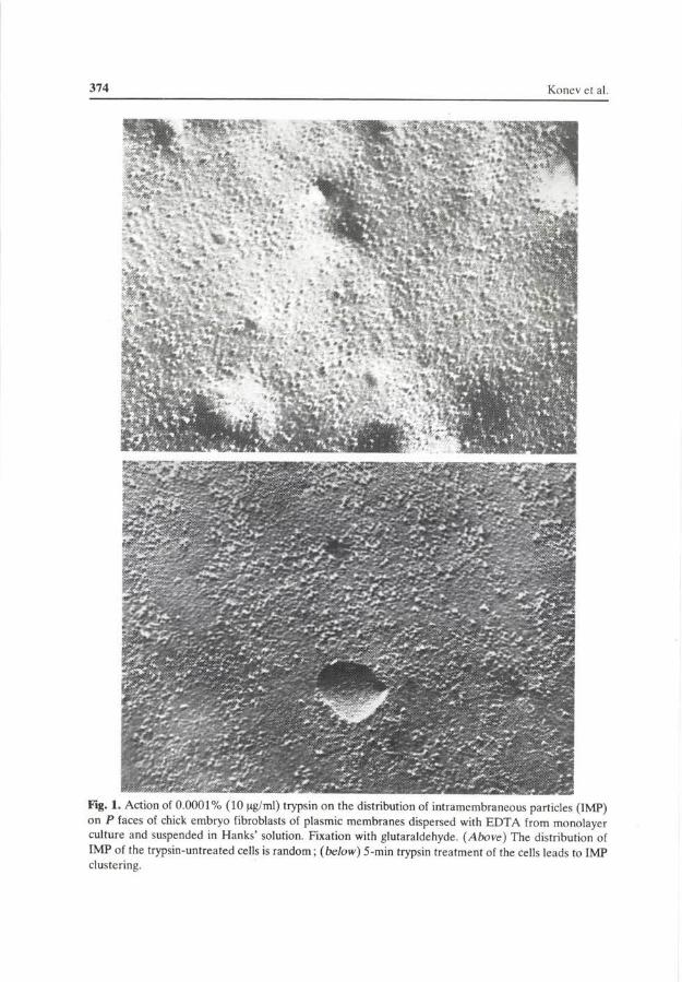
The E face of the membrane contained a much smaller number of IMP as compared with the P face. A short-time treatment of the suspended fibroblasts with trypsin at a final concentration of 10 ^g/ml (0.001%) led to a pronounced change of the IMP distribution pattern, i.e. on the P face the amount of single (dispersed) particles decreased and the number of clusters which contained aggregated IMP sharply rose (Fig. 1, Table 1). A similar behaviour was observed on the E face. In the cells which were not fixed with glutaraldehyde these effects are not so pronounced (cf. Table 1).
A study of the dependence of the IMP distribution pattern for the cells fixed with glutaraldehyde on the trypsin concentration in the suspension has proved that the most distinct changes take place in the range of trypsin concentration from 2 to 10 ng/ml (Fig. 2).
Alongside with IMP clustering, the trypsin-induced structural membrane modification is accompanied by a higher electrophoretic mobility (towards the anode) of trypsinized chick embryo fibroblasts (Fig. 3). It is essential that the most distinct changes of the electrophoretic cell mobility as well as the changes in the
374 Konev et al
Fig. 1. Action of 0.0001% (10 (ig/ml) trypsin on the distribution of intramembraneous particles (IMP) on P faces of chick embryo fibroblasts of plasmic membranes dispersed with EDTA from monolayer culture and suspended in Hanks' solution. Fixation with glutaraldehyde. (Above) The distribution of IMP of the trypsin-untreated cells is random; (below) 5-min trypsin treatment of the cells leads to IMP clustering.

Trypsin Effect on IMP Distribution 375
Table 1. The trypsin effect on the distribution of intramembraneous particles (IMP) of P faces of plasma membranes of chick embryo fibroblasts suspended in Hanks' medium
IMP percentage (±5%)
Observation
number
78 87 87
108
Trypsi-nization 10 itg/ml 5 min, 37 °C
_ + -+
Glutaraldehyde fixation,
2.5% 60 min,
20 °C
+ + --
dispersed
76.1 41.4 48.3 38.9
clustered
aggregates of 3—9 par
ticles
17 37.7 42.5 50
aggregates of 10 particles and
more
6.9 20.9
9.2 11.1
IMP distribution pattern are observed at a low trypsin concentration range (2—10 (ig/ml).
A study of the trypsin action on chick embryo fibroblasts with the aid of the room-temperature tryptophan phosphorescence which is highly sensitive to the structural membrane and protein rearrangements (Mazhul et al. 1976,1979,1980) has revealed that the afterglow decay curve for the untreated fibroblasts suspended in Hanks' solution includes two components with different phosphorenčence times
0 20 40 60 80 100 ug/ml
Fig. 2. The trypsin effect on the IMP distribution pattern on P face of plasmic membranes in chick embryo fibroblasts suspended in Hanks' solution. The cell were treated with different trypsin concentrations for 5 min. Fixation with glutaraldehyde. For the analysis 110 micrographs of the same cell batch were used. O — O, fraction of dispersed IMP; A — A the IMP fraction in aggregates composed of 3 to 9 particles; • — •, the IMP fraction in aggregates composed of 10 and more particles.

376 Konev et al
Fig. 3. The trypsin effect on the electrophoretic mobility of chick embryo fibroblast cells suspended in Hanks' solution. The fibroblasts were treated with different trypsin concentrations for 5 min. The electrophoretic mobility of the untreated cells is taken as 100%. • — • , the cells treated with active trypsin; O — O, the cells treated with contrical-inhibited trypsin.
100
10
1 (
1X
)
\ \ ^s.
\V \ \ \ \ \ \ \ \
<l ' ̂ 1
1 2s
Fig. 4. The trypsin effect on the decay of room-temperature tryptophan phosphorescence of chick embryo fibroblast suspension. (Integral intensity at excitation with A = 297 nm). The cells were treated with trypsin concentration of 50 ug/ml for 10 min. The phosphorescence intensity of trypsin-untreated fibroblasts at zero time is taken as 100 relative units. The total curve of phosphorescence decay of nontrypsinized cell suspension (1) consists of quick (2) and slow (3) components. After trypsinization of the cell decay curve becomes monoexponential (4).

Trypsin Effect on IMP Distribution 377
(Tphosph). After a short time treatment with trypsin at a concentration of 10 to 100 ug/ml (0.001—0.01%) distinct changes in the phosphorescence decay curve are observed: it becomes monoexponential. The lifetime TphoSph changes simultaneously (Fig. 4.)
Discussion
The freeze-etching technique supplies information on the cellular membrane structure and changes in their structural arrangement due to various effects (Konev and Mazhul 1977; Mc Nutt 1977). The IMP observed at the P and E faces are transmembrane proteins or glycoproteins which become exposed upon fracturing membranes along the hydrophobic area of the lipid bilayer (Branton et al. 1975; Edwards et al. 1979).
The present experimental results indicate that a short-time low-concentration trypsin treatment of chick embryo fibroblasts dispersed with EDTA from the monolayer culture and suspended in Hanks' solution gives rise to IMP clustering at P and E faces of the plasmic membrane fractures.
As was demonstrated earlier by the present authors, trypsin brought into chick embryo fibroblast suspension prepared in a similar way causes a pronounced cell aggregation which is completed within a few minutes. Some data were presented which support the suggestion that a structural rearrangement of plasmic membranes which occurs upon cleavage of a small but a critical number of peptide bonds of the proteins at the cell surface is a primary event in the trypsin-induced cell aggregation (Konev and Mazhul 1977; Mazhul et al. 1982).
The changes in the IMP distribution pattern on the E and P faces revealed with electron-microscopic freeze-etching probably reflect the structural rearrangement of plasmic membranes induced by mild proteolysis which leads the cells to a sharp increase of their ability to mutual adhesion. A fair correlation between the curves in the plot of IMP clustering rate and the aggregation versus the trypsin concentration suggests a causal relation between the trypsin-induced rearrangement of plasmic membrane with the cell aggragation. Consequently, the cells with a low adhesive activity (trypsin-untreated) have a random IMP distribution, and IMP of the highly adhesive cells are clustered.
It should be noted that a distinct clustering of IMP was observed only in the glutaraldehyde-fixed cells. Glutaraldehyde, by forming interprotein bridges, prevents probably the protein membrane components from lateral migration when the cells are incubated in glycerol and thus "freezes" the cell surface microrelief at that structural state which occurs just after the trypsin-induced rearrangement was completed.
A change in the distribution pattern of IMP effected by a shorttime (15 min) treatment of 3T3 mice fibroblasts with trypsin (100 Jig/ml) was earlier recorded by

378 Konev et al.
Furcht and Wendelschafer-Crabb (1978). Contrary to the present data, IMP of the control cells were clustered and the particles of the trypsinized fibroblasts were diffusively distributed. The differences in the trypsin effect on the IMP distribution patterns in (Furcht and Wendelschafer-Crabb 1978) and in the present experiments may be attributed to the fact that the 3T3 cells were treated with trypsin in a confluent monolayer culture, whereas the chick embryo fibroblasts were trypsinized after they had been dispersed with EDTA and suspended in Hanks' solution.
As follows from the electrophoretic (towards the anode) mobility measurements the trypsin-induced rearrangement of the plasmic membrane of chick embryo fibroblasts is accompanied by an increase in the negative charge of the cellular surface (Fig. 3).
It was demonstrated earlier that the electrophoretic mobility of the trypsinized (70 ug/ml) 3T3 cells is approximately 25% lower than that of untreated cells (Adam and Adam 1975). However, the electrophoretic mobility of the nontrans-formed BHK-21 cells incubated with trypsin (400 ug/ml) remained unchanged (Latner and Turner 1978). These differences in the trypsin action on the electrophoretic cell properties may probably be explained by different fibroblast types used and unequal experimental conditions.
The increase of the surface charge usually favours the electrostatic cell repulsion but not their aggregation. A sharp increase of the adhesiveness in trypsinized fibroblasts observed in the present experiments support the suggestion that the cells in aggregates are held together by forces other than of electrostatic nature. As was shown earlier by the present authors, the trypsin-induced aggregation of chick embryo fibroblasts is not caused by a mere binding of trypsin (which possesses polycationic properties) to anionic groups of the cell surface. Also the trypsin-induced aggregation is not associated with calcium and magnesium cations or with macromolecules of the aggregation factors (Konev and Mazhul 1977; Mazhul et al. 1982).
The trypsin-induced IMP clustering may be thought to involve formation of the ordered microrelief of "sticky" areas which provide for adhesion of the cells in aggregates. It is possible that the cell contacting involves "additional fitting" of adhesion areas microreliefs which resembles the induced fitting between the active site structure of enzyme and that of substrate upon their interaction.
The trypsin-induced aggregation mechanism will be analyzed in more detail in the subsequent papers of the series.
Investigation of the relationships of the plasmic membrane response to the trypsin action is of particular interest in view of importance of proteases for the cell life activity. Low trypsin concentrations applied to cell surfaces are known to eliminate contact inhibition of reproduction (Burger 1970, 1973; Carney and Cunninghan 1977), to regulate the adenylate cyclase activity (Guirard-Simplot and Colobert 1977; Lacomb et al. 1977; Richert and Ryan 1977), to stimulate the
Trypsin Effect on IMP Distribution 379
deoxyglucose transport (Blumberg and Robbins 1975), to inhibit the lysine transport (Tsan et al. 1973), to suppress the supply of palmitic acid to cells (Machudevan and Sauer 1974), to change the cell volume (Mallucci et al. 1972), to affect the topography and mobility of the lectin receptors and agglutination of the cells by lectin (Hynes 1974; Rapin and Burger 1974; Nicolson 1976). A mild short-time trypsinization removes LETS-protein from the cell surface (Yamada et al. 1976; Mautner and Hynes 1977 ; Chen et al. 1978), causes bulbs to appear on the cell surface, destroys microtubules and microfilaments (Furcht and Wendels-chafer—Crabb 1978; Pollack and Rifkin 1976). It may be thought that the trypsin-like protease effects on the cellular surface bring about malignant transformation of the cells (Reich et al. 1975; Hatcher et al. 1977).
In organism the cellular surface is apparently affected by proteases during the ontogenesis. This is indicated both by association of the proteases (the trypsin-like ones included) with plasmic membranes of normal and transformed cells (Boss-mann 1977; Hatcher et al. 1977), and by quite large trypsin amounts present in serum (Kôstering et al. 1976). However, the cellular surface undergoes the action of lysosomal proteases which can be released in response to various physical, chemical and virus agents (Duve 1976; Pokrovskii and Tutelyan 1976).
Since trypsin does not penetrate inside a cell (Burger 1973; Carney and Cunninghan 1977), all the primary events which result in fundamental shifts take place on the cellular surface. It may be thought that they are underlied by structural rearrangements of plasmic membranes caused by cleavage of a small number of peptid bonds in membrane proteins. It is quite important that the trypsinization time necessary for most of the above mentioned effects to manifest themselves amounts to a few minutes just as in the present experiments. The active trypsin concentrations also agree well.
It seems probable that the trypsin-induced structural rearrangement is quite generalized. It involves the whole plasmic membrane and is manifested by a sharp increase of the cellular surface adhesiveness, a higher ability of the protein components to lateral mobility, exposure of ionogenic groups. The intracellular membranes and the cytoskeleton system are apparently also involved into the structural rearrangement.
That the rearrangement of the cellular membrane system is generalized is also confirmed by the phosphorescence studies of chick embryo fibroblasts. Changes of the kinetic parameters of the room-temperature phosphorescence of fibroblasts after the treatment with low-concentrations of trypsin (Fig. 4) may be interpreted as the changes in the rigidity and polarity of the microenvironment of protein tryptophan residues. Since in the present work a light signal from the whole set of proteins in a cell was recorded, noticeable changes in the phosphorescence decay could be observed only if a considerable amount of the cellular proteins are involved into the structural rearrangement.

380 Konev et al
Thus, knowledge of the relations of the trypsin interaction with the plasmic cell membrane is important not only for elucidating the cellular adhesion mechanism but also for understanding of the role of proteases in the regulation in the functional activity in normal and pathological cells.
References
Adam G., Adam G. (1975): Cell surface change and regulation of cell division of 3T3 cells and transformed derivates. Exp. Cell Res. 93, 71—78
Blumberg P. M., Robbins Ph. W. (1975): Effect of protease on activation of resting chick embryo fibroblasts and on cell surface proteins. Cell 6, 137—147
Bosmann N. B. (1977): Cell surface enzymes: effects on mitotic activity and cell adhesion. Int. Rev. Cytol. 50, 1—22
Branton D., Bullivant S., Gilula N. E„ Karnovsky M. S., Moor H., Miihlethaler K. D. H., Northcote D. H., Packer L., Satir B., Speth V., Staehelin L. A., Steere R., Weinstein R. (1975): Freeze-etching nomenclature. Science 190, 54—56
Burger M. M. (1970): Protoelytic enzymes initiating cell division and escape from contact inhibition of growth. Nature 227, 170—171
Burger M. M. (1973): Surface changes in transformed cells detected by lectins. Fed. Proc. 32,91—101 Carney D. H., Cunningham D. D. (1977): Initiation of chick cell division by trypsin action at the cell
surface. Nature 268, 602—606 Chen L. B., Murray A., Segal R. A., Bushnell A., Walsh M., (1978): Studies on intercellular LETS
glycoprotein matrixes. Cell 14, 377—391 de Duve C. (1976): The role of lysosomes in the pathogeny of disease. Scand. J. Reumatol. 5, suppl. No
12, 63-69 Edwards H. H., Mueller T. J., Morrison M. (1979): Distribution of transmembrane polypeptides in
freeze-fracture. Science 203, 1344—1345 Furcht L. T., Wendelshafer-Crabb G. (1978): Trypsin-induced coordinated alterations in cell shape,
cytoskeleton and intrinsic membrane structure of contact-inhibited cells. Exp. Cell Res. 114,1—14 Guirard-Simplot A., Colobert L. (1977): Adenylat cyclase activation by trypsin in KB cell cultures.
Experientia 33, 899—990 Hatcher V. B., Oberman M. S., Wertheim M. S., Rhee Ch. Y., Tsien G., Burk P. G. (1977): The
relationship between surface protease activity and the rate of cell proliferation on normal and transformed cells. Biochem. Biophys. Res. Commun. 76, 602—608
Hynes R. O. (1974): Role of surface alterations in cell transformations: the importance of proteases and surface proteins. Cell, 1, 147—156
Konev S. V., Mazhul V. M. (1977): Intercellular Contacts Náuka i Tekhnika, Minsk (in Russian) Kôstering H., Haunschild N., Warmann E., Vólker P. (1976): Einflus von Trypsinogen und Trypsin auf
die Blutgerinnung in vitro. Med. Welt 27, 1575—1579 Lacombe M-L., Stengel D., Hanoune J. (1977): Proteolytic activation of adenylate cyclase from rat
liver plasma membranes. FEBS Lett. 77, 159—163 Latner A. L., Turner G. A. (1978): Increased trypsin sensitivity of cell surface macromolecules after
malignant transformation. Exp. Cell. Biol. 46, 290—309 Machadevan S., Sauer F. (1974): Effect of trypsin, phospholipases and membrane-impermeable
reagents on the uptake of palmitic acid by isolated rat liver cells. Arch. Biochem. Biophys. 164, 185—194

Trypsin Effect on IMP Distribution 381
Mallucci L., Wells V., Young M. R. (1972): Effect of trypsin on cell volume and mass. Nature, 239, 53—55
Mautner V., Hynes R. O. (1977): Surface distribution on LETS protein in relation to the cytoskeleton of normal and transformed cells. J. Cell Biol. 75, 743—768
Mazhul V. M., Ermolaev Yu. S., Konev S. V. (1979): Room temperature tryptophan phosphorescence as a new method for the investigation of structural states of cellular membranes and proteins. 26 Nat. Confer. Luminesc. Abstr. pp. 29—30 (in Russian)
Mazhul V. M., Ermolaev Yu. S., Konev S. V. (1980): Room temperature tryptophan phosphorescence as a new method for the investigation of structural states of cellular membranes and proteins. Zhurn. Priklad. Spektroskop. 32, 903—907 (in Russian)
Mazhul V. M., Nikolskaya V. P., Konev S. V. (1982): The trypsin effect on intercellular adhesion of chick embryo fibroblasts. Gen. Physiol. Biophys. 1,
Mazhul V. M, Ermolaev Yu. S., Bobrov V. A., Nikolskaya V. P., Konev S. V. (1976): About the sensitivity of the parameters of room temperature phosphorescence to the conformation of cellular proteins and membranes. Izv. Acad. Náuk BSSR, Ser. Biol. 6, 51—56 (in Russian)
Mc Nutt N. S. (1977): Freeze-fracture techniques and application to the structural analysis of the mammalian plasma membrane. In: Cell Surface Reviews (Eds. G. Poste, G. L. Nicolson), v. 3, pp. 75—126, Elsevier Press, Amsterdam
Moor H., Muhlethaler K. (1963): Fine structure in frozen-etched yeast cells. J. Cell Biol. 17,609—628 Nicolson G. L. (1976): Transmembrane control of the receptors on normal and tumor cells. II. Surface
changes associated with transformation and malignancy. Biochim. Biophys. Acta, 458, 1—72 Pokrovskii A. A., Tutelyan V. A. (1976): Lysosomes. Nauka, Moscow Pollack R., Rifkin D. B. (1976): Modification of mammalian cell shape: redistribution of intercellular
action by SV40 virus, proteases, cytochalasin B and dimethylsulfoxide. In: Cell Motility. Book A. Motility of Muscle and Non-Muscle Cells. (Eds. R. Goldman, T. Pollard, J. Rosenbaum), pp. 389—401, Cold Spring Harbor Press, New York
Rapin A. M., Burger M. M. (1974): Tumor cell surfaces: general alterations detected by agglutinins. Advan. Cancer Res. 20, 1—91
Reich E., Shaw E., Rifkin D. B. (1975): Proteases and cellular proliferation. In: Protease and Biological Control. (Cold Spring Harbor Laboratory), pp. 777—797, Cold Spring Harbor, New York
Richert N. D., Ryan R. J. (1977): Proteolytic enzyme activation of rat ovarian adenylate cyclase. Proc. Nat. Acad. Sci. USA, 74, 4857-^1861
Tsan Min-Fu, Taube R. A., Berlin R. D. (1973): The effect of trypsin on membrane transport of non-electrolytes. J. Cell Physiol. 81, 251—256
Yamada K. M., Yamada S. S., Pastan I. (1976): Cell surface protein partially restores morphology, adhesiveness and contact inhibition of movement to transformed fibroblasts. Proc. Nat. Acad. Sci. USA, 73, 1217—1221
Received April 15, 1982 / Accepted May 24, 1982