tuberculosis infection and tuberculosis disease · 2018-12-18 · in this module, you will learn...
TRANSCRIPT

3Module
National Center for HIV/AIDS, Viral Hepatitis, STD, and TB Prevention Division of Tuberculosis Elimination
Self-Study Modules on Tuberculosis
Targeted Testing and the Diagnosis of Latent Tuberculosis Infection and Tuberculosis Disease


3Self-Study Modules
on Tuberculosis
Module
Targeted Testing and the Diagnosis of Latent Tuberculosis Infection
and Tuberculosis Disease
U.S. DEPARTMENT OF HEALTH AND HUMAN SERVICESCenters for Disease Control and Prevention
National Center for HIV/AIDS, Viral Hepatitis, STD, and TB PreventionDivision of Tuberculosis Elimination
Atlanta, Georgia 2016

viv

3Self-Study Modules
on Tuberculosis
Module
Contents
Background . . . . . . . . . . . . . . . . . . . . . . . . . . . . . . . . . . . . . . . . . . . . . 1
Objectives . . . . . . . . . . . . . . . . . . . . . . . . . . . . . . . . . . . . . . . . . . . . . . 1
New Terms . . . . . . . . . . . . . . . . . . . . . . . . . . . . . . . . . . . . . . . . . . . . . . 2
Targeted Testing . . . . . . . . . . . . . . . . . . . . . . . . . . . . . . . . . . . . . . . . 6
Diagnosis of LTBI . . . . . . . . . . . . . . . . . . . . . . . . . . . . . . . . . . . . . . . . 8
Diagnosis of TB Disease . . . . . . . . . . . . . . . . . . . . . . . . . . . . . . . . . 39
TB Genotyping . . . . . . . . . . . . . . . . . . . . . . . . . . . . . . . . . . . . . . . . . 69
Additional Resources . . . . . . . . . . . . . . . . . . . . . . . . . . . . . . . . . . . 71
Answers to Study Questions . . . . . . . . . . . . . . . . . . . . . . . . . . . . . 73
Case Study Answers . . . . . . . . . . . . . . . . . . . . . . . . . . . . . . . . . . . . 81
Module 3— Targeted Testing and the Diagnosis of Latent Tuberculosis Infection and Tuberculosis Disease
viv

1vi

BackgroundIn this module, you will learn about targeted testing and the diagnosis of latent tuberculosis (TB) infection (LTBI) and TB disease. Targeted testing is a TB control strategy that is used to identify people who have LTBI and are at high risk for developing TB disease and would benefit from treatment. LTBI is diagnosed with the Mantoux tuberculin skin test (TST) or an interferon-gamma release assay (IGRA), such as the QuantiFERON®-TB Gold In-Tube test (QFT-GIT) or the T-SPOT®.TB test (T-Spot).
It is important to medically evaluate people who have symptoms of TB disease; if they are found to have TB disease, they need treatment to be cured and to help stop the transmission of TB to others. For this reason, the diagnosis of TB disease is crucial to controlling the spread of TB in homes and communities. In most cases, TB disease is diagnosed with certain laboratory tests. For patients who may have pulmonary TB disease, a chest x-ray is also useful for diagnosis.
Note: The Self-Study Modules on Tuberculosis are a series of educational modules designed to provide information about TB in a self-study
format. The target audiences include outreach workers, nurses, physicians, administrators, health educators, and students from a variety
of settings. The Modules should not be used as a substitute for guidelines and should not be used for patient care decisions.
ObjectivesAfter working through this module, you will be able to
1. Identify high-risk groups for targeted testing.
2. Describe how to place, read, and interpret a Mantoux tuberculin skin test.
3. Describe how to interpret an interferon-gamma release assay.
4. Discuss considerations for using either the Mantoux tuberculin skin test or an interferon-gamma release assay for diagnosing latent tuberculosis infection.
5. Describe the components of a medical evaluation for diagnosing TB disease.
define
explain
describe
list
1vi
Module 3— Targeted Testing and the Diagnosis of Latent Tuberculosis Infection and Tuberculosis Disease

New TermsNew terms introduced in this module are included below. These terms appear in bold in the module text.
acid-fast bacilli (AFB)—mycobacteria that when stained, retain color even after they have been washed in an acid solution; may be detected under a microscope in a stained smear
anergy—the inability to react to a skin test because of a weakened immune system, often caused by HIV infection or severe illness
antigen—protein substances that can produce an immune response (such as CFP-10, ESAT-6, or those in PPD)
bacteriologic examination—tests done in a mycobacteriology laboratory to aid diagnosis of TB disease; includes examining a specimen under a microscope, culturing the specimen, and testing for drug susceptibility
baseline skin test—a tuberculin skin test (TST) given to employees or residents in certain facilities when they start their job or enter the facility (see TB testing program and two-step testing)
BCG—bacille Calmette-Guérin (BCG), a vaccine for TB disease that is used in many countries but rarely used in the United States; may cause a false-positive reaction to the TST but does not affect interferon-gamma release assay (IGRA) results
boosted reaction—a positive reaction to a TST, due to a boosted immune response from a skin test given up to a year earlier; occurs in people who were infected a long time ago and whose ability to react to tuberculin has lessened. Two-step testing is used in TB testing programs to tell the difference between boosted reactions and reactions caused by recent infection (see booster phenomenon and two-step testing).
booster phenomenon—a phenomenon in which people (especially older adults) who are skin tested many years after becoming infected with M. tuberculosis may have a negative reaction to an initial TST, followed by a positive reaction to a TST given up to a year later; this happens because the first
32
Module 3— Targeted Testing and the Diagnosis of Latent Tuberculosis Infection and Tuberculosis Disease

TST boosts the immune response. Two-step testing is used in TB testing programs to tell the difference between boosted reactions and reactions caused by recent infection (see two-step testing).
bronchoscopy—a procedure used to obtain pulmonary secretions or lung tissue with an instrument called a bronchoscope; used only when patients cannot cough up sputum on their own and an induced specimen cannot be obtained
cavity—a hollow space within the lung, visible on a chest x-ray, that may contain many tubercle bacilli; often occurs in people with severe pulmonary TB disease
CFP-10—one of the antigens used in IGRAs that is found in M. tuberculosis strains but not in BCG vaccine strains
clinician—a physician, physician’s assistant, or nurse
colonies—groups of mycobacteria that have grown on solid media
control—a standard of comparison for checking or verifying the results of an experiment
culture—to grow organisms in media (substances containing nutrients) so that they or the product of this process can be identified; a positive culture for M. tuberculosis contains tubercle bacilli, whereas a negative culture contains no detectable tubercle bacilli
drug susceptibility pattern—the list of antituberculosis drugs to which a strain of tubercle bacilli is susceptible and to which it is resistant
erythema—redness around the site of the injection when a TST is done; erythema is not measured as part of the reaction size because redness does not indicate that a person has TB infection
ESAT-6—one of the antigens used in IGRAs that is found in M. tuberculosis strains but not in BCG vaccine strains
exposure to TB—time spent with or near someone who has infectious TB disease
false-negative reaction—a negative reaction to the TST or IGRA in a person who has TB infection
false-positive reaction—a positive reaction to the TST or IGRA in a person who does not have TB infection
gastric washing—a procedure done by inserting a tube through the patient’s nose and passing it into the stomach; may be useful for obtaining a specimen for culture from children, who produce little or no sputum when they cough
GeneXpert—a semi-automated molecular diagnostic system. See Xpert MTB/RIF assay.
genotype—distinct genetic pattern of an organism
genotyping—a laboratory-based method that can determine the genetic pattern of the strain of M. tuberculosis that caused TB disease in a person
induced sputum—sputum that is obtained by having the patient inhale a saline (salt water) mist, causing the patient to cough deeply; this procedure is used to help patients cough up sputum if they cannot do so on their own
induration—swelling that can be felt around the site of injection after a TST is done; the reaction size is the diameter of the swollen area, measured across the forearm
infiltrate—a collection of fluid and cells in the tissues of the lung; visible on a chest x-ray in people with pulmonary TB disease
32
Module 3— Targeted Testing and the Diagnosis of Latent Tuberculosis Infection and Tuberculosis Disease

interferon-gamma (IFN-γ)—protein that is normally produced by the body in response to infection. IGRA interpretations are based on the amount of IFN-γ that is released or on the number of cells that release IFN-γ.
interferon-gamma release assay (IGRA)— a type of blood test that measures a person’s immune reactivity to M. tuberculosis. In the United States, the QuantiFERON®-TB Gold In-Tube (QFT-GIT) and the T-SPOT®.TB test (T-Spot) are currently available IGRAs.
isolate—a group of organisms isolated or separated from a specimen; in an M. tuberculosis isolate, the organisms have been grown in culture and identified as M. tuberculosis
malaise—a feeling of general discomfort or illness
Mantoux tuberculin skin test (TST)— a method of testing for TB infection; a needle and syringe are used to inject 0.1 ml (5 tuberculin units) of purified protein derivative (PPD) tuberculin solution between the layers of the skin (intradermally), usually on the forearm; the reaction to this test, usually a small swollen area (induration), is measured 48 to 72 hours after the injection and is interpreted as positive or negative depending on the size of the reaction and the patient’s risk factors for TB; the routine methodology for tuberculin skin testing worldwide; supersedes all older methods
media—substances containing special nutrients and used for growing cultures of bacteria found in specimens
medical history—the part of a patient’s life history that is important for diagnosing and treating TB infection or disease, including history of exposure, symptoms, previous diagnosis of TB infection or disease, and risk factors for TB disease
mycobacteriology laboratory—a laboratory that deals specifically with M. tuberculosis and other mycobacteria
nucleic acid amplification (NAA)—a technique that amplifies (copies) DNA and RNA segments. Often used in assays to directly detect microorganisms in sputum specimens.
polymerase chain reaction (PCR)—a type of NAA used to make many copies of a segment of DNA
PPD (purified protein derivative)—antigens such as the type of tuberculin used in the TST (see antigen)
QuantiFERON®-TB Gold In-Tube test (QFT-GIT)—a blood test used to determine TB infection. The QFT-GIT measures the response to simulated TB proteins when they are mixed with a small amount of whole blood.
resistant—an organism’s ability to grow despite the presence of a particular drug
skin test conversion—a change in a skin test reaction from negative to positive between testing intervals
smear—a specimen that has been smeared onto a glass slide, stained, washed in an acid solution, and then placed under the microscope for examination; used to detect acid-fast bacilli in a specimen
sputum—phlegm from deep in the lungs, collected in a sterile container for processing and examination
susceptible—an organism’s ability to be killed by a particular drug
symptoms of TB disease—noticeable conditions caused by TB disease. The symptoms of pulmonary TB disease include coughing, pain in the chest when breathing or coughing, and coughing up sputum or blood. The general symptoms of TB disease (pulmonary or
54
Module 3— Targeted Testing and the Diagnosis of Latent Tuberculosis Infection and Tuberculosis Disease

extrapulmonary) include weight loss, fatigue, malaise, fever, and night sweats. The symptoms of extrapulmonary TB disease depend on the part of the body that is affected by the disease.
targeted testing—a TB control strategy to identify persons at high risk for latent TB infection and persons at high risk for developing TB disease who would benefit from treatment
TB7.7—one of the antigens used in the QFT-GIT
TB testing program—a program in which employees and residents of a facility are periodically tested for TB; done to identify people who have TB infection and possibly TB disease and to determine whether TB is being transmitted in the facility
T-SPOT®.TB Test (T-Spot)—a blood test used to determine TB infection; the T-Spot measures the number of T cells that secrete IFN-γ upon activation by M. tuberculosis antigens
tuberculin—a substance made from tubercle bacilli that have been killed by heating; used to determine whether a person has TB infection. Tuberculin is not a vaccine.
tuberculin skin test (TST)—a test used to detect TB infection (see Mantoux tuberculin skin test)
tuberculin unit—a standard strength of tuberculin used in the United States and Canada; a strength of 5 tuberculin units is used for the Mantoux TST
two-step testing—a strategy used in TB testing programs to distinguish a boosted reaction (caused by TB infection that occurred many years ago) from a reaction caused by recent infection. If a person has a negative reaction to an initial skin test, a second test is given 1 to 3 weeks later; a positive reaction to the second test probably represents a boosted reaction, not recent infection. Two-step testing is used in many TB testing programs for skin testing employees when they start their job.
window period—the time between a person’s last exposure to infectious TB and when a TST or IGRA can reliably detect infection with M. tuberculosis
Xpert MTB/RIF assay—a nucleic acid amplification (NAA) test that simultaneously identifies Mycobacterium tuberculosis complex and rifampin resistance in a sputum sample
54
Module 3— Targeted Testing and the Diagnosis of Latent Tuberculosis Infection and Tuberculosis Disease

3Self-Study Modules
on Tuberculosis
Module
Targeted TestingTargeted testing is the TB control strategy that is used to identify and treat persons who are at high risk for latent TB infection (LTBI) or at high risk for developing TB disease once infected with M. tuberculosis. Identifying persons with LTBI is important to the goal of TB elimination because LTBI treatment can prevent these persons from developing TB disease and thereby stop the further spread of TB to others.
Thus, during routine patient evaluations, health care providers should identify persons who are at high risk for TB and test them for LTBI. However, TB testing activities should be done only when there is a plan for follow-up care to evaluate and treat all individuals diagnosed with LTBI or TB disease. Healthcare agencies or other facilities should consult with the local health department before starting a TB testing program to make sure that any person whose test result is positive will have access to follow-up care.
People who are not at high risk for LTBI generally should not be tested. Testing in low-risk populations can take resources away from other important activities. Also, positive test results in low-risk populations are sometimes inaccurate. However, there may be instances in which health care providers are asked to test individuals who are not generally considered as high risk (for example, daycare center workers, teachers, and college students) because of local policies and procedures.
Identifying High-Risk Groups for TB TestingHigh-risk groups can be divided into two categories:
�� People who are at high risk for exposure to or infection with M. tuberculosis
�� People who are at high risk for developing TB disease once infected with M. tuberculosis
Flexibility should be used in defining high-risk groups for testing. Since the epidemiology of TB can change, the risk of LTBI or TB disease among groups may change over time. Groups that are currently identified as being at low-risk may later be considered high priority. Moreover, because of the differences in populations from one community to another,
Targeted testing is the TB control strategy that is used to identify and treat persons who are at high risk for latent
TB infection (LTBI) or at high risk for developing TB disease once infected
with M. tuberculosis.
People who are not at high risk for LTBI generally
should not be tested.
Flexibility should be used in defining high-risk groups for testing.
76
Module 3— Targeted Testing and the Diagnosis of Latent Tuberculosis Infection and Tuberculosis Disease
76

definitions of high risk should be made at the local (city, county, or state) level according to local demographics and TB epidemiology.
In general, however, high-risk groups listed in both categories of Table 3.1 should be tested.
Table 3.1 Groups at High Risk for TB Infection and TB Disease.
People at High Risk for Exposure to or Infection with
M. tuberculosis
People at High Risk for Developing TB Disease after
Infection with M. tuberculosis
�� Contacts of people known or suspected to have TB disease
�� People who have come to the United States within the last 5 years from areas of the world where TB is common (for example, Asia, Africa, Russia, Eastern Europe, or Latin America)
�� People who visit areas with a high prevalence of TB disease, especially if visits are frequent or prolonged
�� People who live or work in high-risk congregate settings (for example, nursing homes, homeless shelters, or correctional facilities)
�� Health care workers who serve patients who are at increased risk for TB disease
�� Populations defined locally as having an increased incidence of LTBI or TB disease, possibly including medically underserved, low-income populations, or persons who abuse drugs or alcohol
�� Infants, children, and adolescents exposed to adults who are at increased risk for LTBI or TB disease
�� People living with HIV
�� Children younger than 5 years of age
�� People recently infected with M. tuberculosis (within the past 2 years)
�� People with a history of untreated or inadequately treated TB disease
�� Persons who are receiving immunosuppressive therapy such as tumor necrosis factor-alpha (TNF) antagonists, systemic corticosteroids equivalent to/greater than 15 mg of prednisone per day, or immunosuppressive drug therapy following organ transplantation
�� Persons with silicosis, diabetes mellitus, chronic renal failure, leukemia, or cancer of the head, neck, or lung
�� Persons who have had a gastrectomy or jejunoileal bypass
�� Low body weight
�� Cigarette smokers and persons who abuse drugs or alcohol
�� Populations defined locally as having an increased incidence of disease due to M. tuberculosis, including medically underserved, low-income populations.
76
Module 3— Targeted Testing and the Diagnosis of Latent Tuberculosis Infection and Tuberculosis Disease
76

3Self-Study Modules
on Tuberculosis
Module
Diagnosis of LTBICurrently, the available methods of testing for M. tuberculosis infection are the Mantoux tuberculin skin test (TST) and the interferon-gamma release assays (IGRAs) such as the QuantiFERON®-TB Gold In-Tube (QFT-GIT) test or the T-SPOT®.TB test (T-Spot).
The Mantoux Tuberculin Skin Test (TST)The TST is used to determine if a person is infected with M. tuberculosis. In this test, a substance called tuberculin is injected into the skin. Tuberculin contains antigens used for diagnosing TB infection; it is not a vaccine. An antigen is a protein substance that can produce an immune response. Tuberculin is made from proteins derived from tubercle bacilli that have been killed by heating. In most people who have TB infection, the immune system will recognize the tuberculin because it is similar to the tubercle bacilli that caused infection. This will cause a reaction to the tuberculin at the site of the injection. Tuberculin used for the skin test is also known as purified protein derivative, or PPD.
Administering the TSTThe TST is given by using a single dose disposable tuberculin syringe to inject 0.1 ml of 5 tuberculin units of liquid tuberculin between the layers of the skin (intradermally), on the forearm (Figure 3.1). A tuberculin unit is a standard strength of tuberculin. When giving the TST, institutional guidelines for infection control should be followed.
A patient’s forearm should be examined by a trained health care worker 48 to 72 hours after the tuberculin is injected. Health care workers should not ask patients to read their own skin test results. Most people with TB infection will have a positive reaction to the tuberculin. The reaction is an area of induration (swelling that can be felt) around the site of the injection. The diameter of the indurated area is measured in millimeters across the forearm (Figure 3.2); erythema (redness) around the indurated area is not measured, because the presence of erythema does not indicate that a person has TB infection (Figure 3.3).
The TST is used to determine if a person is
infected with M. tuberculosis.
Tuberculin is not a vaccine.
Most people with TB infection have a positive
reaction to the tuberculin.
The reaction is an area of induration, or swelling, around the site of the injection.
98
Module 3— Targeted Testing and the Diagnosis of Latent Tuberculosis Infection and Tuberculosis Disease
98

Figure 3.1 Administering the Mantoux TST.
Figure 3.2 Only the induration is being measured. This is CORRECT.
Figure 3.3 The erythema is being measured. This is INCORRECT.
98
Module 3— Targeted Testing and the Diagnosis of Latent Tuberculosis Infection and Tuberculosis Disease
98

Study Question 3 .1–3 .23.1 What is the TST used for?
3.2 How is the TST given?
Answers to study questions are on pages 73–80
1110
Module 3— Targeted Testing and the Diagnosis of Latent Tuberculosis Infection and Tuberculosis Disease

Study Question 3 .3–3 .43.3 With the TST, when is the patient’s arm examined?
3.4 How is the induration measured?
Answers to study questions are on pages 73–80
1110
Module 3— Targeted Testing and the Diagnosis of Latent Tuberculosis Infection and Tuberculosis Disease

Interpreting the ReactionInterpreting a TST reaction depends on the size of the induration and the person’s risk factors for TB (Table 3.2).
An induration of 5 or more millimeters is considered a positive reaction for the following people:
�� People living with HIV �� Recent contacts of people with infectious TB�� People with chest x-ray findings suggestive of previous
TB disease�� People with organ transplants �� Other immunosuppressed patients (for example, patients
on prolonged therapy with corticosteroids equivalent to/greater than 15mg per day of prednisone or those taking TNF-alpha antagonists)
An induration of 10 or more millimeters is considered a positive reaction for the following people:
�� People who have recently come to the United States (within the last 5 years) from areas of the world where TB is common (for example, Asia, Africa, Russia, Eastern Europe, or Latin America)
�� People who abuse drugs �� Mycobacteriology laboratory workers�� People who live or work in high-risk congregate settings
(for example, nursing homes, homeless shelters, or correctional facilities)
�� People with certain medical conditions that place them at high risk for TB (for example, silicosis, diabetes mellitus, severe kidney disease, certain types of cancer, and certain intestinal conditions)
�� Children younger than 5 years of age�� Infants, children, or adolescents exposed to adults in
high-risk categories
An induration of 15 or more millimeters is considered a positive reaction for people with no known risk factors for TB. However, it is important to remember that testing for TB infection should generally be targeted towards high-risk groups, since test results in low-risk groups can be inaccurate.
Most people who have a positive TST reaction will usually have a positive reaction every time they are tested, regardless of whether they receive treatment. This is because the TST detects the immune response to tuberculin, not the presence of tubercle bacilli in the body.
Interpreting a TST reaction depends on the size of the induration and the
person’s risk factors for TB.
An induration of 15 or more millimeters is considered
a positive reaction for people with no known
risk factors for TB.
1312
Module 3— Targeted Testing and the Diagnosis of Latent Tuberculosis Infection and Tuberculosis Disease

Thus the TST should not be performed on a person who has a documented history of either a positive TST result or treatment for TB disease.
Interpreting the TST Reaction for Occupational ExposureFor people who may be exposed to TB on the job (such as health care workers and staff of nursing homes or correctional facilities), the interpretation of the TST reaction as positive or negative depends on
�� The employee’s individual risk factors for TB�� The risk of exposure to TB in the person’s job
Therefore, in facilities where TB patients receive care, 10 or more millimeters of induration may be considered a positive reaction for employees with no other risk factors for TB. In facilities where the risk of exposure to TB is very low, 15 or more millimeters of induration may be considered a positive reaction for employees with no other risk factors for TB.
Table 3.2 Interpreting the TST reaction.
5 or more millimeters
10 or more millimeters
15 or more millimeters
An induration of 5 or more millimeters is considered positive for
�� People living with HIV
�� Recent contacts of people with infectious TB
�� People with chest x-ray findings suggestive of previous TB disease
�� People with organ transplants
�� Other immunosuppressed patients (for example, patients on prolonged therapy with corticosteroids equivalent to/greater than 15mg per day of prednisone or those taking TNF-alpha antagonists)
An induration of 10 or more millimeters is considered positive for
�� People who have recently come to the United States (within the last 5 years) from areas of the world where TB is common (for example, Asia, Africa, Russia, Eastern Europe, or Latin America)
�� People who abuse drugs
�� Mycobacteriology laboratory workers
�� People who live or work in high-risk congregate settings (for example, nursing homes, homeless shelters, or correctional facilities)
�� People with certain medical conditions that place them at high risk for TB (for example, silicosis, diabetes mellitus, severe kidney disease, certain types of cancer, and certain intestinal conditions)
�� Children younger than 5 years of age
�� Infants, children, and adolescents exposed to adults in high-risk categories
An induration of 15 or more millimeters is considered positive for
�� People with no known risk factors for TB
1312
Module 3— Targeted Testing and the Diagnosis of Latent Tuberculosis Infection and Tuberculosis Disease

Study Questions 3 .5–3 .6
3.5 What two factors determine the interpretation of a skin test reaction as positive or negative? What additional factor is considered for people who may be exposed to TB on the job?
3.6 Name five groups of people for which 5 or more millimeters of induration is considered a positive reaction.
Answers to study questions are on pages 73–80
1514
Module 3— Targeted Testing and the Diagnosis of Latent Tuberculosis Infection and Tuberculosis Disease

Study Questions 3 .7–3 .8
3.7 Name seven groups of people for which 10 or more millimeters of induration is considered a positive reaction.
3.8 For which group of people is 15 or more millimeters of induration considered a positive reaction?
Answers to study questions are on pages 73–80
1514
Module 3— Targeted Testing and the Diagnosis of Latent Tuberculosis Infection and Tuberculosis Disease

Case Study 3 .1
Which of the following patients have a positive TST reaction? Circle the best answer(s).
a) Mr. West, 36 years old, HIV infected, 8 mm of induration
b) Ms. Hernandez, 26 years old, native of Mexico, 7 mm of induration
c) Ms. Jones, 56 years old, has diabetes, 12 mm of induration
d) Mr. Sung, 79 years old, resident of a nursing home, 11 mm of induration
e) Mr. Williams, 21 years old, no known risk factors, 13 mm of induration
f ) Ms. Marcos, 42 years old, chest x-ray findings suggestive of previous TB, 6 mm of induration
g) Ms. Rayle, 50 years old, husband has pulmonary TB, 9 mm of induration
Answers to case studies are on pages 81–84
1716
Module 3— Targeted Testing and the Diagnosis of Latent Tuberculosis Infection and Tuberculosis Disease

False-Positive Reactions TST is a valuable tool, but it is not perfect. Several factors may cause people to have a positive reaction even if they do not have TB infection. This is called a false-positive reaction.
The causes of false positive reactions may include, but are not limited to, the following:
�� Infection with nontuberculous mycobacteria (NTM) (mycobacteria other than M. tuberculosis)
�� BCG vaccination�� Administration of incorrect antigen�� Incorrect measuring or interpretation of the TST reaction
Infection with NTM can sometimes cause a false positive reaction to the TST. Another cause of a false positive reaction is BCG (bacille Calmette-Guérin); a vaccine for TB disease that is used in many countries but is rarely used in the United States because studies have shown that it is not completely effective. People who have been vaccinated with BCG may have a positive reaction to the TST even if they do not have TB infection. There is no reliable way to distinguish a positive TST reaction caused by BCG vaccination from a reaction caused by true TB infection. Thus, when using the TST, people who have been vaccinated with BCG should always be further evaluated for LTBI or TB disease as if they were not vaccinated with BCG.
A false-positive reaction may also occur if an incorrect antigen is used or when the results are not measured or interpreted properly.
False-positive reactions can be caused by infection
with nontuberculous mycobacteria or
vaccination with BCG.
People who have a positive reaction should be further
evaluated for LTBI or TB disease, even if they were
vaccinated with BCG.
1716
Module 3— Targeted Testing and the Diagnosis of Latent Tuberculosis Infection and Tuberculosis Disease
Study Questions 3 .9–3 .10
3.9 Name four factors that may cause false-positive reactions to the TST.
3.10 Is there a reliable way to distinguish a positive tuberculin reaction caused by vaccination with BCG from a reaction caused by true TB infection?
Answers to study questions are on pages 73–80
1918
Module 3— Targeted Testing and the Diagnosis of Latent Tuberculosis Infection and Tuberculosis Disease

Case Study 3 .2
A 30-year-old man who recently immigrated to the United States from India is given a TST and found to have 14 millimeters of induration. He reports that he was vaccinated with BCG as a child. He also says that his wife was treated for pulmonary TB disease last year.
How should this man’s results be interpreted?
What factors make it more likely that this man’s positive reaction is due to TB infection?
Answers to case studies are on pages 81–84
1918
Module 3— Targeted Testing and the Diagnosis of Latent Tuberculosis Infection and Tuberculosis Disease

False-Negative ReactionsSome people have a negative reaction to the TST even though they have TB infection. This is called a false-negative reaction. The reasons for these false-negative reactions may include, but are not limited to the following:
�� Anergy�� Recent TB infection (within the past 8 to 10 weeks)�� Very young age (younger than 6 months)�� Recent live-virus measles or smallpox vaccination�� Incorrect method of giving the TST �� Incorrect measuring or interpretation of TST reaction
A common cause of false-negative reactions is anergy. Anergy is the inability to react to skin tests because of a weakened immune system. HIV infection is an important cause of anergy, but many other conditions, such as cancer, measles or other viral infections, or even a severe case of TB disease, can weaken the immune system and cause anergy.
Another cause of a false-negative reaction is recent TB infection (infection within the past 8 to 10 weeks). It can take 2 to 8 weeks after TB infection for the body’s immune system to be able to react to tuberculin and for the infection to be detected by the TST. The time between a person’s last exposure to infectious TB and when a test can reliably detect infection with M. tuberculosis is referred to as the window period. For this reason, it is recommended that contacts of someone with infectious TB disease who have a negative test result be retested 8 to 10 weeks after the last time they were in contact with the person who has TB disease.
A third cause of false-negative reactions is very young age. Because their immune systems are not yet fully developed, children younger than 6 months of age may have a false-negative reaction to the TST.
Vaccination with live viruses may also lead to a false-negative reaction. The Advisory Committee on Immunization Practices recommends that skin testing be done on either the same day as vaccination with live-virus measles vaccine or 4 to 6 weeks after vaccination to prevent possible false-negative reactions. Also, skin testing should not be done until at least 1 month after a smallpox vaccination.
A false-negative reaction may also occur when the TST is given incorrectly or the results are not measured or interpreted properly.
Anergy is the inability to react to skin tests
because of a weakened immune system.
It takes 2 to 8 weeks after TB infection for the body’s immune system to be able
to react to tuberculin.
Because their immune systems are not yet
fully developed, children younger than 6 months of age may have a false-negative
reaction to the TST.
2120
Module 3— Targeted Testing and the Diagnosis of Latent Tuberculosis Infection and Tuberculosis Disease

Both false-positive and false-negative reactions to the TST are summarized in Table 3.3.
Any patient with symptoms of TB should be evaluated for TB disease, regardless of his or her TST reaction. In fact, people with symptoms of TB should be evaluated for TB disease right away, at the same time that the TST is given. TB symptoms and the diagnosis of disease are discussed later in this module.
Any patient with symptoms of TB should be evaluated
for TB disease, regardless of his or her skin test reaction.
Table 3.3 False-positive and false-negative reactions to the TST.
Type of Reaction Possible Cause People at Risk
False-positive Nontuberculous mycobacteria (NTM) People infected with NTM
BCG vaccination People vaccinated with BCG
Administering incorrect antigen Any person being tested
Incorrect interpretation of TST result Any person being tested
False-negative Anergy HIV-infected people, other people with weakened immune systems, severe TB disease, and some viral illness (e.g., measles and chicken pox)
Recent TB infection People infected with M. tuberculosis within the past 8 to 10 weeks
Very young age Children younger than 6 months of age
Recent live-virus measles or small pox vaccination
Any person who will receive or has recently received a live-virus vaccination
Incorrect method of giving TST Any person being tested
Incorrect interpretation of TST Any person being tested
2120
Module 3— Targeted Testing and the Diagnosis of Latent Tuberculosis Infection and Tuberculosis Disease

Study Questions 3 .11–3 .12
3.11 Name six factors that can cause false-negative reactions to the TST.
3.12 What is anergy?
Answers to study questions are on pages 73–80
2322
Module 3— Targeted Testing and the Diagnosis of Latent Tuberculosis Infection and Tuberculosis Disease

Study Questions 3 .13–3 .14
3.13 After TB germs have been transmitted to someone, how long does it take before TB infection can be detected by the TST?
3.14 What should be done if a patient has a negative TST result, but has symptoms of TB disease?
Answers to study questions are on pages 73–80
2322
Module 3— Targeted Testing and the Diagnosis of Latent Tuberculosis Infection and Tuberculosis Disease

Case Study 3 .3
Mr. Bell comes to the TB clinic for a TST. He believes that he has been exposed to TB, and he knows he is at high risk for TB because he is HIV infected. He is given a TST, and his reaction is read 48 hours later as 0 millimeters of induration.
What are three ways to interpret this result?
Answers to case studies are on pages 81–84
2524
Module 3— Targeted Testing and the Diagnosis of Latent Tuberculosis Infection and Tuberculosis Disease

Interferon-Gamma Release AssaysInterferon-gamma release assays (IGRAs) are blood tests that help diagnose M. tuberculosis infection by measuring a person’s immune reactivity to M. tuberculosis. White blood cells from most people who are infected with M. tuberculosis will release interferon-gamma (IFN-γ) when mixed with antigens derived from M. tuberculosis.
Two IGRAs are currently available in the United States:
�� QuantiFERON®-TB Gold In-Tube test (QFT-GIT) (Figure 3.4); and
�� T-SPOT®.TB test (T-Spot) (Figure 3.5).
The QFT-GIT was approved by the Food and Drug Administration in October 2007 and the T-Spot was approved in 2008. In 2010, the CDC published Updated Guidelines for Using Interferon Gamma Release Assays to Detect Mycobacterium tuberculosis Infection (www.cdc.gov/tb).
Conducting an IGRA To conduct an IGRA, a patient’s blood samples are mixed with antigens and controls. The antigens, testing methods, and interpretation criteria for each of the IGRAs differ (Table 3.4).
Health care workers should be properly trained on how to conduct an IGRA. In general, health care workers should read the instructions from the manufacturer and follow the steps below:
�� Confirm arrangements for testing in a qualified laboratory.�� Arrange for delivery of the blood sample to the laboratory
in the time the laboratory specifies to ensure testing of samples with viable blood cells.
�� Draw a blood sample from the patient according to the test manufacturer’s instructions.
�� Schedule a follow-up appointment for the patient to receive test results.
�� Based on test results, provide follow-up evaluation and treatment as needed.
IGRAs are used to help diagnose
M. tuberculosis infection.
Before conducting an IGRA, health care workers should read the instructions from
the manufacturer and confirm arrangements with
a qualified laboratory.
2524
Module 3— Targeted Testing and the Diagnosis of Latent Tuberculosis Infection and Tuberculosis Disease

Table 3.4 Differences in currently available IGRAs.
QFT-GIT T-Spot
Processing TimeProcess whole blood within 16 hours
Process blood cells within 8 to 30 hours
M. tuberculosis Antigens
ESAT-6, CFP-10, and TB7.7 ESAT-6 and CFP-10
Measurement IFN-γ concentrationNumber of IFN-γ producing cells (spots)
Possible ResultsPositive, negative, indeterminate
Positive, negative, indeterminate, borderline
How IGRAs WorkPatient blood samples are mixed with antigens (protein substances that can produce an immune response) and incubated. The antigens used, ESAT-6, CFP-10, and TB7.7 are found in M. tuberculosis strains. If a person has M. tuberculosis infection, the blood cells in the sample will recognize the antigens and release IFN-γ in response. IFN-γ is a protein that the body produces in response to infections.
Blood samples are also mixed with control substances. These controls are used for comparison purposes to help verify test results and to determine a person’s background level of IFN-γ.
Interpreting IGRA Results QFT-GIT results are based on the amount of IFN-γ that is released in response to the M. tuberculosis antigens and control substances. T-Spot results are based on the number of IFN-γ producing cells (spots) produced.
Laboratories should report both the qualitative and quantitative test results. Qualitative results are reported as positive, negative, indeterminate, or borderline (Table 3.5). Quantitative results are reported as numerical values. Quantitative results may be useful for clinical decision making in combination with the patient’s risk factors.
To calculate the test results, laboratories use software provided by the manufacturer. The laboratory conducting the analysis of the IGRA will then submit a report of the results back to the health care provider who requested the test.
2726
Module 3— Targeted Testing and the Diagnosis of Latent Tuberculosis Infection and Tuberculosis Disease

As with the TST, medical evaluation and additional tests (such as chest x-rays, sputum smears, and culture) are needed to confirm the diagnosis of LTBI or TB disease.
Table 3.5 Interpretation of IGRA results.
IGRA Result Interpretation
Positive M. tuberculosis infection likely
Negative M. tuberculosis infection unlikely, but cannot be excluded especially if
1. Patient has signs and symptoms of TB
2. Patient has a high risk for developing TB disease once infected with M. tuberculosis
Indeterminate The test did not provide useful information about the likelihood of M. tuberculosis infection. Repeating an IGRA or performing a TST may be useful.
Borderline (T-Spot only)
The test did not provide useful information about the likelihood of M. tuberculosis infection. Repeating an IGRA or performing a TST might be useful.
If the IGRA result is positive, then it is likely that the patient has M. tuberculosis infection. There is no reason to follow a positive IGRA result with a TST. However, TB disease should be ruled out by medical evaluation before LTBI is diagnosed.
If the IGRA result is negative, then the patient is unlikely to have M. tuberculosis infection and may not require further evaluation unless he or she has signs and symptoms of TB disease. As with the TST, persons who have a negative test result can still have LTBI.
If the IGRA result is indeterminate or borderline, that means that the test did not provide useful information about the likelihood of M. tuberculosis infection. Repeating an IGRA or performing a TST may be useful.
Health care workers should consider each IGRA result and its interpretation along with other epidemiologic, historical, physical, and diagnostic findings. Regardless of test results, if a patient has signs and symptoms of TB disease or if they are at high risk for developing TB disease, they should receive further evaluation.
Persons who test positive for TB infection should be evaluated for TB disease
and, if disease is ruled out, they should be considered
for LTBI treatment.
2726
Module 3— Targeted Testing and the Diagnosis of Latent Tuberculosis Infection and Tuberculosis Disease

As with the TST, negative IGRA results for contacts to persons with infectious TB should be confirmed with a repeat test 8 to 10 weeks after their last exposure to TB.
IGRA Recommendations An IGRA may be used in place of a TST in all situations in which the TST is recommended. However, there are a few preferences and special considerations when determining which test to use. For example, IGRAs are the preferred method of testing for
�� Groups of people who might be less likely to return for TST reading and interpretation (for example, homeless persons or drug users)
�� Persons who have received the BCG vaccine
The TST is the preferred method of testing for children younger than 5 years of age.
Routine testing using both TST and IGRAs is NOT recommended. However, there are certain situations where results from both tests may be useful:
�� When the initial test is negative and: �� The risk for infection, progression to disease, or a poor outcome is high (for example, persons living with HIV or children younger than 5 years of age are exposed to a person with infectious TB).
�� There is clinical suspicion for TB disease (for example, signs, symptoms, or radiographic evidence suggestive of TB disease) and confirmation of M. tuberculosis infection is desired.
�� When the initial test is positive and: �� Additional evidence of infection is required to encourage the patient’s acceptance and adherence to treatment.
�� The person has a low risk of both infection and progression from infection to TB disease.
In addition, repeating an IGRA or performing a TST might be useful when the initial IGRA result is indeterminate, borderline, or invalid and a reason for testing persists.
IGRAs are the preferred method of testing for
groups of people who have poor rates of return for TST reading and interpretation and for persons who have received the BCG vaccine.
2928
Module 3— Targeted Testing and the Diagnosis of Latent Tuberculosis Infection and Tuberculosis Disease

Advantages of IGRAs There are advantages and limitations to using IGRAs. Below are some advantages to using an IGRA compared to using the TST (Table 3.6).
�� Requires a single patient visit to conduct the test�� Results can be available within 24 hours�� Does not cause the booster phenomenon which can
happen with repeat TSTs (see page 33 of this module for more information on the booster phenomenon)
�� Previous BCG vaccination does not cause a false-positive result
An advantage of using an IGRA compared to the TST is that it only requires one patient visit to conduct the test and results can be available within 24 hours. The TST requires two visits by the patient to obtain test results. The patient must come for their first visit to receive the TST and then for a second visit, 48 to 72 hours later, to have their TST reaction read.
Since IGRAs are blood tests conducted in a laboratory, the patient is not exposed to the antigens. Thus, unlike the TST, there is no booster phenomenon when using an IGRA. Since IGRAs do not cause the booster phenomenon, there is no need to use two-step testing. (The booster phenomenon and two-step testing are discussed in the TB Testing Program section of this module.)
Another advantage of using IGRAs is that there is less chance of error in reading the result since it is a laboratory-based test. The TST requires that the health care worker place, measure, and interpret the test. Thus, there is more of a chance of an incorrect reading of TST results.
The antigens used in IGRAs are not found in the BCG vaccine strains. Therefore, previous vaccination with BCG will not cause false-positive results when using an IGRA.
IGRA results can be available in 24 hours.
IGRAs do not cause the booster phenomenon.
BCG vaccination does not affect IGRA results.
2928
Module 3— Targeted Testing and the Diagnosis of Latent Tuberculosis Infection and Tuberculosis Disease

Table 3.6 Advantages of using an IGRA compared to using the TST.
IGRA TST
Requires one patient visit to conduct the test
Requires at least two patient visits to conduct the test
Results can be available in 24 hours
Results are available 48 to 72 hours later
Does not cause booster phenomenon
Can cause booster phenomenon
Laboratory test not affected by health care worker perception or bias
Reading by health care worker may be subjective
Previous BCG vaccination does not cause false-positive result
Previous BCG vaccination may cause false-positive result
Disadvantages and Limitations of IGRAsThere is still limited laboratory and medical experience with using IGRAs. The following are some known disadvantages and limitations to using IGRAs:
�� Blood samples must be processed within 8 to 30 hours after collection.
�� Errors in collecting or transporting blood specimens or in running and interpreting the test can decrease the accuracy of IGRAs.
�� There is limited data on the use of IGRAs to predict who will progress to TB disease in the future.
�� There are limited data on the use of IGRAs in �� Children younger than 5 years of age,
�� Persons recently exposed to M. tuberculosis,�� Immunocompromised persons, and�� Serial testing.
�� Tests may be expensive.
There is still limited laboratory and
medical experience with using IGRAs.
3130
Module 3— Targeted Testing and the Diagnosis of Latent Tuberculosis Infection and Tuberculosis Disease

Figure 3.4 QFT-GIT testing materials.
Figure 3.5 T-SPOT®.TB testing materials.
3130
Module 3— Targeted Testing and the Diagnosis of Latent Tuberculosis Infection and Tuberculosis Disease

Study Questions 3 .15–3 .18
3.15 What are the steps for conducting an IGRA?
3.16 How are IGRA results interpreted?
3.17 How should a negative IGRA result be interpreted?
3.18 What are five advantages for using IGRAs as compared to the TST?
Answers to study questions are on pages 73–80
3332
Module 3— Targeted Testing and the Diagnosis of Latent Tuberculosis Infection and Tuberculosis Disease

TB Testing ProgramsMany residential facilities, health care facilities, and other settings have TB testing programs. This means that employees and residents are periodically given TSTs or IGRAs. The purposes of the testing programs are to
�� Identify people who have LTBI and possibly TB disease, so that they can be given treatment as needed
�� Determine whether TB is being transmitted in the facility
In a TB testing program, employees or residents are TB tested when they start their job or enter the facility. If they are using a TST, this is called the baseline skin test. If they have a negative test result, the employee or resident may be retested at regular intervals thereafter. In some facilities, repeat testing should be done at least once a year. For more information on TB testing programs at health care and residential facilities, please refer to Module 5, Infectiousness and Infection Control.
Employees or residents whose test results convert from negative to positive between testing intervals may have become infected with M. tuberculosis. These test conversions may indicate that TB is being transmitted in the facility. People with test conversions are at high risk of developing TB disease because they were infected with M. tuberculosis relatively recently. In order to detect TB transmission and identify people who have test conversions, it is important to keep accurate information on every employee’s baseline test, and subsequent tests.
The Booster PhenomenonOne factor that can affect the accuracy of the baseline skin test is the booster phenomenon. The booster phenomenon happens because in some people who have TB infection, the ability to react to tuberculin lessens over time. When these people are skin tested many years after they become infected with M. tuberculosis, they may have a negative reaction. However, if they are tested again within a year of the first test, they may have a positive reaction. This is because the first skin test “jogged the memory” of the immune system, boosting its ability to react to tuberculin. It may appear that these people were infected between the first and second skin tests; however, the second reaction is actually a boosted reaction (due to TB infection that occurred a long time ago). The booster phenomenon occurs mainly among older adults. Figure 3.6 illustrates the booster phenomenon.
Certain facilities have TB testing programs, in which employees and
residents are periodically given TSTs or IGRAs.
In a TB testing program, employees or residents are tested for TB when they start their job or
enter the facility; repeat testing is done at regular
intervals thereafter.
In order to detect TB transmission and
identify people who have test conversions, accurate information must be obtained for
every employee’s tests.
The booster phenomenon can affect the accuracy
of the skin test.
The booster phenomenon happens when a skin test boosts the ability of the immune system to react to tuberculin.
3332
Module 3— Targeted Testing and the Diagnosis of Latent Tuberculosis Infection and Tuberculosis Disease
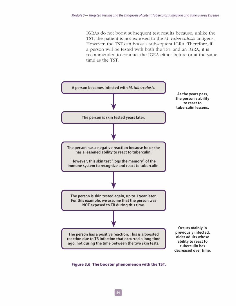
IGRAs do not boost subsequent test results because, unlike the TST, the patient is not exposed to the M. tuberculosis antigens. However, the TST can boost a subsequent IGRA. Therefore, if a person will be tested with both the TST and an IGRA, it is recommended to conduct the IGRA either before or at the same time as the TST.
A person becomes infected with M. tuberculosis.
The person is skin tested years later.
The person has a negative reaction because he or she has a lessened ability to react to tuberculin.
However, this skin test “jogs the memory” of the immune system to recognize and react to tuberculin.
The person is skin tested again, up to 1 year later. For this example, we assume that the person was
NOT exposed to TB during this time.
The person has a positive reaction. This is a boosted reaction due to TB infection that occurred a long time ago, not during the time between the two skin tests.
As the years pass, the person’s ability
to react to tuberculin lessens.
Occurs mainly in previously infected, older adults whose ability to react to
tuberculin has decreased over time.
Figure 3.6 The booster phenomenon with the TST.
3534
Module 3— Targeted Testing and the Diagnosis of Latent Tuberculosis Infection and Tuberculosis Disease

The booster phenomenon can present a problem in TB testing programs. This is because a negative reaction to the baseline skin test, followed by a positive reaction to a subsequent skin test that is given up to a year later, may be caused by either
�� Recent TB infection in a person who was NOT infected at the time of the baseline skin test, or
�� A boosted reaction in a person who WAS already infected before the baseline skin test.
Two-Step Testing with the TSTTo avoid misinterpretation, a strategy called two-step testing has been developed for telling the difference between boosted reactions and reactions caused by recent infection. Two-step testing should be used for the initial skin testing of persons who will be retested periodically, such as health care workers.
If a person has a negative reaction to an initial skin test, he or she is given a second test 1 to 3 weeks later.
�� If the reaction to the second test is positive, it probably is a boosted reaction (due to TB infection that occurred a long time ago).
�� If the reaction to the second test is negative, the person is considered uninfected. In this person, a positive reaction to a skin test given later on will probably be due to recent infection and be considered a skin test conversion.
Because two-step testing provides accurate information about an employee’s baseline skin test reaction, it is used in many TB testing programs for employees when they start their job. In particular, two-step testing is often used in hospitals and nursing homes. The procedure for two-step testing is shown in Figure 3.7.
Two-step testing is not required for IGRAs because IGRA testing does not boost subsequent test results.
Two-step testing is a strategy for telling the
difference between boosted reactions and reactions
caused by recent infection.
Two-step testing is used in many TB testing programs for skin testing employees when they start their job.
3534
Module 3— Targeted Testing and the Diagnosis of Latent Tuberculosis Infection and Tuberculosis Disease

Negative Positive
Retest 1–3 weeks later Person probably has TB infection
Person probably does NOT have TB infection
The reaction is considered a
boosted reaction (due to TB infection
that occurred a long time ago)
Baseline Skin Test
Reaction
Reaction
Repeat at regular intervals; a positive
reaction will probably be due to a recent
TB infection
Retesting not necessary
Negative Positive
Figure 3.7 Two-step testing with the TST.
3736
Module 3— Targeted Testing and the Diagnosis of Latent Tuberculosis Infection and Tuberculosis Disease

Study Questions 3 .19–3 .22
3.19 What is the booster phenomenon?
3.20 What is the purpose of two-step testing?
3.21 In what type of situation is two-step testing used?
3.22 How is two-step testing done?
Answers to study questions are on pages 73–80
3736
Module 3— Targeted Testing and the Diagnosis of Latent Tuberculosis Infection and Tuberculosis Disease

Case Study 3 .4
Ms. Wilson is a 60-year-old nurse. When she started a job at the local hospital, she was given a TST, her first test in 25 years. Her reaction was read 48 hours later as 0 millimeters of induration. Six months later, she was retested as part of the TB testing program in the unit where she works. Her skin test reaction was read 48 hours later as 11 millimeters of induration.
What are two ways to interpret this result?
Answers to case studies are on pages 81–84
3938 3938
Module 3— Targeted Testing and the Diagnosis of Latent Tuberculosis Infection and Tuberculosis Disease

3Self-Study Modules
on Tuberculosis
Module
Diagnosis of TB DiseaseThe key to diagnosing TB is for clinicians to “think TB” when they see a patient with symptoms of the disease or abnormal chest x-ray findings. Because TB is not as common as it was many years ago in the United States, many clinicians do not consider the possibility of TB when making diagnoses for patients who have symptoms of TB. When this happens, the diagnosis of TB may be delayed or even overlooked, and the patient will remain ill and possibly infectious.
Anyone with symptoms of TB or anyone found to have a positive TST or IGRA result should be medically evaluated for TB disease.
There are five components for conducting a complete medical evaluation for diagnosing TB disease.
1. Medical history
2. Physical examination
3. Test for TB infection
4. Chest x-ray
5. Bacteriological examinations
1 . The Medical HistoryA medical history is the part of a patient’s life history that is important for diagnosing and treating the patient’s medical condition. It includes social, family, medical, and occupational information about the patient.
To obtain a medical history, the clinician should ask whether the patient has:
a. Any symptoms of TB disease
b. Been exposed to a person with infectious TB or has risk factors for exposure to TB
c. Risk factors for developing TB disease
d. Had LTBI or TB disease before
Anyone with symptoms of TB or a positive TST or IGRA result should be medically evaluated for TB disease.
Module 3— Targeted Testing and the Diagnosis of Latent Tuberculosis Infection and Tuberculosis Disease
3938 3938

Clinicians should suspect the possibility of TB disease in patients with any of these factors.
a . Symptoms of TB diseasePeople with TB disease may or may not have symptoms. However, most patients with TB disease have one or more symptoms that led them to seek medical care. Occasionally, TB is discovered during a medical examination for an unrelated condition (for example, when a patient is given a chest x-ray before undergoing surgery). Usually, when patients do have symptoms, the symptoms have developed gradually, and they have been present for weeks or even months. General symptoms of TB disease include
�� Fever�� Chills�� Night sweats�� Weight loss�� Appetite loss�� Fatigue �� Malaise
Pulmonary TB disease usually causes one or more of the following symptoms:
�� A cough lasting for 3 or more weeks�� Chest pain when breathing or coughing�� Coughing up sputum (phlegm from deep in the lungs)
or blood
The symptoms of extrapulmonary TB disease depend on the part of the body that is affected by the disease. For example, TB of the spine may cause pain in the back; TB of the kidney may cause blood in the urine.
All of these symptoms may be caused by other diseases, but they should prompt the clinician to suspect the possibility of TB disease.
b . Exposure to TBAnother important part of the medical history is asking the patient about his or her exposure to TB. Patients should be asked whether they have spent time with someone who has infectious TB. Some people may have been exposed to TB in the distant past, when they were children. Others may have been exposed more recently.
People with TB disease may or may not have symptoms.
The symptoms of extrapulmonary TB
disease depend on the part of the body that is affected by the disease.
Some people may have been exposed to TB in the distant past, when
they were children. Others may have been exposed more recently.
4140
Module 3— Targeted Testing and the Diagnosis of Latent Tuberculosis Infection and Tuberculosis Disease

Anyone who has been exposed to TB may have TB infection. Some people become infected with M. tuberculosis without knowing that they were exposed to it. The risk of being exposed to TB is higher for some people. It is important to consider demographic factors (for example, country of origin, age, ethnic or racial group, or occupation) that may increase the patient’s risk for exposure to TB.
c . Risk factors for developing TB disease The third part of the medical history is checking for the patient’s risk factors for developing TB disease, including other medical conditions, especially HIV infection, which can increase the risk of LTBI progressing to TB disease. All patients who do not know their current HIV status should be referred for HIV counseling and testing.
For more information on factors and medical conditions that can increase the risk of patients developing TB disease, see the Targeted Testing section of this module.
d . Previous TB infection or TB diseaseDuring the medical history, the clinician should ask the patient whether he or she has ever been diagnosed with or treated for TB infection or disease.
�� Patients known to have a positive TST or IGRA result probably have TB infection. If they were infected within the past 2 years, they are at high-risk for developing TB disease.
�� Patients who have had TB disease before should be asked when they had the disease and how the disease was treated.
Clinicians may also contact the local health department for information about whether a patient has received TB treatment in the past. If the treatment regimen that was prescribed was inadequate or if the patient did not follow the recommended treatment, the TB disease may come back, and it may be resistant to one or more of the drugs used.
2 . Physical ExaminationA physical examination is an essential part of the evaluation of any patient. It cannot confirm or rule out TB disease, but it can provide valuable information about the patient’s overall condition and factors that may affect how TB disease is treated if it is diagnosed.
Some conditions increase the risk that TB infection will progress to disease.
The clinician should ask the patient whether he or she has ever been diagnosed
with or treated for TB infection or disease.
4140
Module 3— Targeted Testing and the Diagnosis of Latent Tuberculosis Infection and Tuberculosis Disease
3 . Testing for TB InfectionCurrently, there are two methods available for diagnosing TB infection in the United States:
�� Mantoux tuberculin skin test (TST)�� Interferon-gamma release assays (IGRAs)
�� QuantiFERON®-TB Gold In-Tube test (QFT-GIT)�� T-SPOT®.TB test
Patients with symptoms of TB disease are sometimes evaluated by TST or IGRA to detect infection with TB. However, some patients with TB disease may have a negative TST or IGRA result. Patients with symptoms should always be evaluated for TB disease, regardless of their TST or IGRA results. Furthermore, clinicians should not wait for TST or IGRA results before starting other diagnostic tests for patients with symptoms of TB disease. Instead, the clinician should place a TST or order an IGRA at the same time as the other steps in the diagnosis of TB disease.
Patients with symptoms of TB disease should
always be evaluated for TB disease, regardless of their TST or IGRA results.
4342
Module 3— Targeted Testing and the Diagnosis of Latent Tuberculosis Infection and Tuberculosis Disease

Study Questions 3 .23
3.23 What are the five components for conducting a medical evaluation for diagnosing TB disease?
Answers to study questions are on pages 73–80
4342
Module 3— Targeted Testing and the Diagnosis of Latent Tuberculosis Infection and Tuberculosis Disease

Study Questions 3 .24–3 .26
3.24 What parts of a patient’s medical history should lead a clinician to suspect the possibility of TB?
3.25 What are the symptoms of pulmonary TB disease? What are the symptoms of extrapulmonary TB disease?
3.26 For patients with symptoms of TB disease, should clinicians wait for TST or IGRA results before starting other diagnostic tests?
Answers to study questions are on pages 73–80
4544
Module 3— Targeted Testing and the Diagnosis of Latent Tuberculosis Infection and Tuberculosis Disease

4 . The Chest X-RayThe chest x-ray, or radiograph, is useful for diagnosing TB disease because pulmonary TB is the most common form of the disease. Usually, when a person has TB disease in the lungs, the chest x-ray appears abnormal (Figure 3.8). It may show infiltrates (collections of fluid and cells in the tissues of the lung) or cavities (hollow spaces within the lung that may contain many tubercle bacilli).
Chest x-rays are useful for diagnosing TB disease
because pulmonary TB is the most common form of the disease.
Figure 3.8 Abnormal chest x-ray. Arrow points to lower lobe cavity.
The purposes of the chest x-ray are to
�� Help rule out the possibility of pulmonary TB disease in a person who has a positive TST or IGRA result
�� Check for lung abnormalities in people who have symptoms of TB disease
The results of a chest x-ray, however, cannot confirm that a person has TB disease. A variety of illnesses may produce abnormalities whose appearance on a chest x-ray resembles TB.
4544
Module 3— Targeted Testing and the Diagnosis of Latent Tuberculosis Infection and Tuberculosis Disease
Although an abnormality on a chest x-ray may lead a clinician to suspect TB, only a bacteriologic culture that is positive for M. tuberculosis confirms that a patient has TB disease. Moreover, a chest x-ray cannot detect TB infection.
In persons living with HIV, pulmonary TB disease may have an unusual appearance on the chest x-ray. The chest x-ray may even appear entirely normal.
A chest x-ray may be used to rule out the possibility of pulmonary TB in a person who has had a positive TST or IGRA result and no symptoms of disease.
The results of a chest x-ray cannot confirm that a person has TB disease.
In HIV-infected patients, pulmonary TB may have an unusual appearance
on the chest x-ray.
4746
Module 3— Targeted Testing and the Diagnosis of Latent Tuberculosis Infection and Tuberculosis Disease

Study Questions 3 .27–3 .28
3.27 Name two purposes of the chest x-ray.
3.28 Can the results of a chest x-ray confirm that a person has TB disease? Why or why not?
Answers to study questions are on pages 73–80
4746
Module 3— Targeted Testing and the Diagnosis of Latent Tuberculosis Infection and Tuberculosis Disease

5 . The Bacteriologic ExaminationClinical specimens (for example, sputum or urine) are examined and cultured (grown) in the laboratory for the bacteriologic examination. TB bacteriologic examination is done in a laboratory that specifically deals with M. tuberculosis and other mycobacteria (a mycobacteriology laboratory). The bacteriologic examination has five parts:
a. Specimen collection
b. Examination of smears for acid-fast bacilli
c. Direct detection of M. tuberculosis in the specimen using a nucleic acid amplification test
d. Specimen culturing and identification of growth
e. Drug susceptibility testing
a . Specimen collectionSpecimens that will be sent to the laboratory can be obtained in several ways. Usually, patients who are suspected of having pulmonary TB disease simply cough up sputum (phlegm from deep in the lungs) into a sterile container for processing and examination (Figure 3.9). This is the least expensive and easiest procedure. A health care worker should coach and directly supervise the patient when sputum is collected. Patients who are not supervised are not always successful in providing an adequate specimen, especially in their first attempt. The volume and quality of the specimen are critical for ensuring bacteriological examination is successful.
A bacteriologic examination is done in a
laboratory that specifically deals with M. tuberculosis
and other mycobacteria.
Patients suspected of having pulmonary TB
disease may cough up sputum, which is collected in a sterile
container for processing and examination.
4948
Module 3— Targeted Testing and the Diagnosis of Latent Tuberculosis Infection and Tuberculosis Disease

Figure 3.9 A TB patient has coughed up sputum and is depositing it into a sterile container. The patient is sitting in a special sputum collection booth that prevents the spread of tubercle bacilli.
Patients should have at least three consecutive sputum specimens examined, each collected in 8 to 24-hour intervals. At least one should be collected early in the morning.
If a patient cannot cough up sputum on his or her own, other techniques can be used to obtain a specimen. An induced sputum sample can be obtained by having the patient inhale a saline (salt water) mist, which causes the patient to cough deeply. Induced specimens are often clear and watery; they should be labeled “induced specimen” so that they will not be confused with saliva. Laboratories will not accept saliva as a specimen.
Another procedure, bronchoscopy, can be used to obtain pulmonary secretions or lung tissue. In this procedure, an instrument called the bronchoscope is passed through the mouth directly into the diseased portion of the lung, and some sputum or lung tissue is removed. Bronchoscopy should be used only when patients cannot cough up sputum on their own and an induced specimen cannot be obtained.
For an induced sputum sample, the patient inhales a saline mist, causing him
or her to cough deeply.
A bronchoscopy is done to obtain pulmonary
secretions or lung tissue.
4948
Module 3— Targeted Testing and the Diagnosis of Latent Tuberculosis Infection and Tuberculosis Disease

A fourth procedure, gastric washing, involves inserting a tube through the patient’s nose and passing it into the stomach. The idea is to get a sample of gastric secretions that contains sputum that has been coughed into the throat and then swallowed. Gastric washings are done in the morning because patients usually swallow sputum during the night. This procedure is usually used only when patients cannot cough up sputum on their own, an induced specimen cannot be obtained, and bronchoscopy cannot be done. However, gastric washings are often used for obtaining samples from children. Most children produce little or no sputum when they cough.
It is very important for health care workers to use infection control precautions to control the spread of tubercle bacilli during these procedures and any other procedures that may cause persons who have pulmonary TB disease to cough. This is discussed further in Module 5, Infectiousness and Infection Control.
In patients who have extrapulmonary TB disease, specimens other than sputum may be obtained. The specimen obtained from these patients depends on the part of the body that is affected. For example, urine samples are obtained from patients suspected of having TB disease of the kidney, and fluid samples are obtained from the area around the spine in patients suspected of having TB meningitis (TB disease in the membranes surrounding the brain and spinal cord).
The methods of obtaining a sputum specimen are summarized in Table 3.7.
For gastric washing, a tube is inserted through the
patient’s nose and passed into the stomach to collect
a sample of sputum that has been coughed into the
throat and then swallowed.
Health care workers should use precautions to control
the spread of tubercle bacilli during sputum collection procedures.
In patients with extrapulmonary TB
disease, specimens are obtained in various ways, depending on the part of the body that is affected.
5150
Module 3— Targeted Testing and the Diagnosis of Latent Tuberculosis Infection and Tuberculosis Disease

Table 3.7 Methods of obtaining a sputum specimen.
Method Description Advantages Disadvantages
Coughing up sputum Patient coughs up sputum
Inexpensive Easy to do
Patient may not be able to cough up sputum on his or her own, or may spit up saliva instead of sputum
Inducing sputum Patient inhales a saline mist, causing him or her to cough deeply
Easy to do Specimens may be watery and may be confused with saliva (should be labeled “induced specimen”)
Requires special equipment
Bronchoscopy Bronchoscope is passed through the mouth or nose directly into the diseased portion of the lung, and some sputum or lung tissue is removed
Useful for obtaining sputum when coughing or inducing sputum does not work
Most expensive and invasive procedure
Requires special equipment
Must be done by a specialized physician in a hospital or clinic
Gastric washing Tube is inserted through the patient’s nose and passed into the stomach to get a sample of gastric secretions that contain sputum that has been coughed into the throat and then swallowed
Useful for obtaining samples in children, who usually produce little or no sputum when they cough
Must be done as soon as patient wakes up in the morning; patient may be required to stay in hospital
Can be uncomfortable for the patient
5150
Module 3— Targeted Testing and the Diagnosis of Latent Tuberculosis Infection and Tuberculosis Disease

Case Study 3 .5
Mr. Lee has a cough and other symptoms of TB disease, and he is evaluated with a chest x-ray. However, he is unable to cough up any sputum on his own for the bacteriologic examination.
What should be done?
Answers to case studies are on pages 81–84
5352
Module 3— Targeted Testing and the Diagnosis of Latent Tuberculosis Infection and Tuberculosis Disease

b . Examination of AFB smearsBefore the specimen is examined under a microscope, it may undergo processing to break up the sample and kill other organisms that may be present. A sample is smeared onto a glass slide and stained with a dye. This is called a smear. Then laboratory personnel use a microscope to look for acid-fast bacilli (AFB) on the smear (Figure 3.10). AFB are mycobacteria that stay stained even after they have been washed in an acid solution. Tubercle bacilli are one kind of AFB.
When AFB are seen in a smear, they are counted. There is a system for reporting the number of AFB that are seen at a certain magnification. According to the number of AFB seen, the smears are classified as 4+, 3+, 2+, or 1+. In smears classified as 4+, 10 times as many AFB were seen as in smears classified as 3+; in 3+ smears, 10 times as many as in 2+ smears; and in 2+ smears, 10 times as many as in 1+ smears.
Specimens are smeared onto a glass slide and
stained so that they can be examined for acid-fast bacilli (AFB) under a microscope.
Figure 3.10 AFB smear. In this photograph, the AFB (shown in red) are tubercle bacilli.
Smears that are classified as 4+ and 3+ are considered strongly positive; 2+ and 1+ smears are considered moderately positive. If very few AFB are seen, the smear is classified by the actual number of AFB seen (no plus sign). For example, if only 2 AFB were seen in the entire smear, the smear is classified as “2 AFB seen.” Smears classified in this way are considered weakly positive, and the smear should be repeated. Finally, if no AFB are seen, the smear is called negative. But a negative smear does not rule out the possibility of TB because there can be AFB in the smear that were not seen.
When AFB are seen in a smear, they are counted
and classified according to the number of AFB seen.
5352
Module 3— Targeted Testing and the Diagnosis of Latent Tuberculosis Infection and Tuberculosis Disease

It takes only a few hours to prepare and examine a smear. Therefore, the results of the smear examination should be available to the clinician within 1 day.
The results of the smear examination can be used to help determine the infectiousness of the patient. Patients who have many tubercle bacilli in their sputum have a positive smear. Patients who have positive smears are considered infectious because they can cough many tubercle bacilli into the air. (This is discussed in more detail in Module 5, Infectiousness and Infection Control.) However, because AFB are not always tubercle bacilli, patients who have positive smears do not necessarily have TB. Furthermore, as mentioned previously, patients who have negative smears may have TB.
The classification of smears is summarized in Table 3.8.
A negative smear does not rule out the possibility
of TB because there can be AFB in the smear that were not seen.
The results of the smear examination can be
used to help determine the infectiousness
of the patient.
Table 3.8 Smear classifications and results.
Classification of Smear Smear Result Infectiousness of Patient
4+ Strongly positive Probably very infectious
3+ Strongly positive Probably very infectious
2+ Moderately positive Probably infectious
1+ Moderately positive Probably infectious
Actual number of AFB seen (no plus sign)
Weakly positive Probably infectious
No AFB seen Negative May not be infectious*
* The criteria for determining whether a patient may be considered noninfectious are discussed in Module 5, Infectiousness and
Infection Control.
5554
Module 3— Targeted Testing and the Diagnosis of Latent Tuberculosis Infection and Tuberculosis Disease

Study Questions 3 .29–3 .313.29 What are the four ways to collect sputum specimens? Indicate which procedure is the
least expensive and easiest to perform.
3.30 What do laboratory personnel look for in a smear?
3.31 What does a positive smear indicate about a patient’s infectiousness?
Answers to case studies are on pages 81–84
5554
Module 3— Targeted Testing and the Diagnosis of Latent Tuberculosis Infection and Tuberculosis Disease

Case Study 3 .6
Ms. Thompson gave three sputum specimens, which were sent to the laboratory for smear examination and culture. The smear results were reported as 4+, 3+, and 4+.
What do these results tell you about Ms. Thompson’s diagnosis and her infectiousness?
Answers to case studies are on pages 81–84
5756
Module 3— Targeted Testing and the Diagnosis of Latent Tuberculosis Infection and Tuberculosis Disease

Case Study 3 .7
Mr. Sagoo has symptoms of TB disease and a cavity on his chest x-ray, but all of his sputum smears are negative for acid-fast bacilli.
Does this rule out the diagnosis of pulmonary TB disease?
Why or why not?
Answers to case studies are on pages 81–84
5756
Module 3— Targeted Testing and the Diagnosis of Latent Tuberculosis Infection and Tuberculosis Disease

c . Direct detection of M. tuberculosis in the specimen by nucleic acid amplification testNucleic acid amplification (NAA) tests can be used to rapidly detect M. tuberculosis from sputum specimens. (See Figure 3.11). NAA tests, including polymerase chain reaction (PCR) and other methods, rapidly identify the microorganisms in the specimen by amplifying (copying) DNA and RNA segments. NAA testing should be performed on at least one respiratory specimen from a patient with signs and symptoms of pulmonary TB for whom a diagnosis of TB is being considered, and for whom the test result would alter case management or TB control activities.
A patient can be presumed to have TB if he or she has a positive NAA test with an AFB positive smear. If a patient has a negative NAA test with an AFB positive smear, he or she may have a nontuberculous mycobacteria (NTM) infection. NAA test results can help guide clinicians’ decisions for patient therapy and isolation; however, NAA tests do not replace the need for AFB smear, culture, or clinical judgment.
For more information about NAA tests, refer to the CDC’s Updated Guidelines for the Use of Nucleic Acid Amplification Tests in the Diagnosis of Tuberculosis, available from the CDC website (www.cdc.gov/tb).
Xpert MTB/RIF AssayThe Xpert MTB/RIF assay is an NAA test that simultaneously detects M. tuberculosis complex and resistance to rifampin, one of the most effective drugs used to treat TB.
To conduct the Xpert MTB/RIF assay, a sputum sample is mixed with a sterilizing reagent that is provided with the assay, and a cartridge containing the mixture is placed in the GeneXpert machine. Results from the test are available in less than 2 hours.
Results that are positive for M. tuberculosis complex and for rifampin resistance indicate that the bacteria have a high probability of resistance to rifampin. This result should be confirmed by additional rapid testing. If rifampin resistance is confirmed, rapid molecular testing for drug resistance to both first-line and second-line drugs should be performed so that an effective treatment regimen can be selected. Growth-based drug susceptibility testing should be performed as well. Growth-based susceptibility testing is discussed in more detail on page 65 of this module.
Nucleic acid amplification (NAA) tests can be used
to detect M. tuberculosis from sputum specimens.
NAA test results can help guide clinicians’ decisions
for patient therapy and isolation; however, NAA tests do not replace the
need for AFB smear, culture, or clinical judgment.
The Xpert MTB/RIF assay simultaneously
detects Mycobacterium tuberculosis complex
and resistance to rifampin in less than 2 hours.
5958
Module 3— Targeted Testing and the Diagnosis of Latent Tuberculosis Infection and Tuberculosis Disease

Results that are positive for M. tuberculosis complex, but negative for rifampin resistance mean that the bacteria are probably susceptible to rifampin. However, all tests that are positive for M. tuberculosis complex should have growth-based drug susceptibility testing to first-line TB drugs.
Results that are positive for M. tuberculosis complex and indeterminate for rifampin resistance mean that the test could not accurately determine if the bacteria are resistant.
As with other NAA tests, the Xpert MTB/RIF assay should be interpreted along with clinical, radiographic, and other laboratory findings. The Xpert MTB/RIF assay does not replace the need for AFB smear microscopy, culture for mycobacteria, growth-based drug susceptibility testing, and genotyping. Providers and laboratories need to ensure that patient specimens are available for all recommended mycobacterial testing.
For more information on the Xpert MTB/RIF assay, refer to the CDC guidelines, Availability of an Assay for Detecting Mycobacterium tuberculosis, Including Rifampin-Resistant Strains, and Considerations for Its Use—United States, 2013, available from the CDC website (www.cdc.gov/tb).
d . Culturing and identifying growth from the specimen Culturing the specimen means growing the mycobacteria on solid or in liquid media (Figure 3.12). When the mycobacteria have formed colonies (groups), or when there is sufficient growth in the liquid media, they can be identified. All specimens should be cultured, regardless of whether the smear is positive or negative.
Culturing the specimen is necessary to determine if it contains M. tuberculosis and to confirm a diagnosis of TB disease. Additionally, culture is necessary to perform further drug susceptibility testing and genotyping.
In some instances, patients are diagnosed with TB disease on the basis of their clinical presentation (i.e., signs and symptoms, response to treatment), even if their culture is negative.
As with other NAA tests, the Xpert MTB/RIF assay should
be interpreted along with clinical, radiographic, and other laboratory findings.
All specimens should be cultured, regardless of whether the smear is
positive or negative.
Culturing the specimen is necessary to determine if it contains M. tuberculosis
and to confirm a diagnosis of TB disease.
5958
Module 3— Targeted Testing and the Diagnosis of Latent Tuberculosis Infection and Tuberculosis Disease

Figure 3.11 NAA test kit.
Figure 3.12 Colonies of M. tuberculosis growing on solid media.
6160
Module 3— Targeted Testing and the Diagnosis of Latent Tuberculosis Infection and Tuberculosis Disease
The first procedure in culturing the specimen is to detect the growth of the mycobacteria. Mycobacteria grow very slowly. When solid media are used to culture the specimen, it can take as long as 3 to 6 weeks for the growth of the mycobacteria to be detected. However, rapid culturing methods that involve liquid media can decrease this time to 4 to 14 days.
The second procedure is to identify the organism that has grown in the culture. All types of mycobacteria will grow in solid or liquid media. For this reason, laboratory tests must be done to determine whether the organism is M. tuberculosis or one of the nontuberculous mycobacteria. Nucleic acid probes can rapidly identify the type of mycobacteria present in the specimens in 2 to 4 hours. Thus, using liquid media and nucleic acid probes, it is usually possible to culture and to identify M .tuberculosis within three weeks.
When M. tuberculosis is identified in a patient’s culture, the patient is said to have a positive culture for M. tuberculosis. A positive culture for M. tuberculosis, also called an M. tuberculosis isolate, confirms the diagnosis of TB disease.
When M. tuberculosis is NOT identified in a patient’s culture, the patient is said to have a negative culture for M. tuberculosis. A negative culture does not necessarily rule out the diagnosis of TB disease; as mentioned earlier, some patients with negative cultures are diagnosed with TB disease on the basis of their clinical presentation (i.e., signs and symptoms, response to treatment).
Follow-up bacteriological examinations are important for assessing the patient’s infectiousness and response to treatment. Specimens should be obtained monthly until 2 consecutive specimen cultures are negative. Culture conversion is the most important objective measure of response to treatment.
Laboratories should report positive results on smears and cultures within 24 hours by telephone or fax to the primary health care provider and to the state or local TB control program, as required by law. Out-of-state laboratories who receive referral specimens must contact the health care provider in the patient’s state of origin. Follow-up results may be reported by mail. It is the responsibility of the primary health care provider to promptly report all suspected or confirmed cases of TB to the state or local health department, unless state laws indicate otherwise. This will ensure that a contact investigation can be initiated quickly to interrupt the potential ongoing transmission.
The first procedure in culturing the specimen is to detect the growth of the mycobacteria.
The second procedure is to identify the organism
that has grown.
A positive culture for M. tuberculosis confirms
the diagnosis of TB disease.
6160
Module 3— Targeted Testing and the Diagnosis of Latent Tuberculosis Infection and Tuberculosis Disease

The differences between sputum smears and cultures are summarized in Table 3.9.
Table 3.9 Differences between sputum smears and cultures.
Feature Smears Cultures
Equipment needed
Microscope, glass slides, special dyes
Incubators, biosafety cabinet, culture plates or tubes, culture media
Time needed to make report
1 day 4 days to 8 weeks (depending on method used and how quickly the organism grows)
Basis of procedure
Looking for AFB on slide under microscope
Growth and identification of tubercle bacilli or other mycobacteria on culture media
Significance of a negative report
Patient is less likely to be infectious
Does not rule out TB disease (culture may be positive)
No live tubercle bacilli found in the specimen
Does not rule out TB disease (live tubercle bacilli may be in other specimens or in the patient)
Significance of a positive report
Patient is more likely to be infectious (if AFB are tubercle bacilli)
AFB could be nontuberculous mycobacteria
Confirms diagnosis of TB disease
6362
Module 3— Targeted Testing and the Diagnosis of Latent Tuberculosis Infection and Tuberculosis Disease
Criteria for Reporting TB CasesAll 50 states, the District of Columbia, New York City, Puerto Rico, and seven other jurisdictions in the Pacific and the Caribbean report TB cases to the federal Centers for Disease Control and Prevention (CDC) using a standard case report form called the Report of Verified Case of Tuberculosis (RVCT). Each reported TB case is checked to make sure that it meets certain criteria. All cases that meet the criteria, called verified TB cases, are counted each year.
Cases that meet one of these four sets of criteria are counted as verified TB cases:
1. The patient has a positive culture for M. tuberculosis.
or
2. The patient has a positive NAA test for M. tuberculosis.*
or
3. The patient has a positive smear for AFB, but a culture has not been or cannot be obtained or is falsely negative or contaminated.
or
4. If in the absence of laboratory confirmation, the patient meets all of the following criteria:
�� A positive TST or IGRA,�� Other signs and symptoms of TB disease (e.g., abnormal chest x-ray,
fever, night sweats, cough, weight loss, hemoptysis),�� Treatment with two or more anti-TB drugs, and �� A completed diagnostic evaluation.
5. In addition, cases that do not meet any of these sets of criteria (for example, a patient who is anergic and has a negative culture for M. tuberculosis but who has signs and symptoms of TB disease) may be counted as a verified TB case if a health care provider has reported the case and decided to treat the patient for TB disease.
*Nucleic acid amplification (NAA) tests must be accompanied by culture for mycobacteria species. However, for surveillance
purposes, CDC will accept results obtained from NAA tests approved by the Food and Drug Administration (FDA) and used
according to the approved product labeling on the package insert, or a test produced and validated in accordance with
applicable FDA and Clinical Laboratory Improvement Amendments (CLIA) regulations.
6362
Module 3— Targeted Testing and the Diagnosis of Latent Tuberculosis Infection and Tuberculosis Disease

e . Drug susceptibility testingFor all patients, drug susceptibility tests should be done when the patient is first found to have a positive culture for M. tuberculosis (that is, the first isolate of M. tuberculosis). Drug susceptibility tests are done to determine which drugs will kill the tubercle bacilli that are causing disease in a particular patient. Tubercle bacilli that are killed by a particular drug are said to be susceptible to that drug, whereas those that can grow even in the presence of a particular drug are said to be resistant to that drug. The drug susceptibility pattern of a strain of tubercle bacilli is the list of drugs to which the strain is susceptible and to which it is resistant. The initial isolate should be tested for resistance to the first-line anti-TB drugs: rifampin, isoniazid, ethambutol, and pyrazinamide.
Drug-resistant TB can be mono-resistant if the tubercle bacilli are resistant to any one TB treatment drug, or poly-resistant if resistant to at least two TB drugs (but not both isoniazid and rifampin). A patient is diagnosed with multidrug-resistant TB (MDR TB) if the tubercle bacilli are resistant to at least isoniazid and rifampin, the two best first-line TB treatment drugs. A patient is diagnosed with extensively drug-resistant TB (XDR TB) if the tubercle bacilli are resistant to isoniazid and rifampin, plus resistant to any fluoroquinolone and at least one of three injectable anti-TB drugs (such as amikacin, kanamycin, or capreomycin).
The results of drug susceptibility tests can help clinicians choose the appropriate drugs for treating each patient. This is very important. Patients with TB disease who are treated with drugs to which their strain of TB is resistant may not be cured. In fact, their strain of TB may become resistant to additional drugs.
Drug susceptibility tests should be repeated if a patient has a positive culture for M. tuberculosis after 3 months of treatment or if a patient does not seem to be getting better. That way, the clinician can find out whether the patient’s strain of TB has become resistant to certain drugs; if necessary, the clinician may change the drugs used for treating the patient. Clinicians treating patients with drug-resistant TB should always consult with a medical expert who is familiar with the treatment of drug-resistant TB.
Drug susceptibility tests should be done when a patient is first found to have a positive culture
for M. tuberculosis.
Drug susceptibility tests determine which drugs
will kill the tubercle bacilli that are causing disease in a particular patient.
The results of drug susceptibility tests can help clinicians choose the appropriate drugs
for each patient.
Drug susceptibility tests should be repeated if a patient has a positive
culture for M. tuberculosis after 3 months of treatment
or if a patient does not seem to be getting better.
6564
Module 3— Targeted Testing and the Diagnosis of Latent Tuberculosis Infection and Tuberculosis Disease

Growth-Based Drug Susceptibility Testing
Growth-based drug susceptibility testing can be done using a liquid medium or a solid medium method. In a drug susceptibility test, organisms that grow in media containing a specific drug are considered resistant to that drug (Figure 3.13). Liquid medium methods are faster than solid media methods for determining susceptibility to first-line TB medications (Figure 3.14). Usually the susceptibility results can be obtained within 7 to 14 days with the liquid medium method. Traditional solid medium methods can take as long as 21 days.
Molecular Detection of Drug Resistance
Drug resistance is due to the presence of mutations in specific M. tuberculosis genes. There are a variety of tests that can detect mutations associated with drug resistance. The tests are done on patient specimens or isolates from patient specimens. If a mutation thought to be associated with resistance is detected, the bacteria are considered to be drug resistant. Molecular tests can provide results within 24 to 48 hours.
A limitation of molecular testing for drug resistance is that the clinical relevance of some mutations remains unknown. Further, not all biological mechanisms of resistance are known. As a result, if no mutations are detected by the molecular test, drug resistance cannot be ruled out. Therefore, it is essential that conventional growth-based drug susceptibility tests are done and used in conjunction with molecular results.
Molecular tests provide preliminary guidance on effective therapy for TB patients. Molecular detection of drug resistance should be considered for patients with the following characteristics:
�� High risk of rifampin resistance, including MDR TB (e.g., previously treated for TB, contact to someone with MDR TB, or foreign-born from a high-risk country);
�� First-line drug susceptibility results are available and show resistance to rifampin;
�� Infectiousness poses a risk to vulnerable contacts (e.g., infants); and
�� Contraindications to essential first-line medications (e.g., rifampin allergy).
6564
Module 3— Targeted Testing and the Diagnosis of Latent Tuberculosis Infection and Tuberculosis Disease
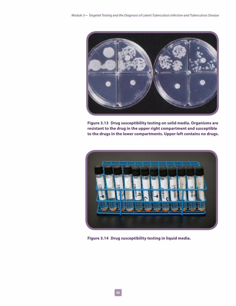
Figure 3.13 Drug susceptibility testing on solid media. Organisms are resistant to the drug in the upper right compartment and susceptible to the drugs in the lower compartments. Upper left contains no drugs.
Figure 3.14 Drug susceptibility testing in liquid media.
6766
Module 3— Targeted Testing and the Diagnosis of Latent Tuberculosis Infection and Tuberculosis Disease

Study Questions 3 .32–3 .353.32 Why is it necessary to culture a specimen?
3.33 What does a positive culture for M. tuberculosis mean? How is this important for the TB diagnosis?
3.34 Why are drug susceptibility tests done?
3.35 How often should drug susceptibility tests be done?
Answers to study questions are on pages 73–80
6766
Module 3— Targeted Testing and the Diagnosis of Latent Tuberculosis Infection and Tuberculosis Disease

Case Study 3 .8
In the public health clinic, you see a patient, Ms. Sanchez, who complains of weight loss, fever, and a cough of 4 weeks duration. When questioned, she reports that she has been treated for TB disease in the past and that she occasionally injects heroin.
What parts of Ms. Sanchez’s medical history lead you to suspect TB disease?
What diagnostic tests should be done?
Answers to case studies are on pages 81–84
6968 6968
Module 3— Targeted Testing and the Diagnosis of Latent Tuberculosis Infection and Tuberculosis Disease

3Self-Study Modules
on Tuberculosis
Module
TB GenotypingTB genotyping is a laboratory-based method that can determine the genetic pattern of the strain of M. tuberculosis that caused TB disease in a person. Each strain has a distinct genetic pattern, or genotype. Genotyping is done for culture-positive cases of TB disease. TB programs should work with laboratories to ensure that TB genotyping results are obtained for all culture-positive cases.
Genotyping has applications at both the population level and the individual patient level. At the population level, genotyping is most commonly used for detecting, refuting, and monitoring TB outbreaks. For more information about the use of genotyping in TB outbreaks, refer to Module 9, Tuberculosis Outbreak Detection and Response.
At the patient level, TB genotyping has three uses:
�� Distinguishing relapse from reinfection�� Detecting false-positive culture results�� Informing contact investigations
Distinguishing Relapse from ReinfectionSome patients previously treated for TB disease may develop TB disease again. This could be due to a relapse or reinfection. Genotyping results can be used to distinguish relapse from reinfection.
Recurrence of TB disease after treatment is most often due to relapse of TB disease. Relapse can occur for several reasons including inappropriate treatment, nonadherence to treatment, or unidentified drug resistance. If genotype information from the first episode of TB is available, it can be compared with genotype information from the second episode. If the genotypes match, it is likely the TB disease was not completely cured the first time it was treated.
If the genotypes from the two episodes of TB disease do not match, it is likely the second episode occurred because the person became infected with a different strain of M. tuberculosis (reinfection).
Module 3— Targeted Testing and the Diagnosis of Latent Tuberculosis Infection and Tuberculosis Disease
6968 6968

Detecting False-Positive CulturesGenotype information can alert laboratory and public health staff to situations where specimens from different patients, processed within the same facility and time period, share genotypes. When this situation occurs, the laboratory can follow a process to determine if an error has been made, and one or more of the cultures are falsely positive. False-positive TB cultures can be caused by cross-contamination of a specimen in the laboratory, clerical error such as mislabeling of specimens, or contaminated equipment such as a bronchoscope used to collect the samples.
Identifying false-positive cultures is important because doing so helps prevent unnecessary treatment and contact investigations for persons who would otherwise be misdiagnosed with TB disease.
Informing Contact InvestigationsTypically, genotype information will not be available during the early stages of a contact investigation; however, when it is available, genotype results can help confirm, disprove, or detect connections among patients. For more information about the use of genotyping in contact investigations, refer to Module 8, Contact Investigations for Tuberculosis
7170 7170
Module 3— Targeted Testing and the Diagnosis of Latent Tuberculosis Infection and Tuberculosis Disease
3Self-Study Modules
on Tuberculosis
Module
Additional Resources1. American Thoracic Society and Centers for Disease Control
and Prevention. Diagnostic standards and classification of tuberculosis in adults and children. Am J Respir Crit Care Med 2000;161:1376–1395. www.cdc.gov/tb/publications/pdf/1376.pdf.
2. American Thoracic Society and Centers for Disease Control and Prevention. Targeted tuberculin testing and treatment of latent tuberculosis infection. Am J Respir Crit Care Med 2000;161;S221–S247. www.cdc.gov/mmwr/preview/mmwrhtml/rr4906a1.htm.
3. CDC. Availability of an Assay for Detecting Mycobacterium tuberculosis, Including Rifampin-Resistant Strains, and Considerations for Its Use—United States, 2013. MMWR 2013; 62 (41). www.cdc.gov/mmwr/preview/mmwrhtml/mm6241a1.htm?s_cid=mm6241a1_e.
4. CDC. Essential components of a tuberculosis prevention and control program. The Americas, 1995. MMWR 1995;44 (No. RR-11):1–16. www.cdc.gov/MMWR/preview/MMWRhtml/00038823.htm.
5. CDC. Guidelines for preventing the transmission of Mycobacterium tuberculosis in health-care settings, 2005. The Americas, 2005. MMWR 2005;54(No.RR-17):1-140. www.cdc.gov/mmwr/preview/mmwrhtml/rr5417a1.htm?s_cid=rr5417a1_e.
6. CDC. Guidelines for the investigation of contacts of persons with infectious tuberculosis. The Americas, 2005. MMWR 2005;54(No. RR-15):1–55. www.cdc.gov/mmwr/preview/mmwrhtml/rr5415a1.htm.
7. CDC. Mantoux Tuberculin Skin Test [DVD]. Atlanta, Ga: Department of Health and Human Services, CDC; March 2005. www.cdc.gov/tb/education/Mantoux/default.htm.
8. CDC. Recommendations for using smallpox vaccine in a pre-event vaccination program. The Americas, 2003. MMWR 2003;52(No. RR-7):1–16. www.cdc.gov/mmwr/preview/mmwrhtml/rr5207a1.htm.
9. CDC. The role of BCG vaccine in the prevention and control of tuberculosis in the United States: a joint statement by the Advisory Council for the Elimination of Tuberculosis and the Advisory Committee on Immunization Practices. The Americas, 1996. MMWR 1996;45(No. RR-4):1–18. www.cdc.gov/MMWR/preview/MMWRhtml/00041047.htm.
10. CDC. Tuberculosis elimination revisited: obstacles, opportunities, and a renewed commitment. The Americas, 1999. MMWR 1999;48 (No. RR-9):1–13. www.cdc.gov/MMWR/preview/MMWRhtml/rr4809a1.htm.
Module 3— Targeted Testing and the Diagnosis of Latent Tuberculosis Infection and Tuberculosis Disease
7170 7170

11. CDC. Updated Guidelines for the Use of Nucleic Acid Amplification Tests in the Diagnosis of Tuberculosis. MMWR 2009; 58 (1). www.cdc.gov/mmwr/preview/mmwrhtml/mm5801a3.htm?scid=mm5801a3_e.
12. CDC. Updated guidelines for using interferon gamma release assays to detect Mycobacterium tuberculosis infection—United States, 2010. MMWR 2010; 59 (RR-5). www.cdc.gov/mmwr/preview/mmwrhtml/rr5905a1.htm?s_cid=rr5905a1_e.
13. Lewinsohn DM, Leonard MK, LoBue PA, Cohn DL, Daley CL, Desmond E, Keane J, Lewinsohn DA, Loeffler AM, Mazurek GH, O’Brien RJ, Pai M, Richeldi L, Salfinger M, Shinnick TM, Sterling TR, Warshauer DM, Woods GL. Official American Thoracic Society/Infectious Diseases Society of America/Centers for Disease Control and Prevention Clinical Practice Guidelines: Diagnosis of Tuberculosis in Adults and Children. Clinical Infectious Diseases 2016: 1–33. www.cdc.gov/tb/publications/guidelines/pdf/cid_ciw694_full.pdf.
7372
Module 3— Targeted Testing and the Diagnosis of Latent Tuberculosis Infection and Tuberculosis Disease
Answers to Study Questions3.1. What is the TST used for?
The TST is used to determine whether a person has TB infection.
3.2. How is the TST given?
The TST is given by using a needle and syringe to inject 0.1 ml of 5 tuberculin units of liquid tuberculin between the layers of the skin (intradermally), usually on the forearm.
3.3. With the TST, when is the patient’s arm examined?
The patient’s arm is examined 48 to 72 hours after the tuberculin is injected.
3.4. How is the induration measured?
The diameter of the indurated area is measured across the forearm; erythema (redness) around the indurated area is not measured, because the presence of erythema does not indicate that a person has TB infection.
3.5. What two factors determine the interpretation of a TST reaction as positive or negative? What additional factor is considered for people who may be exposed to TB on the job?
The two factors that are used to determine the interpretation of a TST include the size of the induration and the person’s risk factors for TB.
For people who may be exposed to TB on the job, an additional factor that is considered is the risk of exposure to TB in the person’s job.
3.6. Name five groups of people for which 5 or more millimeters of induration is considered a positive reaction.
An induration of 5 or more millimeters is considered a positive reaction for
�� People living with HIV
�� Recent contacts of people with infectious TB
�� People with chest x-ray findings suggestive of previous TB disease
�� People with organ transplants
�� Other immunosuppressed patients (for example, patients on prolonged therapy with corticosteroids equivalent to/greater than 15mg per day of prednisone or those taking TNF-alpha antagonists)
7372
Module 3— Targeted Testing and the Diagnosis of Latent Tuberculosis Infection and Tuberculosis Disease

Answers to Study Questions, Continued3.7. Name seven groups of people for which 10 or more millimeters of induration is
considered a positive reaction.
An induration of 10 or more millimeters is considered a positive reaction for
�� People who have recently come to the United States (within the last 5 years) from areas of the world where TB is common (for example, Asia, Africa, Russia, Eastern Europe, or Latin America)
�� People who abuse drugs
�� Mycobacteriology laboratory workers
�� People who live or work in high-risk congregate settings (for example, nursing homes, homeless shelters, or correctional facilities)
�� People with certain medical conditions that place them at high risk for TB (for example, silicosis, diabetes mellitus, severe kidney disease, certain types of cancer, and certain intestinal conditions)
�� Children younger than 5 years of age
�� Infants, children, or adolescents exposed to adults in high-risk categories
3.8. For which group of people is 15 or more millimeters of induration considered a positive reaction?
An induration of 15 or more millimeters is considered a positive reaction for people with no known risk factors for TB.
3.9. Name four factors that may cause false-positive reactions to the TST.
�� Infection with nontuberculous mycobacteria (NTM) (mycobacteria other than M. tuberculosis)
�� BCG vaccination
�� Administration on incorrect antigen
�� Incorrect measuring or interpretation of the TST reaction
3.10. Is there a reliable way to distinguish a positive TST reaction caused by vaccination with BCG from a reaction caused by true TB infection?
No, there is no reliable way to distinguish a positive TST reaction caused by vaccination with BCG from a reaction caused by true TB infection. When using the TST, people with a positive reaction who have been vaccinated with BCG should always be further evaluated for LTBI or TB disease the same as if they were not vaccinated with BCG.
7574
Module 3— Targeted Testing and the Diagnosis of Latent Tuberculosis Infection and Tuberculosis Disease
Answers to Study Questions, Continued3.11. Name six factors that may cause false-negative reactions to the TST.
False-negative reactions may be due to
�� Anergy
�� Recent TB infection (within the past 8 to 10 weeks)
�� Very young age (younger than 6 months of age)
�� Recent live-virus measles or smallpox vaccination
�� Incorrect method of giving the TST
�� Incorrect measuring or interpretation of TST reaction
3.12. What is anergy?
Anergy is the inability to react to skin tests because of a weakened immune system. Many conditions, such as HIV infection, cancer, or severe TB disease itself, may weaken the immune system and cause anergy. HIV infection is an important cause of anergy.
3.13. After TB has been transmitted, how long does it take before TB infection can be detected by the TST?
After TB has been transmitted, it takes 2 to 8 weeks before TB infection can be detected by the TST.
3.14. What should be done if a patient has a negative TST reaction but has symptoms of TB disease?
Any patient with symptoms of TB disease should be evaluated for TB disease, regardless of his or her skin test reaction. In fact, people with symptoms of TB disease should be evaluated for TB disease right away, at the same time that the TST is given.
3.15. What are the steps for conducting an IGRA?
To conduct an IGRA, health care workers should first read the instructions from the manufacturer and then follow the steps below:
�� Confirm arrangements for testing in a qualified laboratory.
�� Arrange for delivery of the blood sample to the laboratory in the time the laboratory specifies to ensure testing of samples with viable blood cells.
�� Draw a blood sample from the patient according to the test manufacturer’s instructions.
�� Schedule a follow-up appointment for the patient to receive test results.
�� Based on test results, provide follow-up evaluation and treatment as needed.
7574
Module 3— Targeted Testing and the Diagnosis of Latent Tuberculosis Infection and Tuberculosis Disease

Answers to Study Questions, Continued3.16. How are IGRA results interpreted?
QFT-GIT results are based on the amount of IFN-γ that is released in response to the M. tuberculosis antigens and control substances. T-Spot results are based on the number of IFN-γ producing cells (spots) produced. Both the standard qualitative test interpretation (positive, negative, or indeterminate) and the quantitative assay measurements (Nil, TB, and Mitogen concentrations or spot counts) should be reported.
3.17. How should negative IGRA results be interpreted?
If the result is negative, then the patient is unlikely to have M. tuberculosis infection and may not require further evaluation unless they have signs and symptoms of TB disease. Moreover, as with the TST, persons who have a negative test result can still have LTBI.
3.18. What are five advantages for using IGRAs as compared to the TST?
�� Requires a single patient visit to conduct the test
�� Results can be available within 24 hours
�� Does not cause the booster phenomenon which can happen with repeat TSTs
�� Laboratory test not affected by health care worker perception or bias
�� BCG vaccination does not affect IGRA results
3.19. What is the booster phenomenon?
The booster phenomenon is a phenomenon in which people (especially older adults) who are skin tested many years after becoming infected with M. tuberculosis may have a negative reaction to an initial skin test, followed by a positive reaction to a skin test given up to a year later. This happens because in some people who have TB infection, the ability to react to tuberculin lessens over time. The first skin test “jogs the memory” of the immune system, boosting its ability to react to tuberculin.
3.20. What is the purpose of two-step testing?
The purpose of two-step testing is to tell the difference between boosted reactions and reactions caused by recent infection. Because it provides accurate information about each employee’s baseline skin test reaction, two-step testing is used in many TB screening programs for skin testing employees when they start their job.
3.21. In what type of situation is two-step testing used?
Two-step testing is used in many TB screening programs for skin testing employees when they start their job.
7776
Module 3— Targeted Testing and the Diagnosis of Latent Tuberculosis Infection and Tuberculosis Disease

Answers to Study Questions, Continued3.22. How is two-step testing done?
If a person has a negative reaction to an initial skin test, he or she is given a second test 1 to 3 weeks later.
�� If the reaction to the second test is positive, it is considered a boosted reaction (due to TB infection that occurred a long time ago)
�� If the reaction to the second test is negative, the person is considered uninfected. In this person, a positive reaction to a TST given later on will probably be due to recent TB infection.
3.23. What are the five components for conducting a complete medical evaluation for diagnosing TB disease?
�� Medical history
�� Physical examination
�� Test for TB infection
�� Chest x-ray
�� Bacteriologic examinations
3.24. What parts of a patient’s medical history should lead a clinician to suspect the possibility of TB?
Clinicians should suspect the possibility of TB disease in patients who have
�� Symptoms of TB disease
�� Been exposed to a person who has infectious TB or has other risk factors for exposure to TB
�� Risk factors for developing TB disease
�� Had TB infection or TB disease before
7776
Module 3— Targeted Testing and the Diagnosis of Latent Tuberculosis Infection and Tuberculosis Disease

Answers to Study Questions, Continued3.25. What are the symptoms of pulmonary TB disease? What are the symptoms of
extrapulmonary TB disease?
General symptoms of TB disease include:
�� Fatigue
�� Malaise
�� Fever
�� Weight loss
�� Night sweats
Symptoms of pulmonary TB disease include:
�� Coughing
�� Pain in the chest when breathing or coughing
�� Coughing up sputum or blood
The symptoms of extrapulmonary TB disease depend on the part of the body that is affected by the disease. For example, TB of the spine may cause pain in the back; TB of the kidney may cause blood in the urine.
3.26. For patients with symptoms of TB disease, should clinicians wait for TST or IGRA results before starting other diagnostic tests?
No. For patients with symptoms of TB disease, clinicians should not wait for TST or IGRA results before starting other diagnostic tests.
3.27. Name two purposes of the chest x-ray.
The purposes of the chest x-ray are to
�� Help rule out the possibility of pulmonary TB disease in a person who has a positive TST or IGRA result and no symptoms of TB
�� Check for lung abnormalities in people who have symptoms of TB disease
3.28. Can the results of a chest x-ray confirm that a person has TB disease? Why or why not?
No, the results of a chest x-ray cannot confirm that a person has TB disease. This is because a variety of illnesses may produce abnormalities whose appearance on a chest x-ray resembles TB. Although an abnormality on a chest x-ray may lead a clinician to suspect TB, only a bacteriologic culture that is positive for M. tuberculosis confirms that a patient has TB disease.
7978
Module 3— Targeted Testing and the Diagnosis of Latent Tuberculosis Infection and Tuberculosis Disease

Answers to Study Questions, Continued3.29. What are the four ways to collect sputum specimens? Indicate which procedure is
the least expensive and easiest to perform.
�� Usually, patients who are suspected of having pulmonary TB disease simply cough up sputum and the sputum is collected in a sterile container for processing and examination. This is the least expensive and easiest procedure. If a patient cannot cough up sputum on his or her own, other techniques can be used to obtain a specimen.
�� An induced sputum sample can be obtained by having the patient inhale a saline (salt water) mist, which causes the patient to cough deeply.
�� Bronchoscopy can be used to obtain pulmonary secretions or lung tissue. In this procedure, an instrument called the bronchoscope is passed through the nose or mouth directly into the diseased portion of the lung, and some sputum or lung tissue is removed.
�� Gastric washing involves inserting a tube through the patient’s nose and passing it into the stomach. The idea is to get a sample of gastric secretions that contain sputum that has been coughed into the throat and then swallowed.
3.30. What do laboratory personnel look for in a smear?
Laboratory personnel use the microscope to look for acid-fast bacilli (AFB) on the smear. AFB are mycobacteria that stay stained even after they have been washed in an acid solution. Tubercle bacilli are one kind of AFB.
3.31. What does a positive smear indicate about a patient’s infectiousness?
The results of the smear examination can be used to help determine the infectiousness of the patient. Patients who have many tubercle bacilli in their sputum have a positive smear. Patients who have positive smears are considered infectious because they can cough many tubercle bacilli into the air.
3.32. Why is it necessary to culture a specimen?
Culturing the specimen is necessary to determine whether the specimen contains M. tuberculosis and to confirm a diagnosis of TB disease. (However, in some cases, patients are diagnosed with TB disease on the basis of their signs and symptoms, even if their specimen does not contain M. tuberculosis.) Additionally, culture is necessary for genotyping and for performing drug susceptibility testing.
7978
Module 3— Targeted Testing and the Diagnosis of Latent Tuberculosis Infection and Tuberculosis Disease

Answers to Study Questions, Continued3.33. What does a positive culture for M. tuberculosis mean? How is this important for
the TB diagnosis?
A positive culture for M. tuberculosis means that M. tuberculosis has been identified in a patient’s culture. A positive culture for M. tuberculosis confirms the diagnosis of TB disease.
3.34. Why are drug susceptibility tests done?
Drug susceptibility tests are done to determine which drugs will kill the tubercle bacilli that are causing disease in a particular patient. The results of drug susceptibility tests can help clinicians choose the appropriate drugs for each patient.
3.35. How often should drug susceptibility tests be done?
Drug susceptibility tests should be done when a patient is first found to have a positive culture for M. tuberculosis. In addition, drug susceptibility tests should be repeated if a patient has a positive culture for M. tuberculosis after 3 months of treatment or if a patient does not seem to be getting better.
8180
Module 3— Targeted Testing and the Diagnosis of Latent Tuberculosis Infection and Tuberculosis Disease

Case Study Answers
3.1 Which of the following patients have a positive TST reaction? Circle the best answer(s).
a) Mr. West, 36 years old, HIV infected, 8 mm of induration
b) Ms. Hernandez, 26 years old, native of Mexico, 7 mm of induration
c) Ms. Jones, 56 years old, has diabetes, 12 mm of induration
d) Mr. Sung, 79 years old, resident of a nursing home, 11 mm of induration
e) Mr. Williams, 21 years old, no known risk factors, 13 mm of induration
f ) Ms. Marcos, 42 years old, chest x-ray findings suggestive of previous TB, 6 mm of induration
g) Ms. Rayle, 50 years old, husband has pulmonary TB, 9 mm of induration
A, C, D, F, and G all have positive TST reactions.
8180
Module 3— Targeted Testing and the Diagnosis of Latent Tuberculosis Infection and Tuberculosis Disease

Case Study Answers, Continued
3.2. A 30-year-old man who recently immigrated to the United States from India is given a TST and found to have 14 millimeters of induration. He reports that he was vaccinated with BCG as a child. He also says that his wife was treated for pulmonary TB disease last year.
How should this man’s results be interpreted?
This man has a positive reaction to the TST (10 or more millimeters is considered a positive reaction for a person from a country where TB is common). Also, he is a contact of person with pulmonary TB (his wife). Because he was vaccinated with BCG there is a possibility that this may be a false-positive reaction. However, since there is no reliable way to distinguish a positive TST reaction caused by BCG or from a true TB infection, this man should be further evaluated for LTBI or TB disease as if he was never vaccinated with BCG.
What factors make it more likely that this man’s positive reaction is due to TB infection?
This man is from an area of the world where TB is common, so he was probably exposed to TB in his native country. Therefore, he is at increased risk for TB infection. Also, his wife has had pulmonary TB, which further increases the probability that he has been exposed to TB. Since there is no reliable way to distinguish a positive TST reaction caused by BCG from a true TB infection, this man should be further evaluated for LTBI or TB disease as if he was never vaccinated with BCG.
3.3. Mr. Bell comes to the TB clinic for a TST. He believes that he has been exposed to TB, and he knows he is at high risk for TB because he is HIV-infected. He is given a TST, and his reaction is read 48 hours later as 0 millimeters of induration.
What are three ways to interpret this result?
There are three possible reasons why Mr. Bell had no reaction to the tuberculin skin test.
�� First, he may not have TB infection. �� Second, he may be anergic. People who are HIV-infected are more likely to be anergic than persons who are not HIV-infected. If Mr. Bell is anergic, he would be unable to react to tuberculin even if he did have TB infection.
�� Third, it may be less than 8 to 10 weeks since he was exposed to TB. After TB has been transmitted, it takes 2 to 8 weeks before TB infection can be detected by the TST. Mr. Bell should be retested 8 to 10 weeks after he was last exposed to TB.
8382
Module 3— Targeted Testing and the Diagnosis of Latent Tuberculosis Infection and Tuberculosis Disease

Case Study Answers, Continued
3.4. Ms. Wilson is a 60-year-old nurse. When she started a job at the local hospital, she was given a TST, her first test in 25 years. Her reaction was read 48 hours later as 0 millimeters of induration. Six months later, she was retested as part of the TB screening program in the unit where she works. Her TST reaction was read 48 hours later as 11 millimeters of induration.
What are two ways to interpret this result?
There are two possible explanations for this result.
�� One explanation is that Ms. Wilson may have been exposed to and infected with M. tuberculosis sometime in the 6 months between her first and second skin tests.
�� The other explanation is the booster phenomenon. If Ms. Wilson was infected with M. tuberculosis many years ago, her ability to react to tuberculin may have decreased. This would explain why she did not react to the first TST. Then the first tuberculin test may have boosted the ability of her immune system to react to tuberculin. This would explain why she had a positive reaction to the second TST, which was given within a year of the first test. If this scenario is true, Ms. Wilson’s positive reaction would not mean that she was recently infected with M. tuberculosis.
This problem in interpreting Ms. Wilson’s reaction would have been avoided if she had been tested with a two-step procedure when she first joined the hospital. In any event, because she has a positive reaction, Ms. Wilson should be evaluated for TB disease.
3.5. Mr. Lee has a cough and other symptoms of TB disease, and he is evaluated with a chest x-ray. However, he is unable to cough up any sputum on his own for the bacteriologic examination.
What should be done?
If a patient cannot cough up a sputum specimen, other techniques can be used to obtain sputum. First, clinicians can try to obtain an induced sputum sample. If they cannot obtain an induced sputum sample, a bronchoscopy or gastric washing may be done.
8382
Module 3— Targeted Testing and the Diagnosis of Latent Tuberculosis Infection and Tuberculosis Disease

Case Study Answers, Continued
3.6. Ms. Thompson gave three sputum specimens, which were sent to the laboratory for smear examination and culture. The smear results were reported as 4+, 3+, and 4+.
What do these results tell you about Ms. Thompson’s diagnosis and her infectiousness?
These results show that Ms. Thompson’s sputum specimens contain many acid-fast bacilli. Because the smears are positive, clinicians should suspect that Ms. Thompson has TB disease. They should also consider her infectious. However, it is possible that these acid-fast bacilli are mycobacteria other than tubercle bacilli. Therefore, the diagnosis of TB disease cannot be proven until the culture results are available.
3.7. Mr. Sagoo has symptoms of TB disease and a cavity on his chest x-ray, but all of his sputum smears are negative for acid-fast bacilli.
Does this rule out the diagnosis of pulmonary TB disease?
No.
Why or why not?
M. tuberculosis may grow in the cultures even though there were no acid-fast bacilli on the smear. Mr. Sagoo’s symptoms and his abnormal chest x-ray suggest that he does have pulmonary TB disease.
3.8. In the public health clinic, you see a patient, Ms. Sanchez, who complains of weight loss, fever, and a cough of 4 weeks’ duration. When questioned, she reports that she has been treated for TB disease in the past and that she occasionally injects heroin.
What parts of Ms. Sanchez’s medical history lead you to suspect TB disease?
Ms. Sanchez has symptoms of TB disease (weight loss, fever, and a persistent cough). Also, in the past she has been treated for TB disease. We don’t know whether she completed therapy, but until we can prove otherwise, we should assume that she has TB disease again. Her history of injecting illegal drugs (heroin) is another risk factor for TB.
What diagnostic tests should be done?
People who have TB symptoms should be evaluated for TB disease. Because she has symptoms of pulmonary TB disease (coughing), Ms. Sanchez should be given a chest x-ray. In addition, a sputum specimen should be collected for smear and culture, blood for an HIV test, and drug susceptibility testing should be done if the culture is positive for M. tuberculosis.
8584
Module 3— Targeted Testing and the Diagnosis of Latent Tuberculosis Infection and Tuberculosis Disease

8584

3Module
CS253781-C