turnover of plasma proteins1 - bmj
TRANSCRIPT
J. clin. Path., 28, Suppl. (Ass. Clin. Path.), 6, 13-21
Turnover of plasma proteins1STIG JARNUM
From Medical Department P, Division of Gastroenterology, Rigshospitalet, Copenhagen, Denmark
Studies of plasma protein turnover have been usedextensively over the last 20 years both under ex-perimental conditions and in clinical medicine. Theyhave shown that disturbed plasma protein. metab-olism is a very common phenomenon in disease,probably as common as an elevated erythrocytesedimentation rate.
Abnormal Serum Total Protein Concentration
Only turnover studies can reveal the mechanismbehind an abnormal serum concentration of agiven protein. Albumin, the major fraction of serumprotein, is usually low when total serum protein islow and hence basic causes of hypoproteinaemia aremuch the same as those of hypoalbuminaemia: (1)increased catabolism which occurs in a variety ofacute conditions such as acute infections, traumaincluding major surgery and myocardial infarction;(2) abnormal protein loss, which may be due toburns, severe proteinuria or gastrointestinal proteinleakage; (3) decreased synthesis which is most oftencaused by liver disease or malnutrition and/or mal-absorption; (4) haemodilution, which occurs inpregnancy, in some cases of cirrhosis of the liver andin surgical patients who have been overhydrated,especially when renal insufficiency is also present.The causes of hyperproteinaemia are fewer, be-
cause it is never due to decreased catabolism orabnormal retention. They include increased synthesisof immunoglobulins, either by normal clones (inchronic inflammatory conditions such as cirrhosisof the liver) or by abnormal clones (myelomatosis),which may produce a significant increase in totalserum protein concentration; and haemoconcentra-tion which is seen in severely dehydrated patients(cholera, pyloric stenosis) and increases haemoglobinas well as serum protein concentration.A normal total serum protein concentration does
not necessarily imply normal concentration ofindividual fractions. It may, for instance, maskhypoalbuminaemia with hypergammaglobulinaemia,'The work was supported by grants from the Danish Medical Re-search Foundation (J. no. 512-1117 and 512-2649), P. Carl Petersen'sFond and King Christian d X's Fond.
13
as seen typically in cirrhosis of the liver. Similarly,normal concentration of a particular protein fractiondoes not necessarily imply normal turnover; inchronic inflammatory bowel disease serum immuno-globulin concentrations are usually normal but theirmetabolism is not (vide infra).The only way to determine the effect of a given
disease on the metabolism of specific plasma pro-teins or to establish the mechanism of an alteredserum concentration is by means of turnover studies.
Methods of Studying Turnover
RADIOIODINE-LABELLED PROTEINSTurnover studies with labelled protein in man werebegun by Sterling in 1951 using 131I which, in manyrespects, turned out to be an ideal label. However,from a theoretical point of view radioiodine-labelledproteins have a major disadvantage in that they onlymeasure protein degradation or breakdown.
Before labelling the protein in question must beisolated in pure, undenatured form, usually by meansof gel filtration. The labelling must be performedgently and according to certain rules which ensurethat the final labelled product remains undenaturedand metabolically homogeneous. One widely adoptedmethod is to use monoiodine-chloride (IC1) asoxidizing agent (McFarlane, 1958). The radioactiveiodide is oxidized to iodine which labels the proteinby binding to tyrosine. After intravenous injectionthe labelled protein should behave like the body'sunlabelled protein as regards both distribution andbreakdown.
Since all methods of calculation are based on thedisappearance rate of the protein label from thebody, thyroid uptake of radioiodide must be pre-vented by administration of stable iodide throughoutthe study; this also minimizes the radiation dose tothe thyroid gland. The renal excretion of the labelmust be rapid in comparison with the rate of break-down of the labelled protein molecule, eg, thenormal metabolic clearance of labelled albumin isabout 200 ml plasma per 24 hours whereas renalclearance of iodide is 200 ml plasma in eight minutes.The practical procedure is common to all
copyright. on January 30, 2022 by guest. P
rotected byhttp://jcp.bm
j.com/
J Clin P
athol: first published as 10.1136/jcp.s1-6.1.13 on 1 January 1975. Dow
nloaded from

Stig Jarnum
clinical turnover studies. A known amount oflabelled protein is given intravenously. Blood iswithdrawn after 10 minutes (for determination ofplasma volume and intravascular protein mass) andthereafter at daily intervals for the duration of thestudy, which may be a few days or several weeksdepending on the purpose of the study. The excre-tion of label is followed either by analysis of urine,and, in patients with gastrointestinal disease, stoolscollected as 24-hour specimens or by using a totalbody counter. Total body counting is simpler, andalso more accurate as even the most meticulouscollection of excreta fails to account for 5 to 10 % ofthe total loss, probably due to some iodide excretionin sweat.
CALCULATIONTurnover data are usually obtained either by measur-ing the loss of label and calculating the clearance orby analysis of the plasma radioactivity curve. Thesemethods do not give true turnover data since onlydegradation is measured. However, provided asteady state is maintained with constant distributionand intravascular mass of the protein during thestudy, then the amount of protein degraded dailymust equal the amount of protein synthesized daily.
Metabolic clearance (fig 1)This is calculated from the daily excretion of radio-iodide in urine and the daily average plasma con-centration of the labelled protein, a calculation whichis analogous to that of many renal function tests. Inpatients with presumed gastrointestinal proteinleakage the iodine lost in the stools must also betaken into account. The metabolic clearance isdetermined daily for eight to 10 days, and theaverage value is taken as the value closest to the truemetabolic clearance.
Analysis of the plasma concentration curveThe plasma disappearance curve of labelled proteincan be treated mathematically in a number of ways,the simplest being the two-compartment model.The labelled protein disappears from plasmapartly by metabolic degradation and partly bytransfer to an extravascular pool. When equilib-rium is attained, the slope of the curve re-flects metabolic breakdown. This model was thefirst to be applied to plasma protein turnover studies(Sterling, 1951). However, the two-compartmentmodel represents an oversimplification because itassumes an even degradation in both intra- andextravascular compartments which is incorrect. Thedegradation takes place within or very close to theintravascular compartment. For this reason the two-compartment model leads to falsely high degrada-
1.00-
WUI)00
0WUWz
CL0 LL.0z0
U.
0.50.
Q05.
0.03
0.015
1Q U
10 15 DAYS
Fig 1 Calculation of metabolic clearance in a studywith radioiodine-labelled protein. All quantities(Q) are expressed as percentages of injected dose.Qu: Daily urinary excretion of radioiodide 'liberated'through protein breakdown. Qp: Plasma concentrationof labelled protein.Qul Qp: Daily plasna clearance of labelled protein, whereQp denotes mean concentration within the 24-hour period.The metabolic clearance is taken as the average of
QuIQp in seven to 10 consecutive days.QR: The amount of labelled protein remaining in the
body = (1 - Y2Qu).QE: Extravascular labelled protein= (QR - QI).QI = total intravascular labelled protein.Intravascular fraction of total protein mass can be
estimatedfrom QPIQR at Te, the time of'equilibrium', where QE attains maximal activity andis horizontal (Campbell et al, 1956; Pearson et al, 1958).
tion values, especially when the degradation isabnormally high.
Matthews (1957) introduced the more correctmulticompartment model in plasma protein turn-over studies (fig 2). She split up the plasma curveinto a number of exponential functions, usually twoor three. From their intercepts and slope constantsthe fractional catabolic rate, ie, that fraction of theintravascular protein mass which is degraded perday, can easily be calculated by a simple formulawhich expresses catabolic rate as the reciprocal ofthe integrated time-concentration area from timezero to infinity (Nosslin, 1966).
Slope constants and intercepts also make possiblethe calculation of a distribution ratio, ie, that frac-tion of the protein which is located intravascularly.When using plasma curve analysis for calculation
of protein degradation, it is essential to prolong thestudy until the final slope of decreasing plasmaactivity can be established with reasonable accuracy.It may take two or three weeks. If daily whole body
U lal
gl l f. i i i l
. . . v v .
14
QRQE
Te =14d
1 r-m 11- QP
I
copyright. on January 30, 2022 by guest. P
rotected byhttp://jcp.bm
j.com/
J Clin P
athol: first published as 10.1136/jcp.s1-6.1.13 on 1 January 1975. Dow
nloaded from

Turnover ofplasma proteins
DEGRADATION RATE
Intercepts: C, :0.:
Fig 2 Calculation offractional catin a multicompartment model (Mattplasma activity curve is solved as aexponential functions each defined bthe y axis at to (c) and its slope conequal to (cl/bl + c2/b2 +
counting can be performed, abodyradioactivitycan be obtaine(fig 3). This slope always equalplasma radioactivity.
14C-CARBONATEAs mentioned above, withproteins only the degradation radetermined. With the introduci'4C-carbonate method (McFarPearson, and Martz, 1963) itmake a direct measurement ofsynthesis of plasma proteins. Foinjection of 14C. carbonate s5combined with ammonia and o
after MATTHEWS cells to give 6-14C-arginine, which now enters bothmicrosomal protein synthesis and urea productionthrough the Krebs-Henseleit cycle. The specificactivities of the guanidine carbon of protein argi-nine and of urea carbon are identical.
bT :0.027 The synthesis rate of a given protein can be calcu-lated when urea production and 14C activity ac-cumulated in urea and in the arginine of the protein
35 C2 0.40 C,: 0.25 under study are measured:Total 14C in urea carbon
FCR. 0.073 day` Mass of urea carbon synthesized in same timeTotal 14C in guanidine carbon of protein arginineMass of corresponding protein carbon synthesized insame time
15 io DAYS The study of the patient can be concluded withinfour to six hours. However, the amount of analytical
rabolic rate (FCR) work involved is so extensive that it has preventedthews, 1957). The the application of the method on a large scale. Thenumber of great advantages of the method are that it provides)y its intercept with a direct determination of synthesis rate, that theistant (b). FCR is synthesis rate of a number of proteins produced in
-ci/b)-1 x day*. the liver can be measured in one study, and that itdetermines the rate of synthesis over a relatively
final slope of total brief period so that rapid changes of protein syn-d within three weeks thesis can be studied. Samuel, Jarnum, andIs the final slope of Jeejeebhoy (1969) found by this method that a four-
fold increase in the rate of albumin synthesis oc-curred in four malnourished Indian patients withtropical sprue when they were treated with intra-
radioiodine-labelled venous amino acid infusions (Aminofusin,te of proteins can be Pfrimmer). When patients are in a steady state, verytion in 1963 of the good agreement has been found between the rate oflane, 1963; Reeve, synthesis of albumin determined directly by 14C-became possible to carbonate and indirectly by means of 131P-labelledthe rate of hepatic albumin. It may be concluded that for most clinicalIlowing intravenous studies radioiodine-labelled proteins are acceptableome of the 14C is for plasma protein turnover studies.trnithine in the liver
Normal Values
Tlh 19.2d
ib0 20 DaYS
Fig 3 Whole body activity curve (WBC) comparedwith plasma activity curve over more than three weeks.The final slopes are identical. Only whole body countingyields the final slope within a week.
The table shows turnover values in normal subjectsfor a number of proteins which are present in plasmain relatively large concentration. The two proteinswhich are normally present in highest concentrationin plasma, albumin and IgG have fractional cata-
4A bolic rates of about 9% of the intravascular mass perday. All other proteins studied have higher catabolicrates. The table also shows that the distribution ratiodepends on the molecular size of the protein;
; proteins with large molecules like IgM and fibrinogenare predominantly located in the intravascularcompartment.The serum concentration and the synthetic rate of
immunoglobulins are much more variable than thoseof albumin and transferrin. The most probable ex-
is
copyright. on January 30, 2022 by guest. P
rotected byhttp://jcp.bm
j.com/
J Clin P
athol: first published as 10.1136/jcp.s1-6.1.13 on 1 January 1975. Dow
nloaded from

Plasma Protein Serum Fractional Catabolic Synthetic Rate Distribution RatioConcentration (gil) Rate as Percentage of (g/175 cm/day) IVM as Percentage of
Intravascular Mass (IVM) Total Massper Day
Albumin 42 (10%)1 9 (9%) 11 (16%) 45 (8%)Transferrin 2-2 (8%) 17 (7%) 1.1 (21%) 49 (5%)IgG 11(17%) 7(21%) 2-1(28%) 58 (12%)IgM 0-8 (35%) 11 (14%) 0.3 (41%) 74 (15%)Fibrinogen 3-6 25 2-2 84
Table Normal turnover values of certain plasma proteins
'Coefficient of variation.
planation is that albumin and transferrin have fairlyconstant functions. Albumin maintains the oncoticpressure of plasma, and transferrin acts as transportmedium of iron from the intestinal tract and ironstores to the bone marrow. In contrast, the synthesisand degradation of immunoglobulins are to a largeextent dependent on the strength of antigenic stimu-lation, which probably varies considerably fromindividual to individual and with social criteria.Thus the IgG in apparently healthy Indians inBombay has been shown to be markedly higher thanin control subjects from western Europe (Jarnum,Jeejeebhoy, and Singh, 1968a), probably due to amuch higher incidence of infectious disease andtherefore a much stronger antigenic stimulation inIndia. Living at high altitudes also seems to affectimmunoglobulin turnover. We studied albumin andIgG turnover in healthy subjects before, during andafter eight days exposure to an altitude of 3456 m inSwitzerland (Westergaard, Jarnum, Preisig, Rams0e,Tauber, and Tygstrup, 1970). Albumin degradationremained constant, whereas the catabolism of IgGwas significantly increased (p < 0.01). The reasonfor this is unknown.Another remarkable effect of the stay at high
altitude was a shift of labelled protein from thevascular bed to extravascular compartments, prob-ably due to the increased capillary permeabilitywhich is known to occur at high altitude. Figure 4shows that on returning to sea level plasma 131kactivity from 1311 albumin was lower and whole-body 131I activity higher than predicted from thelevels found at high altitude; this can only be ex-plained by an extravascular shift of the protein.
Clinical Applications
Clinical studies of plasma protein turnover havebeen popular over the last 20 years, probably be-cause the use of radioiodine-labelled proteins is safe,and numerous reports have appeared on proteinturnover in disease with known disturbances ofprotein metabolism. For the sake of clarity I shall
R 50- _50%1.
30- MIOLE BUDY 1311-ACTIVJTY 30
100- 100
50 ~~~PLASMA 131 1-ACTIVITY 5
200 0 - - -2020~~~~ ~~~~~~~0000
10 20 Days
Fig 4 Whole body andplasma activity of 131I-albuminbefore and after an eight-day stay at high altitude. Onreturn to sea level whole-body activity was higher andplasma activity lower than predicted. The most probableexplanation is that part of the intravascular proteinmass shifted to the extravascular compartments at highaltitude (Westergaard et al, 1970).QR = amount of labelled protein remaining in the bodyQp = plasma concentration of labelled protein
discuss them under two headings: hypoanabolic andhypercatabolic states.
HYPOANABOLIC STATESCongenital deficiency of plasma protein synthesis isusually characterized by a very low plasma con-centration or complete absence of a particular pro-tein. Such disorders usually present clinically asmight be expected, eg, recurrent infections in agam-maglobulinaemia, severe iron deficiency anaemia inatransferrinaemia, or megaloblastic anaemia whentranscobalamin is absent. However, in analbumin-aemia severe oedema would be expected whereas infact these patients may have only slight oedema andbe otherwise unaffected.
Acquired deficiency of plasma protein synthesisis quite common. Thus, a low synthesis rate ofalbumin and transferrin is an almost constant find-ing in severe malnutrition whether due to mal-
16 Stig Jarnum
copyright. on January 30, 2022 by guest. P
rotected byhttp://jcp.bm
j.com/
J Clin P
athol: first published as 10.1136/jcp.s1-6.1.13 on 1 January 1975. Dow
nloaded from
Turnover ofplasma proteins
absorption or dietary deficiency, and may occur alsoin cirrhosis of the liver.Various types of a- or hypo-gammaglobulinaemia
are now well recognized and may be either idiopathicor a complication of another disease, for instance,hypogammaglobulinaemia in lymphatic leukaemia.
HYPERCATABOLIC STATESHypercatabolic hypoproteinaemia may be due to atrue endogenous hypercatabolism or to an abnormalprotein loss.
Endogenous hypercatabolismEndogenous hypercatabolism of albumin is knownto occur in thyrotoxicosis and in Cushing's diseaseor in patients treated with glucocorticosteroids. It isseen very rarely in patients with severe hypogam-maglobulinaemia. We have reported a 54-year-oldman with hypogammaglobulinaemia who died fromgeneralized Pneumocystis carinii infection, and whoseserum albumin was only 2 g per litre due to asevenfold increase in the rate of catabolism; he hadneither proteinuria nor intestinal protein loss(Jarnum, Rasmussen, Ohlsen, and Sorensen, 1968b).An analogous and also unexplained increase of
catabolism of IgG is sometimes found in patientswith myotonic dystrophy (Wochner, Drews, Strober,and Waldmann, 1966).
ABNORMAL PROTEIN LOSS FROM SKIN, KIDNEY OR GUT
Skin diseasesExtensive second and third degree burns have a pro-found effect on plasma protein turnover. The con-centration of serum albumin and of immuno-globulin (IgG, IgA and IgM) falls initially, due to acombination of increased catabolism, exudationthrough the damaged skin and especially extra-vascular accumulation of plasma protein (Birke,Liljedahl, Plantin, and Reizenstein, 1968). Albuminsynthesis is decreased despite the fact that thecolloid osmotic pressure is low and the supply ofcalories and amino acids is high. The modern treat-ment in a warm (320), dry atmosphere increasesalbumin sythesis significantly, but not to normallevels (Birke, 1970). However, the synthesis rate ofimmunoglobulins is markedly increased. The resultis that from 10 to 21 days after burning the serumconcentration of immunoglobulins is elevated, withthe notable exception of patients who ultimately diefrom sepsis as these show no elevation of serum IgG(Birke, 1970).
Renal diseaseThe physiological role of the kidney in plasma pro-
tein turnover is limited. Proteins of molecular weightless than 50 000 which pass the glomerular mem-brane seem to be catabolized by tubular cells. Inboth animals and man radioiodine-labelled lightchain dimer (molecular weight, 44 000) given intra-venously was shown to be catabolized by the kidney(Strober, Mogielnicki, and Waldmann, 1973).Similarly the catabolism of lysozyme (muramidase),with a molecular weight slightly smaller than lightchain, has been shown to occur mainly in the kidney,the catabolic rate in nephrectomized patients beingonly 15% of the normal fractional catabolic rate of76% per hour (Hansen, Karle, Andersen, and0lgaard, 1972).
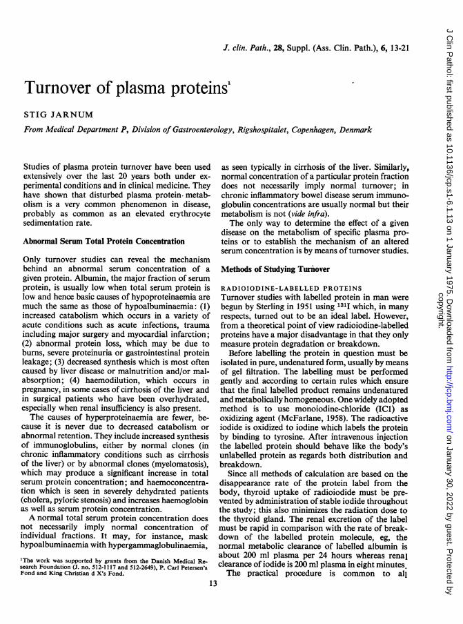
In the nephrotic syndrome the plasma proteinpattern and turnover are profoundly altered, butmost of the changes can be explained by the factthat proteins which are not normally filtered can passthrough the damaged glomerular membrane, espe-cially those of relatively low molecular weight suchas albumin and the smaller immunoglobulins. As aresult, the catabolism of low molecular weight pro-teins such as light chain, which is normally filtered,is unchanged, while the catabolic rate of albumin(molecular weight 68 000) is increased. In a study of30 adult nephrotic patients with a normal or almostnormal glomerular filtration rate we found that theendogenous catabolic rate of albumin, ie, that partof total albumin turnover which is not due to albu-minuria, was increased in 19 (Jensen, Rossing,
iI
.
..
r-0.93* p '0.001
2 4 6 8 10 12ALBIINURIA, g/day.immunhe y~ s
Fig 5 Relation between direct (immunochemicalanalysis of the urine) and indirect (131I-albumin study)determination ofalbuminuria in 18 adult patients withnephrotic syndrome. IVM x FCR x PBI* =intravascular albumin mass x fractional catabolic ratex protein-bound fraction of urinary 1311.
17
copyright. on January 30, 2022 by guest. P
rotected byhttp://jcp.bm
j.com/
J Clin P
athol: first published as 10.1136/jcp.s1-6.1.13 on 1 January 1975. Dow
nloaded from

18
Andersen, and Jarnum, 1967). Furthermore, theincrease in endogenous catabolic rate was positivelycorrelated with the degree of albuminuria. Thisfinding agrees well with the suggestion (Sellers, Katz,andRosenfeld, 1961) that innephrosis filtered proteinsare partly catabolized by tubular cells. The validityof the results of 13'I albumin studies in the nephroticsyndrome is indicated by the close correlation be-tween the amount of albumin uria calculated fromthe plasma 1311 albumin disappearance and theprotein-bound radio-activity of the urine, and thatdetermined directly by immunochemical analysis ofthe urine (Jensen et al, 1967), as shown in figure 5.
Gastrointestinal diseaseThe gastrointestinal tract was not recognized as asite of abnormal protein loss until 1957. In that yearCitrin and his associates showed that the cause ofsevere hypoalbuminaemia regularly present inMenetrier's disease (or giant hypertrophic gastritis)was an abnormal protein leakage through theenormously enlarged rugae of the stomach (Citrin,Sterling, and Halsted, 1957). Two years later anabnormal gastrointestinal protein leakage, demon-strated by means of 1311-labelled polyvinylpyr-rolidone, was reported as the probable cause of therare condition idiopathic hypoproteinaemia(Gordon, 1959; Schwartz and Jarnum, 1959).
Since then a great variety of gastrointestinaldiseases have been shown to be associated with ab-normal gastrointestinal protein loss. They may begrouped according to whether the epithelium isnormal, abnormal or lost (Jarnum, 1963). When theepithelium is lost, as in ulcerative colitis, it is notsurprising that large amounts of protein-richexudate escape to the intestinal lumen.
In Menetrier's disease there is a great increase inthe surface area of the gastric epithelium, which mayexplain the protein loss since, even under normalconditions, some protein leaks through the gastro-intestinal epithelium. Other protein-losing diseaseswith abnormal epithelium are carcinoma of thestomach or colon and non-tropical sprue. However,in sprue protein is not lost in all cases.The largest gastrointestinal protein loss we have
seen has occurred in patients with congenital mal-formations of intestinal lymphatics; stasis produces aclub-shaped dilatation of the small-intestinal villi(fig 6) because the chyle is trapped in the centrallacteals of the villi. The epithelium is normal evenwhen studied by electron microscopy, but it ispossible that multiple tiny ruptures occur so that theprotein-rich chyle escapes into the gut lumen.The same mechanism explains the protein loss
seen in some cases of chronic constrictive pericarditisin which the marked elevation of the central venous
Stig Jarnam
Fig 6 Intestinal villi in a jejunal biopsy from a patientwith intestinal lymphangiectasia. The villi are almostclub-shaped due to dilated lymphatics. The epithelialmembrane may burst (lower left corner) and protein-richinterstitial fluid and chyle leak into the intestinal lumen.
pressure obstructs the outflow of lymph to thevenous system, and causes 'lymphatic hypertension'which again produces stasis in the lacteals of thevilli.
Detection and Quantitation of GastrointestinalProtein Loss
Intestinal loss of protein results in loss of the samefraction of the intravascular mass of all plasmaproteins (Waldmann and Schwab, 1965), and anumber of radioisotope-labelled macromolecularcompounds have been used to detect or measureincreased gastrointestinal protein leakage. Ideallythe test substance should be a plasma proteinlabelled with a radioisotope which does not changethe metabolic behaviour of the protein and is notabsorbed from the gut. Simultaneous determina-tions of the faecal excretion of the isotope and dis-appearance rate of the labelled protein from plasmawould permit calculation of the turnover and distri-bution of the protein, and also of that part of itsdegradation which is due to leakage into the gut. Sofar 67Cu-caeruloplasmin seems to be the onlycompound which fulfils these requirements(Waldmann, Morell, Wochner, Strober, andSternlieb, 1967). Unfortunately, it is not practicablefor clinical studies because of its high cost and thebrief half-life of the isotope (62 hours).When 51CrCI3 is injected intravenously the 51Cr
labels plasma proteins, predominantly transferrin,in a matter of seconds. The subsequent faecal 51Crclearance offers a good estimation of gastrointestinalprotein loss (van Tongeren and Reichert, 1966);this is apparent from the good correlation betweenfaecal 51Cr clearance and the fractional catabolic
copyright. on January 30, 2022 by guest. P
rotected byhttp://jcp.bm
j.com/
J Clin P
athol: first published as 10.1136/jcp.s1-6.1.13 on 1 January 1975. Dow
nloaded from
Tutrnover ofplasma proteins
rate of albumin (Jarnum, Westergaard, Yssing, andJensen, 1968c) which is an indirect indicator of thesize of the protein loss. However, endogenous turn-over cannot be determined with 51Cr-labelled pro-teins because binding of 51Cr to protein is unstable.
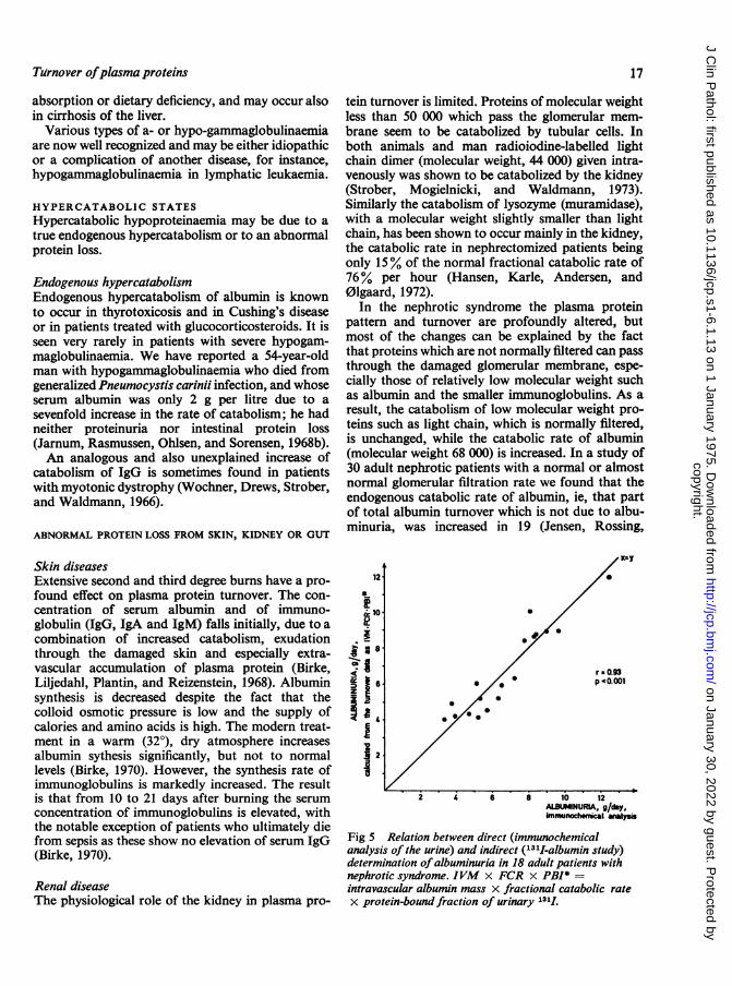
59Fe-labelled iron dextran may also be used todetect and measure gastrointestinal protein loss(fig 7, Jarnum et al, 1968c). The 59Fe is not excretedin the urine, a great advantage when contaminationof the stools with urine is difficult to prevent as, forexample, in infants.
30,
ui gU v 20
g ne10W 10i! '.
not respond (fig 8). The same relationship has beenobserved by de Dombal (1969) in patients withulcerative colitis. We found the same trend inulcerative colitis, but not on a statistically significantlevel.We have performed turnover studies with radio-
iodine-labelled albumin and IgG simultaneously inabout 100 patients with Crohn's disease or ulcerativecolitis. In most of the patients we also estimated theintestinal protein loss either by means of 59Fe-labelled iron-dextran, which could be done simul-taneously owing to the high energy level of gammarays from 59Fe, or by means of 51CrCI3, which hadto be done one week in advance due to the similarityof gamma ray energy of 51Cr and 1311. We foundthat an abnormal but, in most cases, moderate
r-089* 0 p<O 001
/
^~~~~~~~~~_
'O Xx xX
10 20' 30 4i: 50 60FCR-ALBUMIN,60
m. Normal range of intravascular albumin per day* Protein -losing gastroenteropathy
x Children with nephrotic syndrome
Fig 7 Faecal clearance of 59Fe andfractional catabolicrate (FCR) ofalbumin after intravenous injection of59Fe-labelled iron dextran and 131I-albumin in 16patients with protein-losing gastroenteropathy and sevenchildren with the nephrotic syndrome. A high correlationwas present in patients with protein-losinggastroenteropathy (Jarnum et al, 1968).
Protein Turnover and Protein Loss in Chronic Inflam-matory Bowel Disease
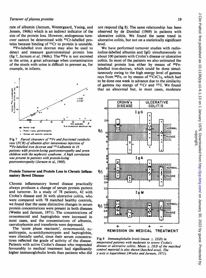
Chronic inflammatory bowel disease practicallyalways produces a change of serum protein patternand turnover. In a study of 78 patients, 42 withCrohn's disease and 36 with ulcerative colitis, whowere compared with 78 matched healthy controls,we found that the same distinctive changes in serumprotein concentrations were present in both diseases(Weeke and Jarnum, 1971). The concentrations oforosomucoid and haptoglobin were increased inmost cases, and the concentrations of albumin,caeruloplasmin and transferrin were depressed.The 'acute phase reactants', orosomucoid, al-
antitrypsin, al-antichymotrypsin and haptoglobin,were clinically useful, since their serum concentra-tions reflected the grade of activity of the disease.Patients with active Crohn's disease who respondedfavourably to medical treatment had significantlyhigher immunoglobulin levels than patients who did
+ -+ -REMISSION ON MEDICAL TREATMENT
Fig 8 Immunoglobulin levels (mean ± 2SD) inunoperated patients with moderate to severe Crohn'sdisease or ulcerative colitis. Mean + 2SD of the matchedcontrol material is also shown (hatched area). They axis is logarithmic (Weeke and Jarnum, 1971).
19
copyright. on January 30, 2022 by guest. P
rotected byhttp://jcp.bm
j.com/
J Clin P
athol: first published as 10.1136/jcp.s1-6.1.13 on 1 January 1975. Dow
nloaded from

20
intestinal protein loss is present in the great majorityof patients and that albumin and IgG turnover areaffected in a similar way in the two diseases. Datawere analysed only from the 48 patients in whom thediagnosis was considered certain (Jarnum andJensen, 1975), namely, 21 patients with ulcerativecolitis and 27 with Crohn's disease. In all of thelatter the diagnosis was confirmed by intestinal re-section (performed one-half to five months later).The catabolic rates of albumin and IgG were elevatedin practically all cases and there was a high degree ofcorrelation between them, in ulcerative colitis (r =0-83, p < 0-001) and in Crohn's disease (r = 0 73,p < 0-001). The data from patients with ulcerativecolitis are given in figure 9.
In the majority of cases the proportional increasein the catabolic rate of IgG was greater than that ofalbumin. Since the excess catabolism is due mainlyto intestinal protein loss, a preferential loss of IgGmight be postulated. However, this is highly un-likely, since intestinal protein loss is 'bulk loss' whichdrains the same fraction of the intravascular mass ofall plasma proteins regardless of their molecularsize (Waldmann and Schwab, 1965). Therefore thehypercatabolism of IgG, which is well known tooccur in other chronic inflammatory conditions suchas cirrhosis of the liver and systemic lupus erythe-matosus (Andersen, 1964), is probably due not onlyto protein loss but also to 'endogenous' hyper-catabolism of IgG. In non-inflammatory protein-losing enteropathies such as intestinal lymphangiec-tasis the catabolic rate of IgG is close to or lowerthan that of albumin (Bendixen, Jarnum, S0ltoft,Westergaard, Weeke, and Yssing, 1968).As mentioned earlier, the serum concentration of
Stig Jarnum
a
L.
00)
-Ocm
.
r=0.83pcO.OO1
FCR of ALBUMIN, % per day
Fig 9 Albumin and IgG degradation in ulcerativecolitis (n = 21).
IgG in ulcerative colitis and Crohn's disease isusually within the normal range, whereas serumalbumin is depressed. We found an inverse relation-ship between serum albumin and catabolic rate dueto the fact that the synthesis rate of albumin islargely unchanged in these conditions. In contrast,serum IgG concentration seems to be quite un-affected by the hypercatabolism which indicates thata significant increase of IgG synthesis has takenplace. We used 59Fe-iron-dextran to assess the size of
Fig 10 The ratio (* GI*A)5 between faecal 125I excretion
from I1-IgG andfaecal 131I£-~4 excretion from 131I-albumin in
ulcerative colitis (UC, n = 21):.;3 and three groups ofpatients with
Crohn's disease (CD, n = 26)a - -2a localized to various sites of the
J.. gilE .- intestinal tract.
CR@ s DIS': * IIm,UP 1-
. :.0U
..
* * :
copyright. on January 30, 2022 by guest. P
rotected byhttp://jcp.bm
j.com/
J Clin P
athol: first published as 10.1136/jcp.s1-6.1.13 on 1 January 1975. Dow
nloaded from

Turnover ofplasma proteins 21
intestinal protein loss. In an earlier study (Jarnumet al, 1968c) we found quite a good correlation be-tween gastrointestinal 59Fe clearance and thefractional catabolic rate of albumin in various typesof protein-losing gastroenteropathy (fig 7). In thisstudy of chronic inflammatory bowel disease asimilar satisfactory correlation was found both inulcerative colitis and in Crohn's disease. Further-more the intestinal protein loss was unrelated to theanatomical extent of the lesion in Crohn's disease.Apparently, protein loss is more dependent on otherfactors such as the depth of the ulceration or thedamage to lymphatics.The faecal excretion of 1311 and 1251 from labelled
albumin and IgG respectively turned out to be auseful guide to the topographic site of the lesion inCrohn's disease (fig 10) since the ratio of faecal 1251from IgG to faecal 1311 from albumin increased asthe location of the lesion became more proximal.This is probably due to a higher intraluminal pro-teolysis of albumin than of IgG. In an earlier study(S0ltoft and Jamum, 1969) we found that faecesfrom patients with chronic inflammatory boweldisease can break down albumin more rapidly thanIgG, whereas faeces from patients with diarrhoeafrom other causes (postgastrectomy syndrome,gluten enteropathy, lactose malabsorption, smallintestinal resection) showed quite the opposite. Wehave no explanation for this phenomenon.
This survey of plasma protein turnover has dealtmainly with clinical aspects. The intention has beento show that such studies, at least so far as the majorplasma protein components are concerned, arerelatively simple and can yield information on thepathogenesis of abnormal plasma protein concentra-tion patterns which are helpful both in diagnosis andin the assessment of therapy.
References
Andersen, S. B. (1964). Metabolism of Human Gamma Globulin(y,v-globulin). Blackwell, Oxford.
Bendixen, G., Jarnum, S., S0ltoft, J., Westergaard, H., Weeke, B.,and Yssing, M. (1968). IgG and albumin turnover in Crohn'sdisease. Scand. J. Gastroent., 3, 481.
Birke, G. (1970). Regulation of protein metabolism in burns. InPlasma Protein Metabolism: Regulation of Synthesis, Distribu-tion and Degradation, edited by M. A. Rothschild and T.Waldmann, p. 415. Academic Press, New York and London.
Birke, G., Liljedahl, S.-O., Plantin, L. 0., and Reizenstein, P. (1968),Studies on burns IX. The distribution and losses through thewound of 1"'I-albumin measured by whole-body counting.Acta chir. scand., 134, 27.
Citrin, Y., Sterling, K., and Halsted, J. A. (1957). The mechanism ofhypoproteinemia associated with giant hypertrophy of thegastric mucosa. New Engl. J. Med., 257, 906.
de Dombal, F. T. (1969). Prognostic value of estimating serum pro-teins in cases of ulcerative colitis in remission. Gut, 10, 491.
Gordon, R. S., Jr. (1959). Exudative enteropathy: abnormal perme-ability of the gastrointestinal tract demonstrable with labelledpolyvinylpyrrolidone. Lancet, 1, 325.
Hansen, N. E., Karle, H., Andersen, V., and 0lgaard, K. (1972).Lysozyme turnover in man. J. clin. Invest., 51, 1146.
Jarnum, S. (1963). Protein-Losing Gastroenteropathy, p. 198. Blackwell,Oxford.
Jarnum, S., Jeejeebhoy, K. N., and Singh, B. (1968a). Dysgamma-globulinaemia in tropical sprue. Brit. med. J., 4, 416.
Jarnum, S., and Jensen, K. B. (1975). Faecal radioiodide excretionfollowing intravenous injection of "311-albumin and 115I-IgGin chronic inflammatory bowel disease. An aid to topographicdiagnosis. Gastroenterology, in press.
Jarnum, S., Rasmussen, E. F., Ohlsen, A. S., and S0rensen, A. W. S(1968b). Generalized pneumocystis carinii infection with severeidiopathic hypoproteinemia. Ann. intern. Med., 68, 138.
Jarnum, S., Westergaard, H., Yssing, M., and Jensen, H. (1968c).Quantitation of gastrointestinal protein loss by means of Fe"'-labeled iron dextran. Gastroenterology, 55, 229.
Jensen, H., Rossing, N., Andersen, S. B., and Jarnum, S. (1967).Albumin metabolism in the nephrotic syndrome in adults.Clin. Sci., 33, 445.
McFarlane, A. S. (1958). Efficient trace-labelling of protein withiodine. Nature (Lond.), 182, 53.
McFarlane, A. S. (1963). Measurement of synthesis rates of liver-produced plasma proteins. Biochem. J., 89, 277.
Matthews, C. M. E. (1957). The theory of tracer experiments with3'I-labelled plasma proteins. Phys. med. Biol., 2, 36.
Nosslin, B. (1966). Applications of tracer theory to protein turnoverstudies. J. nucl. biol. Med., 9, 18.
Reeve, E. B., Pearson, J. R., and Martz, D. C. (1963). Plasma proteinsynthesis in the liver. Method for measurement of albuminformation in vivo. Science, 139, 914.
Samuel, A. M., Jarnum, S., and Jeejeebhoy, K. N. (1969). Influence ofparenteral I-isomeric amino acids on absolute albumin synthesisrates in tropical sprue. Scand. J. Gastroent., 4, Suppi. 3, 51.
Schwartz, M., and Jarnum, S. (1959). Gastrointestinal protein loss inidiopathic (hypercatabolic) hypoproteinaemia. Lancet, 1, 327.
Sellers, A. L., Katz, J., and Rosenfeld, S. (1961). Plasma albumincatabolism in experimental nephrosis. Nature (Lond.), 192, 562.
Sterling, K. (1951). The turnover rate of serum albumin in man asmeasured by I131-tagged albumin. J. clin. Invest., 30, 1228.
Strober, W., Mogielnicki, R. P., and Waldmann, T. A. (1973). Therole of the kidney in the metabolism of serum proteins. InProtein Turnover: Ciba Foundation Symposium 9 (new series)p. 25. Associated Scientific Publishers, tAmsterdam, London,New York.
Soltoft, J., and Jarnum, S. (1969). Faecal proteolysis of albumin andIgG in Crohn's disease and ulcerative colitis. Digestion, 2,282-288.
van Tongeren, J. H. M., and Reichert, W. J. (1966). Demonstrationof protein-losing gastroenteropathy: the quantitative estima-tion of gastrointestinal protein loss using "lCr-labelled plasmaproteins. Clin. chim. Acta, 14, 42.
Waldmann, T. A., Morell, A. G., Wochner, R. D., Strober W., andSternlieb, I. (1967). Measurement of gastrointestinal protein lossusing ceruloplasmin labeled with 'copper. J. clin. Invest., 46,10.
Waldmann, T. A., and Schwab, P. J. (1965). IgG (7 S gamma globulin)metabolism in hypogammaglobulinemia. Studies in patientswith defective gamma globulin synthesis, gastrointestinal pro-tein loss, or both. J. clin. Invest., 44, 1523.
Weeke, B., and Jarnum, S. (1971). Serum concentration of 19 serumproteins in Crohns' disease and ulcerative colitis. Gut, 12, 297
Westergaard, H., Jarnum, S., Preisig, R., Rams0e, K., Tauber, J., andTygstrup, N. (1970). Degradation of albumin and IgG at highaltitude. J. appl. Physiol., 28, 728.
Wochner, R. D., Drews, G., Strober, W., and Waldmann, T. A.(1966). Accelerated breakdown of immunoglobulin G (IgG) inmyotonic dystrophy: a hereditary error of immunoglobulincatabolism. J. clin. Invest., 45, 321.
copyright. on January 30, 2022 by guest. P
rotected byhttp://jcp.bm
j.com/
J Clin P
athol: first published as 10.1136/jcp.s1-6.1.13 on 1 January 1975. Dow
nloaded from