tweed ajo 1946 - the frankfort mandibular plane angle in orthodontic diagnosis, classification,...
TRANSCRIPT
American
Journal of Orthodontics and Oral Surgery
(All rights reserz;Ptl)
VOL. 32 APRIL, 1946 No. 4
Original Articles
THE FRANKFORT-MANDIBULAR PLANE ANGLE IN ORTHODONTIC DIAGNOSIS, CLASSIFICA4TION, TREATMENT PLANNING,
AND PROGNOSIS
CHARLES H. TTVEED, D.D.S., TUCSOX, ARIZ.
T HE material presented here is the result of constant clinical observation covering a period of many years. While not unexpected, it is nevertheless
gratifying to find that in the main my clinical findings, although arrived at in- dependently, bear an extremely high degree of correlation to, and actually pro- vide additional emphasis and concrete evidence of, the validity of the results and conclusions obtained by some of the outstanding scientific laboratory research workers in the field of orthodontics. However, many of our very able research men, after presenting their findings to us, have added to our confusion by dis- regarding, if not actually refuting, their own\ scientific investigations. I refer especially to the fact that they frequently will not or do not relate their statis- tical and laboratory findings to their treatment procedures. This is most unfor- tunate because when clinical findings and scientific research are more closely wedded, only then will many of our complex problems in orthodontics be solved- not before. Let us all remember this fact.
My clinical observations have been focused for many years o,n (1) the position of the mandibular incisors as related to the medullary bone of the body of the mandible-1 have in the past referred to this as basal bone or dental base-and (2) the normal facial esthetics ahd their deviations. My observations have led me to the conviction that in all orthodontic therapy involving Class 1: Class II, and bimaxillary protrusion types of malocclusion, where the growth pattern of the face is not too abnormal, the mandibular incisors must always be positioned upright on the alveolar process and over medullary ‘bone. Further- more, I am convinced that the normal range of variation of the inclination of the mandibular incisor teeth, as reIated to a plane parallel with the lower border
175
176 CHARLES H. TWEED
or base of the mandible in sagittal view, is approximately +5”, with 90” as the norm when the incisors stand at right angles to the plane parallel with the lower border or base of the mandible, i.e., the mandibular plane.
This variation in range of the normal was accepted by me as a result of my study of individuals whom I considered to possess normal occlusion. The nor- mal as I visualize it must have, in addition to correct occlusal relationship, all five of the other qualifications as outlined in the correct interpretation of Angle’s* definition of the line of occlusion. The possessor of all six of the fundamental requisites for normal occlusion as outlined by Angle must have a facial growth pattern normal in its entirety. .
In my opinion, a thorough concept of the normal growth pattern of the child’s face or any face is as important to orthodontists, if not more so, as com- plete mastery of the science of occlusion. Occlusion and facial esthetics, whether normal or abnormal, are so intimately associated and interdependent one upon the other that orthodontics must embrace both equally, because they cannot be dissociated. This thought was originally expressed by Angle. The man un- able to correlate normal oc.clusion with the normal growth pattern of the face is indeed a sorry orthodontist. Let us settle the argument now and forever that it is possible to overemphasize facial esthetics in orthodontics. Normal occlusion in its correct sense is impossible without a normal facial pattern, and a normal facial pattern is none other than the ultimate in balance and harmony of facial esthetics2
I might state here, but shall have -more to say on the subject later, that the +5” normal range of variation in the inclination of the mandibular incisors, as related to t,he plane formed by the lower border or base of the mandible, applies to tho,se cases only in which growth has approximated the general normal pattern of the individual.” It applies also to all those cases where lack of osseous growth has been general and has resulted in a slightly diminished structure of the bones of the maxilla and mandible, without markedly disturbing the directional growth of the jaws. In these cases a discrepancy often can be observed between the mesiodistal configuration of the dental arch and the tooth-bearing bones of the jaws, resulting in crowding and displacement of teeth, and giving the general appearance of too much tooth material for the available bone in which the teeth are to be accommodated. If this condition is pronounced, it becomes necessary to reduce the mesiodistal dimension of the dental arch in order to obtain both nor- mal tooth-bony base relationship and normal facial esthetics.
The foregoing does not apply to true Class III cases or to those cases where lack of growth in the condylar growth centers has perverted the growth pattern vector from the normal downward and forward direction to too much downward and not enough forward. The la& of growth in these growth centers which causes these abnormalities is, in my opinion, more common than most of us realize. We are prone to dismiss this deformity from our problems by calling it type. Perhaps half of all our problem cases fall into this category in vary- ing degree. In its more pronounced forms it is a condition for which the ortho- dontist can do little as far as facial esthetics are concerned. We shall discuss these cases more fully later.
FRANKFORT-NIANDIBULAR PLASE ANGLE 177
Broadbent, Brodie,j Margolis,6 and others have made splendid contributions on t.he growth and development of the head and face of the child. Brodie has demonstrated the angular constancy of the lower border or base of the mandible. By that, I mean he has shown that the plane of the lower border of the mandible when related to any fixed point, and the gonion angle when related to any con- stant plane, always remain virtually the same. In other words, growth is of such a nature that the planes formed by the base of the mandible at variolus ages from 3 months upward are always approximately parallel to one another.”
The parallel growth of the lower border or base of the mandible is due to its angular constancy and is quite obvious to all who have studied facial growth. YIargolis7 deserves credit for being the first to relate the axial inclination of the central mandibular incisor with the sagittal plane tangent to the most de- pendent points on the lower border or base of the mandible. He has named the angle formed by the interception of the long axes of the mandibular incisoras with the plane formed by the lower border of the mandible, the incisor man- dibular plane angle. Margolis8 also ~2~11s attention to the fact that the philosophy which calls for distal movement of the denture, in certain cases of malocclusion, and the maintenance of the denture in normal relation to the rest of the head structukes during orthodontic treatment, depends on the degree to which the mandibular incisors can be placed and maintained in an upright position over the medullary bone of the body of the mandible and that all of this is in ac- cordance with cvqlutionary tendencies of facial growth.”
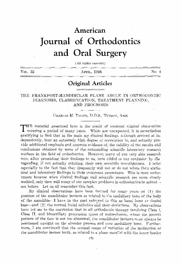
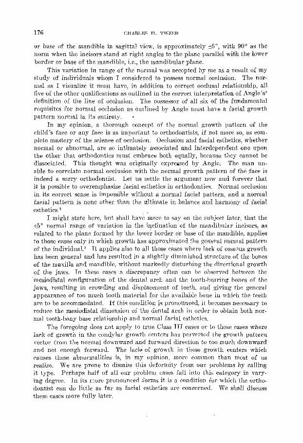
As stated by Margolis :s ‘(Up to this point (Figs. 1 to G), it seems quite evident that there has. been a reduction in the alveolar bone in man as compared with anthropoids and primitive man, so that in modern man a chin has developed. The chin is not so much a for- wa.rd development of the mental eminence but rather the result of a recession of the alveolar boric. It becomes obvious then, that the mandibular incisors have been straightened upward during the process of evolution or better, during the reduction of the alveolar bone and the formation of the chin. ”
In spite of this conclusive evidence, I find some highly regarded scientific workers in the field of orthodontics who contend actually that it is good clip- ical procedure to reverse evolutionary trends in the orthodontic treatment of our patients and to make X in Fig. 9 look like IX.
As man fulfills his evolutionary destiny we find the cranium becoming in- creasingly larger and the face correspondingly smaller in their relative pro- portions to each other in the skull. While this is going on, the mandibular in- cisors are becoming less procumbent. Mandibular prominepce is developing in direct ratio to the diminishing degree of the procumbency of the mandibular in- cisors. The more procumbent the mandibular incisors, the less the mandibular prominence, and, inversely, the more upright the mandibular incisors as related to the lower border of the mandible on the sagittal plane, the more pronounced the mandibular prominence, or chin.
The acceptance of the premise that in normal occlusion the mandibular in.cisors are always upright over medullary or basal bone certainly adds to the
*I am indebted to Dr. Margolis for the use of Figs. 1 to 13 which he has loaned me tg demonstrate our viems.
1’78 CHARLES H. TWEED
Fig. I.-AI? adu degree of PT( )rEc-
Fig. 2.-A 3 wung chimpanzee. Note similarities to Fig. 1 and compare with Figs. 3, 4, 5, and 6.
FRASKFORT-MANDIBULAR PLANE ANGLE 179
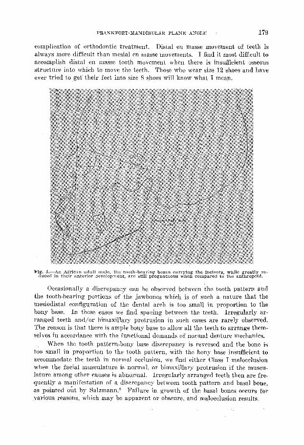
complication of orthodontic treatment. Distal en masse movement of teeth is always more dificult than mesial en masse movements. I find it most difficult to accomplish distal en masse tooth movement when there is insufficient osseous structure into which to move the teeth. Those who wear size 12 shoes and have ever tried to get their feet into size 8 shoes will know what I mean.
Fig. 3.-An African adult male, the tooth-bearing bones carrying the incisors, while grleatly re- duced in their anterior development, are still prognathous when compared to the anthropoid.
Occasionally a discrepancy can be observed between the tooth pattern and the t,ooth-bea,ring portions of the jawbones which is of such a nature that the mesiodistal configuration of the dental arch is too small in proportion to the bony base. In these cases we find spacin g between the teeth. Irregularly ar- ranged teeth and/or bimaxillary protrusion in such cases are rarely observed. The reason is that there is ample bony base to allow all the teeth to arrange them- selves in accordance with the functional demands of normal denture mechanics.
When the tooth pattern-bony base discrepancy is reversed and the bone is too small in proportion to the tooth pattern, with the bony base insufficient to accommodate the teeth in normal occlusion, we find either Class I ma,locclusion when the facial musculature is normal, or bimaxillary protrusion if the muscu- lature among other causes is abnormal. Irregularly arranged teeth then are fre- quently a manifestation of a discrepancy between tooth pattern and basal bone, as pointed out by Salzmann.g Failure in growth of the basal bones occurs for various reasons, which may be apparent or obscure, and malocclusion result,s.
180 -- / CHABLES H. TWEED
The dentition itself can remain quite stable once it reaches a state in mgloc- elusion in which the forces originally responsible for the initiation of the maloc- clusion became neutralized. The proof of this statement can be found in the fact that, as a rule, the condition of malocclusion in the examples referred to in the foregoing do not become progressively worse, once a state of balance in the forces responsible for the malocclusion has been reached. The functional bal- ance, although manifestly existing in a state of malocclusion, resists change. The irregular dentition in balance is, therefore, a far more stable condition than the same dentition treated orthodontically but forced out of balance and into pro- trusive relationship to the medullary portion of the bony base.’ Such ortho- dontic treatment is usually followed by “collapse.”
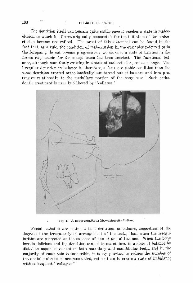
Fig. 4.-A nonprognathous Massachusetts Indian.
Facial esthetics are better with a dentition in balance, regardless of the degree of the irregularity of arrangement of the teeth, than when the irregu- larities are corrected at the expense of loss of dental balance. When the bony base is deficient and the dentition cannot be maintained in a state of balance by distal en masse movement of both maxillary and mandibular teeth, and in the majority of cases this is impossible, it is my practice to reduce the number of the dental units to be accommodated, rather than to create a state of imbalance with subsequent “collapse. ”
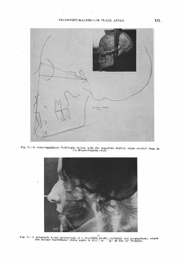
Fig.
FRANKFORT-MASDIBULAR PLANE ASGLE
5.-A nonprogqathous California Indian with the mandible slightly more receded thal the Massachusetts skull.
n in
‘: Fig. %-A composite x-ray photograph of a beautiful profile, certainly not prognathous. where
the incisor mandibular plane angle is 89&O, or - yso in the _+5” formula.
182 CHARLES H. TWEED
As a general rule, teeth are irregular for the same reason that third molars are so often impacted. The reason is, I repeat, that there exists a discrepancy between tooth pattern and basal bone because of a lack of osseous growth over which the orthodontist has no control. Until we recognize and accept this fact, treatment planning will remain obscure and results of treatment will continue to be as indifferent in the future as they so frequently have been in the pa,st. .
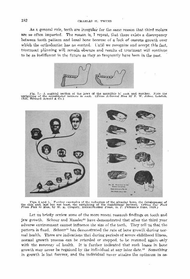
Fig. 7.-A sagittal section of the jaws of the mandible bf man and monkey. Note the uprighting of the mandibular incisors in man. (From A?bowaZ iMan by F. IV. Jones, London, 1926. Edward Arnold & Co.)
Figs. 8 and I).-Further examples of the reduction of the alveolar bone, the development of the chin and, last but not least, the uprighting of the mandibular incisors. (From Our B’aoe From Risiz to Mm-&, by W. K. Gregory, Knickerbocker Press, G. P. Putnam’s Sops, 1929.)
Let us briefly review some of the more recent research findings on tooth and jaw growth. Schour and MassleF have demonstrated that after the third year adverse environment cannot influence the size of the teeth. They tell us that the pa.ttern is fixed. SchouY1 has demonstrated the rat,e of bone growth during nor- mal health. There are indications that during periods of severe childhood illness, normal growth process can be retarded or stopped, to be resumed again only with the recovery of health. It is further indicated that such losses in bone growth may never be regained by the individual at any later date.12 Something in growth is lost forever, and the individual never attains the optimum in os-
FRANKFORT-MANDIBULAR PLANE AXGLE 183
seous growth intended by his genetic pattern. BroadbenP concurs in this opinion.
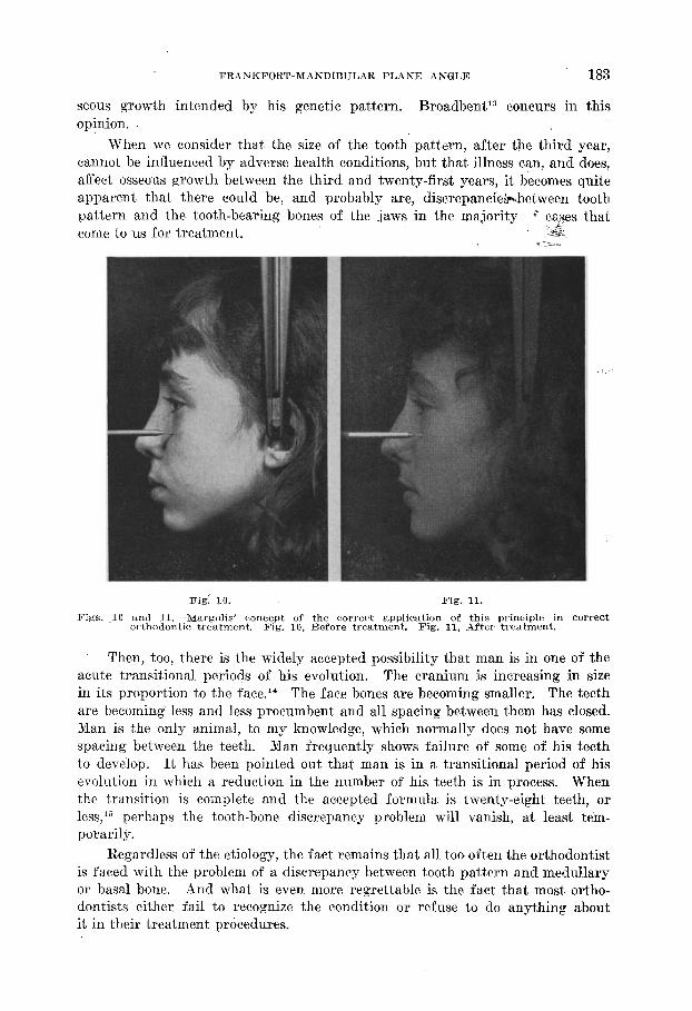
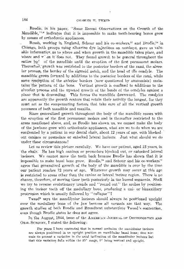
When we consider that the size of the tooth pattern, after the third year, cannot be influenced by adverse health conditions, but that illness can, and1 does, affect osseous growth between the third and twenty-first years: it becomes quite apparent that there could be, and probably are, discrepanciejP+.between tooth pattern and the tooth-bearing bones of the jaws in the majority c cases that come to us for treatment. +,
Fig: 10. Fig. 11.
Figs. ,lO and il.-Margolis’ concept of the correct application of this principle in correct orthodontic treatment. Fig. 10, Before treatment. Fig. 11, After treatment.
Then, too, there is the widely accepted possibility that man is in one of the acute transitional periods of his evolution. The cranium is increasing in size in its proportion to the face.14 The face bones are becoming smaller. The teeth are becoming less and less procumbent and all spacing between them has closed. Man is the only animal, to my knowledge, which normally does not have some spacing between the teeth. Man frequently shows failure of some of his teeth to develop. It has been pointed out that man is in a transitional period. of his evolution in which a reduction in the number of his teeth is in process. When the transition is complete and the accepted formula is twenty-eight teeth, or less,l” perhaps the tooth-bone discrepancy problem will vanish, at least tem- porarily.
Regardless of the etiology, the fact remains that all too often the orthodontist is faced with the problem of a discrepamy between tooth pattern and medullary or basal bone. And what is even more regrettable is the fact that most ortho- dontists either fail to recognize the condition or refuse to do anything about it in their treatment procedures.
184 CHARLES H. TWEED
Brodie, in his paper, “Some Recent Observations on the Growth of the Mandible, “I6 indicates that it is impossible to make tooth-bearing bones grow by means of orthodontic appliances.
Brash, working in England, Schour and his co-workers,17 and Brodie,lg in Chicago, both groups using alizarine dye injections on monkeys, gave us valu- able informatiovi as to where and when growth in the mandible takes place, and where and v - tin it ‘does not. They found growth to be general throughout the entire be’ of the mandible until the eruption of the first permanent molars. Thereafte?;growt,h was restricted to the posterior borders of the rami, the alveo- lar process, &he border of the sigmoid notch, and the head of tKe condyle. The mandible grows forward by additions to the posterior borders of the rami, while some res?rption of the anterior borders (now questioned by anatomists) main- tains ‘the ‘pattern of the bone. Vertical growth is confined to additions to the alvedlar process and the upward growth of the heads of the condyles against a plane that is “descending. This forces the mandible downward. The condyles are apparently the growth centers that retain their activity the longest, for they must act as the compensating factors that take care of all the vertical growth processes of both mandible and maxilla.
Since generalized growth throughout the body of the mandible ceases with the eruption o,f the first permanent molars and is thereafter restricted to the areas mentioned above, and, as Brodie has shown us, we cannot make the body of the jawbone grow with orthodontic appliances, what are we to do when we are confronted by a patient in our dental chair, about 12 years of age, with blocked- out canines or premolars or enlocked lateral incisors. Just what should we do under these circumstances Z
Let us review this pict,ure carefully. We have our patient, aged 12 years, in the chair: He has high canines or premolars blocked out, or enlocked lateral incisors. We cannot move the teeth back because Brodie has shown that it is impossible to make basal bone grow. Brobie,18 and Schour &d his co-workers17 agree that generalized growth of the body of the mandible is over by the time our patient reaches 12 years of age. Whatever ~growth may occur at this age is restricted to areas other than the canine or lateral incisor region. There is no chance, therefore, of moving these teeth posteriorly in the buccal segments. Shall we try to reverse evolutionary trends and “round out” the arches by position- in.g the incisor teeth off the medullary bone, producing a uni- or himaxillary protrusion which is usually followed by “collapse ” ?
Tweed’ says the mandibular incisors should always be positioned upright over the medullary bone of the jaw because all normals are that way. The growth studies of both Brodie and Broadbent substantiate Tweed’s, contention, even though Brodie states he does not agree.
In the August, 1944, issue of the AMERICAN.JOTJRNAL OF ORTHODONTICS AND ORAL SURGERY, I stated the following :
For years I have contended that in normal occlusion the mandibular incisors are always positioned in an upright position on mandibular basal bone; that nor- mals do present a variation in the axial inclination of the mandibular incisors but that this variation falls within the 25” range, 0” being vertical and upright.
FRANKFORT-MANDIBULAR PLANE ANGLE 185
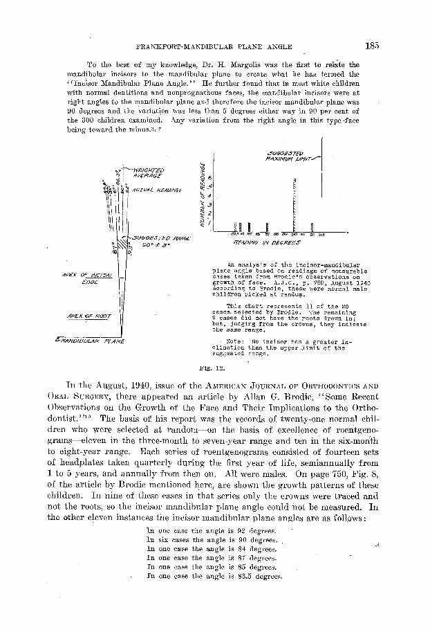
To the best of my knowledge, Dr. H. Margolis was the first to relate the mandibu1a.r incisors to the mandibular plane to create what he has termed the ‘ i Incisor Mandibular Plane Angle. ’ ’ He further found that in most white children with normal dentitions and nonprognathous faces, the mandibular incisors were at right angles to the mandibular plane and therefore the incisor mandibular plane was 90 degrees and the variation was less than 5 degrees either way in 90 per cent of the 300 children examined. Any variation from the right angle in this type -face being toward the minus.3, 7
An analysis of the incisor-mandibular plane angle based on readings of mensurable cases taken from Brodie's observations on growth of face. A.J.O., p. 750, August 1940 According to Srodie, these were nornal male children picked at rendom.
This chart represents 11 of the 20 cases selected by Srodie. The remaining 9 cases did not have the roots drawn in; but, judging from the crowns, they indicate the same range.
Note: No incisor has a &reater in- clination than the upper limit of the sugaested range.
Fig. 12.
In the August, 1940,issue of the AMERICAN JOURNAL OF ORTHODONTICS AND
ORAL SURGERY, there appeared an article by Allan G. Brodie, “Some Recent Observations on the Growth of the Face and Their Implications to the Ortho- dontist. ’ ‘I9 The bas,is of his report was the records of twenty-one normal chil- dren who were selected at random-on the basis of excellence of roentgeno- grams-eleven in the three-month to seven-year range and ten in the six-month to eight-year range. Each series of roentgenograms consisted of fourteen sets of headplates taken quarterly during the first year of life, semiannually from 1 to 5 years, and annually from then on. All were males. On page 750, Fig. 8, of the article by Brodie mentioned here, are shown the growth patterns of these children. In nine of these cases in that series only the crowns were traced and not the roots, so the incisor mandibular plane angle could not be measured. In the other eleven instances the incisor mandibular plane angles are as folllows:
In one case the angle is 92 degrees. In six cases the angle is 90 degrees. , In one case the angle is 84 degrees.
.;i
In one case the angle is 87 degrees. In one case the angle is 85 degrees. In one case the angle is 83.5 degrees.
186 CHARLES H. TWEED
The angular variation of the mandibular incisors with relation to the lower border of the mandible is 8.5” in this group of normals.
The average for these eleven cases is an incisor mandibular plane angle of 88.3”, which is vertical or upright and well within the range of the 15” formula. In fact, it is -1.8”.
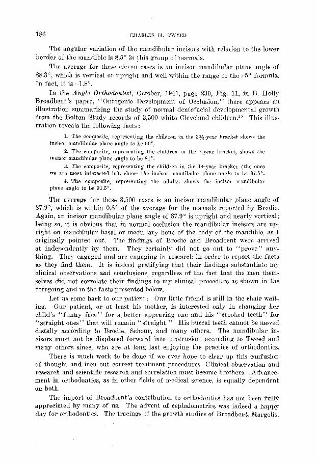
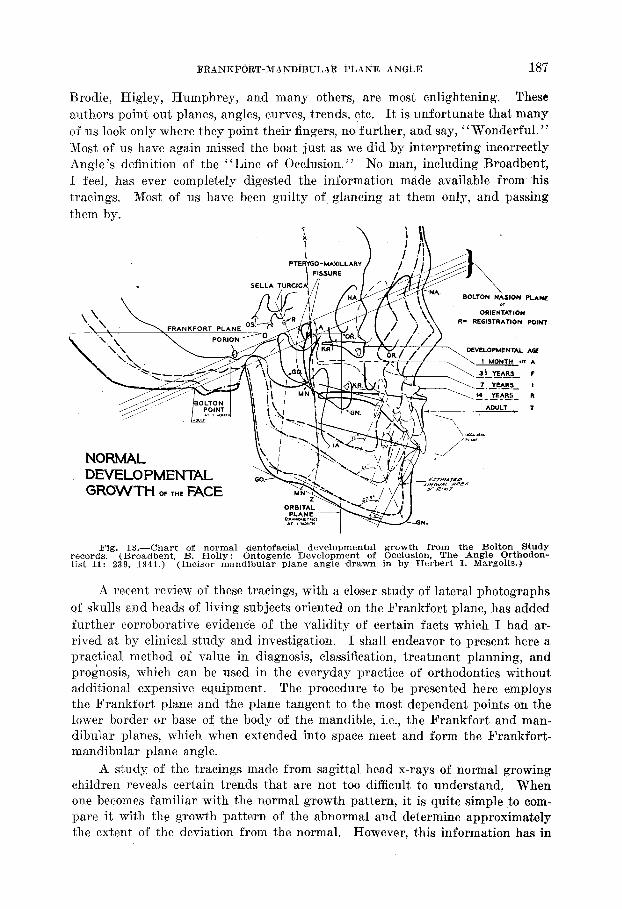
In the Angle Orthodontist, October, 1941, page 239, Fig. 11, in B. Holly Broadbent’s paper, “Ontogenic Development of Occlusion,” there appears an illustration smnmarizing the study of normal dentofacial developmental growth from the Bolton Study records of 3,500 white Cleveland children.20 This illus- tration reveals the following facts :
1. The composite, representing the children in the 3%.year bracket shows the incisor mandibular plane angle to be 90’;
2. The composite, representing the children in the 7-year bracket, shows the incisor mandibular plane angle to be 81”.
3. The composite, representing the children in the 14-year bracket (the ones we are most interested in), shows the incisor mandibular plane angle to be 87.5”.
4. The composite, representing the adults, shows the incisor mandibular plane angle to be 91.5”.
The average for these 3,500 cases is an incisor mandibular plane angle of 87.9”, which is within 0.8” of the average for the normals reported by Brodie. Again, an incisor mandibular plane angle of 87.9” is upright and nearly vertical; being so, it is obvious that in normal occlusion the mandibular incisors are up- right on mandibular basal or medullary bone of the body of the mandible, as I originally pointed out. The findings of Brodie and Broadbent were arrived at independently by them. They certainly did not go out to “prove” any- thing. They engaged and are engaging in research in order to report the facts as they find them. It is indeed gratifying that their findings substantiate my clinical observations and conclusions, regardless of the fact that the men them- selves did not correlate their findings to my clinical procedure as shown in the foregoing and in the facts presented below.
Let us come back to our patient: Our little friend is still in the chair wait- ing. Our patient, or at least his mother, is interested only in changing her child’s “funny face” for a better appearing one and his “crooked teeth” for “straight ones” that will remain “straight.” His buccal teeth cannot be moved distally according to Brodie, Schour, ,and many others. The mandibular in- cisors must not be displaced forward into protrusion, according to Tweed and many others Gnce, who are at long last enjoying the practice of orthodontics.
There is much work to be done if we ever hope to clear up this confusion of thought and iron out correct treatment procedures. Clinical observation and research and scientific research and correlation must become brothers. Advance- ment in orthodontics, as in other fields of medical science, is equally dependent on both.
The import of Broadbent’s contribution to orthodontics has not been fully appreciated by many of us. The advent of cephalometrics was indeed a happy
day for orthodontics. The tracings of the growth studies of Broadbent, Margolis,
j?kArjKi@kT-MANDfBULAk PjLArjE ANGLE 18’7
Brodie, Higley, Humphrey, and many others, are most enlightening. These authors point out planes, angles, .curves, trends, etc. It is unfortunate that many of us look only where they point their fingers, no further, and say, “Wonderful. ” Most of us have again missed the boat just as we did by interpreting incorrectly Angle’s definition of the “Line of Occlusion.” No man, including Broadbent, I feel, has ever completely digested the information made available from his tracings. Most of us have been guilty of glancing at them only, and passing them by.
DEVELOPMENTi9L GROWTH OF T,.,E FACE
Fig. 13.-Chart of normal dentofacial developmental growth from the Bolton Study records. (Broadbent, B. Holly : Ontogenic Development of Occlusion, The Angle Orthodon- tist 11: 239, 1941.) (Incisor mandibular plane angle ,drawn in by Herbert I. Margolis.)
A recent review of these tracings, with a closer study of lateral photographs of skulls and heads of living subjects oriented on the Frankfort plane, has added further corroborative evidence of the validity of certain facts which I had ar- rived at by clinical study and investigation. I shall endeavor to present here a practical method of value in diagnosis, classification, treatment planning, and prognosis, which can be used in the everyday practice of orthodontics without additional expensive equipment. The procedure to be presented here employs the Frankfort plane and the plane tangent to the most dependent points on the lower border or base of the body of the mandible, i.e., the Frankfort and man- dibular planes, which when extended into space meet and form the Frankfort- mandibular plane angle.
A study of the tracings made from sagittal head x-rays of normal growing children reveals certain trends that are not too difficult to, understand. When one becomes familiar with the normal growth pattern, it is quite simple to oom- pare it with the growth pattern of the abnormal and determine approximately the extent of the deviation from the normal. However, this information has in
188 CHARLES H. TWEED
the past been regarded as being of academic interest only. It need no longer remain so, because, as we shall,show here, it has a deep and vital significance in our daily practice.
As shown in the foregoing, Brodiel” and others have demonstrated that once the growth pattern of the facial bones is established, whether normal or ab- normal, it is virtually constant and resists change. For instance, in those cases where there has been injury to the condylar growth centers that temporarily retards growth in these centers, we find short rami, and growth is diverted more downward and less forward than in the normal growth vector, which is down- ward and forward. This loss in the normal, proportional bone pattern is never regained by the individual by future growth spurts. This statement is sup- ported by the fact thjt the parallelism of the lower borders of the mandible continues in the abnormal direction during the subsequent growth of these in- dividuals. It is obvious to all of us that such a case is to be avoided because there is little other than correction of occlusal relationships that the orthodontist can do for these patients. Yes, we all recognize tha.t, but I wonder how many of us realize that a great many of our problem cases are but variations of this same condition. And, because we fail to detect a faulty growth pattern, we be- come befuddled. Were we more observant, we would recognize these cases at once and realize that, because of the abnormal growth pattern, we shall never be able to attain all of our orthodontic objectives, regardless of how we treat the case.
A classificatioh of prognosis” that will pick out these cases should be of great value to all of us.. My purpose is to make a start in that direction, with the hope that others will consider the problem worthy of further study.
In using this diagnostic method, we can employ Salzmann’fi Maxillato? and take our measurements directly on the patient, or we may employ a sagittal head x-ray or profile phot0graph.t
When sagittal x-rays or photographs are used, the Frankfort plane is ex- tended posteriorly through the back of the head. The plane formed by the lower border of the mandible is likewise extended po,steriorly until it intercepts the Frankfort plane. The degree of the angle form”ed by the intercepting of these two planes will determine the location of the point of interception of these planes, which is somewhere distal to the auditory meatus. We shall refer to this angle as the Frankfort-mandibular plane angle when the Frankfort plane is used ; the Bolton-mandibular plane angle when the Bolton plane is used. We shall use the Frankfort plane, because for our purposes.it is more practical.
If the Frankfort-mandibular plane angle (these figures are at present ap- proximations) is betwgen 16” and 28”, the growth vector has been downward and forward to a degree which is normal. These cases will frequently have
*BY “prognosis” I &San the attaining Of, or as nearly as possible the attaining of, all four orthodontic objectives which, I feel, all orthodontists should strive for. These are:
1. The best possible in facial esthetics. 2. Permanency of end result. 3. An efficient masticating apparatus. 4. Longevity of the dentition. ;Add 5 to 7 degrees to the above figures when measuring on sagittal head x-,rays.
FRANKFORT-MANDIBULAR PLANE ANGLE 189
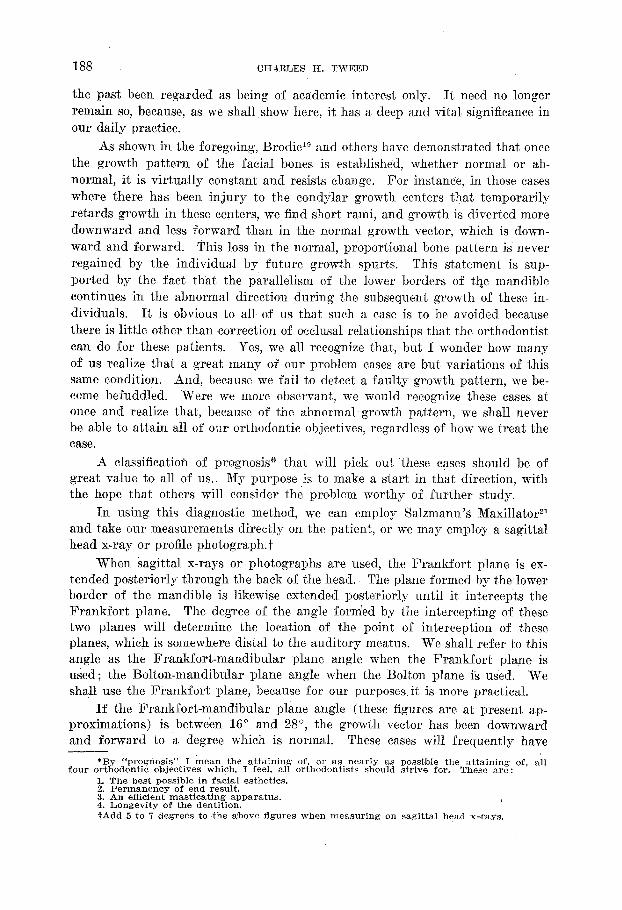
normal occlusion or an osseous growth pattern of but slight deviation from normal, even though the malocclusion is of a severe nature. The orthodontist can be reasonably certain of a permanent result with excellent facial esthetics if he will reduce the tooth pattern so that it is in keeping with the osseous bulk
The basa obje
Fig. 14.-An excellent face pattern in which the Frankfort-mandibular plane angle is case is a Class II malocclusion complic_ated by a discrepancy between tooth pattern
tl bone. If this discrepancy is corrected, there is no reason why all four of the orthodc ctives cannot be attained. Prognosis is excellent.
20”. and
mtic
190 CHARLES H. TWEED
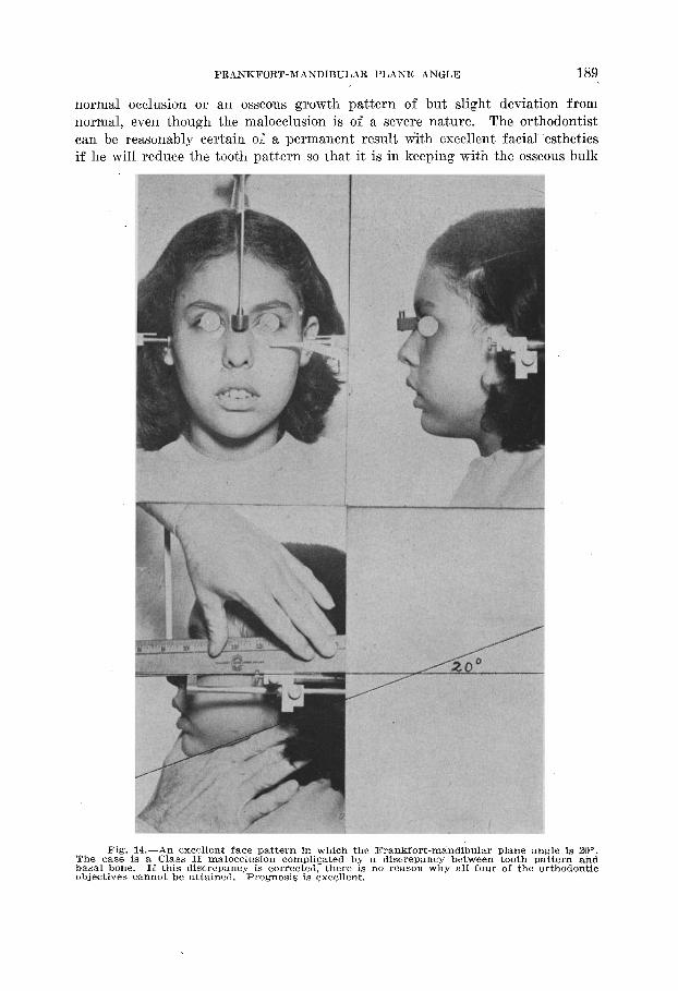
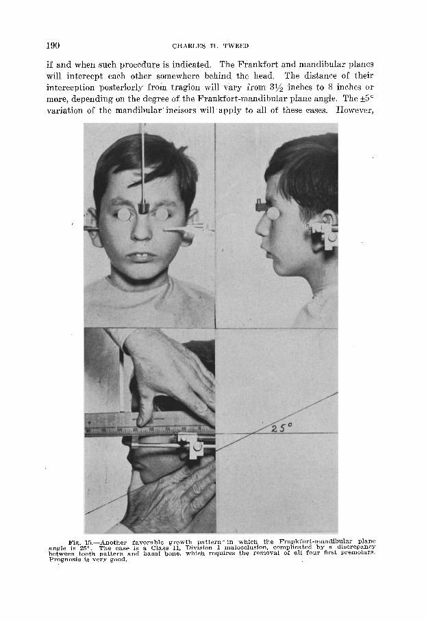
if and when such procedure is indicated. The Frankfort and mandibular planes will intercept each other somewhere behind the head. The distance of their interception posteriorly from tragion will vary from 31/z inches to 8 inches or more, depending on the degree of the Frankfort-mandibular plane angle. The +5” variation of the mandibular incisors will apply to all of these cases. However,
angle betwe PlWgl
Fig. l&-Another favorable growth pattern- in which the FrankfortmandibUlar Pl is 25”. The case is a Class II, Division 1 malocclusion, complicated by a discrepa
!en tooth pattern and basal bone, which requires the removal of all four first PremOl msis is very good,
lane. .ncy ars.
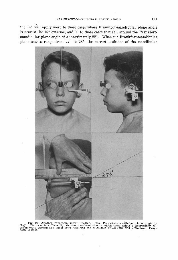
FRANKFORT-MANDIBULAR PLANE ANGLE 191 'I , the +5” will apply more to those cases whose Frankfort-mandibular plane angle is nearest the 16” extreme, and 0” to those cases that fall around the Fran.kfort- mandibular plane angle of approximately 22”. When the Frankfort-mandibular plane angles range from 22” to 28”, the correct positions of the mandibular
Fig. 16.-Another favorable growth pattern. The Frankfort-mandibular plane angle is 271/2”. The case is a Class II, Division 1 malocclusion in which there exists a discrepancy be- tween tooth pattern and basal bone requiring the extraction of all four first premolars. Prog- nosis is good.
192 CHARLES H. TWEED
incisors will vary from 0” when the Frankfort-mandibular plane angle is 22”, to -5” when that angle increases to 28”. My opinion is that about 60 per cent of all malocclusions will fall within a 16” to 28” range of the Frankfort-mandibu- lar plane angle when measurements are taken from profile photographs. More than half of these cases will require reduction of tooth pattern if proportions approximating the normal are to be realized.
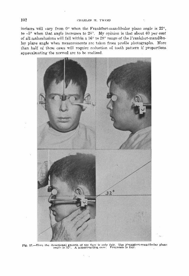
Fig. 17.-Here the directional growth of the face iS only fair. The Frankfort-mandibular plane angle is 32”. A nonextraction case. Prognosis is fair.
men the stan
and
FRANKFORT-MANDIBULAR PLANE ASGLE* 193 ., ,.
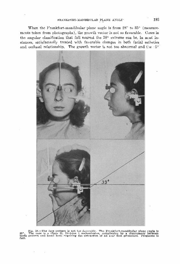
When the Frankfort-mandibular plane angle is from 28” to 35” (measure- tts taken from photographs), the growth vector is not so favorable. Cases in angular classification that fall nearest the 28” extreme can be, in most in- Lees, satisfactorily treated with favorable changes in both facial esthetics occlusal relationship. The growth vector is not too abnormal and the +5”‘
33”. tooth fair.
Fig. l&--The face pattern is not too favorable. The Frankfort-mandibular plane angle is The case is a Class II, Division 1 malocclusion, complicated by a discrepancy between
, pattern and basal bone requiring the extraction of all four first premolars. Prognosis is
194 CHARLES H. TWEED
formula can be applied, but it will be found that the inclination of the mandibu- lar incisors must fall near the -5” extreme. Prognosis is good. A larger per- centage of these cases will require extraction of teeth, as procumbent tendencies are more pronounced. When the Frankfort-mandibular plane angle approaches
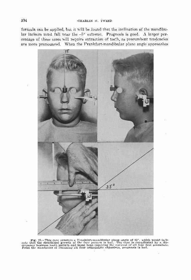
Fig. lg.-This case presents a Frankfort-mandibular plane angle of 35”. which would indi- cate that the directional growth of the face pattern is bad. The case is complicated by a dis- crepancy between tooth pattern and basal bone requiring the removal of all four first premolars. From the standpoint of obtaining all four orthodontic objectives, prognosis is bad.
FRANKFORT-MANDIBULAR PLANE ANGLE 1%
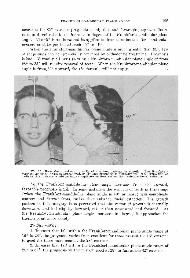
nearer to the 35” extreme, prognosis is only fair, and favorable prognosis dimin- ishes in direct ratio to the increase in degree of the Frankfort-mandibular plane angle. The +5” formula cannot be applied to these cases because the man~dibular incisors must be positioned from -5” to -10”.
When the Frankfort-mandibular plane angle is much greater tha,n 35”, few of these cases can be appreciably benefited by orthodontic treatment. Prognosis is bad. Virtually all cases showin g a Frankfort-mandibular plane angle of from 28” to 35” will require removal of teeth. When the Frankfort-mandibular plane angle is from 30” upward, the +5” formula will not apply. *
Fig. 20.-Here the directional growth of the face pattern is chaotic. The Frankfort- mandibular plane angle is approximately 41” and prognosis is virtually nil. The extraction of teeth in this instance would perhaps complicate matters rather than enhance facial esthetics.
As the Frankfort-mandibular plane angle increases from 35” upward, favorable prognosis is nil. In some instances the removal of teeth in this range (when the Frankfort-mandibular plane angle is 40” or more) will complicate matters and detract from, rather than enhance, facial esthetics. The growth pattern in this category is so perverted that the vector of growth is virtually downward and but slightly forward, rather than downward and forward. As the Frankfort-mandibular plane angle increases in degree, it approaches the tragion point more closely.
1. In cases that fall within the Frankfort-mandibular plane angle range of 16” to 28”, the prognosis varies from excellent for those nearest the 16” extreme to good for those cases nearest the 28” extreme.
2. In cases that fall within the Frankfort-mandibular plane angle range of 28” to 32”, the prognosis will vary from good at 28” to fair at the 32" extreme.
196 CHARLES H. TWEED
32”
35” as 4
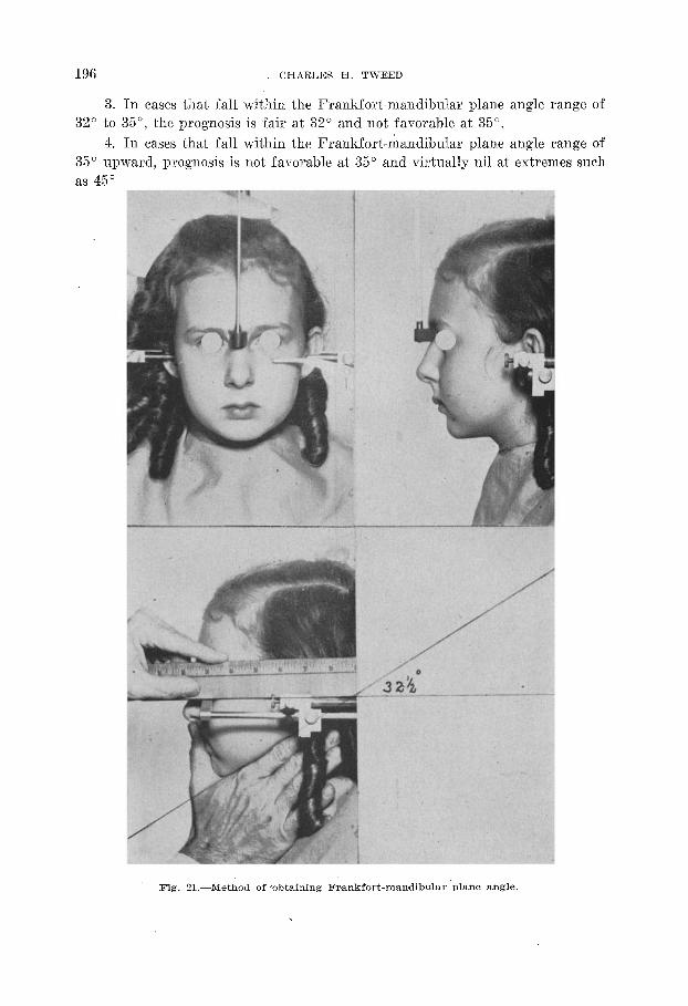
3. In cases that fall within the Frankfort-mandibular plane angle range of to 35”, the prognosis is fair at 32” and not favorable at 35”. 4. In cases tha,t fall within the Frankfort-mandibular plane angle range of upward, prognosis is not favorable at 35” and virtually nil at extremes such
cio to 55”.
Fig. 21.-Method of ‘obtaining Frankfort-man,dibular plane angle.
FRANKFORT-MANDIBULAR PLAKE ASGLE 197
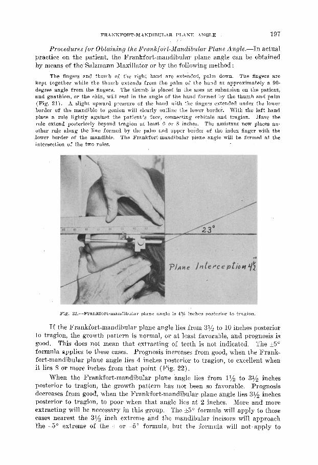
Procedures for Obtaining the FranTcfo,rt-rMa?zdibular Plane Angle.-In actual practice on the patient, the Frankfort-mandibular plane angle can be obtained by means of the Salzmann Maxillator or by the following method :
The fingers and thumb of the right hand are extended, palm down. The fingers are kept together while the thumb extends from the palm of the hand at approximately a 90- degree angle from the fingers. The thumb is placed in the area at subnasion on the patient, and gnathion, or the chin, will rest in the angle of the hand formed by the thumb and palm (Fig. 21). A slight upward pressure of the hand with the fingers extended under the lower border of the mandible to gonion will clearly outline the lower border. With the left hand place a rule lightly against the patient’s face, connecting orbitale and tragion. Have the rule extend posteriorly beyond tragion at least 6 or 8 inches. The assistant now places an- other rule along the line formed by the palm and upper boider of the index finger with the lower border of the mandible. The Frankfort-mandibular plane angle will be formed at the intersection of the two rules.
F’ig. 22.-Frankfort-mandibular plane angle is 41/z inches posterior to tragion.
If the Frankfort-mandibular plane angle lies from 3192 to 10 inches posterior to tragion, the growth pattern is normal, or at least favorable, and prognosis is good. This does not mean that extracting of teeth is not indicated, The +5” formula applies to these cases. Prognosis increases from good, when the Frank- fort-mandibular plane angle lies 4 inches posterior to tragion, to excellent when it lies 8 or more inche.s from that point (Fig. 22).
When the Frankfort-mandibular plane angle lies from 11/2 to 31/ inches posterior to tragion, the growth pattern has not been so favorable. Prognosis decreases from good, when the Frankfort-mandibular plane angle lies 39” inches posterior to tragion, to poor when that angle lies at 2 inches. More and more extracting will be necessary in this group. The i5” formula. will apply to those cases nearest the 314 inch extreme and the mandibular incisors will approach the -5” extreme of the + or -5” formula, but the formula will not apply to
198 CHARLES H. TWEED
those cases nearest the 11/z inch extreme. It becomes quite apparent, because of , the angular variation of the lower border of the mandible, that it would be much
better and more simple to relate the inclinations of the mandibular incisors to either the Frankfort or Bolton plane rather than the lower border of the man- dible. The reason is that the 15” formula can be applied to the lower border only when the face pattern is normal or nearly so.
When the Frankfort-mandibular plane angle falls closer than 11/2 inches posterior to tragion, the growth pattern is so abnormal that there is little the orthodontist can do for these sled-runners and Class 111’s.
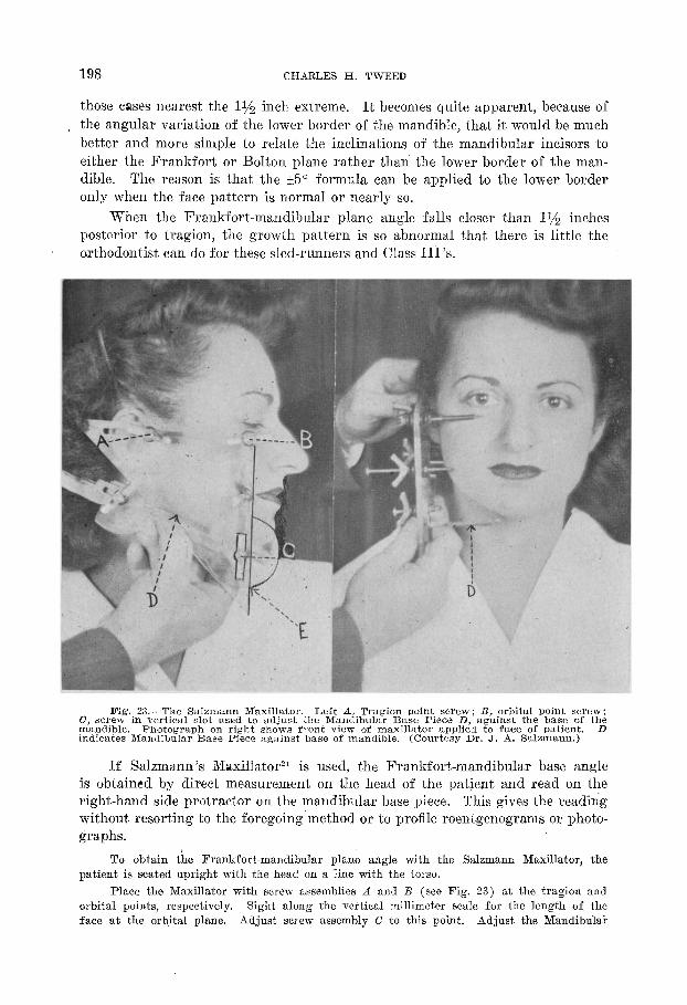
Fig. 23.-The Salzmann Maxillator. Left A, Tragion point screw; B, orbital point screw; 0, screw in vertical slot used to adjust the Mandibular Base Piece D, against the base of th; mandible. Photograph on right shows front view of maxillator applied to face of Patient. indicates Mandlbulnr Base Piece against base of mandible. (Courtesy Dr. J. A. Salzmann.)
If Salzmann’s iNaxillator21 is used, the Frankfort-mandibular base angle is obtained by direct measurement on the head of the patient and read on the right-hand side protractor on the mandibular base piece. This gives the reading without resorting to the foregoin g‘method or to profile roentgenograms or photo- graphs.
To obtain the Frankfort-mandibular plane angle with the Salzmann Maxillator, the patient is seated upright with the head on a line with the torso.
Place the Maxillator with screw assemblies A and B (see Fig. 23) at the tragion and orbital points, respectively. Sight along the vertical millimeter scale for the length of the face at the orbital plane. Adjust screw assembly C to this point. Adjust the Mandibula,E
FRANKFORT-MANDIBULAR PLANE 4NGLE 199
Base Piece D so that it lies flat and is pressed against the lower border of the mandible, while screw assemblies A and B are maintained at their respective points on the face, indicating the Frankfort plane.
The Frankfort-mandibular plane angle is the angle formed by the Frankfort plane and the plane tangent to the base of the body of the mandible and is shown on the Maxillator on the protractor at the bottom of the circumference of the circle on the right side of the Mandibular Base Piece, E, where the latter is crossed by the edge of the vertical B-inch side of the Maxillator.
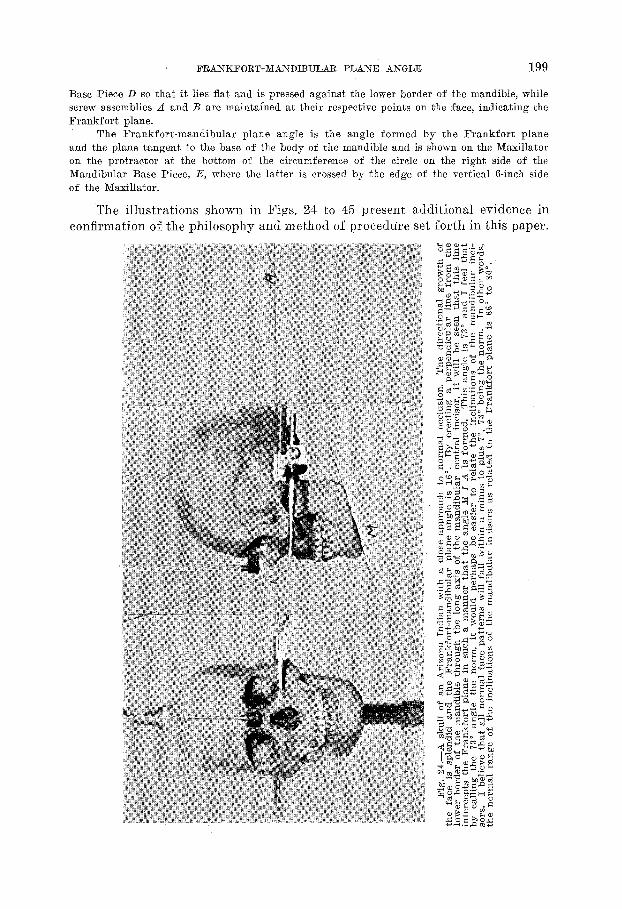
The illustrations shown in Figs. 24 to 45 present additional evidence in confirmation of the philosophy and method of proceddre set forth in this paper.
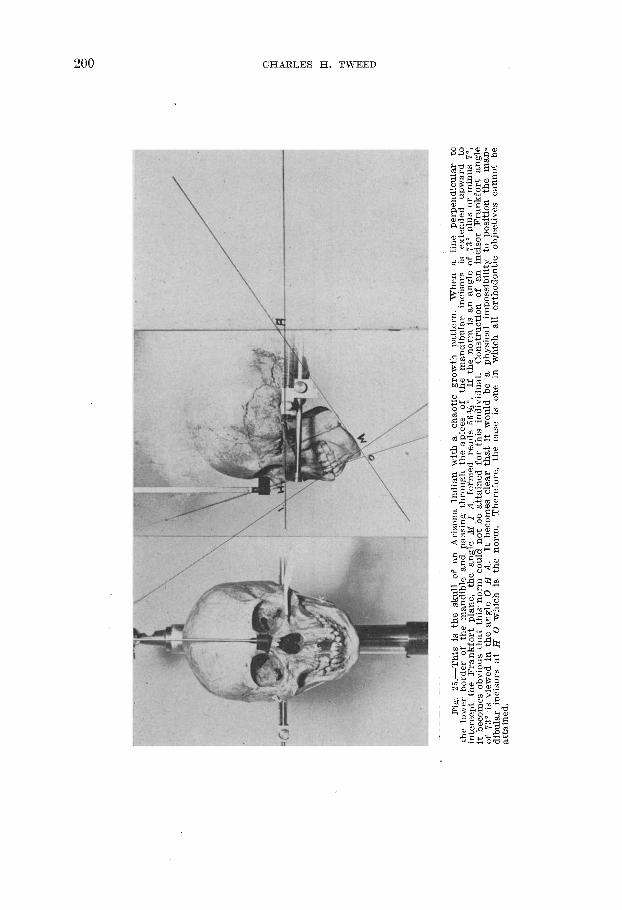
Fig.
25
.-This
is
the
skull
of
an
Ar
izona
ln
dian
wi
th
a ch
aotic
gr
owth
pa
ttern
. W
hen
a lin
e pe
rpen
dicu
lar
to
the
lowe
r bo
rder
of
th
e m
andi
ble
and
pass
ing
thro
ugh
the
apice
s of
th
e m
andi
bula
r inc
isors
is
exte
nded
up
ward
to
in
terc
ept
the
Fran
kfort
plan
e,
the
angl
e M
I
A
form
ed
read
s 56
%“.
If th
e no
rm
is an
an
gle
of
73”
plus
or
m
inus
7”
, it
beco
mes
ob
vious
th
at
this‘
no
rm
coul
d no
t be
at
tain
ed
for
this
indi
vidua
l. Co
nstru
ction
of
an
inc
isor
Fran
kfort
angl
e of
73
” is
viewe
d in
th
e an
gle
0 H
A.
It
beco
mes
cle
ar
that
it
woul
d be
a
phys
ical
impo
ssib
ility
to
posit
ion
the
man
- di
bula
r inc
isors
at
H
0 wh
ich
is th
e no
rm.
Ther
efor
e,
the
case
is
one
in
which
al
l or
thod
ontic
ob
jectiv
es
cann
ot
be
atta
ined
.
FRANKFORT-MISNDIBULliR PLANE ASGLE 201
CHARLES H. TWEED
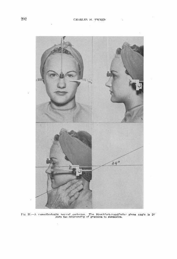
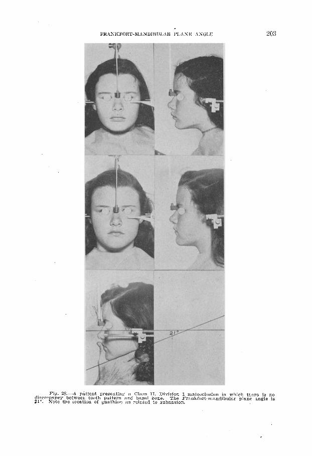
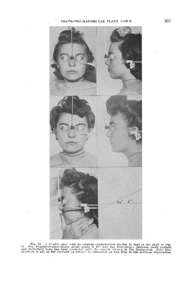
Fig. 27.--A nonorthodontic normal occlusion. The Frankfort-mandibular plane angle is 24’ Note the relationship of gnathion to subnasion.
FRANKFORT-MANDIBULAR PLAXE ASGLE 203
Fig. 26.-A patient presenting a Class II, Division 1 malocclusion in which there is no discrepancy between tooth pattern and basal bone. 21”.
The Frankfort-mandibular plane angle is Note the location of gnathion as related to subnasion.
204 CHARLES H. TWEED
205
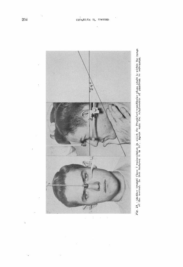
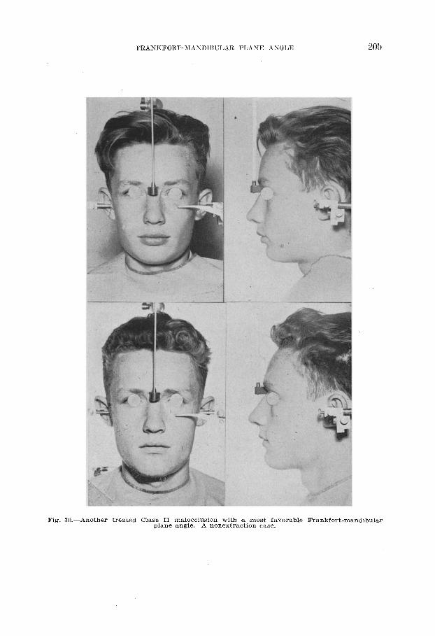
Fig. -Another treated Class II malocclusion with a most favorable Frankfort-ma plane angle. A nonextraction case.
.ndib
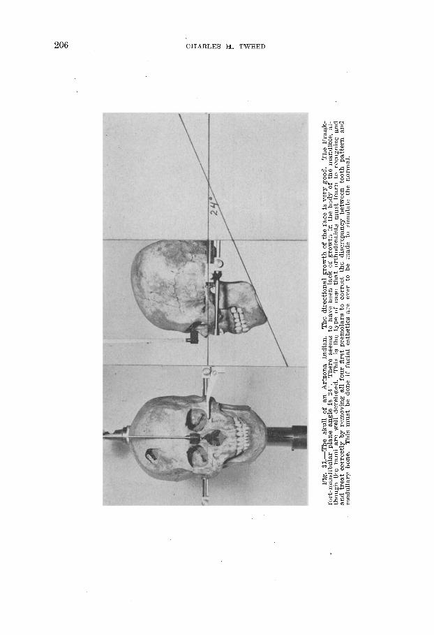
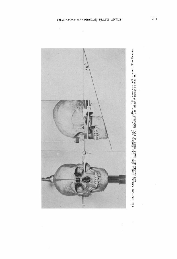
Fig.
tl.
-The
sk
ull
of
an
Arizo
na
Indi
an.
The
dire
ctio
nal
grow
th
of
the
face
is
very
good
. Th
e Fr
ank-
fort-
man
dibu
lar
plan
e an
gle
is 24
”. Th
ere
seem
s to
ha
ve
been
lac
k of
gr
owth
in
th
e bo
dy
of
the
man
dibl
e,
al-
thou
gh
the
ram
i ar
e we
ll ,d
evel
oped
. Th
is is
the
type
of
case
th
at
orth
odon
tists
m
ust
lear
n to
re
cogn
ize
and
and
treat
co
rrectl
y by
re
mov
ing
all
four
fir
st pr
emol
ars
to
corre
ct th
e dis
crep
ancy
be
twee
n to
oth
patte
rn
and
med
ulla
ry
bone
. Th
is m
ust
be
done
if
facia
l es
thet
ics
are
ever
to
be
m
ade
to
simul
ate
the
norm
al.
FRAXITFORT-MANHBULAR PLANE ASGLE 207
Fig. 32.-A treated case with an osseous configuration similar to that of the skull in The Frankfort-mandibular plane angle is 24” and the discrepancy between tooth pal medullary bone has been corrected with the results viewed in the- illustration. Note
thion is not as far forward as related to subnasion as was true in the previous illustra
Fig. .tern that tion.
208 CHARLES H. TWEED
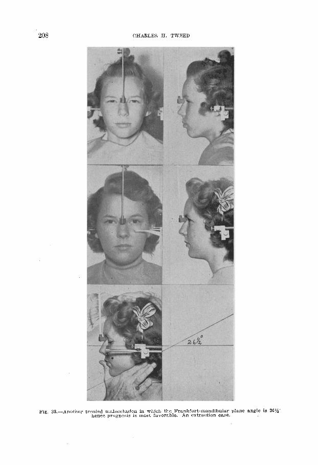
Fig. 33.-Another treated malocclusion in which the Frankfort-mandibular plane angle is %I$$ herice prognosis is most favorable. An extraction case.
FRAXKFORT-MANDIBULAR PLXXE ASGLE
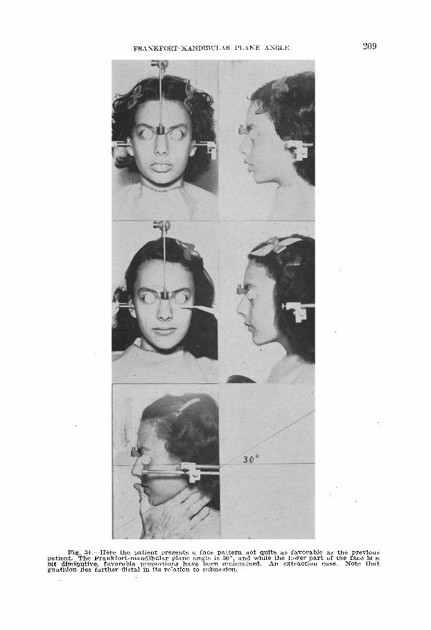
Fig. 34 .-Here the patient presents a face pattern not quite as favorabl Cent. The and while the lower par
dimiputix Frankfort-mandibular plane angle is 30”!
ie, favorable proportions have been mamtained. An extraction athion lies farther distal in its relation to subnasion.
e as the ,t of the c a.23 3. P
Gous is a that
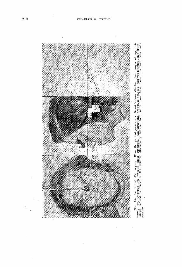
Fig.
35
.-An
orth
odon
tic
trage
dy.
mat
ely
19”.
Here
th
e pa
tient
pr
esen
ts a
Fran
kfor
t-man
dibu
lar
plan
e an
gle
of
appr
oxi-
Gna
thio
n lie
s dir
ectly
un
der
subn
asio
n.
The
dire
ctio
nal
grow
th
is be
autif
ul
and
yet
beca
use
som
e or
tho-
do
ntist
re
fuse
d to
re
cogn
ize
the
exist
ing
disqr
epan
cy
betw
een
toot
h pa
ttern
an
d ba
sal
bone
, th
is re
sult
was
mad
e po
ssib
le.
FRANKFORT-MANDIBULAR BLASE ANGLE 211
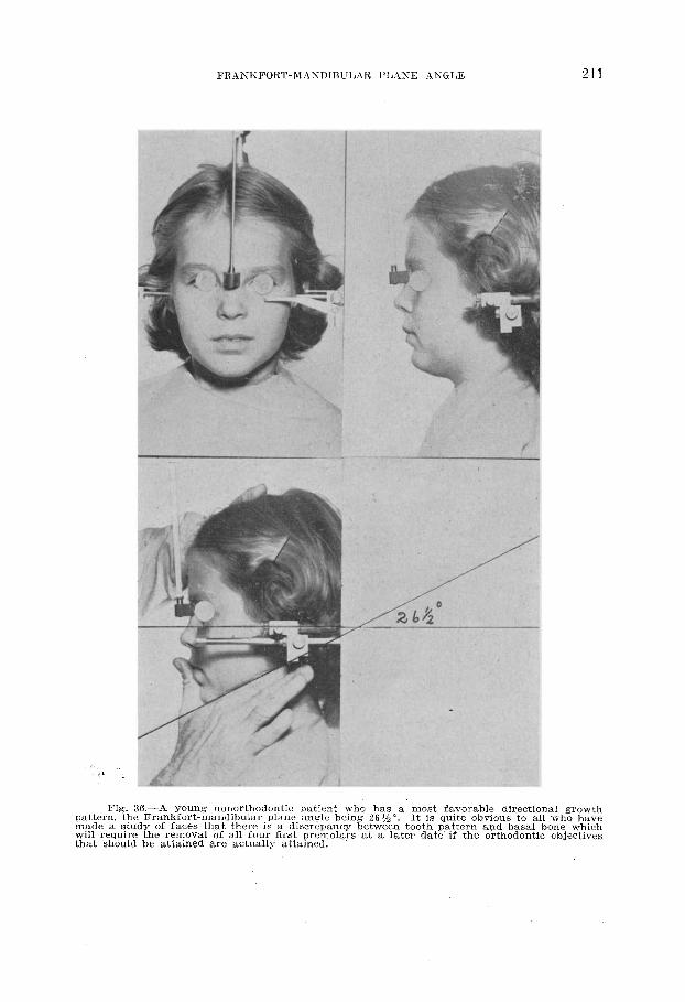
oatter:,i made a will req that she
g. 36.-A young nonorthodontic patient who has a most favorable directional g :rowth the Frankfort-mandibular plane angle being 26 Y2”. It is quite obvious to all whc ) have study of faces that there is a discrepancy between tooth pattern and basal bone which
uire the removal of all four first ~remolars at a later date if the orthodontic objt xtives mid be a.ttained we actually attained.
212 CHARLES Ii. TWEED
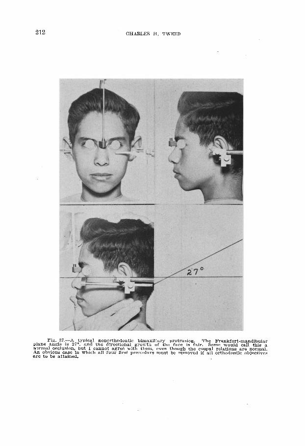
Fig. 37.-A typical nonorthodontic bimaxillary protrusion. plane angle is Z?“,
The Frankfort-mandibular and the directional growth of the face is fair. Some would call this a
nxmal occlusion, but I cannot agre6 with them, even though the cuspal relations are normal. An obvious case in which all four first premolars must be removed if all orthodontic objectives are to be attained.

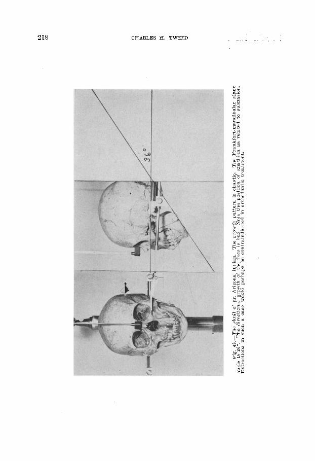
Pig.
38
.-A
skull
of
an
Ar
izona
In
dian
. Th
e gr
owth
ve
ctor
is to
o m
uch
down
ward
an
d no
t en
ough
fo
r- wa
rd.
The
Fran
kfor
t-man
dibu
lar
plan
e an
gle
is 32
”. No
te
that
gn
athi
on
lies
a bi
t po
sterio
r to
su
bnas
ion.
Vi
r- tu
ally
all
case
s pr
esen
ting
the
abov
e sy
mpt
oms
*ill
requ
ire
the
rem
oval
of
al
l fo
ur
first
wem
olar
s.
Whe
n th
e Fr
ankf
ort-m
andi
bula
r pl
ane
angl
e ra
nges
fro
m
28
to
32”,
or
to
the
degr
ee
seen
in
th
is sk
ull,
the
-f5”
form
ula
will
not
appl
y.
Thes
e ca
ses
can
be
grea
tly
bene
fited
by
or
thod
ontic
tre
atm
ent,
but
they
ca
n by
no
m
eans
be
co
nside
red
norm
al
beca
use
ther
e ha
s be
en
a fa
ult
in
grow
th
that
ha
s al
tere
d th
e no
rmal
gr
owth
pa
ttern
.
214 CHARLES H. TWEED
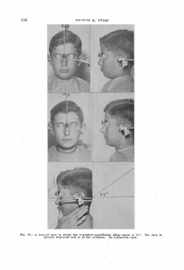
Fig. 39.- -A tr .ed case in which the Frankfort-mandibular plane angle is :reatly improved and so is the occlusion. An extraction case
34”. The face is
FRANKFORT-MANDIBULAR PLANE ANGLE 215
patte angle
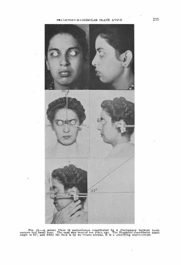
Fig. 40.-A severe Class II malocclusion complicated by a discrepancy r;,ar&io basal bone. The case, was treated ten years ago., The l3++fort-ma
, and while the face IS by no means normal, It IS a gralxfymg nnpl
between tooth mdibular plane -0vement.
216 CHARLES H. TWEED
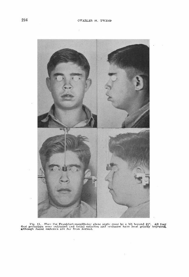
Fig. 41.-Here the Frankfort-mandibular plane angle must be a bit beyond 35”. All four flrst premolars were extracted and facial esthetics and occlusion have been greatly improved, although facial esthetics are far from normal.
’ FRASKFORT-K4SDIBULAR PLANE ANGLE 217
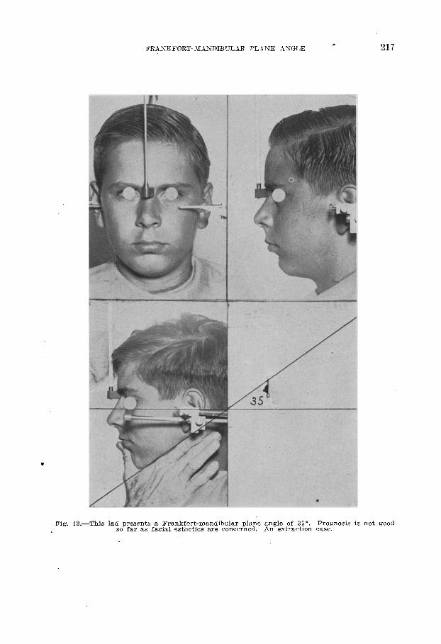
Fig. 42.-This lad presents a Frankfort-mandibular plane angle of 35”. Prognosis is not good /? so far as facial esthetics are concerned. An extraction case.
CHARLES H. TWEED _ _.. . -
FRANKFORT-MANDIBULAR PLANE ANGLE 219
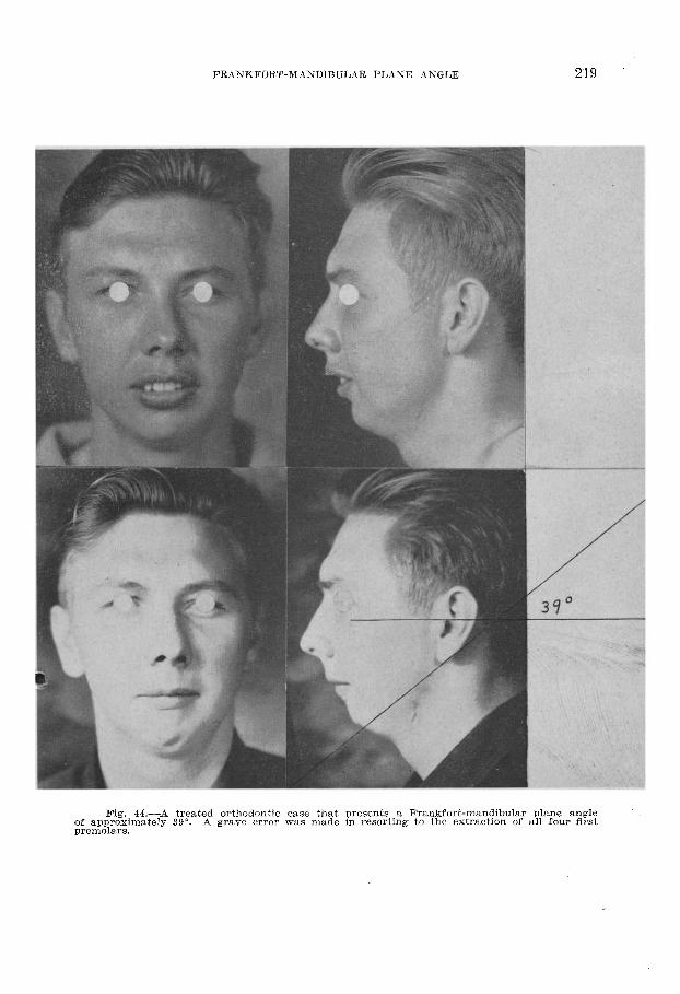
Fig. 44-A treated orthodontic case that presents a Frankfort-mandibular plane angle of approximately 39”. A grave error was made in resortin, e to the extraction of all four first premolars.
220 CHARLES H. TWEED
FRANKFORT-l\ilANDIBULAR PLANE ANGLE 221
I wish to ac.knowledge with thanks the assistance of Dr. J. A. Salzmann in the prepara- tion of this paper.
1.
2.
3.
4. 3.
6.
7.
8. 9.
1’0.
11.
12.
13.
14.
15.
16.
17.
18.
19.
20.
21.
REFERENCES
Angle, Edward H.: Malocclusion of the Teeth, ed. 7, 19Oi, S. S. White Dental1 Manu- facturing Co.
Tweed, Charles H.: The Application of -the Principles of the Edgewise Arch in the Treatment of Malocclusion.
Tweed, Charles H.: I, II, Angle Orthodontist 11: 5, 12, 1941.
Indications for the Extraction of Teeth in Orthodontic Pro- cedure, Ant. J. OKTHOI>ONTICS ANU 0~~1;SURG. 30: 405, 1944.
Broadbent, B. Holly: The Face of the Normal Child, Angle Orthodontist 7: 183, 1937. Brodie, Allan G.: On the Growth Pattern of the Human Head From the Third Month
to the Eighth Year of Life, Am. J. Anat. 68: 209, 1941. Margolis, Herbert I.: A Plastic and Graphic Technique for Recording Dental ‘Changes
and Facial Growth, Anf. J. ORTHODONTICS AND ORAL SURG. 25: 1027, 1939; Stand- ardized X-Ray Cephalographics, Ibid. 26: 725, 1940. See also Salzmann, J. A.: Principles of Orthodontics, Philadelphia, 1943, J. B. Lippincott Co., pp, 9697.
Margolis, Herbert I. : The dxial Inclination of the Mandibular Incisors, AM. J. ORTHODONTICS AP;D ORBL SURG. 29: 571,1943.
Margolis, Herbert I. : Unpublished material. (See Discussion.) Salzmann, J. 9.: The Rationale of Extraction as an Adjunct to Orthodontic Mechano-
therapy and the Sequelae of Extraction in the Absence of Orthodontic Guidance, AM. J. ORTHODONTICS AND ORAL SURG. 31: 181, 1945; Principles of Orthodontics, Philadelphia, 1943, J. B. Lippincott Co., pp. 320-385.
&hour, I., and Massler, Maury: The Growth. Pattern of Human Teeth. Part 1, J. Am. Dent. A. 27: 1778, 1940; Part 2, J. Am. Dent. A. 27: 1918, 1940.
&hour, I. : Rate of Growth of Alveolar Bone Measured by Alizarine Injections, J. Dent. Research 15: 329, 1936; The Tooth as an Index of the Constitutional Pat- tern of the Child, Proc. Third Bien. Meeting, Society Res. Child Dev. Nat. Res. Council, 109-110, 1939.
Salzmann, J. A.: Principles of Orthodontics, Philadelphia, 1943, J. B. Lippincott Co., pp. 26-28.
Boadbent, B. Holly: The Orthodontic Value of Studies in Facial Growth, Physical and Mental Adolescent Growth, Proc. of Conf. on Ado]. Cleveland, 37-39, 1930; Ontogenetic Development of Occlusion,, In: Development of Occlusion, Phil- adelphia, 1941, University of Pennsylvanra Press, pp. 31-48.
Gregory, William K.: The Evolution of Dental Occlusion From Fish to Man, In: De- velopment of Occlusion, Philadelphia, 1941, University of Pennsylvania Press, pp. l-30.
Gregory, William K.: The Origin and Evolution of the Human Dentition, Baltimore, 1922, Williams & Wilkins Co.
Brodie, Allan G.: Some Recent Observatiorrs on the Growth of the Mandible, Angle Orthodontist 10: 63, 1940.
&hour, I., Hoffman, M. M., Sarnat, B. G., and Engel, M. B.: Vital Staining of Grow- ing Bones and Teeth With Alizarine Red S, J. Dent. Research 20: 411, 1941; Brash, James C.: The Growth of the Jaws, Normal and Abnormal in Health and Disease, London, 1924, The Dental Board of the United Kingdom.
Brodie, Allan G.: On the Growth of the Jaws and the Eruption-of the Teeth, Angle Orthodontist 12: 102, 1942.
Brodie, Allan G.: Some Recent Observations on the Growth of the Face and Their Implications to the Orthodontist, Alt. J. ORTHODO~VTICS AND 0~~1, SURG. 26: 741, 1940.
Broadbent, B. Holly: Ontogenic Development of Occlusion, Angle Orthodontist 11: 223. 1941.
Salzmann J. A.: The Maxillator: A New Instrument for Measuring the Frankfort- Man&bular Base Angle, the Incisor-Mandibular Base Angle, and Other Com- ponent Parts of the Face and Jaws, AM. J. ORTHODONTICS AND ORAL SURG. 31: 608, 1945.
222 CHARLES H. TWEED
DISCUSSIOIS
Dv. Herbert Margolis, Boston, Mass.-1 consider it a privilege to be asked to discuss Dr. Tweed’s latest contribution to orthodontics. However, I am not unmindful of my inability to perform adequately the task assigned.
The paper can be divided into two parts: first, a -,fuller explanation of the funda- mental principles underlying the positioning of the mandibular incisors as related to diag- nosis and planning of treatment; second, a projection into the field of prognosis ‘as re- lated to orthodontic treatment.
Dr. Tweed suggests that when-research and clinical practice are more closely wedded, t,hcn orthodontics will pass out of the realm of necromancy, the horizon will be enlarged; and the service rendered will be not only improved but also greatly increased. This union is of vital concern to the clinician and is steeped in, and correlated with, objective scien- tific investigation. Although often mentioned, it is unfortunately not common practice in orthodonties.
I should like’to discuss first the orientation of the mandibular incisors. It is a mat- ter of record that during a display of a number of case reports by Dr. Tweed some years ago, the editor of the Angle Orthodontist called that display the Snest clinical demonstra- tion he had ever seen. However, always the objeetive clinician, Dr. Tweed was dissatisfied with his results; and he changed, rather drastically, his whole method of procedure. It appears that two things influenced this change: first, the facial contours of some patients were not improved as a result of treatment, -but rather were more prognathous; and, sec- ond, in some other cases the teeth again became irregular after retention. In the first case, teeth were aligned but the face distorted; in the other, the teeth were crowded but the face remained normal.
Dr. Tweed made the discovery for which all orthodontists and patients should be everlastingly grateful : that, in the course of so-called orthodox treatment, very often the mandibular incisors were tipped forward. The result was either a prognathous face or crowded teeth. There remained the convenient excuse of pressure from the third molars; and then there was the more remote refuge of heredity or the endocrines. If the third molars were congenitally absent or were removed surgically, and if the internist gave a negative report, then we blamed the resultant failure on type.
The fact remained, however, that these incisors had been tipped forward during treat- ment. They were more procumbent after treatment than before treatment. Dr. Tweed was, indeed, not alone in accomplishing this; every orthodontist in the country was bliss- fully pushing these teeth forward. Published case reports indicate that. Scientific records, only a few years ago, of treated cases also indicated just that. Many of ,these cases were then considered successful by authorities in this specialty. When Dr. Tweed began teach- ing the uprighting of the mandibular incisors, from a clinical standpoint, he was censured by some rather severely; chiefly, because he did not express the uprighting in an accepted scientific term; and, also, because in the opinion of some critics, he had considered facial contour from a single preconceived ideal. From my investigations, I found that what Dr. Tweed was doing in uprighting the incisors was in direct harmony with evolutionary trends in the development of man and that tipping these teeth forward by the orthodontist is, in my opinion, ‘ ‘ evolution in reverse. ”
The resultant failure had nothing to do with remote causes in many cases.. We orthodontists were responsible and it will not serve the profession to deny the responsi- bility. Many orthodontists have since accepted that principle of “uprighting” the man- dibular incisors, or at least of not increasing their procumbency.
In the evolution of man there is an increase in the size of the brain, associated with a reduction of the snout. All available material indicates that during the process of the reduction of the lower third of the face, the incisors have been rotated lingually, on a horizontal axis, with the apex of the incisor at or near to the fulcrum. The clinical results demonstrated by Dr. Tweed are in direct harmony with that scientific fact.
FRA4NKFORT-MANDIBULAR PLANE ANGLE 223
There is another anatomic correlation with the uprighting of the mandibular incisors not previously mentioned that I should like to suggest at this time: starting with thg anthropoid, we find prominent supraorbital ridges with a receding frontal bone associated with procumbent mandibular incisors. From the standpoint of mechanical advantage, that is admirably efficient. Because of the lack of vertical forehead in the ape, these heavy supraorbital ridges serve as buttresses to withstand the impact of occlusion (Fig. 1). In modern man there is an increase,in the parietal areas of the brain. Also the fron%al bones become more upright and a forehead is developed. The supraorbital ridges of the ape have disappeared in man. Likewise, the mandibular incisors have become more upright.
Fig. 1. Fig. 2.
Fig. l.-“One of ou? nearest living relatives. Female Chimpanzee and Young.” (From A4Znsost nz~nzcvn by R. M. Perkes; The Century Co.)
Note prognathous mouth, large supraorbital ridges, receding frontal bone.
Fig. 2-c Ma% awxZ His Forerz~waers by Buttel-Reepier.) Note the correlation of procumbent incisors with the supraorbital ridges in the mature
gorilla. These ridges serve as buttresses for the massive jaws and procumbent teeth. With
the uprighting of the incisors in modern man, there is an uprighting of the frontal bone. The baby gorilla (left) is more nearly like modern man.
With the recession of the snout and the uprighting of the incisors, what is ther’e then in the cranium to withstand impact of occlusion in the anterior segment in man? Exami- nation of the mechanics involved would indicate that the ridges are no longer necessary. The vertically elongated forehead, due to the uprighting of the frontal bone, serves beau- tifully as a mechanical buttress for the stress of occlusion for the lower third of the face reduced by the uprighting of the mandibular incisors. (Fig. 2.)
I should like to make this point clearer in the following manner:
If a vertical blow were directed at the lower edge of a board, inclined at an angle of 45 degrees or more, a horizontal beam or some reinforcement would be necessary to resist the stress. However, if the impact were in direct line with the board, so that edge strength were utilized, no reinforcement would be necessary. Likewise, vve all know how
CHARLES H. TWEED
much more resistant to bending a rectangular piece of wire is on its larger dimension than on its lesser dimension. Therefore, in the evolution of man, concomitant with an increase in the size of the brain, there is an uprighting of the frontal bones, creating the forehead or the lofty brow and the consequent disappearance of the supraorhital ridges. Together with the uprighting of the frontal bone there is decrease in the tooth bearing structure, an uprighting of the mandibular incisors, and the development of a chin following the loss of the simian shelf. All this is an impressive demonstration of functional adaptation.
In orthodontic therapy, with the advent of intermaxillary elastics, the orthodontist has often thrown out of harmony one of those forces when he increased the angle of incli- nation of the mandibular incisors, increasing the facial prognathism. It is one thing for a child to have a prognathous face normally; it is quite another matter for the orthodontist to create one. (Fig. 3.)
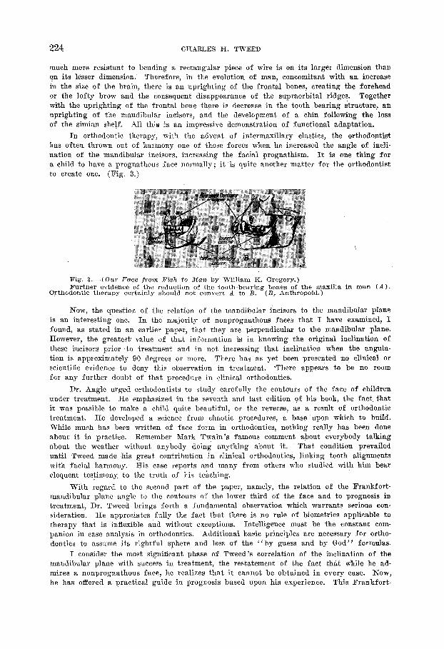
Fig. 3.-( Our Face fro?% Fish to Man by William K. Gregory.) Further evidence of the reduction of the tooth-bearing bones of the maxilla in man (A).
Orthodontic therapy certainly should not convert A to B. (I?, Anthropoid.)
Now, the question of the relation of the mandibular incisors to the mandibular plane is an interesting one. In the majority of nonprognathous faces that I have examined, I found, as stated in an earlier paper, that they are perpendicular to the mandibular plane. However, the greatest value of that information is in knowing the original inclination of these incisors prior to treatment and in not increasing that inclination when the angula- tion is approximately 90 degrees or more. There has as yet been presented no clinical or scientific evidence to deny this observation in treatment. -There appears to be no room for any further doubt of that procedure in clinical orthodontics.
Dr. Angle urged orthodontists to study carefully the contours of the face of children under treatment. He emphasized in the seventh and last edition of his book, the fact that it was possible to make a child quite beautiful, or the reverse, as a result of orthodontic treatment. He developed a science from chaotic procedures, a base upon which to build. While much has been written of face form in orthodontics, nothing really has been done about it in practice. Remember Mark Twain’s famous comment about everybody taking about the weather without anybody doing anything about it. That condition prevailed until Tweed made his great contribution in clinical orthodontics, linking tooth alignments with facial harmony. His case reports and many from others who studied with him bear eloquent testimony. to the truth of his teaching.
With regard to the second part of the paper, namely, the relation of the Frankfort- mandibular plane angle to the contours of the lower third of the face and to prognosis in treatment, Dr. Tweed brings forth a fundamental observation which warrants serious con- sideration. He appreciates fully the fact that there is no rule of biometrics applicable to therapy that is inflexible and without exceptions. Intelligence must be the constant com- panion in case analysis in orthodontics. Additional basic principles are necessary for ortho- dontics to assume its rightful sphere and less of the “by guess and by God” formulas.
I consider the most significant phase of Tweed’s correlation of the inclination of the mandibular plane with success in treatment, the restatement of the fact that while he ad- mires a nonprognathous face, he realizes that it cannot be obtained in every case. Now, he has offered a practical guide in prognosis based upon his experience. This Frankfort-
FRANKFORT-MASDIBULAR PLANE ANGLE 225
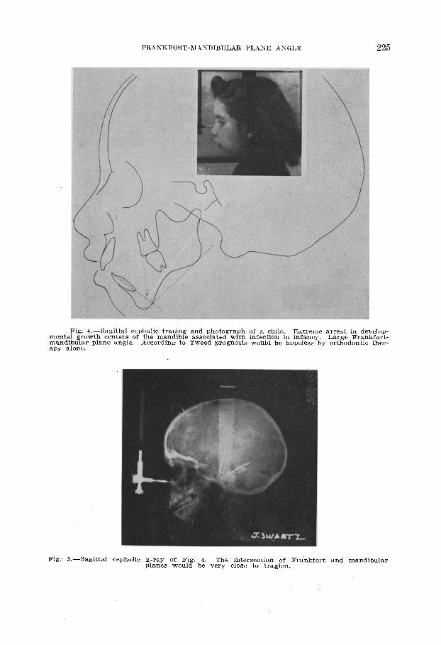
Fig. 4.-Sagittal cephalic tracing and photograph of a child. Extreme arrest in develop- mental growth centers of the mandible associated with infection in infancy. mandibular plane angle.
Large Frankfert-
apy alone. According to Tweed prognosis would be hopeless by orthodontic ther-
Fig: 5.-Sagittal cephalic x-ray of Fig. 4. The intersection of Frankfort and mandibular planes would be very close to tragion.
226 CHARLES H. TWEED
mandibular plane angle will serve as a guide and a red light when discussing prognosis, prior to starting treatment. Obviously, it is much healthier for all concerned to have that knowl- edge available before undertaking a case than to apologize after treatment.
That further elaboration of this correlative factor is necessary is quite apparent to Tweed, who suggests in this paper that he hopes others will investigate his study based upon their own clinical experience. He has provided another important germinal seed for use in practice.
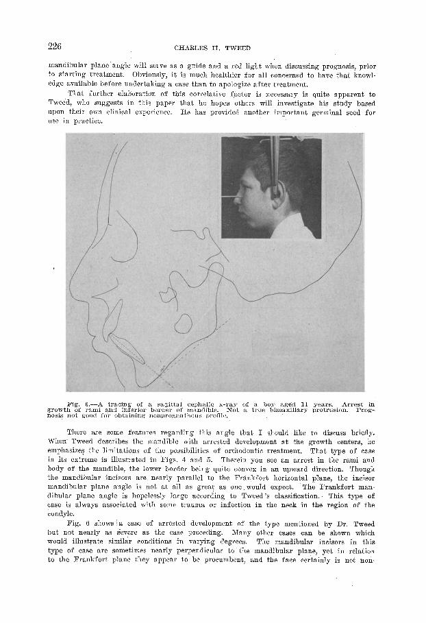
Fig. 6.-A tracing of a. sagittal cephalic x-ray of a boy aged 11 years. Arrest in ,growth of rami and inferior border of mandible. Not a true bimaxillary protrusion. Prog- nosis not good for obtaining nonprognathous profile.
There are some features regarding this angle that I should like to discuss briefly. When Tweed describes the mandible with arrested development at the growth centers, he emphasizes the limitations of the possibilities of orthodontic treatment. in its extreme is illustrated in Figs. 4 and 5.
That type of case Therein you see an arrest in the rami and
body of the mandible, the lower border being quite convex in an upward direction. Though the mandibular incisors are nearly parallel to the Frankfort horizontal plane, the incisor mandibular plane angle is not at all as great as one.would expect. The Frankfort man- dibular plane angle is hopelessly large according to Tweed’s classification. This type of case is always associated with some trauma or infection in the neck in the region of the condyle.
Fig. G shows ia case of arrested development of the type mentioned by Dr. Tweed but not nearly as severe as the case preceding. Many other cases can be shown which would illustrate similar conditions in varying degrees. The mandibular incisors in this type of case are sometimes nearly perpendicular to the mandibular plane, yet in relation to the Frankfort plane they appear to be procumbent, and the face certainly is not non-
FRASKFORT-MASDIBULAR PLANE ANGLE 227
prognathous. I believe this type of case has been wrongly classified as bimaxillary pro- trusion. It would appear, in this paradoxical situation, that here indeed is a condition where the correlation of incisor mandibular plane angle is entirely wrong, for the man- dibular incisors are sometimes like those of a sheep, and the face is certainly not non- prognathous.
Wherein lies the error of interpretation?
The inclination of the incisors in those cases may very well be correct as related to the mandible; the deformity, homever, lies mithin the mandible itself. Tipping back the incisors, in that type of ease, is not the solution, for they may be correct or nearly so in incli- nation. If that patient is to obtain a normal face, it would be necessary to lengthen the body of the mandible, mostly at the border, and to lengthen the ascending rami. An orthodontic appliance can never do that. This condition, though extreme, demonstrates the necessity for consideration of the entire anatomic field with which the orthodontist is concerned, not simply the teeth and alveoli.
A. B.
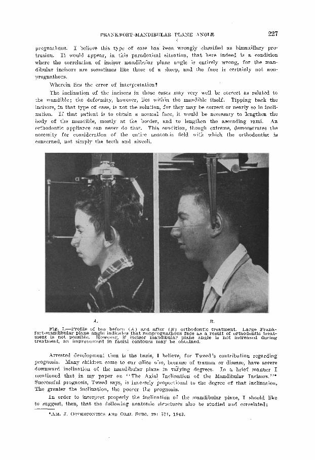
Fig. 7.-Profile of boy before (a) and after (B) orthodontic treatment. Large Frank- fort-mandibular plane angle indicates that nonprognathous face as a result of orthodont,ic treat- merit is not possible. However, if incisor mandibular plane angle is not increased during treatment, an improvement in facial contours may be obtained.
Arrested development then is the basis, I believe, for Tweed’s contribution regarding prognosis. Many children come to our office who, because of trauma or disease, have severe domnward inclination of the mandibular plane in vaTying degrees. In a brief manner I mentioned that in my paper on C ‘The Axial Inclination of the Mandibular Incisors.“* Successful prognosis, Tweed says, is inversely proportional to the degree of that inclination, The greater the inclination, the poorer the prognosis.
In order to interpret properly the inclination of the mandibular plane, I should like to suggest, then, that the following anatomic structures also be studied and correlated:
“AM. J. ORTHODONTICS AND ORAL SURG. 29: 571, 1943.
228 CHARLES H. TWEED
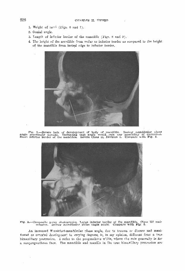
1. Height of rami (Figs. G and 7).
2. Genial angle. 3. Length of inferior border of the mandible (Figs. 8 and 9).
4. The height of the mandible from molar to inferior border as compared to the height of the mandible from in&al edge to inferior border.
Fig. X.-Severe lack of development of body of mandible. Incisor mandibular plane angle practically normal. Increasing that angle would ruin any possibility of correction. Short inferior border of the mandible. Severe Class II, Division 1. Compare with Fig. 9.
Fig. 9.-Composite x-ray photographs. Large inferior border of the mandible. Class III mal- oclusion. Incisor mandibular plane angle acute. Compare with Fig. 8.
an increased Frankfort-mandibular plane angle, due to trauma or disease and mani- fested as arrested development in varying degrees, is, in my opinion, different from a true bimaxillary protrusion. I refer to the prognathous white, where the rule generally is for a nonprognathous face. The mandible and maxilla in the true bimaxillary protrusion are
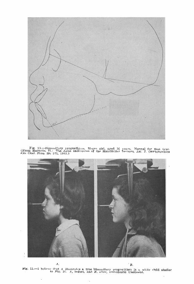
Fig. lO.-Bimaxillary lxvgnathism. Negro girl, aged 25 years. Normal for that type. (From Margolis, H.: The Axial Inclination of the Mandibular Incisors, AM. J. ORTHODONTICS
AND ORAL SURG. 29: 571, 1943.)
A. B.
Fig. 11.-I believ;othF:t A illustrates a true bimaxillary progoathism in a while child similar g. 10. A, before, and B, after, orthodontic treatment.
230 CHARLES H. TWEED
well developed and proportional. The teeth related to the mandibular plane are procumbent as compared to the nonprognathous white, and they are stable. (Fig. 10.)
A true bimaxillary prognathous condition in a white, I think, may be termed “delayed evolution’ ’ or L L incomplete evolution. ’ ’
Double protrusion, when the result of tipping the mandibular incisors forward, is differ- ent from a true bimaxillary prognathism in that the former is one created by the ortho- dontist-the face of the child before treatment being nonprognathous; in the latter, the true bimaxillary, the child presents normal cuspal relationship of the teeth but a prognath- ous face. The former is i (evolution in reverse” the latter, perhaps, i I delayed evolution. ” Examples of “delayed evolution” may be seen in the retention in man of the platysma myoides muscle so vital to the horse to chase off flies, but of little importance to man. The appendix in the rabbit is of great functional value but as yet little proof has been shown of its value to man. And so the prognathous white is also an evidence of incomplete or “.delayed evolution. ” Whether orthodontists should treat that type of case has already been answered by Tweed. Fig. 11 is an illustration. of the facial changes in the profile of my concept of a true bimaxillary protrusion which I have treated.
May I close with my expression of gratitude for the privilege of reading and dis- cussing Dr. Tweed’s paper. The study clubs named in his honor are thankful that the Lord has spared him from his recent severe illness. We ark proud that even then he car- ried on, and we pray he may be with us for many years to enjoy the rewards he so strongly merits and so constantly avoids.