twenty-second annual progress report interactive graphics
TRANSCRIPT
Twenty-second Annual Progress Report
Interactive Graphics For Molecular
Studies /'
TR96-055 1996
William V. Wright Russell M. Taylor II
Frederick Brooks, Jr.
Department of Computer Science University of North Carolina at Chapel Hill
Chapel Hill, NC 27599-3175
National Center For Research Resources GrantRR02170-13, National Institutes of Health UNC is an Equal Opportunity/ Affirmative Action Institution.
DEPARTMENT OF HEALTH AND HUMAN SERVICES PUBLIC HEALTH SERVICE
NATIONAL INSTITUTES OF HEALTH
NATIONAL CENTER FOR RESEARCH RESOURCES BIOMEDICAL RESEARCH TECHNOLOGY PROGRAM
ANNUAL PROGRESS REPORT
l. PHS GRk'-iTNUlviBER: P41RR02170-l3
2. NAME OF RECIPIENT INSTITUTION: L'NIVERSITY OF NORTH CAROLINA
3. HEALTH PROFESSIONAL SCHOOL: DEP ARTlvlENT OF COfviPUTER SCIENCE
4. REPORTING PERIOD:
A. FROM MAY 1, 1995
B. TO APRIL 30, 1996
5. PRINCIPAL INVESTIGATOR:
A. FREDERICK P. BROOKS, JR.
B. TITLE: KENANrl3f~s:pR OF COlv!P
C. SIGNATl:JRE: \J ~j ~ 6. DATE SIGI\i"ED: MARCH 1, 1996
7. TELEPHO!'<"E !\'UMBER: (919) 962-1931 t
8. FACSIMILE ]',1JMBER: (919) 962-1799
9. ELECTRONIC MAIL ADDRESS: [email protected]
PART II. NARRATIVE DESCRIPTION
II.A. SUMMARY OF RESEARCH PROGRESS
GRANT NUMBER: P41 RR02170-13
REPORT PD: 5/1/95-4/30/96
We harness computer and graphics technology to help biochemists understand the structure and function of life molecules and to help biologists image and manipulate biological microstructures. Understanding macro-molecular behavior is crucial for understanding disease and for designing drugs.
Objectives for Molecular Graphics: Our ambitious goal for the 1990's decade is a unified 3-D meter-cubed virtual model with good physical behavior. Over the past year we have refined this goal to accommodate two users working side-by-side on a shared molecular model.
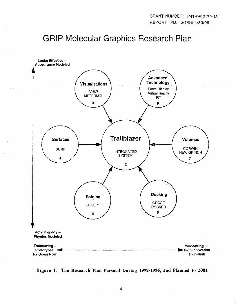
This goal has many technical components. Our strategy has been to pursue independent subprojects which would be separately useful and will come together to compose our goal molecular system. Each subproject has one or two graduate students. Figure I summarizes our molecular graphics research plan for the coming years, showing the central Trailblazer system and the component technology subsysteras:
• real time molecule docking, folding, and other interactions. • better ways to visualize electron density maps and other 3-D scalar functions. • better ways to visualize solvent-accessible and other surface representations of molecules. • new molecular visualizations. exploiting virtual environments technology. • exploring both the technical means for force display and its molecular structure uses. • experiments with advanced graphics technology, to adapt it to molecular problems.
Controlled manipulation with scanning probe microscopes: In addition to our program: in molecular graphics, we have continued our thrust into probe microscopy. Two of the students work this area; one investigating enhanced force feedback (friction, adhesion, direct Z control of the tip) and the other working closely with the collaborators to improve the user interface (providing precise measurement tools, direct control over lighting and viewing parameters, and enhanced visualizations such as contour lines and display of multiple data sets).
At the sarue time, the Nanomanipulator subproject has received funding through the NSF to support four more students and to provide a complete graphics and force-feedback system in the physics lab. This funding will provide a PixeiFlow graphics system to allow exploration of advanced shading techniques and vastly improved graphics performance over present systeras. This Pixe!Flow system (which uses the same OpenGL graphics language as SGI equipment) will be available for use by the molecular graphics projects.
We met our objective of controlled dissection of viruses, using the Nanomanipulator to derive the mechanical properties of individual Tobacco Mosaic Virus particles. Our immediate future goals for the project are to:
• Shift to Adenovirus particles for adhesion studies, to gain insight into their function as vectors in gene therapy.
• Record and display multiple simultaneous data sets during an experhnent (adhesion, compliance, conductance, lateral force).
2

GRANT NUMBER: P41RR02170-13
REPORT PD: 5/1/95-4/30/96
• Build a custom-designed nanoWorkbench (NSF funding), with registered visual and force display, and ergonomics appropriate for the application.
The accomplishments and plans for all our subprojects are presented in Part ill of this report. Four notable accomplishments of the Resource are highlighted in the next section.
3
GRANT NUMBER: P41RR02170-13
REPORT PO: 5/1/95-4/30/96
GRIP Molecular Graphics Research Plan
Looks Effective -Appearance Modeled
Surfaces
SURF
Acts Properly -Physics Modeled
VIFm METERMOL
Folding
SCULPT
Trailblazer
INTEGRATED SYSTEM
Force Display Virtual Reality
PIT
Docking
GROPE DOCKER
Volumes
CORWIN NEWGR!NCH
Trailblazing- Wildcatting -Prototypes .... ---------------------I ... High Innovation
for Users Now High·Risk
Figure 1. The Research Plan Pursued During 1992·1996, and Planned to 2001
4
II.B. IDGHLIGHTS
GRANT NUMBER: P41 RR02170-13
REPORT PD: 5/1/95-4/30/96
1. A Virtual-reality interface for a scanning probe microscope
Tapping/contact mode. In order to provide the widest range of forces between imaging and modification modes, and in order to better quantify the forces used during modification, we have developed Tapping/contact mode operation. This places the AFM in non-contact tapping mode for imaging and feeling the surface, then switches it into contact mode for modification. Tapping mode is a standard microscope technique that oscillates the tip at a high frequency (around 100 kHz) and measures differences in amplimde to determine surface height. In this mode, the tip lightly taps the surface, rather than being pulled across it. This has allowed us to non-destructively image such delicate samples as virus particles, while still being able to push them around. To-do this, we have augmented the feedback circuitry of the instrument so that the computer can switch its feedback on the fly.
Acquiring multiple data sets. Using the newly-acquired Topometrix SPM, we have acquired force curves for each point on a scan, from which we have extracted the adhesion between the tip and sample at each point on the surface. Adhesion information from a charged tip may allow us to map the surface charges of virus particles, providing information about their structure. The same force-curve data contains information about the compliance of the surface, and the instrument is capable of producing lateral force and conductance information at each location on the surface. We have added a laser to onr existing SPM, which means that we can acquire near-field optical (NFOM) information, mapping the transmissivity of the surface at well below the wavelength of light. These data sets give us additional information about the surface and tip-surface interactions.
Improved data display. Onr data display has gone forward on two fronts: providing more quantitative information and displaying multiple data sets overlaid on topography. To provide indications of relative heights and slopes, we have added constant-height contour lines to the surface. We have also added a tool that extracts cross-sections through the data along paths drawn by the user. This tool allows the user to do measurements along the centerline of a bent TMV particle, for example. Additional data sets {NFOM and adhesion so far) can be mapped to color and friction. Color is useful for displaying scanned information, while friction provides feedback while the user is feeling the surface. We intend to further explore the display of multiple data sets using Pixe!Flow, as described in the next section.
Move to common hardware. The Nanomanipulator system was initially developed using the Pixel-Planes 5 graphics supercomputer, the Argonne-ill Remote Manipulator arm and an STM that was built at UCLA with custom electronics and control hardware designed by the project. None of these systems were available at other lahs, but were part of our advanced hardware as a wildcatting development site. Over the past two years, we have ported the system to also run on common offthe-shelf hardware. The system now runs on SGI graphics platforms (Indigo2 Maximum Impact or Onyx) and the PHANToM force-feedback device from SensAble Devices. We have begun the process of modifying the control software for Topometrix SPMs so that we can use their hardware out of the box with our software, and we have reached the point where we can scan the surface and select areas of interest. We expect to have the system fully ported by May 1996.
Pili Fibers. Katrina Forest from Scripps Institute visited our site, bringing with her samples of Pili fibers. These fibers are the grappling hooks that bacteria use to attach themselves to cells, so measurements of their adhesive properties and understanding of their structure is of direct relevance to understanding their function. During the half-day initial experiment, we were able to scan the fibers at high resolution, feel the fibers, and do gross modification.
Adeno virus. Forrest Ferrari, a graduate student under Jude Samulski at the UNC center for Gene Therapy, has been using the Nanomanipu!ator system to examine Adenovirus particles.
5
GRANT NUMBER: P41RR02170-13
REPORT PD: 5/1/95-4/30/96
These soccer ball-shaped particles can be used as containers for genetic material, and are used as vectors to infuse the material into cells. We have scanned samples under both TEM and SPM to compare sizes, and have been able to controllably position and probe the virus. In one experiment, we studied the adhesion of the particles to each other by pressing them together and then using the tip to pry them apart. In another, we pressed the tip on top of a particle until we forced a dimple; as we scanned, the dimple slowly disappeared. Understanding the adhesion between particles and the different proteins found on the surface of cells may reveal how and where the virus particles enter the cell to do their work.
2. Additional funding for the Nanomanipulator
The Nanomanipulator has received three grants from the National Science Foundation to explore different areas of system development. This means that the GRIP Research Resource support of the project can be focused on supporting students to work with outside scientists who come to use the system. This section describes the goals of the work that is funded outside the Resource that will add system capabilities that may be used by visiting scientists and local collaborators alike.
Complete system in the lab. An Academic Research Initiative grant has enabled us to purchase a Topometrix SPM and a PHi\ .. 'IT oM force-feedback device. This equipment and an SGI Onyx with an RE2 graphics engine, purchased with this NIH grant, have been placed in the SPM lab in the Physics deparunent as a complete, full-time experiment station. This allows unrestricted and unshared access to the dedicated Nanomanipulator set-up by the project. It also puts the equipment right in the laboratory where sample and tip preparation occurs. The grant will also allow us to upgrade to the next generation of graphics hardware on the Onyx in the corning year.
Pixe!Flow. While we are accelerating experiments on the existing system, an HPCC grant supports the continued investigation of leading-edge hardware and software techniques on the Pix elF! ow graphics engine. Pixe!Flow will allow us to increase the number of displayed surface data points from 80x80 to more than 256x256, allowing the detection of both fme details and large-scale features in the same image. This will be important for the study of biological samples, where comparison of surface features on several different particles can help reveal differences in orientation and size. In addition to improved display speed, Pixe!Flow will also offer programmable shading. This will provide us with several more channels on which to display multiple data sets, including surface roughness, noise texture, density of local pits and translucent overlays. It will also enable exploration of more realistic lighting effects, including Phong shading (or even bi-directional reflectance shading) and shadows. Pixe!Flow's scalability and flexibility will allow us to trade these capabilities against each other to determine which are most important to understanding surfaces.
nano Workbench. The HPCC grant will also support the development of a custom-designed workbench for nanomanipulation. An up-projected stereo image coincident in space with the PHAt'IToM force display will provide a surface that can be seen and felt. A separate control monitor will provide control over system, data, and display parameters and will allow quantitative measurements on data crosssections. A two-axis stylus will provide direct and natural control over light direction. This station, together with the virtual tip technology we are developing, will provide a workbench much like that of a woodcarver where the scientist can interact directly with a surface that looks and feels real.
Remote operation. PixelFlow and the nanoWorkbench will be located in the graphics laboratory connected to the microscope through a fiber-optic network. We will also explore connections to more remote sites with Topometrix microscopes. The most likely places for such remote connections are Topometrix labs and HP's new basic science laboratory. Such connections will require us to face and solve problems that relate to all remote visualization systems where the scientist interacts with either remote instrumentation or remote simulation. This would allow a scientist visiting here to use our new interface to connect to and operate the microscope in their own lab, with sample and tip prepared by their own students.
6
GRANT NUMBER: P41RR02170-13
REPORT PO: 5/1/95-4/30/96
3. CORWIN ·· Reciprocal-View crystallographic system
The goal of this project is the near-real-time construction of 3D contour maps from x-ray crystallographic data to represent electron density distributions in protein crystals. This system will enable a user to change his model of a protein structure and observe immediately the effect of his change on various types of hybrid maps which are based on both the current model and crystallographic data. The project name, CORWIN, derives from coupled reciprocal windows and reflects a system feature that enables it to represent crystallographic data simultaneously in both the real-space and frequency domains.
Over the past year researeh assistant Jim Van Verth has made a number of improvements to this . system incorporating suggestions from several potential users, our Advisory Committee, and the NIH committee that reviewed our competitive proposal for the next five years. Among many improvements the system now handles the crystal space groups most commonly found for proteins and is extensible to all space groups. The model modifications available to the user have been extended, and the system performance has been substantially improved.
We have begun developing a community of users both academic and commercial, and we are currently installing copies of the system in the departments of biochemistry both on our campus and at Duke University. We are continuing to enhance the system with new tools for manipulation of models and new representations of the crystallographic data.
4. PIT --Protein Interaction Theater
The PIT is a computer graphics workplace especially designed for two collaborating users who are studying a three-dimensional structure. Both users will see a stereo image of the structure. The motion of each user's head will be tracked, allowing the image to be modified to give the illusion of a structure fixed in space. Both users will see the images as being in the same position and orientation in laboratory space, and as a result, they will be able to supplement their conversation with hand gestures to the shared image. The users will scale, position, and orient the image so that they can see and "touch" the elements of current interest. Input devices will enable the users to interact with the model of the structure to select elements and to deform the structure. Some of these devices will also display the forces generated by the molecule models. Each user will have a separate text screen and keyboard with which to access and edit shared and separate documents. Although we will test the design on molecular applications, we expect it to have a wider range of usefulness.
PART III. DESCRIPTION OF PROGRAM ACTIVIDES
III.A. SCIENTIFIC SUBPROJECTS
In this section, we identify 14 subprojects, give the requested facts and a brief report on each, and summarize this data for the entire project.
7
SCIENTIFIC SUBPROJECT
BRTPUNIT: T
GRANT NUMBER: P41 RR02170·13
REPORT PD: 5/1/95·4/30/96
TITLE: Nanomanipulator - Interactive Scanning Probe Microscopes
KEYWORDS: scanning probe microscope, atomic force microscope, teleoperation, virtual environment, scientific visualization
AXIS 1: 9 11
AXIS II:
lNVESTi: DEGREEi: DEPTi: NONHOST1:
INVEST2: DEGREE2: DEPT2: NONHOST2:
INVEST3: DEGREES: DEPT3: NONHOST3:
INVEST4: DEGREE4: DEPT4: NONHOST4:
INVESTS: DEGREES: DEPTS: NONHOSTS:
% BRTP $:
42 63 70
Taylor, Russell M. Ph.D. Computer Science
Brooks, Frederick P. Ph.D. Computer Science
Washburn, Sean Ph.D. Physics & Astronomy
Superfine, Richard Ph.D. Physics & Astronomy
Chi, Vernon L BS .. Computer Science
2
ABSTRACT: Towards the ideal user interface for scanning probe microscopes. We have continued to Improve both system capabilities and user interface. In order to provide the widest range of forces between imaging and modification modes, we have developed Tapping/contact mode operation. This places the AFM in non-contact tapping mode for imaging and feeling the surface, then switches It into contact mode for modification.
We have incorporated multiple Input data sets (near-field optical data, surface-tip adhesion data) as additional inputs that are registered with topography. These additional data sets are displayed as surface color when scanning and surface friction when feeling. We will incorporate lateral force, compliance and surface conductance as additional input channels; surface texture, roughness, stickiness, firmness, and sound as additional output channels. Our goal is to provide many simultaneous non-conflicting channels of information from the instrument to the user, In real time during modification and exploration experiments.
8
SCIENTIFIC SUBPROJECT GRANT NUMBER: P41RR02170·13
REPORT PD: 5/1/95-4/30/96
We have ported the system from high-end, specially-designed hardware onto commonlyavailable devices, building a complete Nanomanipulator system In the SPM lab from off-the· shelf components. At the same time, we continue to explore using the advanced capabilities of state-of-the-art hardware, maintaining remote connections to the laboratory over a high-speed network. This meets our goal as a trailblazing facility that explores the most useful future techniques on today's most advanced hardware, developing a system which becomes affordable for widespread use In several years. Close, daily collaboration between computer scientists as tool builders and physical scientists as tool users ensures that the right tools are built, and that the tools become available outside our facility. The system has been presented at both computer science and physics conferences, describing both the user interface work and the science done using the system. Reports published in the proceedings and international speaking engagements spread word of the system usefulness to the broader scientific community.
9
•
SCIENTIF-1C SUBPROJECT
BRTPUNIT: C
TITLE: Quantum Device Fabrication
GRANT NUMBER: P41RR02170·13
REPORT PD: 5/1/95·4/30/96
KEYWORDS: scanning-tunneling microscope, quantum interference, solid-state device
AXIS I: 28 (physics)
AXIS II:
INVEST1: DEGREE1: DEPT1: NONHOST1:
INVEST2: DEGREE2: DEPT2: NONHOST2:
% BRTP $:
92 (surface science}
Washburn, Sean Ph.D. Physics & Astronomy
Taylor, Russell M. Ph.D. Computer Science
1 5
ABSTRACT: Fabricating Quantum Devices. Devices fabricated at a scale small enough to respond to quantum-mechanical effects are of interest not only for developing smaller and faster computers, but also for advanced sensors. These devices are exquisitely sensitive to small fluctuations in voltage potential and magnetic field strength, such as those produced by cells and organisms. We are Investigating techniques for fabricating and testing such tiny devices, in order to understand which device characteristics are critical to function and which can be compromised to allow ease of fabrication.
In the previous year, we used the Nanomanipulator to form 30-nm gaps in gold wire and then repair the gaps, restoring conductivity. We maneuvered a small gold colloid into such a gap (a task that we feel could not have been done without the force feedback present in the Nanomanipulator system). We made .external contact to the wire, measuring the resistance changes during manipulation.
This past year has been spent preparing technique and gathering equipment !or further studies. One difficulty we encountered was with the large amount of gold filrn we had to remove from our 5 micron-wide wires In order to bring them down to a small enough scale. The material would form large clumps around the site, blocking access to the tip and probably causing a large capacitance at the gap. We have found at IBM a source of 1 OOnm wires, which are much better starting templates for our device fabrication.
We have also been specifying and ordering measurement equipment that will be capable of determining the impedance of gaps in the wire without inducing voltages large enough to damage the structure. Once we have a small gap, we will place the device In our liquid helium Dewar and do electron transport measurements. We also plan to Insert colloids In the gaps and perform tunneling spectroscopy on them.
10
SCIENTIFIC SUBPROJECT
BRTP UNIT: C
TITLE: Manipulation of biological particles
GRANT NUMBER: P41RR02170-13
REPORT PO: 5/1/95-4/30/96
KEYWORDS: scanning probe microscope, tobacco mosaic virus, fruit-fly chromosome, adena virus, pili fibers
AXIS 1: 3 7b
AXIS II: 52 59
INVEST1: Henderson, Eric DEGREE1: Ph.D. DEPT1: Zoology and Genetics NONHOST1: Iowa State University
INVEST2: Samulski, R. Jude DEGREE2: Ph.D. DEPT2: Gene Therapy Center NONHOST2:
INVEST3: Forest, Katrina DEGREE3: Ph.D. DEPT3: Molecular Biology NONHOST3: Scripps Research Ins!.
INVEST4: Superfine, Richard DEGREE4: Ph.D. DEPT4: Physics & Astronomy NONHOST4:
% BRTP $: 20
ABSTRACT: We have continued or initiated several collaborations aimed at exploring and manipulating biological samples with the Nanomanipulator. We continue to work with Eric Henderson, who has brought fruit-fly chromosome samples and examined them. He will send a student here with a sample this spring to attempt to cut the chromosomes at user-specified locations.
We have continued our investigations into tobacco-mosaic virus particles. We have experimentally determined parameters for a model that describes the mechanical stiffness and resilience of the particles. We are writing up these results for publication, which will conclude our studies into this particular virus unless another collaborator shows interest in continuing the work.
We have begun collaboration with Forrest Ferrari (a student under Jude Samulski at the UNC Center for Gene Therapy). We have imaged and manipulated adena virus particles; pushing them together and separating them, denting them, and forming clusters. Adena virus particles are used as vectors in gene therapy: they can be filled with genetic material which they deposit inside cells. We will study the adhesion of the particles to different parts of cells in order to better understand how and where they enter the cells.
A new collaboration with Katrina Forest from Scripps Institute is studying pili fibers, which are the fibers used by bacteria to attach themselves to cells. Her group Is studying the
11
SCIENTIFIC SUBPROJECT GRANT NUMBER: P41RR02170-13
REPORT PD: 5/1/95-4/30/96
structure of these fibers, in order to determine the packing order of the components that make up the fibers. Using adhesion mapping with a functionalized tip, we hope to be able to determine the distance between endpoints of these units, thus determining the packing density. We will also study the adhesion of the fibers to each other and to different substrates, using the lateral force measurement capabilities of our new Topometrix AFM.
The ability of the atomic force microscope to image in fluid allows us to examine virus particles in vitro; we are pushing forwards to achieve the ability to do non-contact mode imaging in liquid.
12
SCIENTIFIC SUBPROJECT
BRTPUNIT: T
GRANT NUMBER: P41RR02170-13
REPORT PD: 5/1/95-4/30/96
TITLE: Trailblazer ·- Next-generation Molecular Graphics System
KEYWORDS: software engineering, object-oriented, workbench
AXIS 1: 11
AXIS II: 42
INVEST1: Brooks, Frederick P. .DEGREE1: PhD. DEPT1: Computer Science NONHOST1:
INVEST2: Wright, William V. DEGREE2: Ph.D. DEPT2: Computer Science NONHOST2:
% BRTP $: 1 8
ABSTPACT: This is a continuing subproject to build an infrastructure for supporting our development of new technology and to enable us to unify our efforts so that all appropriate tools we build can be brought to bear on every client's problem. During the previous year, our research assistants Jason Preibe and Kim Jones developed objectoriented software facilities for capturing a user's input and for displaying results. During the past summer and fall terms Kim focused her attention an our library lor controlling manipulator devices which we use lor six-axis input and for displaying force feedback to the user. Her work is described below under the GROPE subproject. Unfortunately, Kim left our department at the end of the fall term and Jason Preibe did not return for the fall term as expected. Consequently we have recruited two new students for this subproject, Jayant Kolhe and Sumedh Barde. They have returned to the development of C++ objects for representing molecular structures and have built a simple system for manipulating a protein structures by rotating the chemical bonds. Currently they are beginning work on new implementation of our DOCKER system using the object-oriented libraries we have been developing.
13
SCIENTIFIC SUBPROJECT
BRTP UNIT: T
TITLE: PIT -- Protein Interaction Theater
KEYWORDS: molecular modeling, virtual reality
AXIS 1: 11
AXIS II: 42
INVEST1: Brooks, Frederick P. DEGREE1: Ph.D. DEPT1: Computer Science NONHOST1:
INVEST2: Wright, William V. DEGREE2: Ph.D. DEPT2: Computer Science NONHOST2:
% BRTP $: 1 2
GRANT NUMBER: P41 RR02170-13
REPORT PO: 5/1/95-4/30/96
ABSTRACT: The PIT is a computer graphics workplace especially designed for two uses who are collaborating in the study of a three-dimensional structure. Both users will see a stereo image of the structure. Each user's head is tracked, and his image is modified to give the illusion of a structure fixed in space. Both users see their images in the same position and orientation laboratory space and thus they can supplement their conversation with hand gestures to this shared image. The image can be scaled and positioned so that the users can see and "touch" all elements of interest. Input devices enable the users to interact with the image: to change its position, scale, and orientation, to select its elements, and to deform the structure. Some of these devices are also able to display forces on parts of the structure generated by an underlying mathematical model. Each user has a separate text screen and keyboard to access and edit shared and separate documents. Although we will test our design of this workplace on molecular applications, we expect it to have a wider range of usefulness.
Research assistant Mike Meehan who joined the project last fall is working on this project. We have purchased a suitable display device, an Ampro projector. The second projector needed to support the second user will be purchased under another grant that will be sharing this facility with the GRIP project. Meehan has demonstrated that we can use this projector to display an image of a molecule that appears to be floating about 18 inches in front of the screen as is required, and he is currently working on facilities for tracking the positions of the users' heads.
14
SCIENTIFIC SUBPROJECT
BRTP UNIT: T
TITLE: CORWIN -- Coupled Reciprocal Windows
GRANT NUMBER: P41 RR02170-13
REPORT PD: 5/1/95-4/30/96
KEYWORDS: Fourier transform, structure factors, contour maps
AXIS 1: 9
AXIS II:
INVEST1: DEGREE1: DEPT1: NONHOST1:
INVEST2: DEGREE2: DEPT2: NONHOST2:
% BRTP $:
42
Brooks, Frederick P. Ph.D. Computer Science
Wright, William V. Ph.D. Computer Science
9
ABSTRACT: This system is the work of our research assistant Jim Van Verth. It is designed to support refinement of molecular models. The goal is the near-real-time construction of 3D contour maps from x-ray crystallographic data to represent electron density distributions in protein crystals. This system will enable a user to change his model of a protein structure and observe immediately the effect of his change on various hybrid maps which are based on both the current model and crystallographic data. Among many improvements added over the past year, the system now handles the crystal space groups most commonly found for proteins and is extendible to all space groups. The model modifications available to the user have been extended as well. In addition to translations, Mr. Van Verth has added facilities for rotating molecular fragments either freely in space or constrained to rotated about the axis of a chemical bond. The system performance has been substantially improved.
The system also supplements the display of maps and models in real space with the simultaneous display of the underlying structure factors in a second window. The project name, CORWIN, derives from "coupled reciprocal windows" and reflects this system feature that enables it to represent crystallographic data in both the real-space and frequency domains. Each structure factor is represented by a spot or a vector located at the hkl indices of the factor. The sizes of these spots or the brightness of the vectors represent the magnitudes of the structure factors. Their colors (and the directions of the vectors) represent the phase angles.
We have begun developing a community of users both academic and commercial, and we are currently installing copies of the system in the departments of biochemistry both on our campus and at Duke University. During the past year we have begun a collaboration with Dr. Charles Carter of the Biochemistry Department on our campus that is described as the. next subproject.
15
SCIENTIFIC SUBPROJECT
BRTP UNIT: C
TITLE: Tryptophanyl !RNA synthetase (TrpRS)
KEYWORDS: enzyme, protein synthesis, antibiotic
AXIS 1: 7a
AXIS II: 6 6
INVEST1: Carter, Charles W., Jr. DEGREE1: Ph.D. DEPT1: Biochemistry NONHOST1:
% BRTP $: 2
GRANT NUMBER: P41RR02170-13
REPORT PO: 5/1/95-4/30/96
ABSTRACT: Tryptophanyl !RNA synthetase (TrpRS) is one of the crucial enzymes that participates in protein synthesis. The structural study of TrpRS (from Bucillus stearothermophilus) may not only illuminate understanding of the fundamental biochemistry of protein synthesis, but also provide a basis for rational drug design (e.g. antibiotic indolmycin).
We have begun working with Dr. Carter and his student Yuhui Yin on the refinement of their model for the structure of this protein.
16
SCIENTIFIC SUBPROJECT
BRTP UNIT: T
GRANT NUMBER: P41 RR02170-13
REPORT PO: 5/1/95-4/30/96
TITLE: NEW GRINCH -- ab-initio interpretation of electron density data
KEYWORDS: critical points, branch points, ridge lines
AXIS 1:9
AXIS II: 42
INVEST1: Brooks, Frederick P. DEGREE1: Ph.D. DEPT1: Computer Science NONHOST1:
JNVEST2: Eberly, David DEGREE2: Ph.D. DEPT2: NONHOST2: SAS, Inc.
INVEST3: Kryger, Gitay DEGREE3: Ph.D. DEPT3: Structural Biology NONHOST3: Weizmann Institute
INVEST 4: Wright, William V. DEGREE4: Ph.D. DEPT4: Computer Science NONHOST4:
% BRTP $: 12
ABSTRACT: In the early 1980s Tom Williams, while a member of the GRIP project, built the GRJNCH system for the ab-initio interpretation of electron density maps. Williams' system was based on work by Drs. Jonathan Greer, Carroll Johnson, and Stanley Swanson for representing electron density by marking the ridges of high local density. At least 50 copies of this system were disseminated to the crystallographic community.
Juraj Horacek of the current GRIP project team is re-examining this ridge line approach to abinitio electron density interpretation using new, more powerful ridge line finding mathematics developed in our department by David Eberly for the analysis of medical images. Our new approach is distinguished from prior work in that we do not simply connect critical points but actually trace the ridges of high density through an interpolated function. This should help us avoid some kinds of common errors in map interpretation. We expect this work to lead to both new representations for electron density and to new methodologies for its interpretation as a molecular structure. We have been consulting with Dr. Gitay Kryger to identify what facilities might be useful in this system.
17
SCIENTIFIC SUBPROJECT
BRTP UNIT: T
GRANT NUMBER: P41 RR02170-13
REPORT PD: 5/1/95-4/30/96
TITLE: GROPE -- Interaction combining force display and visual display
KEYWORDS: teleoperation, force feedback
AXIS 1: 11
AXIS II: 4 2
INVEST1: Brooks, Frederick P. DEGREE1: Ph.D. DEPT1: Computer Science NONHOST1:
INVEST2: Wright, William V. DEGREE2: Ph.D. DEPT2: Computer Science NONHOST2:
% BRTP $: 2
ABSTRACT: The two students who worked on this subproject last year have left the GRIP team. Bill Mark has selected a dissertation topic in another area, and Scott Randolph left the department when he completed his MS degree. Research assistant Kimberly Jones continued the work on this subproject during the past summer and fall terms when she also left the department. Her major accomplishments were to add simulations of friction and surface texture when the input device is moved tangential to a simulated surface.
18
SCIENTIFIC SUBPROJECT
BRTP UNIT: D
GRANT NUMBER: P41RR02170-13
REPORT PO: 5/1/95-4/30/96
TITLE: SCULPT -- Interactive Constrained Manipulation of Molecular Structures
KEYWORDS: molecular conformation, molecular deformation, energy minimization
AXIS!: 9
AXIS II:
INVEST1:. DEGREE1: DEPT1: NONHOST1:
INVEST2: DEGREE2: DEPT2: NONHOST2:
INVEST3: DEGREE3: DEPT3: NONHOST3:
INVEST4: DEGREE4: DEPT4: NONHOST4:
% BRTP $:
42
Brooks, Frederick P. Ph.D. Computer Science
Richardson, David C. Ph.D. Biochemistry Duke University
Richardson, Jane S. Ph.D. Biochemistry Duke University
Surles, Mark C. Ph.D.
Interactive Simulations, Inc.
2
ABSTRACT: The SCULPT system enables a chemist to change molecular conformations in near real time while the bond geometry of the structure is constrained to canonical values and the free-energy is minimized. Dr. Mark Surles, who developed this system as a research assistant on the GRIP project, continued its development as a post doctoral researcher at the San Diego Supercomputer Center, and has now formed a company, Interactive Simulations, Inc., for marketing the system as a commercial product. We have granted him a non-exclusive license to the code developed while a student here freeing him to do this. We are also serving as a beta test site for his product system.
More recently, Drs. Surles and Amitabh Varshney, also a graduate of our department and former research assistant on the GRIP project, have proposed adding Dr. Varshney's algorithms for calculating the solvent accessible surface of a molecule to the SCULPT system. We are negotiating with them to grant them the appropriate license to do this.
19
SCIENTIFIC SUBPROJECT
BRTP UNIT: D
GRANT NUMBER: P41 RR02170-13
REPORT PD: 5/1/95-4/30/96
TITLE: SURF -- Fast calculation of solvent accessible surfaces
KEYWORDS: convex hulls, alpha hulls, parallel algorithms
AXIS 1:9
AXIS II:
INVEST1: DEGREE1: DEPT1: NONHOST1:
INVEST2: DEGREE2: DEPT2: NONHOST2:
% BRTP $:
42
Brooks, Frederick P. Ph.D. Computer Science
Varshney, Amitabh Ph.D. Computer Science State University of New York - Stony Brook
1
ABSTRACT: During the past year Dr. Varshney has published one more paper stemming from his work on the calculation of solvent accessible surfaces while a research assistant on the GRIP project. In addition, he is working with Dr. Mark Surles, as mentioned above, to incorporate his algorithms in Dr. Surles' SCULPT product program.
20
SCIENTIFIC SUBPROJECT
BRTP UNIT: D
GRANT NUMBER: P41 RR02170-13
REPORT PO: 5/1/95-4/30/96
TITLE: VIEW -- Visualization Impromptu Evaluation Workbench
KEYWORDS: Data base, molecular geometry, computer graphics, script languages
AXIS 1:9
AXIS II:
INVEST1: DEGREE1: DEPT1: NONHOST1:
INVEST2: DEGREE2: DEPT2: NONHOST2:
% BRTP $:
42
Brooks, Frederick P. Ph.D. Computer Science
Bergman, Larry Ph.D. T. J. Watson Laboratory IBM Corporation
1
ABSTRACT: The IBM Corporation has requested a license to further develop the VIEW system built by Dr. Bergman under this grant and to market the resulting system as a commercial product. We are currently negotiating a non-exclusive, no fee license with IBM for this purpose, similar to our license with Interactive Simulations, Inc. for the SCULPT system. If successful, this project will make the VIEW system much more widely available than would possible within our resources.
In addition, during the past year Dr. Bergman has publish one paper stemming from his work on the GRIP project, and Dr. Kim Gernert of Duke University has published a paper citing results obtained with the VIEW system. Both these papers are listed in the bibliography.
21
SCIENTIFIC SUBPROJECT
BRTP UNIT: T
TITLE: Molecular dynamics display
GRANT NUMBER: P41RR02170-13
REPORT PD: 5/1/95-4/30/96
KEYWORDS: molecular conformation, molecular deformation, energy minimization
AXIS 1:9
AXIS II:
INVEST1: DEGREE1: DEPT1: NONHOST1:
INVEST2: DEGREE2: DEPT2: NONHOST2:
INVEST3: DEGREE3: DEPT3: NONHOST3:
INVEST4: DEGREE4: DEPT4: NONHOST4:
INVESTS: DEGREES: DEPT5: NONHOST5:
% BRTP $:
42
Prins, Jan F. Ph.D. Computer Science
Richardson, David C. Ph.D. Biochemistry Duke University
Richardson, Jane S. Ph.D. Biochemistry Duke University
Hermans, Jan Ph.D. Biochemistry
Brooks, Frederick P. Ph.D. Computer Science
1
ABSTRACT: Jonathan Leech, Dr. Prins' graduate student, is continuing work on this project which is supported by the NIH/NCRR grant for a Parallel Computing Resource for Structural Biology whose principal investigator is Dr. Jan Hermans. During the past year an SGI Onyx workstation obtained under Dr. Hermans grant has been installed in our laboratory and is shared with the GRIP project. We expect that this project will make substantial use of our PIT display station when it becomes available.
22
SCIENTIFIC SUBPROJECT
BRTP UNIT: D
GRANT NUMBER: P41RR02170-13
REPORT PD: 5/1/95-4/30/96
TITLE: Software Dissemination ··DOCK, VIEW, SURF, GROPE
KEYWORDS: molecular graphics, molecular modeling, force display
AXIS 1: 1 1
AXIS II:
INVEST1: DEGREE1: DEPT1: NONHOST1:
INVEST2: DEGREE2: DEPT2: NONHOST2:
INVEST3: DEGREE3: DEPT3: NONHOST3:
INVEST4: DEGREE4: DEPT4: NONHOST4:
% BRTP $:
42
Brooks, Frederick P. Ph.D. Computer Science
Wright, William V. Ph.D. Computer Science
Bergman, Larry Ph.D. T. J. Watson Laboratory IBM Corporation
Varshney, Amitabh Ph.D. Computer Science SUNY, Stony Brook
3
ABSTRACT: We are continuing to make available the source code for the DOCK, VIEW, and SURF systems that have been developed by the Research Resource via anonymous ftp from the site ftp.cs.unc.edu (152.2.128.159). All the files needed to create copies of these systems can be found in the directory /pub/projects/GRIP. Dr. Bergman continues to help with the dissemination of the VIEW system that he developed, and Dr. Varshney is helping with his SURF code. Dr. Surles is handling the dissemination of his SCULPT system as described above.
We also make available the source code we have developed under the GROPE subproject for controlling manipulator and force display devices. This code can be found on the same ftp site in the directory /pub/packages/GRIP.
We do not capture the names of all who access our ftp site, but we do know from other communications that a number of people have obtained copies of the GRIP-developed systems in the past year.
23
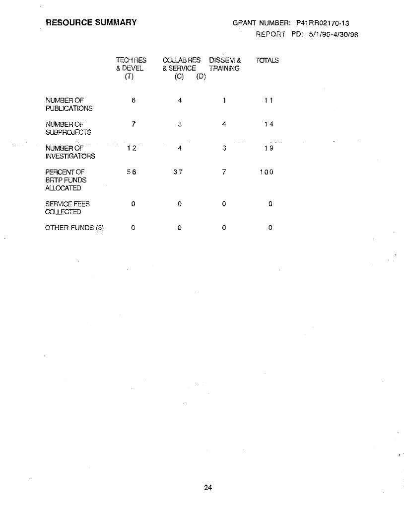
RESOURCE SUMMARY GRANT NUMBER: P41RR02170·13
REPORT PD: 5/1/95·4/30/96
TECH RES OOLLABRES DISSEM& TOTALS &DEVEL &SERVICE TRAINING
(T) (C) (D)
NUMBER OF 6 4 1 1 1 PUBLICATIONS
NUMBER OF 7 3 4 1 4 SUBPROJECTS
NUMBER OF 1 2 4 3 1 9 INVESTIGATORS
PERCENT OF 56 37 7 100 BRTPFUNDS ALLOCATED
SERVICE FEES 0 0 0 0 COUECTED
OTHER FUNDS ($) 0 0 0 0
24
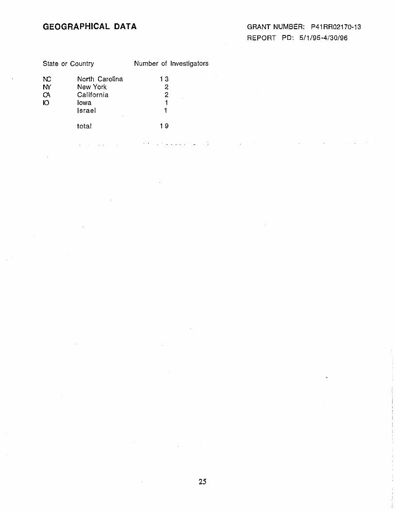
GEOGRAPHICAL DATA
State or Country
1\C NY CA 10
North Carolina New York California Iowa Israel
total
Number of Investigators
1 3 2 2 1 1
1 9
25
GRANT NUMBER: P41 RR02170-13
REPORT PD: 5/1/95-4/30/96
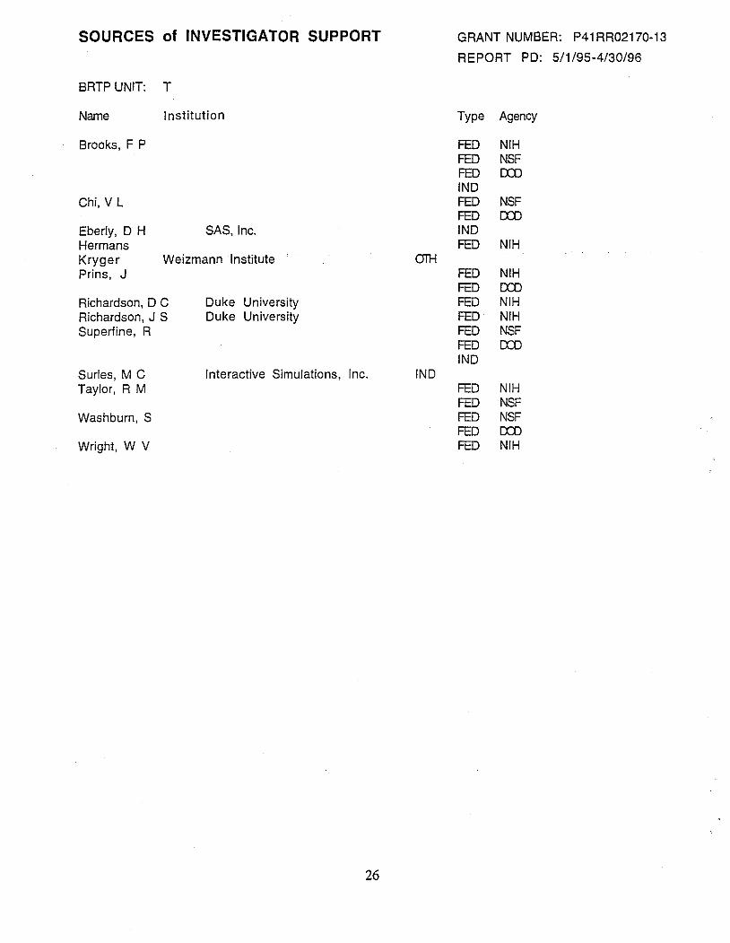
SOURCES of INVESTIGATOR SUPPORT
BRTP UNIT: T
Name Institution
Brooks, F P
Chi, V L
Eberly, D H SAS, Inc. Hermans Kryger Weizmann Institute Prins, J
Richardson, D C Duke University Richardson, J S Duke University Superfine, R
Surles, M C Interactive Simulations, Inc. Taylor, R M
Washburn, S
Wright, W V
26
OIH
INO
GRANT NUMBER: P41RR02170-13
REPORT PO: 5/1/95-4/30/96
Type Agency
FED NIH FED NSF FED em IND FED NSF FED em IND FED NIH
FED NIH FED em FED NIH FED NIH FED NSF FED em INO
FED NIH FED NSF FED NSF FED em FED NIH

SOURCES of INVESTIGATOR SUPPORT
BRTP UNIT: c
Name Institution
Carter, C. Samulski, J. Forest, K. Scripps Research Institute Henderson, E Iowa State University
27
FED
GRANT NUMBER: P41RR02170-13
REPORT PO: 5/1/95-4/30/96
Type Agency
FED NIH FED NIH FED NIH NIH
SOURCES of INVESTIGATOR SUPPORT
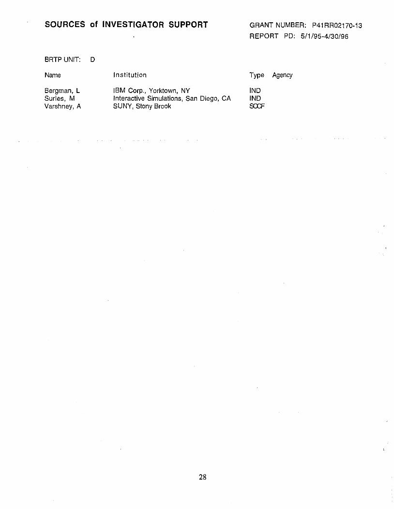
BRTP UNIT: D
Name
Bergman, L Surles, M Varshney, A
Institution
IBM Corp., Yorktown, NY Interactive Simulations, San Diego, CA SUNY, Stony Brook
28
GRANT NUMBER: P41RR02170-13
REPORT PO: 5/1/95-4/30/96
Type Agency
!NO !NO SXF
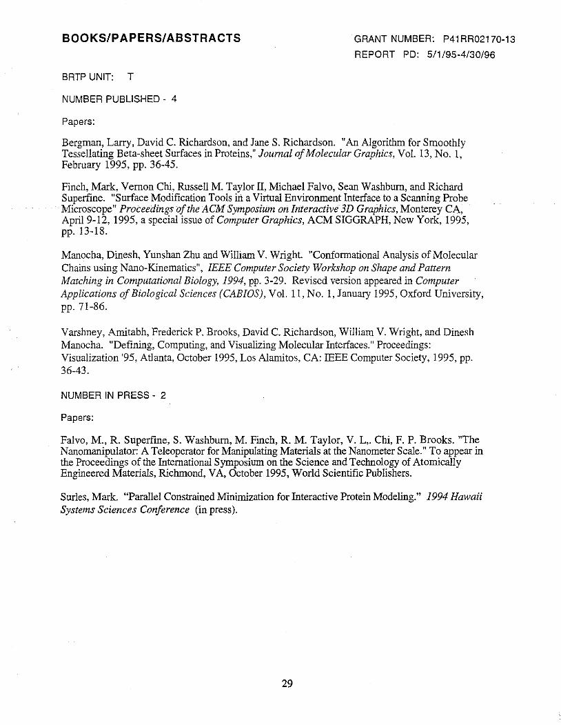
BOOKS/PAPERS/ABSTRACTS
BRTP UNIT: T
NUMBER PUBLISHED - 4
Papers:
GRANT NUMBER: P41RR02170-13
REPORT PO: 5/1/95-4/30/96
Bergman, Larry, David C. Richardson, and JaneS. Richardson. "An Algorithm for Smoothly Tessellating Beta-sheet Surfaces in Proteins," Journal of Molecular Graphics, Vol. 13, No. 1, February 1995, pp. 36-45.
Finch, Mark, Vernon Chi, Russell M. Taylor II, Michael Falvo, Sean Washburn, and Richard Superfine. "Surface Modification Tools in a Virtual Environment Interface to a Scanning Probe Microscope" Proceedings of the ACM Symposium on Interactive 3D Graphics, Monterey CA, April9-12, 1995, a special issue of Computer Graphics, ACM SIGGRAPH, New York, 1995, pp. 13-18.
Manocha, Dinesh, Yunshan Zhu and William V. Wright. "Conformational Analysis of Molecular Chains using Nano-K.inematics", IEEE Computer Society Workshop on Shape and Pattern Matching in Computational Biology, 1994, pp. 3-29. Revised version appeared in Computer Applications of Biological Sciences (CABIOS), Vol. 11, No.1, January 1995, Oxford University, pp. 71-86.
Varshney, Amitabh, FrederickP. Brooks, David C. Richardson, William V. Wright, and Dinesh Manocha. "Defining, Computing, and Visualizing Molecular Interfaces." Proceedings: Visualization '95, Atlanta, October 1995, Los Alamitos, CA: IEEE Computer Society, 1995, pp. 36-43.
NUMBER IN PRESS - 2
Papers:
Falvo, M., R. Superfine, S. Washburn, M. Finch, R. M. Taylor, V. L,. Chi, F. P. Brooks. "The Nanomanipulator: A Teleoperator for Manipulating Materials at the Nanometer Scale." To appear in the Proceedings of the International Symposium on the Science and Technology of Atomically Engineered Materials, Richmond, VA, October 1995, World Scientific Publishers.
Surles, Mark. "Parallel Constrained Minimization for Interactive Protein Modeling." 1994 Hawaii Systems Sciences Conference (in press).
29
BOOKS/PAPERS/ABSTRACTS
BRTP UNIT: C
NUMBER PUBLISHED - 2
Papers:
GRANT NUMBER: P41RR02170-13
REPORT PO: 5/1/95-4/30/96
Albrecht, Kurt, John Hart, Alex Shaw, and A. Keith Dunker. "Quaternion Contact Ribbons: a New Tool for Visualizing Intra- and Intermolecular Interactions in Proteins," Proceedings: Pacific Symposium on Biocomputing 1996, ed. Lawrence Hunter and Teri Klein, World Scientific 1995. ISBN 981-02-2578-4, pp. 41-52.
Gernert, K. M .. , B. D. Thomas, J. C. Plurad, J. S. Richardson, D. C. Richardson, and L. D. Bergman. "Puzzle Pieces Defined: Locating Common Packing Units in Tertiary; Protein Contacts,"· Proceedings: Pacific Symposium on Biocomputing 1996, ed. Lawrence Hunter and Teri Klein, World Scientific 1995. ISBN 981-02-2578-4, pp. 331-349.
NUMBER IN PRESS - 2
Papers:
Falvo, Michael and Richard Superfine. "Atomic Force Microscopy for Nanometer-scale Definition of Gold on Insulators," in preparation for American Journal of Physics.
Falvo, Michael, eta!. "Controlled manipulation of tobacco mosaic virus using an atomic force microscope." In preparation for submission to Biophysical Journal.
30
BOOKS/PAPERS/ABSTRACTS
BRTP UNIT: D
NUMBER PUBLISHED -
Book Chapter:
GRANT NUMBER: P41RR02170-13
REPORT PD: 5/1/95-4/30/96
Bearman, Gerry and Dick Sharp, eds. "Drug Design by Computer: The Future." (Section 2.4). of Book 4 Sensational Chemistry: Our Chemical Environment, Glasgow: Bath Press, 1995, pp. 158-162.
31
PART IV. ADMINISTRATIVE DATA
IV.A. ALLOCATION OF RESOURCE ACCESS
GRANT NUMBER: P41RR02170-13
REPORT PO: 5/1/95-4/30/96
We welcome to use the resources supported by this grant all scientists engaged in work for which our systems are suitable and whose results will be made available to the scientific community and the general public. Should the demand for our services exceed our capacity to work with the applicants, we reserve to right to select those clients whose needs our systems will best serve. We did not decline to work with any client during the past year.
Our Advisory Committee helped us set this policy for resource allocation. Several members of the committee have also made use of our resources and established contacts for us with potential users. Dr. Dagmar Ringe, a member of the committee who reviewed our proposal last June, put us in contact with Dr. Gitay Kryger.
The Advisory Committee met once during the past year on January 26, 1996. The next meeting is planned for January 1997. This year we changed the membership of the Advisory Committee somewhat to better reflect the changing emphasis of the project. The current committee members are:
Vivian Cody, Hauptman-Woodward Medical Research Institute
Richard Colton, Naval Research Laboratory
Michael Cory, Glaxo Wellcome, Inc.
Richard DuBois (ex officio), National Institutes of Health
David DuChamp, Pharmacia & Upjohn, Inc.
Elizabeth Getzoff, Tbe Scripps Research Institute
Michael Pique, Tbe Scripps Research Institute
George Rose, Johns Hopkins University
Stanley Williams, Hewlett Packard Laboratories, and University of California at Los Angeles
Drs. Cody, Cory, DuChamp, Getzoff, and Rose are biochemists. Drs. Colton and Williams are
materials scientists, and Mr. Pique is a computer scientist.
In addition to the annual meeting, we met informally or conversed with several of the committee members on several occasions during the year to discuss how to direct our system development efforts.
IV.B. DISSEMINATION OF INFORMATION
Software. During past year we have made the source code for our DOCK, VIEW and SURF molecular graphics systems available via anonymous ftp. We believe that at least three copies of DOCK, thirty-three copies of VIEW, and seven copies of SURF have been acquired by other sites. We also make available via ftp the source code the software library that we use to support force reflecting devices.
Dr. Mark Surles has formed a company, Interactive Simulations, Inc., to continue development of the SCULPT molecular modeling system he built as a research assistant on the GRIP project. He has now brought an improved version of his system to market on Silicon Graphics workstations and on the Macintosh. We have also entered into negotiations with Dr. Surles and his company to
32
GRANT NUMBER: P41RR02170-13
REPORT PD: 5/1/95-4/30/96
incorporate into SCULPT the work done on representation of molecular surfaces by Dr. Amitabh Varshney, another alumnus of the GRIP project. We have also entered into negotiations with ffiM to license them to develop and market the VIEW system that was developed by Dr. Larry Bergman when he was supported by the Research Resource.
Demonstrations to Visitors. An important means for disseminating the work of the Resource is demonstrations to visitors from industry, universities, the media, and government agencies, and to local groups from schools, colleges, and professional societies. Approximately once a month we devote a day to demonstrations to an assembled diverse group of people who have inquired about our work and facilities. Between 50 and 75 people attend each of these demonstrations. Many, more professionally oriented demonstrations are scheduled for individuals and small groups, some of whom are potential users of our facilities. In all we have demonstrated the work of the Resource to approximately 1200 visitors in the past year. · ·
Papers, Dissertations, and Talks. A number of papers published by team members and talks presented are listed in section III. of this report.
IV.C. TRAINING
The GRIP Resource carried out its education and training role in three ways during the 1995-1996 project year: we trained computer science graduate students in molecular graphics, we helped train chemistry graduate students in molecular graphics through their work in our laboratories, and we collaborated with the UNC School of Pharmacy in their course, Medicinal Chemistry 275/276. Dr. Wright will give one of the lectures for the Medicinal Chemistry course this year.
In past reports, we have identified eighteen computer science students supported by the Resource who have gone on to employment in molecular graphics or scientific visualization. Of these Dr. Russell Taylor has taken a permanent research faculty position in our department. He is now taking an increasingly active and important role in the day-to-day operation of the Resource including management of the Nanomanipu1ator project.
33