twin anemia polycythemia sequence: current views on … · expectant management, and intra-uterine...
TRANSCRIPT

Twin Research and Human GeneticsVolume 19 Number 3 pp. 222–233 C© The Author(s) 2016 doi:10.1017/thg.2016.18
Twin Anemia Polycythemia Sequence: CurrentViews on Pathogenesis, Diagnostic Criteria,Perinatal Management, and Outcome
Lisanne S. A. Tollenaar,1 Femke Slaghekke,1 Johanna M. Middeldorp,1 Frans J. Klumper,1
Monique C. Haak,1 Dick Oepkes,1 and Enrico Lopriore2
1Division of Fetal Medicine, Department of Obstetrics, Leiden University Medical Centre, Leiden, the Netherlands2Division of Neonatology, Department of Pediatrics, Leiden University Medical Centre, Leiden, the Netherlands
Monochorionic twins share a single placenta and are connected with each other through vascular anasto-moses. Unbalanced inter-twin blood transfusion may lead to various complications, including twin-to-twintransfusion syndrome (TTTS) and twin anemia polycythemia sequence (TAPS). TAPS was first describedless than a decade ago, and the pathogenesis of TAPS results from slow blood transfusion from donor torecipient through a few minuscule vascular anastomoses. This gradually leads to anemia in the donor andpolycythemia in the recipient, in the absence of twin oligo-polyhydramnios sequence (TOPS). TAPS mayoccur spontaneously in 3–5% of monochorionic twins or after laser surgery for TTTS. The prevalence ofpost-laser TAPS varies from 2% to 16% of TTTS cases, depending on the rate of residual anastomoses.Pre-natal diagnosis of TAPS is currently based on discordant measurements of the middle cerebral arterypeak systolic velocity (MCA-PSV; >1.5 multiples of the median [MoM] in donors and <1.0 in recipients).Post-natal diagnosis is based on large inter-twin hemoglobin (Hb) difference (>8 g/dL), and at least oneof the following: reticulocyte count ratio >1.7 or minuscule placental anastomoses. Management includesexpectant management, and intra-uterine blood transfusion (IUT) with or without partial exchange trans-fusion (PET) or fetoscopic laser surgery. Post-laser TAPS can be prevented by using the Solomon lasersurgery technique. Short-term neonatal outcome ranges from isolated inter-twin Hb differences to severeneonatal morbidity and neonatal death. Long-term neonatal outcome in post-laser TAPS is comparablewith long-term outcome after treated TTTS. This review summarizes the current knowledge after 10 yearsof research on the pathogenesis, diagnosis, management, and outcome in TAPS.
� Keywords: monochorionic twins, twin anemia polycythemia sequence, anemia, polycythemia
Monochorionic twin pregnancies are at increased risk foradverse outcome compared to dichorionic twin pregnan-cies and singletons. This is primarily due to the fact thatalmost all monochorionic twins share a single placenta,with inter-twin anastomoses allowing blood to flow bidi-rectionally between the two fetuses. Unbalanced net inter-twin blood transfusion may lead to various complications,including TTTS and twin anemia polycythemia sequence(TAPS). TTTS was first described in the 19th century andresults from imbalanced inter-twin blood flow causing hy-povolemia and oligohydramnios in the donor and hyperv-olemia and polyhydramnios in the recipient twin, the so-called TOPS. TAPS is a newly described form of chronicand slow inter-twin blood transfusion characterized bylarge inter-twin Hb differences without signs of TOPS. Thepathogenesis of TAPS is based on the presence of a fewminuscule vascular anastomoses. TAPS may occur sponta-
neously in monochorionic twin pregnancies or may occurin TTTS cases after incomplete laser surgery due to a fewsmall residual anastomoses. The post-laser form of TAPSwas first described in 2006 (Robyr et al., 2006), whereas thespontaneous form of TAPS, as well as the acronym TAPS,was first described in 2007 (Lopriore, Middeldorp et al.,2007). In the past decade, over 100 studies have been pub-lished on TAPS, and our knowledge and awareness havegradually increased. This review focuses on the epidemi-ology, pathogenesis, diagnostic criteria, management op-
RECEIVED 19 January 2016; ACCEPTED 1 February 2016. First pub-lished online 12 April 2016.
ADDRESS FOR CORRESPONDENCE: E. Lopriore, Department of Pe-diatrics, Leiden University Medical Center, J6-S, Albinusdreef 2,2333 ZA Leiden, the Netherlands. E-mail: [email protected]
222
https://www.cambridge.org/core/terms. https://doi.org/10.1017/thg.2016.18Downloaded from https://www.cambridge.org/core. IP address: 54.39.106.173, on 31 Jul 2020 at 03:22:04, subject to the Cambridge Core terms of use, available at
Twin Anemia Polycythemia Sequence
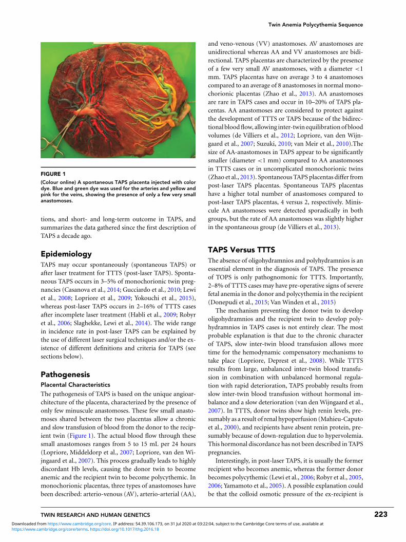
FIGURE 1
(Colour online) A spontaneous TAPS placenta injected with colordye. Blue and green dye was used for the arteries and yellow andpink for the veins, showing the presence of only a few very smallanastomoses.
tions, and short- and long-term outcome in TAPS, andsummarizes the data gathered since the first description ofTAPS a decade ago.
EpidemiologyTAPS may occur spontaneously (spontaneous TAPS) orafter laser treatment for TTTS (post-laser TAPS). Sponta-neous TAPS occurs in 3–5% of monochorionic twin preg-nancies (Casanova et al., 2014; Gucciardo et al., 2010; Lewiet al., 2008; Lopriore et al., 2009; Yokouchi et al., 2015),whereas post-laser TAPS occurs in 2–16% of TTTS casesafter incomplete laser treatment (Habli et al., 2009; Robyret al., 2006; Slaghekke, Lewi et al., 2014). The wide rangein incidence rate in post-laser TAPS can be explained bythe use of different laser surgical techniques and/or the ex-istence of different definitions and criteria for TAPS (seesections below).
PathogenesisPlacental Characteristics
The pathogenesis of TAPS is based on the unique angioar-chitecture of the placenta, characterized by the presence ofonly few minuscule anastomoses. These few small anasto-moses shared between the two placentas allow a chronicand slow transfusion of blood from the donor to the recip-ient twin (Figure 1). The actual blood flow through thesesmall anastomoses ranges from 5 to 15 mL per 24 hours(Lopriore, Middeldorp et al., 2007; Lopriore, van den Wi-jngaard et al., 2007). This process gradually leads to highlydiscordant Hb levels, causing the donor twin to becomeanemic and the recipient twin to become polycythemic. Inmonochorionic placentas, three types of anastomoses havebeen described: arterio-venous (AV), arterio-arterial (AA),
and veno-venous (VV) anastomoses. AV anastomoses areunidirectional whereas AA and VV anastomoses are bidi-rectional. TAPS placentas are characterized by the presenceof a few very small AV anastomoses, with a diameter <1mm. TAPS placentas have on average 3 to 4 anastomosescompared to an average of 8 anastomoses in normal mono-chorionic placentas (Zhao et al., 2013). AA anastomosesare rare in TAPS cases and occur in 10–20% of TAPS pla-centas. AA anastomoses are considered to protect againstthe development of TTTS or TAPS because of the bidirec-tional blood flow, allowing inter-twin equilibration of bloodvolumes (de Villiers et al., 2012; Lopriore, van den Wijn-gaard et al., 2007; Suzuki, 2010; van Meir et al., 2010).Thesize of AA-anastomoses in TAPS appear to be significantlysmaller (diameter <1 mm) compared to AA anastomosesin TTTS cases or in uncomplicated monochorionic twins(Zhao et al., 2013). Spontaneous TAPS placentas differ frompost-laser TAPS placentas. Spontaneous TAPS placentashave a higher total number of anastomoses compared topost-laser TAPS placentas, 4 versus 2, respectively. Minis-cule AA anastomoses were detected sporadically in bothgroups, but the rate of AA anastomoses was slightly higherin the spontaneous group (de Villiers et al., 2013).
TAPS Versus TTTSThe absence of oligohydramnios and polyhydramnios is anessential element in the diagnosis of TAPS. The presenceof TOPS is only pathognomonic for TTTS. Importantly,2–8% of TTTS cases may have pre-operative signs of severefetal anemia in the donor and polycythemia in the recipient(Donepudi et al., 2015; Van Winden et al., 2015)
The mechanism preventing the donor twin to developoligohydramnios and the recipient twin to develop poly-hydramnios in TAPS cases is not entirely clear. The mostprobable explanation is that due to the chronic characterof TAPS, slow inter-twin blood transfusion allows moretime for the hemodynamic compensatory mechanisms totake place (Lopriore, Deprest et al., 2008). While TTTSresults from large, unbalanced inter-twin blood transfu-sion in combination with unbalanced hormonal regula-tion with rapid deterioration, TAPS probably results fromslow inter-twin blood transfusion without hormonal im-balance and a slow deterioration (van den Wijngaard et al.,2007). In TTTS, donor twins show high renin levels, pre-sumably as a result of renal hypoperfusion (Mahieu-Caputoet al., 2000), and recipients have absent renin protein, pre-sumably because of down-regulation due to hypervolemia.This hormonal discordance has not been described in TAPSpregnancies.
Interestingly, in post-laser TAPS, it is usually the formerrecipient who becomes anemic, whereas the former donorbecomes polycythemic (Lewi et al., 2006; Robyr et al., 2005,2006; Yamamoto et al., 2005). A possible explanation couldbe that the colloid osmotic pressure of the ex-recipient is
TWIN RESEARCH AND HUMAN GENETICS 223
https://www.cambridge.org/core/terms. https://doi.org/10.1017/thg.2016.18Downloaded from https://www.cambridge.org/core. IP address: 54.39.106.173, on 31 Jul 2020 at 03:22:04, subject to the Cambridge Core terms of use, available at

Lisanne S. A. Tollenaar et al.
strongly increased prior to and shortly after laser therapy,which attracts excess fluid from the maternal blood to therecipient’s fetal blood. This source of increased fetal plasmavolume followed by amniotic fluid production may delaythe development of oligohydramnios in the ex-recipientwho becomes the TAPS donor (van den Wijngaard et al.2007).
TAPS Versus UncomplicatedMonochorionic TwinsIn addition to the small anastomoses that characterize TAPSplacentas, TAPS placentas also show a remarkable differ-ence in placental shares. In uncomplicated monochorionictwins, the placental share is the principal element affectingfetal growth and final birth weight, that is, the smaller twinusually has a relatively smaller placental share (Lewi et al.,2007). In TAPS twins, the donor twin is usually smallerthan the recipient twin but often has a paradoxically largerplacental share compared to its co-twin. In contrast to un-complicated monochorionic twins, fetal growth in TAPSappears to be primarily determined by inter-twin bloodtransfusion rather than placental share. A relatively largerplacental share may enable the fetal survival of the anemictwin in TAPS (Zhao et al. 2014). In addition, TAPS donortwins also have hypoalbuminemia and hypoproteinemiadue to loss of not only blood cells, but also proteins andnutrients, which may partly also affect their fetal growth(Verbeek et al., 2013).
DiagnosisTAPS can be diagnosed either antenatally or post-natally.TAPS is characterized by Hb differences with the absence ofantenatal ultrasound signs of polyhydramnios in the recipi-ent and oligohydramnios in the donor. Antenatal diagnosisis based on ultrasound abnormalities whereas the post-nataldiagnosis is derived from large inter-twin hematologic dis-cordances and characteristic placental angioarchitecture.
Antenatal Criteria
Antenatal diagnosis of TAPS is based on Doppler ultrasoundabnormalities. MCA-PSV measurement, a non-invasivetest, has become the standard test for the prediction of fetalanemia in singletons in a variety of fetal diseases. In TAPS,this test will show an increased MCA-PSV in the donortwin, suggestive of fetal anemia, and decreased velocities inthe MCA-PSV in the recipient, suggestive of polycythemia.During the past decade, different MCA-PSV values for thediagnosis of TAPS have been proposed. Robyr et al. (2006)initially suggested the use of a MCA-PSV >1.5 MoM for thedonor twin and <0.8 MoM for the recipient twin. However,Slaghekke et al. (2010) showed that a MCA-PSV between 0.8and 1.0 MoM in the recipient was also frequently found inpost-natally diagnosed TAPS cases and suggested thereforethe currently used cut-off of MCA-PSV <1.0 MoM in the
recipient twin and >1.5 MoM in the donor twin. This cut-off level is characterized by high sensitivity and specificity ofMCA-PSV for both anemia and polycythemia, confirmingthe clinical usefulness of this non-invasive test (Slaghekkeet al., 2010). The MCA-PSV measurement has a high di-agnostic accuracy for predicting abnormal Hb levels in fe-tuses with TAPS (Slaghekke, Pasman et al., 2015; Veujozet al. 2015). However, the high predictive value of MCA-PSV measurements reported in the study from Slaghekkeet al. (2010) was determined in a highly selected group ofTAPS-only cases followed in a specialized fetal therapy cen-ter. In a recent study, Fishel-Bartal et al. (2015) did not finda similar predictive value of MCA-PSV for the detectionof polycythemia in monochorionic twins. MCA-PSV mea-surement in polycythemic twins did not differ comparedto normal twins. Fishel-Bartal et al. (2015) did find a sig-nificant correlation between a large inter-twin difference inMCA-PSV (delta MCA-PSV) and large inter-twin hemat-ocrit difference and proposed the alternative use of a deltaMCA-PSV >0.5 MoM instead of the currently used fixedcut-off levels of <1.0 MoM and >1.5 MoM for the pre-nataldiagnosis of TAPS.
In some TAPS cases, additional ultrasound findings havebeen reported. In several cases of spontaneous TAPS, a strik-ing difference in placental thickness and echodensity onultrasound examination was detected (Figure 2; Movva &Rijhsinghani, 2014; Slaghekke et al., 2010; Stritzke et al.,2014). This difference can be explained by the hydropicand echogenic character of the anemic placental share incontrast to the normal appearance of the polycythemic pla-cental share. Another ultrasound finding described in TAPSis the so-called ‘starry sky liver’ (Figure 3; Soundararajan &Howe, 2014). Starry sky appearance refers to a sonographicpattern of the liver, characterized by clearly identified portalvenules (stars) and diminished parenchymal echogenicity(sky) that accentuates the portal venule walls. Starry skyliver is usually reported in acute hepatitis, but other condi-tions like heart failure can lead to this typical finding as well.More studies are needed to further investigate the validityand significance of these antenatal ultrasound findings forthe diagnosis of TAPS.
Since diagnosis of TAPS at an earlier gestational age is as-sociated with more favorable outcomes (Rossi & Prefumo,2014), we recommend the routine use (at least biweekly) ofMCA-PSV Doppler ultrasound measurements in all mono-chorionic pregnancies to timely detect TAPS.
Postnatal Criteria
In 40–63% of the cases, TAPS is not detected antenatally,but is only diagnosed after birth (de Villiers et al., 2013;Slaghekke et al., 2010). Therefore, post-natal diagnosticcriteria have been proposed. These criteria are based onthe presence of (chronic) anemia in the donor and poly-cythemia in the recipient twin (Figure 4) and the charac-teristic angioarchitecture of the placenta. Different cut-off
224 TWIN RESEARCH AND HUMAN GENETICS
https://www.cambridge.org/core/terms. https://doi.org/10.1017/thg.2016.18Downloaded from https://www.cambridge.org/core. IP address: 54.39.106.173, on 31 Jul 2020 at 03:22:04, subject to the Cambridge Core terms of use, available at

Twin Anemia Polycythemia Sequence
FIGURE 2
(Colour online) Ultrasound image of a TAPS placenta showing a difference in placental thickness and echodensity. On the left side ofthe image the hydropic and echogenic placental share of the anemic donor and on the right side the normal aspect of the placenta ofthe recipient is depicted.
FIGURE 3
(Colour online) Ultrasound image showing a starry sky liver in a TAPS recipient with clearly identified portal venules (stars) and diminishedparenchymal echogenicity (sky) that accentuates the portal venule walls.
TWIN RESEARCH AND HUMAN GENETICS 225
https://www.cambridge.org/core/terms. https://doi.org/10.1017/thg.2016.18Downloaded from https://www.cambridge.org/core. IP address: 54.39.106.173, on 31 Jul 2020 at 03:22:04, subject to the Cambridge Core terms of use, available at
Lisanne S. A. Tollenaar et al.
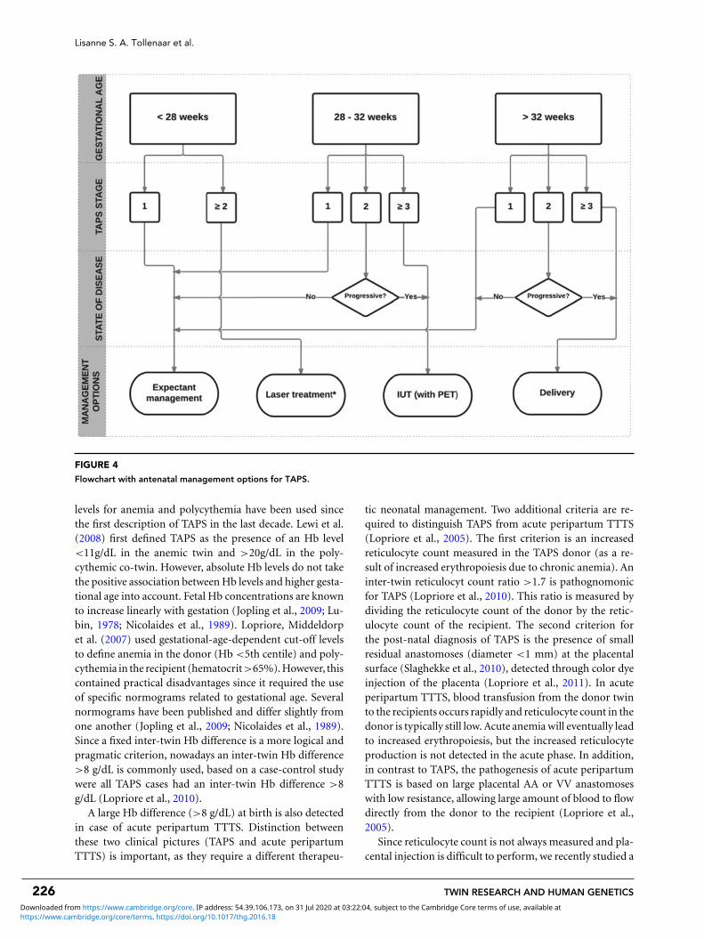
FIGURE 4
Flowchart with antenatal management options for TAPS.
levels for anemia and polycythemia have been used sincethe first description of TAPS in the last decade. Lewi et al.(2008) first defined TAPS as the presence of an Hb level<11g/dL in the anemic twin and >20g/dL in the poly-cythemic co-twin. However, absolute Hb levels do not takethe positive association between Hb levels and higher gesta-tional age into account. Fetal Hb concentrations are knownto increase linearly with gestation (Jopling et al., 2009; Lu-bin, 1978; Nicolaides et al., 1989). Lopriore, Middeldorpet al. (2007) used gestational-age-dependent cut-off levelsto define anemia in the donor (Hb <5th centile) and poly-cythemia in the recipient (hematocrit>65%). However, thiscontained practical disadvantages since it required the useof specific normograms related to gestational age. Severalnormograms have been published and differ slightly fromone another (Jopling et al., 2009; Nicolaides et al., 1989).Since a fixed inter-twin Hb difference is a more logical andpragmatic criterion, nowadays an inter-twin Hb difference>8 g/dL is commonly used, based on a case-control studywere all TAPS cases had an inter-twin Hb difference >8g/dL (Lopriore et al., 2010).
A large Hb difference (>8 g/dL) at birth is also detectedin case of acute peripartum TTTS. Distinction betweenthese two clinical pictures (TAPS and acute peripartumTTTS) is important, as they require a different therapeu-
tic neonatal management. Two additional criteria are re-quired to distinguish TAPS from acute peripartum TTTS(Lopriore et al., 2005). The first criterion is an increasedreticulocyte count measured in the TAPS donor (as a re-sult of increased erythropoiesis due to chronic anemia). Aninter-twin reticulocyt count ratio >1.7 is pathognomonicfor TAPS (Lopriore et al., 2010). This ratio is measured bydividing the reticulocyte count of the donor by the retic-ulocyte count of the recipient. The second criterion forthe post-natal diagnosis of TAPS is the presence of smallresidual anastomoses (diameter <1 mm) at the placentalsurface (Slaghekke et al., 2010), detected through color dyeinjection of the placenta (Lopriore et al., 2011). In acuteperipartum TTTS, blood transfusion from the donor twinto the recipients occurs rapidly and reticulocyte count in thedonor is typically still low. Acute anemia will eventually leadto increased erythropoiesis, but the increased reticulocyteproduction is not detected in the acute phase. In addition,in contrast to TAPS, the pathogenesis of acute peripartumTTTS is based on large placental AA or VV anastomoseswith low resistance, allowing large amount of blood to flowdirectly from the donor to the recipient (Lopriore et al.,2005).
Since reticulocyte count is not always measured and pla-cental injection is difficult to perform, we recently studied a
226 TWIN RESEARCH AND HUMAN GENETICS
https://www.cambridge.org/core/terms. https://doi.org/10.1017/thg.2016.18Downloaded from https://www.cambridge.org/core. IP address: 54.39.106.173, on 31 Jul 2020 at 03:22:04, subject to the Cambridge Core terms of use, available at

Twin Anemia Polycythemia Sequence
FIGURE 5
(Colour online) Spontaneous TAPS twins at birth. On the left, the plethoric polycythemic recipient and on the right the pale anemicdonor.
FIGURE 6
(Colour online) Maternal side of the TAPS placenta showing thedifference in color between the plethoric share of the recipient(left side of the placenta) and the anemic share of the donor (rightside of the placenta).
new additional criterion for post-natal diagnosis of TAPS. Inanalogy with the difference in skin color of the TAPS twins atbirth (the anemic donor is pale and the polycythemic recipi-ent is plethoric), the maternal side of the TAPS placenta alsoshows a striking color difference (Figure 5 & Figure 6). Wedeveloped a new quick and easy tool to determine the colordifference ratio (CDR) between the two placental sharesusing digital pictures (Tollenaar et al., 2016). We foundthat TAPS placentas had a significantly higher CDR (>1.5)compared to uncomplicated monochorionic twin placen-tas. Whether this method can eventually be added to the
list of post-natal criteria requires further investigations inlarger series of placentas with and without TAPS to deter-mine the sensitivity and specificity of the test. Importantly,CDR measurements should also be investigated in placen-tas with acute TTTS to determine whether this method canhelp distinguish TAPS cases from cases with acute peri-partum TTTS, since in both situations large inter-twin Hbdifferences are present at birth.
ClassificationSince TAPS is a heterogeneous disease, a staging system canbe helpful to discriminate between the various forms. Inaddition, a staging system may also prove to be useful in thefuture to compare and analyze TAPS cases (including effectof treatment) between the various centers. We, therefore,recently proposed both an antenatal and post-natal classi-fication system (Tables 1 and 2). Whether this classificationhas an additional value to adequately stage and treat TAPSrequires further investigation.
Perinatal Management and OutcomeThe optimal perinatal management for TAPS is not clear.Options include expectant management, induction of labor,IUTin the donor, with or without PET in the recipient,selective feticide, and (repeat) fetoscopic laser surgery.
Expectant management consists of close monitoringwith ultrasound including Doppler measurements of MCA-PSV. Close monitoring can be considered in less severe casesof TAPS, such as stage 1 and 2. Whether close monitoringis safe enough in these TAPS stages needs to be evaluatedin combination with validating the staging system. When
TWIN RESEARCH AND HUMAN GENETICS 227
https://www.cambridge.org/core/terms. https://doi.org/10.1017/thg.2016.18Downloaded from https://www.cambridge.org/core. IP address: 54.39.106.173, on 31 Jul 2020 at 03:22:04, subject to the Cambridge Core terms of use, available at

Lisanne S. A. Tollenaar et al.
TABLE 1
Antenatal TAPS Classification
Antenatal stage Findings at Doppler ultrasound examination
Stage 1 MCA-PSV donor >1.5 MoM and MCA-PSV recipient < 1.0 MoM, without other signs of fetal compromise.Stage 2 MCA-PSV donor >1.7 and MCA-PSV recipient < 0.8 MoM, without other signs of fetal compromise.Stage 3 As stage 1 or 2, with cardiac compromise of the donor, defined as critically abnormal flow.Stage 4 Hydrops of donor.Stage 5 Intrauterine demise of one or both fetuses preceded by TAPS.
TABLE 2
Post-natal TAPS Classification
Post-natal stage Inter-twin Hffigureb difference, g/dL
Stage 1 >8.0Stage 2 >11.0Stage 3 >14.0Stage 4 >17.0Stage 5 >20.0
TAPS stage 1 quickly progresses to stage 2 or stage �3, intra-uterine intervention or termination of pregnancy should beconsidered. However, both management options may leadto pre-mature delivery and its associated risks of perinatalmorbidity and mortality.
Intrauterine Blood Transfusion
Treatment with IUT in the donor can be performed ei-ther intravascularly or intraperitoneal. Intraperitoneal IUTis preferred, since intraperitoneal transfusion may allowslower absorption of red blood cells into the fetal circula-tion, preventing rapid loss of transfused blood in the circu-lation of the recipient twin (Herway et al., 2009). Althoughtreatment with IUT has often been reported, it is not acausal treatment and is only a temporary solution. Further-more, a potential side effect of IUT treatment is worseningof the polycythemia hyperviscosity syndrome in the recipi-ent. Robyr et al. (2006) reported skin necrosis of the leg inthe recipient twin of a TAPS case treated with several IUTs.To reduce the risk of increasing polycythemia hyperviscos-ity, a combination procedure of IUT in the donor and PETin the recipient can be of additional value. The rationalebehind this therapy is that PET may help to decrease theviscosity of the blood of the polycythemic recipient. Genovaet al. (2013) reported on three different TAPS cases treatedwith IUT with PET. We recently developed a computationalmodel to evaluate the effect of IUT with and without PETin post-laser TAPS cases, and showed the beneficial effectof PET (Slaghekke, van den Wijngaard et al., 2015).
Since TAPS twins share their blood circulation and there-fore have exactly the same blood cell characteristics, it maybe of additional value to transfuse the anemic twin withthe recipient’s whole blood as a donor source instead offoreign donor blood. Recently, Yarci et al. (2014) reporteda case of TAPS in which the anemic donor was success-fully transfused after delivery with blood obtained from the
polycythemic co-twin during PET. The main advantage ofthis new therapeutic method is avoidance of donor expo-sure and of the risk of blood product infections (Yarci et al.,2014). Whether this new approach may lead to decreasedmorbidity in TAPS pregnancies requires further investiga-tion.
Fetoscopic Laser Coagulation
The only causal treatment of TAPS is (repeated) fetoscopiclaser coagulation of the (residual) anastomoses at the vas-cular equator of the placental. Fetoscopic laser coagulationin TAPS is more challenging than in TTTS, since the ab-sence of oligo-polyhydramnios sequences and therefore awavering inter-twin membrane makes the visualization ofthe vascular equator more difficult (Slaghekke et al., 2010).Moreover, placental anastomoses in TAPS are known tobe only few and minuscule and may therefore be missedduring fetoscopy (Slaghekke et al., 2010). Different casereports show the feasibility of fetoscopic laser coagulationin TAPS placentas (Abdel-Sattar et al., 2014; Assaf et al.,2011; Diehl et al., 2013; Groussolles et al., 2012; Ishii et al.,2014). In a retrospective study where laser treatment forantenatally detected TAPS is compared to IUT or expectantmanagement, laser therapy appeared to improve perinataloutcome by prolonging pregnancy and reducing respiratorydistress syndrome (Slaghekke, Favre et al., 2014). The me-dian time between diagnosis and birth was 11 weeks in thelaser group compared to 5 weeks after intrauterine transfu-sion, and 8 weeks after expectant management. In the lasergroup, no residual anastomoses were found after color dyeinjection. Larger, adequately randomized controlled stud-ies are required to determine the optimal management andto evaluate the possible additional value of fetoscopic lasercoagulation for the treatment of TAPS. When performinglaser coagulation in TAPS placentas, we recommend usingthe Solomon technique to reduce the risk of residual anas-tomoses and recurrent TAPS (Slaghekke, Lewi et al., 2014).In the Solomon technique, a line is drawn from one pla-centa margin to the other, connecting the individual laserspots (Slaghekke, Lopriore et al., 2014).
In unique circumstances, spontaneous resolution of an-tenatal TAPS may also occur. Spontaneous resolution hasbeen reported once (Lopriore, Hecher et al., 2008) andwas presumably caused by thrombosis of the residual AV-anastomosis. Whether expectant management would lead
228 TWIN RESEARCH AND HUMAN GENETICS
https://www.cambridge.org/core/terms. https://doi.org/10.1017/thg.2016.18Downloaded from https://www.cambridge.org/core. IP address: 54.39.106.173, on 31 Jul 2020 at 03:22:04, subject to the Cambridge Core terms of use, available at
Twin Anemia Polycythemia Sequence
to spontaneous resolution in other TAPS cases is unknownand should be considered unlikely.
Proposal for Antenatal Management
In the absence of evidence on optimal management, wesuggest that management decisions should be made aftercareful evaluation of different factors, including TAPS stage,gestational age, and the feasibility of the different types ofintra-uterine intervention. TAPS stage 1 and possibly stage2 can be observed with close monitoring. In case TAPSprogresses quickly to stage 2 or in case of stage �3, inter-vention should be considered. If gestational age is below 28weeks and laser treatment is feasible, laser treatment shouldbe considered, since this is the only causal treatment forTAPS and is known to prolong the pregnancy (Slaghekke,Favre et al., 2014). When laser treatment is not feasibleand gestational age is below 30 to 32 weeks of pregnancy,intra-uterine transfusion should be considered. When re-peated intra-uterine transfusions are expected or in case ofsevere polycythemia in the recipient, PET of the recipientshould be envisaged. A management proposal for antenatalTAPS is presented in the flowchart in Figure 4. Whetherthis flowchart is useful in current practice and will improveoutcome still needs to be validated.
Prevention of Post-Laser TAPS
Post-laser TAPS is caused by residual anastomoses at theplacental surface after fetoscopic laser surgery for TTTS.In order to reduce the number of residual anastomoses,the Solomon technique was introduced. The Solomon ran-domized trial by Slaghekke, Lewi et al. (2014) showed asignificant reduction of post-laser TAPS of 16% in the stan-dard treatment group to 3% in the Solomon group. TheSolomon technique did not appear to be associated with anincrease in any identifiable short-term adverse outcome orcomplications. A study investigating the neurodevelopmen-tal outcome at 2 years in TAPS survivors randomized for theSolomon trail showed no difference in the risk of neurode-velopmental impairment between the groups treated withthe Solomon technique and the standard laser technique(van Klink et al., 2015). The Solomon technique shouldtherefore be used in all TTTS cases to reduce the risks ofresidual anastomoses and prevent the occurrence of post-laser TAPS.
Neonatal and Pediatric OutcomeData of perinatal mortality and morbidity rates in TAPS arescarce and mostly based on case reports and small series.The neonatal outcome in TAPS may vary from isolated largeinter-twin Hb differences to severe neonatal morbidity, in-cluding cerebral injury and neonatal death (Luminoso et al.,2013).
Short-Term Neonatal Outcome
Hematological complications are commonly seen in TAPSdonors and recipients, requiring blood transfusion or PET,respectively. TAPS recipients may develop polycythemia hy-perviscosity syndrome, which may possibly lead to necrosisof the skin and multiple limb ischemia (Robyr et al., 2006;Stranak et al., 2015). In addition, recipients are at increasedrisk of thrombocytopenia, probably due to impaired pro-duction secondary to tissue hypoxia and slow spleen bloodflow (Lopriore et al., 2010; Sarkar & Rosenkrantz, 2008).Platelet count at birth was inversely related to the severityof polycythemia in recipients (Lopriore et al., 2010). In addi-tion to lower Hb levels, donor twins with TAPS also have sig-nificantly lower albumin and total protein levels comparedto recipient twins, suggesting that the inter-twin transfusionprocess does not only concern red blood cells but also pro-teins and albumin (Verbeek et al., 2013). Chronic inter-twintransfusion in TAPS may also cause short-term renal dys-function: Verbeek et al. (2015) found that donor twins withTAPS have higher creatinine levels than recipients, probablydue to chronic renal hypoperfusion. Whether donor twinsmay also have permanent renal damage and long-term re-nal complications is not known. Chronic severe anemiain donor twins and polycythemia in recipient twins maytheoretically also lead to cerebral injury. Several small casestudies report on severe cerebral injury leading to fatal out-come in TAPS (Genova et al., 2013; Lopriore et al., 2013).Genova et al. (2013) described a TAPS case, in which de-spite treatment with IUT with PET, the anemic twin died ofextensive cerebral injury including numerous large cysts inthe basal ganglia, bilateral white matter injury, and multi-ple microbleeds. Lopriore et al. (2013) reported on a spon-taneous TAPS case delivered after an emergency cesareansection at 33 weeks gestation. The recipient twin sufferedfrom severe cerebral injury due to massive hemorrhage andinfarctions and died on day 3 after withdrawal of intensivecare.
Long-Term Neurodevelopmental Outcome
The long-term neurodevelopmental outcome in survivingTAPS infants is not well known and data is based on smalluncontrolled case series. Severe long-term morbidity suchas bilateral deafness and spastic paralysis has recently re-ported (Taniguchi et al., 2015). In another recent studyon long-term neurodevelopmental outcome in post-laserTAPS, Slaghekke, van Klink et al. (2014) detected neu-rodevelopmental impairment or mild to moderate cogni-tive delay in 9% and 17% of TAPS survivors, respectively.No difference in impairment was found between donorsand recipients. The rate of impairment in TAPS seems tobe comparable to the rate of impairment in children withTTTS after laser surgery. Risk factors for decreased cog-nitive scores in the study from Slaghekke et al. were lowgestational age at birth and low birth weight, as well as in-trauterine transfusion. In a recent small study, neonatal
TWIN RESEARCH AND HUMAN GENETICS 229
https://www.cambridge.org/core/terms. https://doi.org/10.1017/thg.2016.18Downloaded from https://www.cambridge.org/core. IP address: 54.39.106.173, on 31 Jul 2020 at 03:22:04, subject to the Cambridge Core terms of use, available at
Lisanne S. A. Tollenaar et al.
outcome in monochorionic twins affected by TAPS ap-peared to be comparable to gestational age-matched un-complicated monochorionic twins. However, only 10 TAPScases were included with mild TAPS (stage 1 and 2), lim-iting the conclusions (Ashwal et al., 2015). To date, thereare no studies reporting on neurological, motor, and cog-nitive outcomes of TAPS twins in childhood and ado-lescence. Data from these studies could provide us witha more complete view of the long-term consequencesof TAPS.
ConclusionTAPS is recently described form of feto-fetal transfusionthrough small (diameter <1 mm) anastomoses that may oc-cur in monochorionic twins spontaneously or in TTTS casesafter laser surgery (post-laser TAPS). In the past decade,our knowledge on pathogenesis, diagnostic criteria, man-agement options, and short- and long-term outcome hasgreatly increased. However, further studies are required todetermine the optimal diagnostic criteria. Whether a deltaMCA-PSV >0.5 (comparable to a delta Hb postnatally) isa better alternative criterion than the currently used fixedcut-off levels of <1.0 and >1.5 MoM of MCA-PSV requiresfurther investigation. In addition, we recently introduceda new diagnostic criterion based on the color difference ofthe maternal side of the placenta. Whether this criterion caneventually be added to the list of post-natal criteria requiresfurther investigations in larger series of placentas with andwithout TAPS.
Although different management options have been pro-posed, optimal treatment for TAPS is still unclear and re-mains a challenging problem due to lack of randomizedtrials. In this review, we proposed a stage and gestationalage-based flow chart for the treatment of TAPS. Large ran-domized controlled trials are needed to test the clinicalusefulness of this proposed flowchart.
Studies on long-term outcome in TAPS survivors showthat neurodevelopmental outcome is similar to TTTS twins,but data on neurological, cognitive, and motor function forchildhood and adulthood are still not available. Long-termfollow-up studies comparing TAPS to TTTS and uncom-plicated monochorionic twins are required to determinewhether TAPS twins have an increased risk for develop-ing adverse long-term neurodevelopmental outcome at anolder age.
Since TAPS is a rare disease, collaboration between in-ternational fetal therapy centers is of utmost importance toincrease sample size and quality of the studies. To facilitatethis purpose, we have recently created a web-based reg-istry (www.TAPSregistry.org) to gather information on theshort- and long-term outcome in TAPS. This informationwill provide us crucial information to set up well-designedstudies and investigate the optimal management in thefuture.
ReferencesAbdel-Sattar, M., Platt, L. D., DeVore, G., Porto, M.,
Benirschke, K., & Chmait, R. H. (2014). Treatment of com-plicated spontaneous twin anemia-polycythemia sequencevia fetoscopic laser ablation of the vascular communica-tions. Fetal Diagnosis and Therapy, 38, 233–237.
Ashwal, E., Yinon, Y., Fishel-Bartal, M., Tsur, A., Chayen, B.,Weisz, B., & Lipitz, S. (2015). Twin anemia-polycythemiasequence: Perinatal management and outcome. Fetal Diag-nosis and Therapy. Advance online publication.
Assaf, S. A., Benirschke, K., & Chmait, R. H. (2011). Spon-taneous twin anemia-polycythemia sequence complicatedby recipient placental vascular thrombosis and hydrops fe-talis. Journal of Maternal-Fetal and Neonatal Medicine, 24,549–552.
Casanova, J., Paiva, C., Carvalho, C., & Cunha, A. C. (2014).Twin anemia polycythemia sequence: A report of threecases. Journal of Reproductive Medicine, 59, 596–598.
de Villiers, S. F., Slaghekke, F., Middeldorp, J. M., Walther, F. J.,Oepkes, D., & Lopriore, E. (2012). Arterio-arterial vascularanastomoses in monochorionic placentas with and withouttwin-twin transfusion syndrome. Placenta, 33, 652–654.
de Villiers, S. F., Slaghekke, F., Middeldorp, J. M., Walther,F. J., Oepkes, D., & Lopriore, E. (2013). Placental charac-teristics in monochorionic twins with spontaneous versuspost-laser twin anemia-polycythemia sequence. Placenta,34, 456–459.
Diehl, W., Glosemeyer, P., Tavares De Sousa, M., Hollwitz,B., Ortmeyer, G., & Hecher, K. (2013). Twin anemia-polycythemia sequence in a case of monoamniotic twins.Ultrasound in Obstetrics & Gynecology, 42, 108–111.
Donepudi, R., Papanna, R., Snowise, S., Johnson, A.,Bebbington, M., & Moise, K. J. (2015). Does anemia-polycythemia complicating twin-twin transfusion syn-drome (TTTS) affect outcomes after fetoscopic lasersurgery? Ultrasound in Obstetrics & Gynecology, 47 , 340–344.
Fishel-Bartal, M., Weisz, B., Mazaki-Tovi, S., Ashwal, E.,Chayen, B., Lipitz, S., & Yinon, Y. (2015). Can middlecerebral artery peak systolic velocity predict polycythemiain monochorionic diamniotic twins? Evidence from aprospective cohort study. Ultrasound in Obstetrics & Gy-necology. Advance online publication.
Genova, L., Slaghekke, F., Klumper, F. J., Middeldorp, J. M.,Steggerda, S. J., Oepkes, D., & Lopriore, E. (2013). Man-agement of twin anemia-polycythemia sequence using in-trauterine blood transfusion for the donor and partial ex-change transfusion for the recipient. Fetal Diagnosis andTherapy, 34, 121–126.
Groussolles, M., Sartor, A., Connan, L., & Vayssiere, C. (2012).Evolution of middle cerebral artery peak systolic veloc-ity after a successful laser procedure for iatrogenic twinanemia-polycythemia sequence. Ultrasound in Obstetrics &Gynecology, 39, 354–356.
Gucciardo, L., Lewi, L., Vaast, P., Debska, M., De Catte, L., VanMieghem, T., . . . Deprest, J. (2010). Twin anemia poly-cythemia sequence from a prenatal perspective. PrenatalDiagnosis, 30, 438–442.
230 TWIN RESEARCH AND HUMAN GENETICS
https://www.cambridge.org/core/terms. https://doi.org/10.1017/thg.2016.18Downloaded from https://www.cambridge.org/core. IP address: 54.39.106.173, on 31 Jul 2020 at 03:22:04, subject to the Cambridge Core terms of use, available at

Twin Anemia Polycythemia Sequence
Habli, M., Bombrys, A., Lewis, D., Lim, F. Y., Polzin, W.,Maxwell, R., & Crombleholme, T. (2009). Incidence of com-plications in twin-twin transfusion syndrome after selectivefetoscopic laser photocoagulation: A single-center experi-ence. American Journal of Obstetrics and Gynecology, 201,e411–e417.
Herway, C., Johnson, A., Moise, K., & Moise, K. J. (2009). Fe-tal intraperitoneal transfusion for iatrogenic twin anemia-polycythemia sequence after laser therapy. Ultrasound inObstetrics & Gynecology, 33, 592–594.
Ishii, K., Hayashi, S., Mabuchi, A., Taguchi, T., Yamamoto, R.,Murata, M., & Mitsuda, N. (2014). Therapy by laser equato-rial placental dichorionization for early-onset spontaneoustwin anemia-polycythemia sequence. Fetal Diagnosis andTherapy, 35, 65–68.
Jopling, J., Henry, E., Wiedmeier, S. E., & Christensen,R. D. (2009). Reference ranges for hematocrit and bloodhemoglobin concentration during the neonatal period:Data from a multihospital health care system. Pediatrics,123, e333–337.
Lewi, L., Cannie, M., Blickstein, I., Jani, J., Huber, A., Hecher,K., . . . Deprest, J. (2007). Placental sharing, birthweightdiscordance, and vascular anastomoses in monochorionicdiamniotic twin placentas. American Journal of Obstetricsand Gynecology, 197 , e581–e588.
Lewi, L., Jani, J., Blickstein, I., Huber, A., Gucciardo, L.,Van Mieghem, T., . . . Deprest, J. (2008). The outcomeof monochorionic diamniotic twin gestations in the eraof invasive fetal therapy: A prospective cohort study.American Journal of Obstetrics and Gynecology, 199, e511–e518.
Lewi, L., Jani, J., Cannie, M., Robyr, R., Ville, Y., Hecher, K., . . .Deprest, J. (2006). Intertwin anastomoses in monochori-onic placentas after fetoscopic laser coagulation for twin-to-twin transfusion syndrome: Is there more than meetsthe eye? American Journal of Obstetrics and Gynecology, 194,790–795.
Lopriore, E., Deprest, J., Slaghekke, F., Oepkes, D., Middeldorp,J. M., Vandenbussche, F. P., & Lewi, L. (2008). Placentalcharacteristics in monochorionic twins with and withouttwin anemia-polycythemia sequence. Obstetrics & Gynecol-ogy, 112, 753–758.
Lopriore, E., Hecher, K., Vandenbussche, F. P., van den Wijn-gaard, J. P., Klumper, F. J., & Oepkes, D. (2008). Fetoscopiclaser treatment of twin-to-twin transfusion syndrome fol-lowed by severe twin anemia-polycythemia sequence withspontaneous resolution. American Journal of Obstetrics andGynecology, 198, e4–e7.
Lopriore, E., Middeldorp, J. M., Oepkes, D., Kanhai, H. H.,Walther, F. J., & Vandenbussche, F. P. (2007). Twin anemia-polycythemia sequence in two monochorionic twin pairswithout oligo-polyhydramnios sequence. Placenta, 28, 47–51. doi: 10.1016/j.placenta.2006.01.010.
Lopriore, E., Slaghekke, F., Kersbergen, K. J., de Vries, L. S.,Drogtrop, A. P., Middeldorp, J. M., . . . Benders, M. J.(2013). Severe cerebral injury in a recipient with twinanemia-polycythemia sequence. Ultrasound in Obstetrics &Gynecology, 41, 702–706.
Lopriore, E., Slaghekke, F., Middeldorp, J. M., Klumper, F. J.,Oepkes, D., & Vandenbussche, F. P. (2009). Residual anas-tomoses in twin-to-twin transfusion syndrome treated withselective fetoscopic laser surgery: Localization, size, andconsequences. American Journal of Obstetrics and Gynecol-ogy, 201, 66, e1–e4.
Lopriore, E., Slaghekke, F., Middeldorp, J. M., Klumper, F. J.,van Lith, J. M., Walther, F. J., & Oepkes, D. (2011). Accurateand simple evaluation of vascular anastomoses in mono-chorionic placenta using colored dye. Journal of VisualizedExperiments, (55), e3208.
Lopriore, E., Slaghekke, F., Oepkes, D., Middeldorp, J. M.,Vandenbussche, F. P., & Walther, F. J. (2010). Hematologicalcharacteristics in neonates with twin anemia-polycythemiasequence (TAPS). Prenatal Diagnosis, 30, 251–255.
Lopriore, E., Sueters, M., Middeldorp, J. M., Vandenbussche,F. P., & Walther, F. J. (2005). Haemoglobin differences atbirth in monochorionic twins without chronic twin-to-twin transfusion syndrome. Prenatal Diagnosis, 25, 844–850.
Lopriore, E., van den Wijngaard, J. P., Middeldorp, J. M.,Oepkes, D., Walther, F. J., van Gemert, M. J., &Vandenbussche, F. P. (2007). Assessment of feto-fetal trans-fusion flow through placental arterio-venous anastomosesin a unique case of twin-to-twin transfusion syndrome.Placenta, 28, 209–211.
Lubin, B. (1978). Neonatal anaemia secondary to blood loss.Clinical Haematology, 7 , 19–34.
Luminoso, D., Figueira, C. O., Marins, M., & Peralta, C. F.(2013). Fetal brain lesion associated with spontaneous twinanemia-polycythemia sequence. Ultrasound in Obstetrics &Gynecology, 42, 721–722.
Mahieu-Caputo, D., Dommergues, M., Delezoide, A. L.,Lacoste, M., Cai, Y., Narcy, F., . . . Gubler, M. C. (2000).Twin-to-twin transfusion syndrome. Role of the fetal renin-angiotensin system. American Journal of Pathology, 156,629–636.
Movva, V. C., & Rijhsinghani, A. (2014). Discrepancy in pla-cental echogenicity: A sign of twin anemia polycythemiasequence. Prenatal Diagnosis, 34, 809–811.
Nicolaides, K. H., Thilaganathan, B., & Mibashan, R. S. (1989).Cordocentesis in the investigation of fetal erythropoiesis.American Journal of Obstetrics and Gynecology, 161, 1197–1200.
Robyr, R., Lewi, L., Salomon, L. J., Yamamoto, M., Bernard,J. P., Deprest, J., & Ville, Y. (2005). Recurrence of twin-twintransfusion syndrome (TTTS) and feto-fetal hemorrhage:Two complications of laser treatment with distinct ultra-sound features. Ultrasound in Obstetrics & Gynecology, 26,433–434.
Robyr, R., Lewi, L., Salomon, L. J., Yamamoto, M., Bernard,J. P., Deprest, J., & Ville, Y. (2006). Prevalence and man-agement of late fetal complications following success-ful selective laser coagulation of chorionic plate anas-tomoses in twin-to-twin transfusion syndrome. Amer-ican Journal of Obstetrics and Gynecology, 194, 796–803.
TWIN RESEARCH AND HUMAN GENETICS 231
https://www.cambridge.org/core/terms. https://doi.org/10.1017/thg.2016.18Downloaded from https://www.cambridge.org/core. IP address: 54.39.106.173, on 31 Jul 2020 at 03:22:04, subject to the Cambridge Core terms of use, available at
Lisanne S. A. Tollenaar et al.
Rossi, A. C., & Prefumo, F. (2014). Perinatal outcomes of twinanemia-polycythemia sequence: A systematic review. Jour-nal of Obstetrics and Gynaecology Canada, 36 , 701–707.
Sarkar, S., & Rosenkrantz, T. S. (2008). Neonatal poly-cythemia and hyperviscosity. Seminars in Fetal and NeonatalMedicine, 13, 248–255.
Slaghekke, F., Favre, R., Peeters, S. H., Middeldorp, J. M.,Weingertner, A. S., van Zwet, E. W., . . . Lopriore, E. (2014).Laser surgery as a management option for twin anemia-polycythemia sequence. Ultrasound in Obstetrics & Gyne-cology, 44, 304–310.
Slaghekke, F., Kist, W. J., Oepkes, D., Pasman, S. A.,Middeldorp, J. M., Klumper, F. J., . . . Lopriore, E. (2010).Twin anemia-polycythemia sequence: Diagnostic criteria,classification, perinatal management and outcome. FetalDiagnosis and Therapy, 27 , 181–190.
Slaghekke, F., Lewi, L., Middeldorp, J. M., Weingertner, A. S.,Klumper, F. J., Dekoninck, P., . . . Lopriore, E. (2014).Residual anastomoses in twin-twin transfusion syndromeafter laser: The Solomon randomized trial. American Jour-nal of Obstetrics and Gynecology, 211, 285, e1–e7.
Slaghekke, F., Lopriore, E., Lewi, L., Middeldorp, J. M., vanZwet, E. W., Weingertner, A. S., . . . Oepkes, D. (2014).Fetoscopic laser coagulation of the vascular equator ver-sus selective coagulation for twin-to-twin transfusion syn-drome: An open-label randomised controlled trial. Lancet,383, 2144–2151.
Slaghekke, F., Pasman, S., Veujoz, M., Middeldorp, J. M., Lewi,L., Devlieger, R., . . . Oepkes, D. (2015). Middle cerebralartery peak systolic velocity to predict fetal hemoglobinlevels in twin anemia-polycythemia sequence. Ultrasoundin Obstetrics & Gynecology, 46, 432–436.
Slaghekke, F., van den Wijngaard, J. P., Akkermans, J., vanGemert, M. J., Middeldorp, J. M., Klumper, F. J., . . .Lopriore, E. (2015). Intrauterine transfusion combinedwith partial exchange transfusion for twin anemia poly-cythemia sequence: Modeling a novel technique. Placenta,36 , 599–602.
Slaghekke, F., van Klink, J. M., Koopman, H. M., Middeldorp,J. M., Oepkes, D., & Lopriore, E. (2014). Neurodevelop-mental outcome in twin anemia-polycythemia sequenceafter laser surgery for twin-twin transfusion syndrome. Ul-trasound in Obstetrics & Gynecology, 44, 316–321.
Soundararajan, L. P., & Howe, D. T. (2014). Starry sky liver intwin anemia-polycythemia sequence. Ultrasound in Obstet-rics & Gynecology, 43, 597–599.
Stranak, Z., Korcek, P., Hympanova, L., Kyncl, M., & Krofta,L. (2015). Prenatally acquired multiple limb ischemia in avery low birth weight monochorionic twin. Fetal Diagnosisand Therapy. Advance online publication.
Stritzke, A., Thomas, S., & Somerset, D. (2014). Placental di-chotomy: A hint in twin anemia polycythemia sequence.Journal of Obstetrics and Gynaecology Canada, 36 , 1097–1100.
Suzuki, S. (2010). Twin anemia-polycythemia sequence withplacental arterio-arterial anastomoses. Placenta, 31, 652.
Taniguchi, K., Sumie, M., Sugibayashi, R., Wada, S., Matsuoka,K., & Sago, H. (2015). Twin anemia-polycythemia sequence
after laser surgery for twin-twin transfusion syndrome andmaternal morbidity. Fetal Diagnosis and Therapy, 37 , 148–153.
Tollenaar, L. S. A., Zhao, D., Middeldorp, J. M., Slaghekke, F.,Oepkes, D., & Lopriore, E. (2016). Color difference in pla-centas with twin anemia polycythemia sequence: An ad-ditional diagnostic criterion? Fetal Diagnosis and Therapy.Advance online publication.
van den Wijngaard, J. P., Lewi, L., Lopriore, E., Robyr, R.,Middeldorp, J. M., Vandenbussche, F. P., . . . van Gemert,M. J. (2007). Modeling severely discordant hematocritsand normal amniotic fluids after incomplete laser therapyin twin-to-twin transfusion syndrome. Placenta, 28, 611–615.
van Meir, H., Slaghekke, F., Lopriore, E., & van Wijngaarden,W. J. (2010). Arterio-arterial anastomoses do not preventthe development of twin anemia-polycythemia sequence.Placenta, 31, 163–165.
van Klink, J. M., Slaghekke, F., Balestriero, M. A., Scelsa, B.,Introvini, P., Rustico, M., . . . Lopriore, E. (2015). Neu-rodevelopmental outcome at 2 years in twin-twin trans-fusion syndrome survivors randomized for the Solomontrial. American Journal of Obstetrics and Gynecology, 24, 113.e1–7.
Van Winden, K. R., Quintero, R. A., Kontopoulos, E. V., Korst,L. M., Llanes, A., & Chmait, R. H. (2015). Pre-operativetwin anemia/polycythemia in the setting of twin-twin trans-fusion syndrome (TTTS). Fetal Diagnosis and Therapy, 37 ,274–280.
Verbeek, L., Slaghekke, F., Favre, R., Vieujoz, M., Cavigioli,F., Lista, G., . . . Lopriore, E. (2015). Short-term postnatalrenal function in twin anemia-polycythemia sequence. FetalDiagnosis and Therapy. Advance online publication.
Verbeek, L., Slaghekke, F., Hulzebos, C. V., Oepkes, D., Walther,F. J., & Lopriore, E. (2013). Hypoalbuminemia in donorswith twin anemia-polycythemia sequence: A matched case-control study. Fetal Diagnosis and Therapy, 33, 241–245.
Veujoz, M., Sananes, N., Severac, F., Meyer, N., Weingertner,A. S., Kohler, M., . . . Favre, R. (2015). Evaluation of pre-natal and postnatal diagnostic criteria for twin anemia-polycythemia sequence. Prenatal Diagnosis, 35, 281–288.
Yamamoto, M., El Murr, L., Robyr, R., Leleu, F., Takahashi, Y.,& Ville, Y. (2005). Incidence and impact of perioperativecomplications in 175 fetoscopy-guided laser coagulationsof chorionic plate anastomoses in fetofetal transfusion syn-drome before 26 weeks of gestation. American Journal ofObstetrics and Gynecology, 193, 1110–1116.
Yarci, E., Alyamac Dizdar, E., Oncel, M. Y., Kose Cetinkaya, A.,Derme, T., Canpolat, F. E., . . . Dilmen, U. (2014). Success-ful management of twin anemia/polycythemia sequence bysyngeneic partial exchange transfusion. Fetal Diagnosis andTherapy, 36, 251–254.
Yokouchi, T., Murakoshi, T., Mishima, T., Yano, H., Ohashi, M.,Suzuki, T., . . . Torii, Y. (2015). Incidence of spontaneoustwin anemia-polycythemia sequence in monochorionic-diamniotic twin pregnancies: Single-center prospectivestudy. Journal of Obstetrics and Gynaecology Research, 41,857–860.
232 TWIN RESEARCH AND HUMAN GENETICS
https://www.cambridge.org/core/terms. https://doi.org/10.1017/thg.2016.18Downloaded from https://www.cambridge.org/core. IP address: 54.39.106.173, on 31 Jul 2020 at 03:22:04, subject to the Cambridge Core terms of use, available at

Twin Anemia Polycythemia Sequence
Zhao, D., Slaghekke, F., Middeldorp, J. M., Duan, T.,Oepkes, D., & Lopriore, E. (2014). Placental shareand hemoglobin level in relation to birth weight intwin anemia-polycythemia sequence. Placenta, 35, 1070–1074.
Zhao, D. P., de Villiers, S. F., Slaghekke, F., Walther, F. J.,Middeldorp, J. M., Oepkes, D., & Lopriore, E. (2013).Prevalence, size, number and localization of vascular anas-tomoses in monochorionic placentas. Placenta, 34, 589–593.
TWIN RESEARCH AND HUMAN GENETICS 233
https://www.cambridge.org/core/terms. https://doi.org/10.1017/thg.2016.18Downloaded from https://www.cambridge.org/core. IP address: 54.39.106.173, on 31 Jul 2020 at 03:22:04, subject to the Cambridge Core terms of use, available at