twist propagation in dinucleosome arrays
TRANSCRIPT
Twist Propagation in Dinucleosome Arrays
Irina V. Dobrovolskaia,6 Martin Kenward,6 and Gaurav Arya*Department of NanoEngineering, University of California at San Diego, La Jolla, California
ABSTRACT We present a Monte Carlo simulation study of the distribution and propagation of twist from one DNA linker toanother for a two-nucleosome array subjected to externally applied twist. A mesoscopic model of the array that incorporatesnucleosome geometry along with the bending and twisting mechanics of the linkers is employed and external twist is appliedin stepwise increments to mimic quasistatic twisting of chromatin fibers. Simulation results reveal that the magnitude andsign of the imposed and induced twist on contiguous linkers depend strongly on their relative orientation. Remarkably, the rela-tive direction of the induced and applied twist can become inverted for a subset of linker orientations—a phenomenon we refer toas ‘‘twist inversion’’. We characterize the twist inversion, as a function of relative linker orientation, in a phase diagram andexplain its key features using a simple model based on the geometry of the nucleosome/linker complex. In addition to twist inver-sion, our simulations reveal ‘‘nucleosome flipping’’, whereby nucleosomes may undergo sudden flipping in response to appliedtwist, causing a rapid bending of the linker and a significant change in the overall twist and writhe of the array. Our findings shedlight on the underlying mechanisms by which torsional stresses impact chromatin organization.
INTRODUCTION
During transcription and replication, the unzipping ofdouble-stranded DNA can produce strong torsional forces(1,2). For instance, an advancing RNA polymerase can exerttorques as large as ~1.25 kBT/rad (where kBT is the thermalenergy) (3), resulting in over- and undertwisted DNA aheadand behind it, respectively (4). Such torsional forces maylocally distort the DNA structure (5), and, if sufficientlysevere, induce conformational changes, such as that fromB- to A- and Z-DNA (6). Concurrently, applied torsioncould have global effects leading to the formation of DNAloops, solenoids, and plectonemes (7).
The local and global torsional response of DNA canstrongly impact its biological activity. Structural distortionsof DNA can alter its binding affinity for proteins such astranscription factors, thus influencing gene transcription(8). Twist-dependent protein/DNA binding has also beensuggested as an indirect-readout mechanism for protein/DNA recognition (9,10). DNA supercoiling can alter theaccessibility of DNA to protein binding (8,9,11) and thejuxtaposition probabilities of distant DNA sites, potentiallyaffecting genetic recombination. Given its importance, it isnot surprising that DNA supercoiling is tightly regulatedinside cells. In fact, an entire class of enzymes, the topoiso-merases, are devoted to removing excess positive andnegative supercoils from DNA (12,13). Emerging evidencealso suggests that supercoiling is not an entirely unfavorablebyproduct of transcription; it may instead serve to signalprotein binding over long genomic distances (2,14).
Significant progress has been made in understanding thetorsional behavior of naked DNA. The topology of circularor closed DNA is described by two variables: the twist Twand the writhe Wr. The linking number Lk provides a rela-tionship between Tw and Wr. For closed or end-constrainedDNA, Lk is topologically invariant and equal to the sum ofthe twist and the writhe, Lk ¼ Tw þ Wr (15). A key issue inthe field has been understanding how torsionally stressedDNA, characterized by the deviation of Lk from its relaxedvalue Lk0, distributes its stresses internally through changesin twist and writhe. Torsional stresses in open DNA canresult from external twisting of its ends and in circularDNA through internal cutting, crossing, and rejoining ofsingle strands by topoisomerases (12,13,16). Extensivestudies have examined the relationship between Tw andWr during the relaxation of torsionally-stressed DNAincluding the effects of torsional and bending rigidity andthe presence of binding proteins (10). A large body ofwork has also elucidated various forms of local structuralchanges associated with twisted DNA, by using both exper-imental (17,18) and theoretical approaches (15,19–21).
The majority of studies have primarily focused on thetorsional properties of naked (prokaryotic) DNA. Therefore,most studies are not relevant to eukaryotic DNA, which israrely present in its naked form. Eukaryotic DNA is insteadwrapped around histone octamers to form nucleosomes(22). Naked DNA is only present in short interveningsections between nucleosomes, typically 20–70 basepairs(bp) in length, and referred to as linker DNAs or linkers.Under physiological conditions, the chain of nucleosomesfolds into a compact, 30-nm thick chromatin fiber, whichin turn compacts into higher-order structures (23).
We ask, how are the torsional properties of DNA affectedby its organization into chromatin?
Submitted July 13, 2010, and accepted for publication September 28, 2010.6Irina Dobrovolskaia and Martin Kenward contributed equally to thiswork.
*Correspondence: [email protected]
Editor: Ruth Nussinov.
! 2010 by the Biophysical Society0006-3495/10/11/3355/10 $2.00 doi: 10.1016/j.bpj.2010.09.055
Biophysical Journal Volume 99 November 2010 3355–3364 3355

First, the strong electrostatic binding of DNA to thehistone octamer is expected to critically hinder the propa-gation of torsional stresses between consecutive linkersalong the wound DNA (2). Second, as a result of hinderedtwist propagation, chromatin may instead relax its torsionalstress through rigid-body-like nucleosome rotations ornucleosome flipping. The latter has been predicted to occurin the single-chromatin fiber twisting experiments ofBancaud et al. (3,24). Third, the DNA within chromatin issubjected to significant steric constraints from nucleosomesand internucleosomal interactions mediated by histone tails.Moreover, the strong twist/bend coupling in DNA may beexacerbated in chromatin due to such steric constraints,thus modulating chromatin torsional stresses in an as-yet-unknown manner.
To date, few studies have examined the torsional behaviorof chromatin (3,24,25). Bancaud et al. (3,24) used magnetictweezers to twist individual reconstituted nucleosome arraysat fixed stretching forces. The arrays were found to accom-modate large torsional stresses without significant changesin its length, in sharp contrast to DNA. Moreover, the lengthvariations were found to be highly asymmetric with respectto applied twist direction. The authors proposed a modelfor these variations based on nucleosome flipping, whichmodulated the entry/exit linker conformation (3,24). Recentsingle-molecule twisting studies (26) have confirmed chro-matin’s lower torsional rigidity compared to naked DNA.Other studies have examined the functional consequencesof transcription-generated DNA supercoiling in eukaryoticorganisms. For example, Kouzine et al. (14) have shownthat torsional stresses can propagate over kbp domains inchromatin, promoting formation of non-B-DNA structuresthat signal binding of specific proteins. The above studiesrepresent only the tip-of-the-iceberg, and undoubtedly,many more investigations will provide additional quantita-tive results on the microscopic torsional mechanics of chro-matin and its regulatory roles in biology.
In this study, we show that simple computational modelscan be used to obtain new insights into the torsionalbehavior of chromatin. Specifically, we examine thepropagation of torsional stress from one linker to the next,across a single nucleosome. Our simulations yield twointriguing findings. First, the magnitude and sign ofapplied-versus-induced twist in the linkers is dictated bytheir relative orientation. A subset of these orientations leadsto opposite twist direction in consecutive linkers, a phenom-enon we refer to as ‘‘twist inversion’’. We propose a simplephysical explanation of twist inversion based on the geom-etry of the linker DNA/nucleosome complex. Second, weobserve a phenomenon analogous to buckling in twistedsemirigid rods, whereby the nucleosome undergo suddenflipping in response to continued applied twist. Nucleosomeflipping can induce drastic changes in the conformation ofthe dinucleosome and its overall writhe. We discuss thepotential relevance of our findings to chromatin’s ability
to absorb applied twist, higher-order folding of chromatin,and functional regulation of chromatin through mechanicalstresses.
MODEL AND SIMULATION METHODS
Linker and nucleosome model
Our model system, shown schematically in Fig. 1, containstwo nucleosomes and two DNA linkers, which we refer to asa dinucleosome array or a dinucleosome. One end (thenucleosome) is held fixed and the other end (the linker) istwisted to examine how twist propagates from one linkerto the next across the central nucleosome. The dinucleo-some thus represents the most fundamental unit of chro-matin for examining its torsional properties. It allows usto extract the essential physics governing propagation oftwist across the chromatin fiber that is not affected by othermore complex effects such as internucleosomal interactionsand torsional forces arising from fluctuations in neighboringnucleosomes.
We model the dinucleosome based on previous work(27,28), but simplify some aspects of it. The linker iscomposed of six contiguous charged beads to mimic the60-bp linkers of chicken erythrocyte chromatin (27,28).Each linker bead (Fig. 1) represents a 3-nm-long sectionof double-stranded DNA. Linkers are ascribed interactionpotentials that account for: salt-dependent electrostatics,stretching, bending, and twisting mechanics, and excludedvolume interactions between other linker and nucleosomes.
a
b
FIGURE 1 (a) Dinucleosome array showing the orientation of the twolinkers (red), as defined by the entry/exit angle q and azimuthal angle f.The rightmost nucleosome (blue), penultimate (orange), and last linkerbeads are spatially and rotationally constrained in our twisting simulations.(b) Nucleosome and linker bead coordinate systems. See text for details.
Biophysical Journal 99(10) 3355–3364
3356 Dobrovolskaia et al.
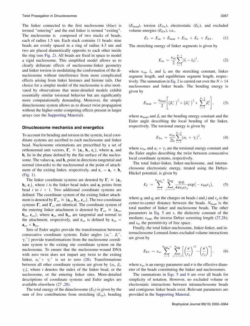
The linker connected to the first nucleosome (blue) istermed ‘‘entering’’ and the end linker is termed ‘‘exiting’’.The nucleosome is composed of two stacks of beads,each of radius 1.5 nm. Each stack contains 11 beads: ninebeads are evenly spaced in a ring of radius 4.3 nm andtwo are placed diametrically opposite to each other insidethe ring (see Fig. 2). All beads are fixed in space to modela rigid nucleosome. This simplified model allows us toclearly delineate effects of nucleosome-linker geometryand linker torsion in modulating the conformation of the di-nucleosome without interference from more complicatedeffects arising from linker histones and histone tails. Ourchoice for a simpler model of the nucleosome is also moti-vated by observations that more-detailed models exhibitessentially similar torsional behavior but are significantlymore computationally demanding. Moreover, the simpledinucleosome system allows us to dissect twist propagationwithout the higher-order competing effects present in largerarrays (see the Supporting Material).
Dinucleosome mechanics and energetics
To account for bending and torsion in the system, local coor-dinate systems are ascribed to each nucleosome and linkerbead. Nucleosome orientations are prescribed by a set oforthonormal unit vectors, Gc h {ac, bc, cc}, where ac andbc lie in the plane defined by the flat surface of the nucleo-some. The values ac and bc point in directions tangential andnormal (inwards) to the nucleosomal at the point of attach-ment of the exiting linker, respectively, and cc ¼ ac # bc(Fig. 1).
The linker coordinate systems are denoted by Gi h {ai,bi, ci}, where i is the linker bead index and ai points frombead i to i þ 1. Two additional coordinate systems aredefined. The coordinate system of the exiting linker attach-ment is denoted byGexh {aex, bex, cex}. The two coordinatesystems Gc and Gex are identical. The coordinate system ofthe entering linker attachment is denoted by Gen h {aen,ben, cen}, where aen and ben are tangential and normal tothe attachment, respectively, and cen is defined by cen ¼aen # ben.
Sets of Euler angles provide the transformation betweenconsecutive coordinate systems. Euler angles {ai
þ, biþ,
giþ} provide transformations from the nucleosome coordi-
nate system to the exiting site coordinate system on thenucleosome. To ensure that the nucleosome-wound DNAwith zero twist does not impart any twist to the exitinglinker, ai
þþ giþ is set to zero (28). Transformations
between all other coordinate systems are given by {ai, bi,gi}, where i denotes the index of the linker bead, or thenucleosome, or the entering linker sites. More-detaileddescriptions of coordinate systems and Euler angles areavailable elsewhere (27 ,28).
The total energy of the dinucleosome (ET) is given by thesum of five contributions from stretching (Estr), bending
(Ebend), torsion (ETw), electrostatic (EC), and excludedvolume energies (EEV), i.e.,
ET ¼ Estr þ Ebend þ ETw þ EC þ EEV: (1)
The stretching energy of linker segments is given by
Estr ¼ kstr
2
XN$1
i¼ 1
ðli $ l0Þ2; (2)
where kstr, li, and l0 are the stretching constant, linkersegment length, and equilibrium segment length, respec-tively. The summation in Eq. 2 is carried out over the N¼ 14nucleosomes and linker beads. The bending energy isgiven by
Ebend ¼ kbend
2
"!bþ1
"2 þ!bþ2
"2 þXN$2
i¼ 1
b2i
#
; (3)
where kbend and bi are the bending energy constant and theEuler angle describing the local bending of the linker,respectively. The torsional energy is given by
ETw ¼ kTw
2l0
XN$2
i¼ 1
ðai þ giÞ2; (4)
where kTw and ai þ gi are the torsional energy constant andthe Euler angles describing the twist between consecutivelocal coordinate systems, respectively.
The total linker-linker, linker-nucleosome, and internu-cleosome electrostatic energy, treated using the Debye-Huckel potential, is given by
EC ¼XNbead$1
i¼ 1
XNbead
j¼ iþ 2
qiqj4pe0erij
exp!$ kDHrij
"; (5)
where qi and qj are the charges on beads i and j and rij is thecenter-to-center distance between the beads. Nbead is thetotal number of linker and nucleosome beads. The otherparameters in Eq. 5 are e, the dielectric constant of themedium; kDH, the inverse Debye screening length (27,28);and e0, the permittivity of free space.Finally, the total linker-nucleosome, linker-linker, and in-
ternucleosome Lennard-Jones excluded volume interactionsare given by
EEV ¼ kevXNbead$1
i¼ 1
XNbead
j¼ iþ 2
#$s
rij
%12
$$s
rij
%6&; (6)
where kev is an energy parameter and s is the effective diam-eter of the beads constituting the linker and nucleosomes.
The summations in Eqs. 5 and 6 are over all beads forsimplicity of notation. However, no excluded volume orelectrostatic interactions between intranucleosome beadsand contiguous linker beads exist. Relevant parameters areprovided in the Supporting Material.
Biophysical Journal 99(10) 3355–3364
Twist Propagation in Dinucleosomes 3357

Monte Carlo twisting protocol
We implement quasistatic twisting of the dinucleosomearray using a Monte Carlo (MC) approach. The initial linkerand nucleosome positions are similar to that in Fig. 1, fora particular q and f. The entry/exit angle q is defined asthe angle between the two linker attachment points on thenucleosome and f is the angle made by the exiting linkerwith the plane of the two nucleosomes. The rightmost nucle-osome (blue) in Fig. 1 a is torsionally and translationallyconstrained; the other (green) nucleosome is free to moveand rotate. The penultimate and last linker beads labeled13 and 14, respectively, in Fig. 2 are also fixed in space.
Before twist application, the dinucleosome is first al-lowed to equilibrate (relax), subject to the constraintsmentioned above. All equilibration phases are carried outusing the Metropolis MC method at constant temperature.Two MC moves are used (27): The first is rotation ofa randomly chosen bead or nucleosome by a random anglein interval [$dr, dr] (dr ¼ 0.3 rad). While linker bead rota-tions are only implemented about its ai axis, nucleosomerotations are chosen randomly about its ac, bc, or cc axis.The second is a translational move by a distance chosenrandomly in the interval [$dt, dt] (dr ¼ 0.3 nm) alonga randomly chosen direction of a random bead or nucleo-some.
Both types of moves are accepted or rejected using thestandard Metropolis criterion based on changes in the totalenergy of the system DE. That is, if DE < 0, the move isaccepted; and if DE > 0, the move is accepted witha probability exp($DE/kBT), where kB is the Boltzmannconstant and T is the temperature. In all cases, we allowthe system to equilibrate for Neq MCmoves before the appli-cation of twist. We use 2# 106 MC steps for each equilibra-tion period.
The average twist in the exiting and entering linkers afterequilibration is Tw0
ex and Tw0en, respectively. After equili-
bration, the penultimate linker bead (bead 13 in Fig. 2, leftpanel) coordinate system is rotated about its local a axisby an angle U0 ¼ 5p/4. The system is then equilibrated
again using the equilibration protocol. Rotations are appliedunidirectionally, in either the clockwise or counterclockwisedirection, which we denote by Uþ and U$, respectively.Subsequent twist is applied using the same stepwise incre-ments and equilibration phases. Fig. 2 shows a plot of thetypical applied and induced twist-versus-MC steps. Ourtwisting method ensures sufficient time for relaxationbetween each subsequent twisting event, thus emulatingthe slow quasistatic twisting of chromatin in experiments.
We carry out a series of such twisting simulations atvarying relative linker orientations, q and f, to probe theireffects on the twisting response of the dinucleosome arrays.These angles are varied in increments of dq ¼ p/12 anddf ¼ p/6, in the range of q ˛(0, 2p), f ˛ [$p/3, p/3].
RESULTS
Twist inversion
We define two parameters, ken and kex, to characterize thefraction of imposed twist stored per linker segment in theexiting and entering linkers,
km ¼2p
!Twm $ Tw0
m
"
NmnU0
; (7)
where m ¼ ex,en, and where Twex and Twen are the averagetwist in the exiting and entering linkers after n applicationsof twist, respectively. The twist in the two linkers is calcu-lated as
Twex ¼ 1
2p
*X12
i¼ 8
ðai þ giÞ
+
; (8)
Twen ¼ 1
2p
*X7
i¼ 1
ðai þ giÞ
+
: (9)
Note that the number of segments that absorb twist in thetwo linkers are different (Nex ¼ 5 and Nen ¼ 7).
Fig. 3 shows kex and ken versus q for several f for positiveapplied twist Uþ during the second phase of twist applica-tion n ¼ 2. Both kex and ken are f-invariant to first-order,but depend strongly on q. Here, kex > 0 for all q- andf-values while, interestingly, ken is negative for a subsetof q. Regions where kex and ken are opposite in sign implyinverted twist. ken¼ 0 for a narrow range of q correspondingto zero twist propagation.
Twist propagation can be more clearly characterized bydefining L ¼ Sgn(ken # kex), where Sgn(x) is the sign of xand Sgn(0) ¼ 0, and L > 0, L ¼ 0, and L < 0 correspondto conserved twist direction; zero twist propagation; andtwist inversion, respectively. Fig. 4 shows twist in a dinu-cleosome with f ¼ $p/3 subjected to negative appliedtwist. The upper panels of Fig. 4 (left to right) show repre-sentative plots of twist in the dinucleosome for q ¼ 3p/2,
FIGURE 2 (Left panel) Twist is applied quasistatically by rotating thelocal coordinate system of the penultimate linker bead about its a axis byan angle U. (Right panel) Schematic of the applied and induced twistfrom the MC simulations.
Biophysical Journal 99(10) 3355–3364
3358 Dobrovolskaia et al.

q¼ p, and q¼ p/6, respectively. Both cases forL> 0 showthat the magnitude of twist in the exiting and enteringlinkers are increasing as the applied total twist increases.The center panel corresponding to L < 0 shows that themagnitude of twist in the exiting and entering linker areincreasing, although in opposite directions. This corre-sponds to twist inversion in the dinucleosome.
We now construct phase diagrams which show relativelinker twist-direction as functions of q and f. These phasediagrams are shown in the two lower panels of Fig. 4 forpositive and negative applied twist. We define three twistpropagation zones. Zones I and III (L > 0) are both twist-direction conserving. Zone II (L < 0), is twist-inverting.Fig. 4 also shows schematic linker conformations in eachzone. Remarkably, the phase diagrams are invariant to fand direction of applied twist. Phase boundaries shown bydashed lines illustrate zero twist propagation, L ¼ 0.
We provide a simple explanation of twist inversion thataccounts for relative linker orientation and nucleosomegeometry. Our Ansatz is that nucleosomes rotate as rigidbodies, resulting from the strong DNA/nucleosome binding.Applied rotations on the exiting linker induce nucleosomerotations, which, in turn, induce rotations in the enteringlinker. We define Gi ¼ ai; bi; ci and G0
i ¼ a0i; b0i; c
0ig
'('to
be the coordinate systems before and after rotation of the
exiting linker, respectively. As shown in Fig. 5,G0 is the coor-dinate system of the entering linker bead, which we assumeto be fixed for our argument; G1 is the coordinate system ofthe entering linker attachment point; G2 is the coordinatesystem of the exiting linker attachment point; and G3 is thecoordinate system of exiting bead being rotated anticlock-wise. That is, the axes b03 and c
03 are rotated while a3 remains
unchanged. A rotation is induced inG2 about the a2 axis, thusrotating the nucleosome. The induced rotation about a2 isalways smaller in magnitude than the applied rotation, result-ing from the linker’s finite torsional rigidity.
Fig. 5, a–c, shows the effect of applied rotation of G3 forthree entry/exit angles: q ¼ 180', 360', and 270', respec-tively. For q¼ 180', the nucleosome rotation causes rotationof G1 about the a1 axis. The relative coordinate system rota-tions and directions required to go from G0 /G1
0 and G20/
G30 are also shown. In fact, the above two rotations are by
definition described by the angles (a0 þ g0) and (a2 þ g2),the twists in the entering and exiting linkers, respectively.Clearly, the two linker twists are opposite in sign, thus re-sulting in twist inversion across the central nucleosome.Similar arguments apply for q ¼ 360' (and 0') as demon-strated in Fig. 5 b. Here, the direction of rotations (a0 þg0) and (a2 þ g2) required to transform G0 / G1
0 andG2
0/ G3
0, respectively, have the same sign. Thus, thetwists in the two linkers are of the same sign. Finally,Fig. 5 c shows the q ¼ 270' (and 90') case. By definition,the Euler angles a0 and g0 can be calculated from the rela-tionship (27)
FIGURE 3 Plots of kex and ken for various f as a function of q for positiveapplied twist, Uþ, for a total applied twist of p/2. Twist inversion occurswhen kex and ken are opposite in sign.
FIGURE 4 The top panel shows representative twist propagation resultsfrom our MC simulations for zones I (q ¼ 3p/2), II (q ¼ p), and III(q ¼ p/6) for counterclockwise applied twist U$ and f ¼ $p/3. Zone IIis twist-inverting. (Solid blue and dashed green lines) Twist in the exitingand entering linker, respectively. (Black and red lines) Applied and inducedtotal twist, respectively. (Bottom panel) Computed regimes of positive (redtriangles) and negative (blue triangles) relative twists L for both directionsof twist.
Biophysical Journal 99(10) 3355–3364
Twist Propagation in Dinucleosomes 3359
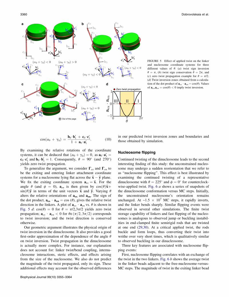
cosða0 þ g0Þ ¼ b0$b01 þ c0$c01
1 þ a0$a01: (10)
By examining the relative rotations of the coordinatesystems, it can be deduced that ða0 þ g0Þ ¼ 0; as a0$a01 ¼c0$c01 and b0$b
01 ¼ 1: Consequently, q ¼ 90' (and 270')
yields zero twist propagation.To generalize the argument, we consider Gex and Gen to
be the exiting and entering linker attachment coordinatesystems for a nucleosome lying flat across the bx $ by plane.We fix the exiting coordinate system aex ¼ bx. For theangle q (and f ¼ 0), aen is then given by cosðqÞbxþsinðqÞby in terms of the unit vectors bx and by. Varying qalters the relative orientations of aex and aen. The sign ofthe dot product, aex $ aen ¼ cos (q), gives the relative twistdirection in the linkers. A plot of aex $ aen vs. q is shown inFig. 5 d: cos(q) ¼ 0 for q ¼ p/2,3p/2 yields zero twistpropagation; aex $ aen < 0 for q˛ðp=2; 3p=2Þ correspondsto twist inversion; and the twist direction is conservedotherwise.
Our geometric argument illustrates the physical origin oftwist inversion in the dinucleosome. It also provides a goodfirst-order approximation of the dependence of the angle qon twist inversion. Twist propagation in the dinucleosomeis actually more complex. For instance, our explanationdoes not account for: linker twist/bend coupling, internu-cleosome interactions, steric effects, and effects arisingfrom the size of the nucleosome. We also do not predictthe magnitude of the twist propagated, only its sign. Theseadditional effects may account for the observed differences
in our predicted twist inversion zones and boundaries andthose obtained by simulation.
Nucleosome flipping
Continued twisting of the dinucleosome leads to the secondinteresting finding of this study: the unconstrained nucleo-some may undergo a sudden reorientation that we refer toas ‘‘nucleosome flipping’’. This effect is best illustrated byexamining the continued twisting of a representativedinucleosome with q ¼ 225' and f ¼ 0' for counterclock-wise-applied twist. Fig. 6 a shows a series of snapshots ofthe dinucleosome conformation versus MC steps. Initially,the unconstrained nucleosome’s orientation remainsunchanged. At ~1.5 # 107 MC steps, it rapidly inverts,and the linker bends sharply. Similar flipping events wereobserved in several other simulations. The finite twiststorage capability of linkers and fast flipping of the nucleo-somes is analogous to observed jump or buckling instabil-ities in end-clamped finite semirigid rods that are twistedat one end (29,30). At a critical applied twist, the rodsbuckle and form loops, thus converting their twist intowrithe over very short times, which is qualitatively similarto observed buckling in our dinucleosome.
Three key features are associated with nucleosome flip-ping events:
First, nucleosome flipping correlates with an exchange ofthe twist in the two linkers. Fig. 6 b shows the average twistin the linker beads adjacent to the free-nucleosome-versus-MC steps. The magnitude of twist in the exiting linker bead
a b
c d
FIGURE 5 Effect of applied twist on the linkerand nucleosome coordinate systems for threedifferent values of q: (a) twist sign inversionq ¼ p, (b) twist sign conservation q ¼ 2p, and(c) zero twist propagation example for q ¼ p/2.(d) Twist inversion zones obtained from a calcula-tion of the dot product of aex $ aen ¼ cos(q). Valuesof aex,aen ¼ cos(q) < 0 imply twist inversion.
Biophysical Journal 99(10) 3355–3364
3360 Dobrovolskaia et al.

increases with larger applied twist. At ~1.5# 107 MC steps,twist propagates from the exiting to the entering linkers.This results in a steep change in the exiting and enteringlinker twists, respectively. Clearly, this exchange is thelargest for cases where the two twists are of opposite signwith respect to each other. Indeed, we observe nucleosomeflipping to be most abrupt for intermediate q-values, corre-sponding to the observed range of twist inversion, andmore gradual for small and large q-values.
Second, an exchange of energy between the differentmodes accompanies nucleosome flipping. Fig. 7 shows theevolution of stretching (Estr), twisting (ETw), bending(Ebend), and Coulombic-plus-excluded-volume (EEVC)energy contributions versus MC steps for the above system.Before flipping (<107 MC steps), ETw increases in the samesteplike manner as the applied twist. The step heightincreases quadratically, as a result of ETw’s quadratic depen-dence on applied twist. We also observe slight increases inEbend, Estr, and EEVC, likely due to the coupling betweenthe bending and twisting modes, the stretching of bentlinkers, and increased electrostatic repulsion between thenucleosomes, respectively. At the onset of flipping, Ebend
rapidly increases while ETw stabilizes. Here, the twistingenergy penalty begins to exceed that associated with thesharp bending of the linkers, thus relieving some of thelinker torsional stress. At this point, linker buckling alsoinduces the abrupt nucleosome flipping, followed byadditional equilibration of the bending and twisting ener-gies. Upon flipping, EEVC also decreases, possibly as a resultof increased separation of the linkers and thereby reducedelectrostatic repulsion. We further note that flipping occursat ~ETw ¼ 22 kcal/mol (~37 kBT). Other simulations withdifferent q and f, in which nucleosome flipping occur, yieldsimilar energy barriers, perhaps suggesting a universality inthe energy required for nucleosome flipping. Note that theabove value is a very rough estimate of the free energy
barrier, as it excludes various contributions includingconformational entropy. It would be interesting to examinethe energetics and kinetics of nucleosome flipping in moredetail.
Third, the imposed twist is not equal to the net observedtwist, which can be interpreted as a change in the overallwrithe of the system. Fig. 6 c shows the average appliedand induced twist in the system along with the total twistin both linkers. The difference between the induced andapplied twist can be explained in terms of the linkingnumber, Lk, given by the sum of the twist and writhe,Lk ¼ TwþWr: Although, strictly speaking, this relation-ship is only applicable to closed curves, we apply it to the
a
b c
FIGURE 6 Nucleosome flipping dynamics fordinucleosomes with q ¼ 225' and f ¼ 0'. (a)Series of snapshots from the MC simulationsshowing evolution of the dinucleosome conforma-tions and associated nucleosome flipping andlinker buckling. (b) Applied and induced twist inlinkers on the exiting (red) and entering linkers(black), respectively. (c) Total applied and inducedtwist (solid black and red lines) and the total twiston exiting and entering linker beads adjacent to thenucleosome (dashed green and solid blue lines).
FIGURE 7 Dinucleosome energy contributions from twist ETw, bendingEbend, stretching Estr, and excluded volume plus Coulombic interactionsEEVC for dinucleosome simulations with q ¼ 225' and f ¼ 0'.
Biophysical Journal 99(10) 3355–3364
Twist Propagation in Dinucleosomes 3361

dinucleosome under the assumption that the constrainedends are connected by a phantom curve. Similar approacheshave been used by others to estimate writhe in open curves(15,31,32). The change in linking number DLk is then equalto DTwþ DWr $ Twap; where Twap is the applied twist.Because DLk ¼ 0 for closed curves, DWr ¼ Twap $ DTwand a numerical estimate of twist-to-writhe conversion isthe difference in the applied and induced twist.
To better illustrate this point, we present an additionalsystem with q ¼ 90' and f ¼ 60' that undergoes a largerconformation change (Fig. 8). As with the previousexample, applied and induced twist are not equal. The spon-taneous occurring twist during the equilibration of the arrayis denoted Tw0ð¼ Tw0
ex þ Tw0enÞ: At the end of the simula-
tion DWrx$ 0:64; illustrating the significant change inthe overall writhe. The change in writhe is correlated witha conformational change of the dinucleosome. Fig. 8 alsoshows the initial and final conformation of the dinucleo-some. The dashed red line indicates that the dinucleosomeis treated as a closed loop. We approximate the directionalwrithe as the sum of the signed crossings of the projectionof the linker path onto a plane. The writhes for the initialand final conformations are Wr ¼ 0 and Wr ¼ $1, yieldingDWr ¼ $1. Thus, the directional writhe change computedfrom the above approximate approach exhibits the samesign as that computed from the difference in the measured
and applied twists—further confirming twist-to-writheconversion in nucleosome flipping.
DISCUSSION
Our simulations furnish two intriguing observations abouttwist propagation in dinucleosome arrays:
First, applied twist may lead to inverted twist in contig-uous linkers. This phenomenon is a result of strong DNA/octamer binding that inhibits twist propagation along thewound DNA; twist propagation instead occurs via rigid-body-like rotations of the entire nucleosome. Hence, twistinversion is strongly dependent on the relative orientationof contiguous linkers at their point of entry and exit to thenucleosome.
Second, continued twisting in dinucleosome arrays maylead to nucleosome flipping and associated linker buckling.The latter occurs to relieve excessive stored twist in onelinker; nucleosome flipping events facilitate quick transferbetween contiguous linkers and also results in conversionof twist to writhe.
Our twist inversion finding leads to the obvious question:Can twist inversion occur in real chromatin fiber?
Our simulations suggest that a single parameter, the entry/exit angle q, dictates twist inversion, and that angles in therange 90')q)220' are required for twist inversion tooccur. The entry/exit angles in the nucleosome crystal struc-ture, q0, are in the range 108'–126' (22), corresponding to1.65–1.7 turns of wound DNA, well within the predictedrange of twist inversion. However, q is dependent on saltconditions, and even at fixed salt conditions, q exhibits largevariations due to spontaneous unwrapping of DNA (33). Atlow salt concentrations, chromatin exhibits an unfoldedconformation. The strong repulsion between the enteringand exiting linkers causes them to diverge, resulting ina large increase in q, potentially exceeding the upper anglelimit of twist inversion. In contrast, at high salt conditions,chromatin is tightly folded and the wound DNA likely main-tains its original entry/exit angle q0. Hence, we expectunfolded chromatin at low salt concentrations to exhibitunidirectional twist and strongly folded chromatin at phys-iological salt conditions to exhibit twist inversion (bimodaltwist). Because the nucleosomes exhibit more restricteddynamics in chromatin fibers due to packing and histonetail interactions, it is difficult to extrapolate how far theunidirectional and inverted twists propagate along the fiberfrom our single-nucleosome results.
What are the possible implications of twist inversion?One possibility is that twist inversion could allow creation
of alternate regions of overwound and underwound DNA,which could potentially have specific roles in chromatinfunction. Some proteins are known to preferentially bindto either over- or underwound DNA (9,34–36). For example,the activity of the 434 repressor is dictated by the degree towhich DNA is wound at its operator site (36). Also,
FIGURE 8 Total applied and induced twist (solid black and red lines) andthe total twist on exiting and entering linker beads adjacent to the nucleo-some (dashed green and solid blue lines) for q¼ 90' and f¼ 60'. Tw0 is thespontaneously occurring twist in the linker after equilibration and DWr isthe change in writhe of the system at the end of the simulation. Also shownare the starting and final dinucleosome conformations and the projections oftheir linker paths onto a plane for counting the number of positive and nega-tive crossings for directional writhe calculations. (Dashed red line)Phantom curve drawn to close the linker trajectory.
Biophysical Journal 99(10) 3355–3364
3362 Dobrovolskaia et al.

architectural proteins such as SRY prefer binding to over-twisted DNAwhile zinc finger proteins prefer undertwistedDNA (35). Opposite twists on contiguous linkers couldtherefore potentially enhance interactions between over-and underwound DNA-binding proteins. Twist inversioncould also play a role in chromatin dynamics, especiallyunder external twisting, as the direction of nucleosome rota-tion depends on the twists in the entering and exiting linkers.In addition, coupling between twist-inverting and non-twist-inverting zones could modulate local nucleosome rotations,providing a method to modify local chromatin conformationand to locally over- or under-twist chromatin, depending onits topology.
The continued twisting of dinucleosomes induces sharpbending of the linker. Because some DNA sequences aremore susceptible to bending than others, such torsion-drivenbuckling may provide a mechanism to modulate site-specific bending in chromatin (37). Localized buckling couldsynergize the binding of specific regulatory proteins thatpreferentially bind to curved DNA. In particular, TATAsequences that play a key role in transcription by bindingto various transcriptional factors are prone to bending.Hence, torsional forces from various cellular machinerycould potentially instigate conformational changes nearstress-sensitive sites, such as TATA boxes, to regulate thetranscriptional activity of chromatin (38). If these torquesinduce nucleosome flipping and buckling of linker, it mayprovide a method of governing local protein binding thatcould regulate protein binding and transcription. The conver-sion of twist to bending via local buckling of DNA may alsoserve as a transient reservoir or dynamic buffer to absorbtwist in chromatin without causing drastic changes in itsconformation (38). The dependence of the abruptness ofnucleosome flipping on linker orientation may also play anas-yet-unknown role in chromatin’s ability to transientlyabsorb torsional stress.
Our energetic analysis indicates that significant twist canbe applied before inducing nucleosome flipping. If, forexample, the applied twist originates from an advancingpolymerase that exerts torques of ~1.25 kBT/rad, flippingwould occur after 56 polymerase rotations, correspondingto the ~37 kBT energy barrier observed in our simulations.In our example, the ~180' nucleosome flipping transitionoccurs after ~270' of twist has been applied. The highenergetic barrier of flipping indicates that dinucleosomescan transiently absorb applied twist and act as reservoirsfor applied twist. Chromatin’s strong twist storing capacitycontrasts sharply with that of DNA, testifying to chromatin’sstructural stability. Larger chromatin arrays will undoubt-edly exhibit a more complex interplay among twist, bend-ing, and internucleosome interactions, and much remainsto be understood about their fundamental physics, includinghow they propagate twist.
In summary, we expect that twist inversion and nucleo-some flipping are real phenomena that could play important
roles in chromatin function. It may be possible to verifytheir existence experimentally using, for instance, singlemolecule techniques (24).
CONCLUSIONS
We have presented a systematic Monte Carlo study of thetorsional response of end-constrained dinucleosome arrays.Our results suggest that the propagation of twist withina dinucleosome strongly depends on the relative orientationof contiguous DNA linkers, a subset of which leads to in-verted twist. Our intuitive explanation of twist inversionillustrates how simple constraints on DNA imposed by thenucleosome lead to very unexpected consequences for twistpropagation along DNA in nucleosome arrays. Our resultsalso demonstrate how dinucleosomes subjected to continuedtwisting can undergo dramatic conformational changes,nucleosome flipping. This flipping occurs at energy barriersthat indicate chromatin’s significant ability to transientlystore twist. Our study thus demonstrates how simplemodeling and simulations provide an effective tool to probenucleosome dynamics and to uncover previously unknownproperties of chromatin. Continued modeling efforts, atvarying levels of complexity, will undoubtedly uncovermore fundamental details of chromatin dynamics. Specifi-cally, our future work will focus on the torsional propertiesof large chromatin arrays containing tens to hundreds ofnucleosomes, with the aim of detailing how applied torsionaffects chromatin higher-order structure.
SUPPORTING MATERIAL
One table and two figures are available at http://www.biophysj.org/biophysj/supplemental/S0006-3495(10)01205-1.
Computer time on the Granite cluster of the Bioengineering department atthe University of California at San Diego is acknowledged. The authorsacknowledge various discussions with Dario Meluzzi, Balaji Iyer, DarrenYang, and Arijit Maitra.
G.A. thanks the Hellman Fellowship and the Chancellor’s InterdisciplinaryCollaboratories program at the University of California at San Diego forfunding.
REFERENCES
1. Alberts, B., A. Johnson, ., P. Walter. 2002. Molecular Biology of theCell. Garland Science, New York.
2. Lavelle, C. 2008. DNA torsional stress propagates through chromatinfiber and participates in transcriptional regulation. Nat. Struct. Mol.Biol. 15:123–125.
3. Bancaud, A., G. Wagner,., A. Prunell. 2007. Nucleosome chiral tran-sition under positive torsional stress in single chromatin fibers. Mol.Cell. 27:135–147.
4. Liu, L. F., and J. C. Wang. 1987. Supercoiling of the DNA templateduring transcription. Proc. Natl. Acad. Sci. USA. 84:7024–7027.
5. Strick, T. R., J. F. Allemand,., V. Croquette. 1998. Behavior of super-coiled DNA. Biophys. J. 74:2016–2028.
Biophysical Journal 99(10) 3355–3364
Twist Propagation in Dinucleosomes 3363

6. Ng, H.-L., and R. E. Dickerson. 2002. Mediation of the A/B-DNA helixtransition by G-tracts in the crystal structure of duplex CATGGGCC-CATG. Nucleic Acids Res. 30:4061–4067.
7. Charvin, G., J.-F. Allemand, ., V. Croquette. 2004. Twisting DNA:single molecule studies. Contemp. Phys. 45:383–403.
8. Imbalzano,A.N., H.Kwon,., R. E.Kingston. 1994. Facilitated bindingof TATA-binding protein to nucleosomal DNA. Nature. 370:481–485.
9. Hatfield, G. W., and C. J. Benham. 2002. DNA topology-mediatedcontrol of global gene expression in Escherichia coli. Annu. Rev. Genet.36:175–203.
10. Lynch, T. W., E. K. Read, ., P. A. Rice. 2003. Integration host factor:putting a twist on protein-DNA recognition. J. Mol. Biol. 330:493–502.
11. Mazur, A. K. 2010. Anharmonic torsional stiffness of DNA revealedunder small external torques. Phys. Rev. Lett. 105:018102.
12. Schoeffler, A. J., and J. M. Berger. 2008. DNA topoisomerases: har-nessing and constraining energy to govern chromosome topology. Q.Rev. Biophys. 41:41–101.
13. Nitiss, J. L. 2009. DNA topoisomerase II and its growing repertoire ofbiological functions. Nat. Rev. Cancer. 9:327–337.
14. Kouzine, F., S. Sanford, ., D. Levens. 2008. The functional responseof upstream DNA to dynamic supercoiling in vivo. Nat. Struct. Mol.Biol. 15:146–154.
15. Rossetto, V., and A. C. Maggs. 2003. Writhing geometry of open DNA.J. Chem. Phys. 21:9864–9874.
16. Meluzzi, D., D. E. Smith, and G. Arya. 2010. Biophysics of knotting.Annu. Rev. Biophys. 39:349–366.
17. Smith, S. B., Y. Cui, and C. Bustamante. 1996. Overstretching B-DNA:the elastic response of individual double-stranded and single-strandedDNA molecules. Science. 271:795–799.
18. Strick, T. R., J.-F. Allemand, ., V. Croquette. 1996. The elasticity ofa single supercoiled DNA molecule. Science. 271:1835–1837.
19. Wereszczynski, J., and I. Andricioaei. 2006. On structural transitions,thermodynamic equilibrium, and the phase diagram of DNA andRNA duplexes under torque and tension. Proc. Natl. Acad. Sci. USA.103:16200–16205.
20. Klenin, K., and J. Langowski. 2000. Computation of writhe inmodeling of supercoiled DNA. Biopolymers. 54:307–317.
21. Merlitz, H., K. Rippe, ., J. Langowski. 1998. Looping dynamics oflinear DNA molecules and the effect of DNA curvature: a study byBrownian dynamics simulation. Biophys. J. 74:773–779.
22. Davey, C. A., D. F. Sargent, ., T. J. Richmond. 2002. Solvent medi-ated interactions in the structure of the nucleosome core particle at1.9 A resolution. J. Mol. Biol. 319:1097–1113.
23. Felsenfeld, G., and M. Groudine. 2003. Controlling the double helix.Nature. 421:448–453.
24. Bancaud, A., N. Conde e Silva, ., J. L. Viovy. 2006. Structural plas-ticity of single chromatin fibers revealed by torsional manipulation.Nat. Struct. Mol. Biol. 13:444–450.
25. Gupta, P., J. Zlatanova, and M. Tomschik. 2009. Nucleosome assemblydepends on the torsion in the DNA molecule: a magnetic tweezersstudy. Biophys. J. 97:3150–3157.
26. Celedon, A., I. M. Nodelman, ., S. X. Sun. 2009. Magnetic tweezersmeasurement of single molecule torque. Nano Lett. 9:1720–1725.
27. Arya, G., Q. Zhang, and T. Schlick. 2006. Flexible histone tails ina new mesoscopic oligonucleosome model. Biophys. J. 91:133–150.
28. Beard, D. A., and T. Schlick. 2001. Computational modeling predictsthe structure and dynamics of chromatin fiber. Structure. 9:105–114.
29. Neukirch, S., G. H. M. van der Heijden, and J. M. T. Thompson. 2002.Writhing instabilities of twisted rods: from infinite to finite length.J. Mech. Phys. Solids. 50:1175–1191.
30. van der Heijden, G. H. M., S. Neukirch, ., J. M. T. Thompson. 2003.Instability and self-contact phenomena in the writhing of clamped rods.Int. J. Mech. Sci. 45:161–196.
31. van der Heijden, G. H. M., M. A. Peletier, and R. Planque. 2007. On-end rotation for open rods undergoing large deformations. Q. Appl.Math. 65:385–402.
32. Starostin, E. L. 2005. On the writhe of non-closed curves. Physical andNumerical Models in Knot Theory. World Scientific, Singapore, pp.525–545.
33. Polach, K. J., and J. Widom. 1995. Mechanism of protein access tospecific DNA sequences in chromatin: a dynamic equilibrium modelfor gene regulation. J. Mol. Biol. 254:130–149.
34. Remus, D., E. L. Beall, and M. R. Botchan. 2004. DNA topology, notDNA sequence, is a critical determinant for Drosophila ORC-DNAbinding. EMBO J. 23:897–907.
35. Rohs, R., X. Jin,., R. S. Mann. 2010. Origins of specificity in protein-DNA recognition. Annu. Rev. Biochem. 79:233–269.
36. Koudelka, G. B. 1998. Recognition of DNA structure by 434 repressor.Nucleic Acids Res. 26:669–675.
37. Kouzine, F., J. Liu, ., D. Levens. 2004. The dynamic response ofupstream DNA to transcription-generated torsional stress. Nat. Struct.Mol. Biol. 11:1092–1100.
38. Lavelle, C., J.-M. Victor, and J. Zlatanova. 2010. Chromatin fiberdynamics under tension and torsion. Int. J. Mol. Sci. 11:1557–1579.
Biophysical Journal 99(10) 3355–3364
3364 Dobrovolskaia et al.