two new subfamilies of dna mismatch repair proteins (muts
TRANSCRIPT
ORIGINAL ARTICLE
Two new subfamilies of DNA mismatch repairproteins (MutS) specifically abundant in themarine environment
Hiroyuki Ogata1, Jessica Ray2, Kensuke Toyoda3,4, Ruth-Anne Sandaa2, Keizo Nagasaki3,Gunnar Bratbak2 and Jean-Michel Claverie1
1Information Genomique et Structurale, CNRS-UPR2589, Institut de Microbiologie de la Mediterranee,Parc Scientifique de Luminy, Aix-Marseille Universite, Marseille Cedex 9, France; 2Department of Biology,University of Bergen, Bergen, Norway and 3Harmful Algal Bloom Division, National Research Instituteof Inland Sea, Fisheries Research Agency, Hiroshima, Japan
MutS proteins are ubiquitous in cellular organisms and have important roles in DNA mismatch repairor recombination. In the virus world, the amoeba-infecting Mimivirus, as well as the recentlysequenced Cafeteria roenbergensis virus are known to encode a MutS related to the homologsfound in octocorals and e-proteobacteria. To explore the presence of MutS proteins in other viralgenomes, we performed a genomic survey of four giant viruses (‘giruses’) (Pyramimonas orientalisvirus (PoV), Phaeocystis pouchetii virus (PpV), Chrysochromulina ericina virus (CeV) andHeterocapsa circularisquama DNA virus (HcDNAV)) that infect unicellular marine algae. Our analysisrevealed the presence of a close homolog of Mimivirus MutS in all the analyzed giruses. These viralhomologs possess a specific domain structure, including a C-terminal HNH-endonuclease domain,defining the new MutS7 subfamily. We confirmed the presence of conserved mismatch recognitionresidues in all members of the MutS7 subfamily, suggesting their role in DNA mismatch repair ratherthan DNA recombination. PoV and PpV were found to contain an additional type of MutS, which wepropose to call MutS8. The MutS8 proteins in PoV and PpV were found to be closely related tohomologs from ‘Candidatus Amoebophilus asiaticus’, an obligate intracellular amoeba-symbiontbelonging to the Bacteroidetes. Furthermore, our analysis revealed that MutS7 and MutS8 areabundant in marine microbial metagenomes and that a vast majority of these environmentalsequences are likely of girus origin. Giruses thus seem to represent a major source of theunderexplored diversity of the MutS family in the microbial world.The ISME Journal (2011) 5, 1143–1151; doi:10.1038/ismej.2010.210; published online 20 January 2011Subject Category: evolutionary geneticsKeywords: mimivirus; girus; virus; DNA repair; MutS
Introduction
Large DNA viruses carry genes for their own DNArepair apparatus to enhance the accuracy of genomereplication (Furuta et al., 1997; Srinivasan andTripathy, 2005; Redrejo-Rodriguez et al., 2009;Bogani et al., 2010). The amoeba-infecting Mimi-virus (Acanthamoeba polyphaga mimivirus, APMV)with the largest genome (1.2 Mb) of all knownviruses encodes eight putative genes for DNA repairenzymes capable of correcting mismatches or errorsinduced by oxidation, UV irradiation and alkylating
agents (Raoult et al., 2004). Most of these genes werenever found in a viral genome until their discoveryin Mimivirus. One of these corresponds to a MutShomolog (open reading frame (ORF) L359) predictedto function in DNA mismatch repair (MMR) orrecombination. MMR recognizes and corrects base–base mismatches and small insertion or deletionloops introduced during replication, leading to50- to 1000-folds enhancement of replication fidelityin cellular organisms (Schofield and Hsieh, 2003;Iyer et al., 2006). The best-studied MMR system isthe Escherichia coli MutS–MutL–MutH pathway.In the first step in this pathway, the MutS homodimerbinds the site of a mismatch (or a loop) in double-strand DNA. The MutS protein recruits the ‘linkerprotein’ MutL and together activates the endonu-clease MutH, which nicks specifically the newlysynthesized DNA strand to initiate DNA excisionand resynthesis pathway. Homologs of E. coli MutShave been found in many species of bacteria,archaea and eukaryotes, and together classified in
Received 28 September 2010; revised 9 December 2010; accepted12 December 2010; published online 20 January 2011
Correspondence: H Ogata, Information Genomique et Structurale,CNRS-UPR2589, Institut de Microbiologie de la Mediterranee,Parc Scientifique de Luminy, Aix-Marseille Universite, 163Avenue de Luminy, Case 934, Marseille Cedex 9 13288, France.E-mail: [email protected] address: Department of Botany, Keio University, Hiyoshi,Kohoku-ku, Yokohama, Kangawa 223-8521, Japan.
The ISME Journal (2011) 5, 1143–1151& 2011 International Society for Microbial Ecology All rights reserved 1751-7362/11
www.nature.com/ismej

the MutS family (Eisen, 1998; Lin et al., 2007).In viruses, MutS homologs have only been foundin Mimivirus, the closely related Mamavirus(La Scola et al., 2008; Yutin et al., 2009), and morerecently in the giant marine virus, Cafeteriaroenbergensis virus (CroV) with a 730-kb genome(Fischer et al., 2010).
The phyletic distribution of the close homologs ofMimivirus MutS is notable. The Mimivirus MutShomolog is most closely related to the homologsfound in the mitochondrial genomes of a groupof animals (that is, octocorals) and several genomesof the e-Proteobacteria such as Sulfurimonas,Nitratiruptor and Arcobacter (Claverie et al., 2006,2009). Octocorals (phylum Cnidaria, class Antho-zoa, subclass Octocorallia) include diverse speciesof corals (for example, soft corals, sea fans, seapens), representing important members of marinecommunities from shallow tropical coral reefs to thedeep sea (McFadden et al., 2006). A mutS homologhas been found encoded in the mitochondria of alloctocorals, including the three major orders Alcyon-cea, Helioporacea and Pennatulacea, but not in themitochondrial genomes of any other eukaryotes,including those of the sister subclass Hexacorallia(for example, stony corals, sea anemones) (Pont-Kingdon et al., 1995; Brugler and France, 2008). Thee-proteobacteria Sulfurimonas and Nitratiruptor aresulfur-oxidizing chemoautotrophs and often foundin deep-sea hydrothermal vent or coastal sediments(Nakagawa et al., 2007; Sievert et al., 2008).Arcobacter includes species of water-borne patho-gens and taxonomically close to Campylobacterjejuni and Helicobacter pylori (Miller et al., 2007).The common origin of these MutS homologs isfurther suggested by their atypical domain organiza-tion. Distinct from all other MutS family proteins,the MutS homologs in Mimivirus, octocorals and thee-proteobacteria are fused with a C-terminal HNHnicking endonuclease domain (Malik and Henikoff,2000; Claverie et al., 2009). The domain fusion waspredicted to make these enzymes a ‘self-contained’single polypeptide having both mismatch reco-gnition (MutS) and nicking (MutH) functions(Malik and Henikoff, 2000). The distributionof these unique MutS homologs is thus limited toa few totally unrelated lineages (that is, Mimivirus, asingle subclass of animals, and the e-Proteobacteria)and suggests the occurrence of gene transferbetween their ancestors (Claverie et al., 2009).We introduce the MutS7 subfamily to denote thisspecific group of MutS proteins.
DNA viruses with genomes greater than 300 kb upto 1.2 Mb are being discovered with increasingfrequency from diverse ecosystems, with many ofthem now being subject to genome sequencinganalysis (La Scola et al., 2010; Van Etten et al.,2010). The double-stranded DNA (dsDNA) genomesof these giant viruses (often called ‘giruses’ (Claverieet al., 2006; Claverie and Ogata, 2009)) show a highcoding potential with more than several hundred of
genes densely packed in their genomes. To investi-gate the presence of Mimivirus-like mutS genein other giruses, we have undertaken a genomicsequencing survey of four giruses previously iso-lated from marine environments. The four girusesinvestigated are Pyramimonas orientalis virus (PoV-01B, 560-kb genome), Phaeocystis pouchetii virus(PpV-01B, 485-kb genome), Chrysochromulina ericinavirus (CeV-01B, 510-kb genome) and Heterocapsacircularisquama virus (HcDNAV, 356-kb genome)(Jacobsen et al., 1996; Sandaa et al., 2001; Tarutaniet al., 2001). The hosts of these viruses arephylogenetically distant and ecologically distinctunicellular marine algae. C. ericina and P. pouchetiiare haptophytes classified in different orders, thatis, Prymnesiales and Phaeocystales, respectively.C. ericina has a worldwide distribution; it occursmost commonly in low numbers but has been observedto form blooms together with other Chrysochromulinaspecies (Simonsen and Moestrup, 1997). P. pouchetiimay both be a free-swimming flagellated celland a non-flagellated cell embedded in gelatinouscolonies that form dense blooms in polar andsub-polar regions. P. orientalis is a non-bloomingprasinophyte belonging to the green algae (Chloro-phyta). H. circularisquama is a small thecatedinoflagellate which frequently forms large-scalered tides in Japan causing mass mortality of shellfish(Tarutani et al., 2001). These four giruses are all lyticviruses belonging to the nucleo-cytoplasmic largeDNA virus (NCLDV) superfamily (Yutin et al., 2009).Phylogenetic analysis of DNA polymerase and majorcapsid protein sequences has revealed that PoV, PpVand CeV form a monophyletic clade that clusterstogether with Mimivirus (Larsen et al., 2008; Monieret al., 2008). In contrast, the DNA polymerasesequence of HcDNAV has been found to be closelyrelated to that of African swine fever virus (Ogataet al., 2009), suggesting that HcDNAV is phylo-genetically distant from the other viruses includedin this study.
With the accumulation of genome sequencesand the following phylogenetic studies during thelast decade, a significant advance has been maderegarding the classification of diverse MutS proteins(Eisen, 1998; Lin et al., 2007). In this study, we usethe following naming of the MutS subfamilies, whichis adapted from the recent study by Lin et al. (Linet al., 2007). In total, 12 subfamilies were previouslydescribed to compose the MutS family: ‘MutS1/MSH1’ including E. coli MutS and the mitochon-dria-targeted fungal MutS homolog 1 (MSH1);‘MutS2’, known to inhibit recombination in H. pylori(Pinto et al., 2005) and to possess a C-terminalendonuclease domain called the small MutS-related(Smr) domain (Moreira and Philippe, 1999; Fukuiet al., 2008); ‘MSH2’, ‘MSH3’, ‘MSH4’, ‘MSH5’ and‘MSH6/7’, found in most eukaryotes (with theexception of MSH7 being a plant-specific paralogousgroup of MSH6 (Wu et al., 2003)); another plant-specific MSH1 (called ‘plt-MSH1’ hereafter) with the
Girus-encoded MutS in marine environmentH Ogata et al
1144
The ISME Journal

GIY-YIG endonuclease domain at their C-terminus(Abdelnoor et al., 2006); ‘MutS3’, ‘MutS4’ and‘MutS5’, recently described but functionally unchar-acterized prokaryotic homologs (Lin et al., 2007), andthe above mentioned ‘MutS7’ subfamily representedby the Mimivirus MutS homolog.
In this report, we analyze the MutS sequencesnewly identified in four giruses, and assess theabundance of their homologs in environmentalsequence databases.
Materials and methods
Girus MutS sequencesAs part of an ongoing genome-sequencing project,we obtained assembled contigs for three previouslyisolated dsDNA viruses, Pyramimonas orientalisvirus (PoV-01B, 141 contigs), Phaeocystis pouchetiivirus (PpV-01B, 287 contigs) and Chrysochromulinaericina virus (234 contigs, CeV-01B) (Larsen et al.,2008). This sequence information will be publishedelsewhere. In this study presented here, we scannedthese girus contigs for the presence of MutShomologs. Two complete MutS ORFs were readilyidentified in each of the PoV and PpV contigs. Partof a contig corresponding to the CeV MutS ORF wastargeted for PCR amplifications using overlappingsets of primers and re-sequenced to resolveambiguities in the contig (see Supplementary TableS1). A fragmented ORF for the HcDNAV MutS wasidentified in the previously-described low coverageshotgun sequencing data (Ogata et al., 2009).We obtained a complete ORF for the HcDNAV MutSafter several trials of TAIL-PCR and sequencing.These sequences were submitted to public DNAdatabases (DDBJ: AB587728; EMBL: FR691705-FR691709). The MutS sequence from CroV (crov486,YP_003970119), that only became recently avail-able, was partially included in our analysis.
Bioinformatics analysisReference MutS sequences except the girus sequencesdetermined in this study were retrieved from theUniProt protein sequence database (as of April 27,2010) (UniProtConsortium, 2010). The selection ofsequences was performed to maximize the coverageof diverse MutS subfamilies, referring to previ-ous publications (Eisen, 1998; Lin et al., 2007),through iterative process involving clustering byBLASTCLUST (Altschul et al., 1997), inspection ofsequence alignment and phylogenetic reconstruc-tion. We used T-Coffee version 8.06 (Notredameet al., 2000) for multiple sequence alignment.We used ClustalX (Larkin et al., 2007) for thevisualization of alignments. All gap-containing siteswere removed from the alignments for the followingphylogenetic analyses. Maximum-likelihood phylo-genetic analyses were performed using PhyMLversion 3.0 (Guindon and Gascuel, 2003) using LG
substitution matrix (Le and Gascuel, 2008) and agamma low (four rate categories). We used ProtTestversion 2.2 (Abascal et al., 2005) to determinethe best substitution model (that is, LG) for ourphylogenetic reconstruction based on the MutSdomain V sequences. Phylogenetic trees were drawnusing MEGA version 4 (Kumar et al., 2008). For thedelineation of the sequence domains, we usedHMMER/HMMSEARCH version 2.3.2 (Eddy, 1996)and PSI-BLAST (Altschul et al., 1997). The assign-ment of environmental sequences on the MutS7 andMutS8 subtrees was performed using a maximum-likelihood method implemented in the ‘phylo-genetic placement’ software developed by Matsenet al. (pplacer version 1.0; http://matsen.fhcrc.org/pplacer/). The results were visualized usingArchaeopteryx version 0.957 (http://www.phylosoft.org/archaeopteryx/) (Han and Zmasek, 2009). Corre-spondence analysis of codon usages was performedusing CodonW version 1.3 (http://codonw.sourceforge.net/).
Results
Two types of MutS homologs in girusesWe identified six ORFs similar to known MutSfamily proteins in the analyzed viral genomicsequences. These ORFs were classified into twogroups according to their length and sequencesimilarity. The first group of ORFs was relativelylong and was found in all the analyzed giruses (PoV,910 amino-acid residues (aa); PpV, 1004 aa;CeV, 1043 aa; HcDNAV, 953 aa). When searchedagainst the NCBI non-redundant protein sequencedatabase using BLAST (Altschul et al., 1997), thesegirus MutS homologs showed the most significantsequence similarities to the MutS7 homologs inMimivirus (amino-acid sequence identity 31–38%;alignment coverage 34–99%; E-value¼ 10�63
B10�120), e-proteobacteria (29–37%; 95–99%;10�100B10�153) and octocorals (26–28%; 96–99%;10�67B10�84). Like previously reported MutS7homologs, these four predicted proteins were foundto possess a C-terminal HNH endonuclease domain(Supplementary Figure S1). The second group ofshorter ORFs similar to MutS proteins was foundin PoV (539 aa) and PpV (600 aa). These PoV andPpV MutS homologs showed the most significantsequence similarity in ‘Candidatus Amoebophilusasiaticus’ (Aasi_0916; amino-acid sequence identity38%; alignment coverage 39%; E-value¼ 2� 10�26)and Clostridium perfringens (YP_694765.1; 34%;32%; 2� 10�17), respectively.
Girus MutS homologs correspond to two distinctsubfamiliesTo classify the newly identified girus MutS homo-logs, we compiled a reference sequence set contain-ing 150 MutS homologs, representing diverse MutSsubfamilies, and performed phylogenetic analyses.
Girus-encoded MutS in marine environmentH Ogata et al
1145
The ISME Journal
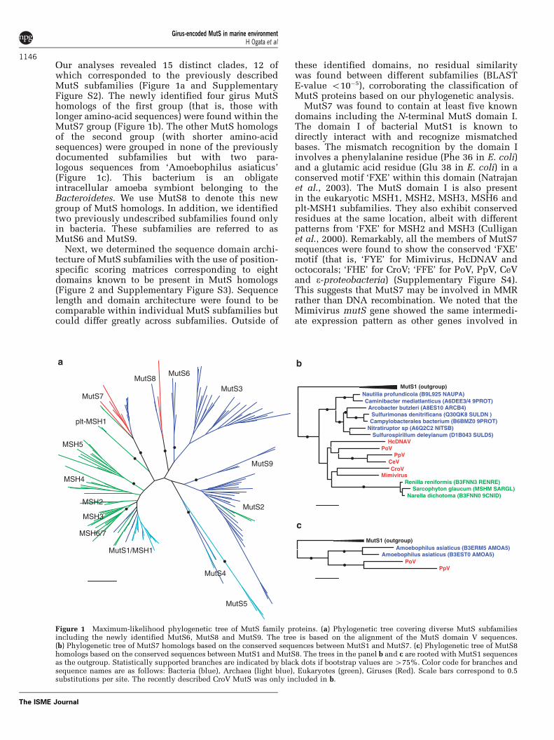
Our analyses revealed 15 distinct clades, 12 ofwhich corresponded to the previously describedMutS subfamilies (Figure 1a and SupplementaryFigure S2). The newly identified four girus MutShomologs of the first group (that is, those withlonger amino-acid sequences) were found within theMutS7 group (Figure 1b). The other MutS homologsof the second group (with shorter amino-acidsequences) were grouped in none of the previouslydocumented subfamilies but with two para-logous sequences from ‘Amoebophilus asiaticus’(Figure 1c). This bacterium is an obligateintracellular amoeba symbiont belonging to theBacteroidetes. We use MutS8 to denote this newgroup of MutS homologs. In addition, we identifiedtwo previously undescribed subfamilies found onlyin bacteria. These subfamilies are referred to asMutS6 and MutS9.
Next, we determined the sequence domain archi-tecture of MutS subfamilies with the use of position-specific scoring matrices corresponding to eightdomains known to be present in MutS homologs(Figure 2 and Supplementary Figure S3). Sequencelength and domain architecture were found to becomparable within individual MutS subfamilies butcould differ greatly across subfamilies. Outside of
these identified domains, no residual similaritywas found between different subfamilies (BLASTE-value o10�5), corroborating the classification ofMutS proteins based on our phylogenetic analysis.
MutS7 was found to contain at least five knowndomains including the N-terminal MutS domain I.The domain I of bacterial MutS1 is known todirectly interact with and recognize mismatchedbases. The mismatch recognition by the domain Iinvolves a phenylalanine residue (Phe 36 in E. coli)and a glutamic acid residue (Glu 38 in E. coli) in aconserved motif ‘FXE’ within this domain (Natrajanet al., 2003). The MutS domain I is also presentin the eukaryotic MSH1, MSH2, MSH3, MSH6 andplt-MSH1 subfamilies. They also exhibit conservedresidues at the same location, albeit with differentpatterns from ‘FXE’ for MSH2 and MSH3 (Culliganet al., 2000). Remarkably, all the members of MutS7sequences were found to show the conserved ‘FXE’motif (that is, ‘FYE’ for Mimivirus, HcDNAV andoctocorals; ‘FHE’ for CroV; ‘FFE’ for PoV, PpV, CeVand e-proteobacteria) (Supplementary Figure S4).This suggests that MutS7 may be involved in MMRrather than DNA recombination. We noted that theMimivirus mutS gene showed the same intermedi-ate expression pattern as other genes involved in
MutS1/MSH1
MSH2
MSH5
MSH6/7
MSH3
MSH4
plt-MSH1
MutS7
MutS8MutS6
MutS3
MutS5
MutS2
MutS4
MutS9
MutS1 (outgroup)Amoebophilus asiaticus (B3ERM5 AMOA5)
Amoebophilus asiaticus (B3EST0 AMOA5)PoV
PpV
MutS1 (outgroup)Nautilia profundicola (B9L925 NAUPA)Caminibacter mediatlanticus (A6DEE3/4 9PROT)
Arcobacter butzleri (A8ES10 ARCB4)Sulfurimonas denitrificans (Q30QK8 SULDN )Campylobacterales bacterium (B6BMZ0 9PROT)
Nitratiruptor sp (A6Q2C2 NITSB)Sulfurospirillum deleyianum (D1B043 SULD5)
HcDNAVPoV
PpVCeVCroV
MimivirusRenilla reniformis (B3FNN3 RENRE)
Sarcophyton glaucum (MSHM SARGL)Narella dichotoma (B3FNN0 9CNID)
Figure 1 Maximum-likelihood phylogenetic tree of MutS family proteins. (a) Phylogenetic tree covering diverse MutS subfamiliesincluding the newly identified MutS6, MutS8 and MutS9. The tree is based on the alignment of the MutS domain V sequences.(b) Phylogenetic tree of MutS7 homologs based on the conserved sequences between MutS1 and MutS7. (c) Phylogenetic tree of MutS8homologs based on the conserved sequences between MutS1 and MutS8. The trees in the panel b and c are rooted with MutS1 sequencesas the outgroup. Statistically supported branches are indicated by black dots if bootstrap values are 475%. Color code for branches andsequence names are as follows: Bacteria (blue), Archaea (light blue), Eukaryotes (green), Giruses (Red). Scale bars correspond to 0.5substitutions per site. The recently described CroV MutS was only included in b.
Girus-encoded MutS in marine environmentH Ogata et al
1146
The ISME Journal

DNA replication (with the highest level of expres-sion between 3 and 5 h after infection) (Legendreet al., 2010). The newly identified MutS8, MutS6and MutS9 lacked the MutS domain I but theypossess the domain III and V. A similar domainconfiguration can be seen in the members of thepreviously described MutS3 subfamily of unknownfunction.
MutS7 and MutS8 are abundant in marinemetagenomic sequence data setsWe next used the 150 reference MutS sequencesto assess the abundance of the MutS subfamiliesin a standard protein sequence database (that is,UniProt), as well as in an environmental sequencecollection (that is, NCBI/Env_Nr) using BLAST. Wefirst collected MutS homologs from UniProt with theuse of a position-specific scoring matrices corre-sponding to the MutS domain V sequences extractedfrom the reference sequence set. This resulted in aset of 4028 MutS homologs including the six MutS
homologs from PoV, PpV, CeV and HcDNAV. These4028 sequences were searched against the 150reference sequences with BLASTP (E-value o10�5),and best hits were used for subfamily assignment.The relative abundance of the predicted subfamiliesis shown in Figure 3 and Supplementary Table S2.Being consistent with their ubiquitous presence inprokaryotes, the most abundant subfamily wasthe MutS1/MSH1 subfamily (45%), which wasfollowed by MutS2 representing 27% of MutShomologs in UniProt. Each of the remaining 13subfamilies accounted for less than 5% of the totalMutS subfamily assignments. The two subfamilies,MutS7 and MutS8, containing viral homologs wereranked at twelfth (0.7%) and fifteenth (0.1%),respectively. This analysis also confirmed the pre-sence of MutS7 exclusively in giruses, thee-Proteobacteria and octocoral mitochondria.The MutS8 subfamily was found to contain onlyPpV, PoV and ‘Amoebophilus asiaticus’ sequences.MutS6 was found exclusively in the Bacteroidetes(Bacteroides, Chitinophaga, Dyadobacter, Pedobacter,Sphingobacterium). MutS9 was found in theBacteroidetes, Firmicutes (Clostridia), Fusobacteria,Thermotogae and ‘Candidatus Cloacamonas (candi-date division WWE1)’. Eukaryotic MutS sequenceswere found in nine subfamilies (that is, MutS1/MSH1, MSH2, MSH3, MSH4, MSH5, MSH6/7,plt-MSH1, MutS2, MutS7). Bacterial sequenceswere present in eight subfamilies (that is, MutS1/MSH1, MutS2, MutS3, MutS4, MutS6, MutS7,MutS8, MutS9). Archaeal MutS sequences werefound in three subfamilies (that is, MutS1/MSH1,MutS4, MutS5).
As the current database is highly biased towardsmodel organisms that have been cultured andtargeted for genomic analysis, we applied the sameprocedure to an environmental protein sequencedata set (NCBI/Env_Nr) to reduce such a bias.
MutS1
MSH3
MSH2
MSH4
MSH5
MSH6/7
plt-MSH1
MutS2
MutS3
MutS4
MutS5
MutS6
MutS7
MutS8
MutS9
MutS domain I GIY-YIG
Smr
HNH
MutS domain II
MutS domain III
MutS domain IV
MutS domain V
Figure 2 Domain architecture of MutS family proteins. Thedrawing represents the typical sequence domain organizations ofMutS subfamilies (approximately scaled). A larger set ofsequences is depicted in Supplementary Figure S3. Position-specific scoring matrices used for the delineation of sequencedomains are as follows: MutS domain I (pfam01624), II(pfam05188), III (pfam05192), IV (pfam05190), V (pfam00488),GIY-YIG endonuclease (pfam01541), Smr (pfam01713) and HNH-endonuclease (pfam01844).
0%
BLAST hits (NCBI/Env_Nr)
0%
MutS1/MSH1
MSH2
MSH3
MSH4
MSH5
MSH6/7
plt-MSH1
MutS2
MutS3
MutS4
MutS5
MutS6
MutS7
MutS8
MutS9
BLAST hits (UniProt, Girus)
60%40%20% 60%40%20%
Figure 3 Representation of the different MutS subfamilies in thecurated UniProt database (left panel) versus the environmentalsequence data set, NCBI/Env_Nr (right panel).
Girus-encoded MutS in marine environmentH Ogata et al
1147
The ISME Journal
The position-specific scoring matrices correspondingto the MutS domain V identified 1568 MutS homo-logs in NCBI/Env_Nr. The subfamily assignmentsof these environmental sequences are shown inFigure 3 and Supplementary Table S2. AgainMutS1/MSH1 (62%) and MutS2 (15%) subfamilieswere the most highly represented groups. However,the MutS7 and MutS8 subfamilies, which includegiral MutS homologs, were now ranked at third (176environmental protein sequences; 11%) and fourth(106 environmental protein sequences; 7%), respec-tively. Each of the remaining 11 subfamiliesaccounted for less than 2% of the total assignments.The environmental protein sequences classified inMutS7 or MutS8 were all from a marine microbialmetagenomic study, the global ocean samplingexpedition (GOS) (Rusch et al., 2007). The GOSreads associated with these protein sequences(441 reads for MutS7; 262 reads for MutS8) werefound to originate in different geographical sam-pling sites (38 sites for MutS7; 35 sites for MutS8;Supplementary Table S3). Therefore, the MutS7 andMutS8 subfamily members are relatively abundantin marine microbial communities, and presentlyunderrepresented in the curated sequence database(that is, UniProt).
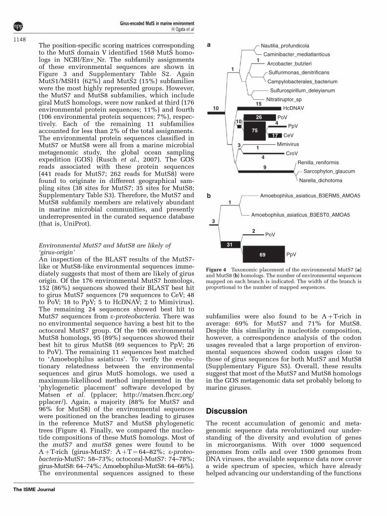
Environmental MutS7 and MutS8 are likely of‘girus-origin’An inspection of the BLAST results of the MutS7-like or MutS8-like environmental sequences imme-diately suggests that most of them are likely of girusorigin. Of the 176 environmental MutS7 homologs,152 (86%) sequences showed their BLAST best hitto girus MutS7 sequences (79 sequences to CeV; 48to PoV; 18 to PpV; 5 to HcDNAV; 2 to Mimivirus).The remaining 24 sequences showed best hit toMutS7 sequences from e-proteobacteria. There wasno environmental sequence having a best hit to theoctocoral MutS7 group. Of the 106 environmentalMutS8 homologs, 95 (89%) sequences showed theirbest hit to girus MutS8 (69 sequences to PpV; 26to PoV). The remaining 11 sequences best matchedto ‘Amoebophilus asiaticus’. To verify the evolu-tionary relatedness between the environmentalsequences and girus MutS homologs, we used amaximum-likelihood method implemented in the‘phylogenetic placement’ software developed byMatsen et al. (pplacer; http://matsen.fhcrc.org/pplacer/). Again, a majority (88% for MutS7 and96% for MutS8) of the environmental sequenceswere positioned on the branches leading to girusesin the reference MutS7 and MutS8 phylogenetictrees (Figure 4). Finally, we compared the nucleo-tide compositions of these MutS homologs. Most ofthe mutS7 and mutS8 genes were found to beAþT-rich (girus-MutS7: AþT¼ 64–82%; e-proteo-bacteria-MutS7: 58–73%; octocoral-MutS7: 74–78%;girus-MutS8: 64–74%; Amoebophilus-MutS8: 64–66%).The environmental sequences assigned to these
subfamilies were also found to be AþT-rich inaverage: 69% for MutS7 and 71% for MutS8.Despite this similarity in nucleotide composition,however, a correspondence analysis of the codonusages revealed that a large proportion of environ-mental sequences showed codon usages close tothose of girus sequences for both MutS7 and MutS8(Supplementary Figure S5). Overall, these resultssuggest that most of the MutS7 and MutS8 homologsin the GOS metagenomic data set probably belong tomarine giruses.
Discussion
The recent accumulation of genomic and meta-genomic sequence data revolutionized our under-standing of the diversity and evolution of genesin microorganisms. With over 1000 sequencedgenomes from cells and over 1500 genomes fromDNA viruses, the available sequence data now covera wide spectrum of species, which have alreadyhelped advancing our understanding of the functions
10
9
1
1
15
26
7517
410
4
13
69
31
2
3
1
Nautilia_profundicola
Caminibacter_mediatlanticus
Arcobacter_butzleri
Sulfurimonas_denitrificans
Campylobacterales_bacterium
Sulfurospirillum_deleyianum
Nitratiruptor_sp
HcDNAV
PoV
PpV
CeV
Mimivirus
CroV
Renilla_reniformis
Sarcophyton_glaucum
Narella_dichotoma
Amoebophilus_asiaticus_B3ERM5_AMOA5
Amoebophilus_asiaticus_B3EST0_AMOA5
PoV
PpV
Figure 4 Taxonomic placement of the environmental MutS7 (a)and MutS8 (b) homologs. The number of environmental sequencesmapped on each branch is indicated. The width of the branch isproportional to the number of mapped sequences.
Girus-encoded MutS in marine environmentH Ogata et al
1148
The ISME Journal

and evolution of protein families such as the MutSfamily (Eisen, 1998; Lin et al., 2007). However, giventhe huge diversity of girus genomes (Ogata andClaverie, 2007), they seem to be still underrepre-sented in this sequencing effort (Claverie et al.,2006; Claverie and Abergel, 2010); out of the1500 available viral genomes, only a handful ofgenomes exceed 350 kb (for example, Mimivirus(1.2 Mb), CroV (730 kb), Emiliania huxleyi virus(407 kb), Paramecium bursaria Chlorella virusNY2A (369 kb), Marseillevirus (368 kb), Canarypoxvirus (360 kb)). In this study, we analyzedfour distantly related marine giruses representing arelatively large class of giruses with estimatedgenome size from 356 kb up to 560 kb and identifiednew MutS homologs in all of the four giruses.
We showed that these girus-encoded MutSproteins fell into two subfamilies: MutS7 andMutS8. The recently reported MutS sequence fromthe largest marine girus, CroV, was classified in theMutS7 subfamily (Figure 1b) and was found to sharethe typical domain organization of this subfamily.Most unexpectedly, close homologs of the girus-encoded MutS7 and MutS8 were found to be highlyabundant in marine metagenomic sequence datasets. Giruses thus seem to represent one of the majorsources of the diversity of MutS family proteins. Ourphylogenetic reconstruction strongly suggests theoccurrence of horizontal gene transfers betweengiruses and cellular organisms for both the MutS7and MutS8 subfamilies. The abundance of ‘girus-like’ MutS7 in the marine environment favors thepreviously proposed scenario that an ancestor ofmarine giruses had a central role in transferringMutS7 to the octocoral mitochondrial genome.Consistently, the branch to the octocoral MutS7sequences was placed within the girus clade inthe MutS7 phylogenetic tree (Figure 1b). Theself-contained nature of the mutS7 gene (with bothrecognition and cutting functions) might havefacilitated such a gene transfer between distantlyrelated organisms. Similar gene transfer from a virusto the ancestor of mitochondria has been proposedfor the mitochondrial RNA/DNA polymerases andDNA primase; in this case, the source is likely to bea cryptic prophage (related to T3/T7) and themitochondrial enzymes are encoded in the nucleargenome (Filee and Forterre, 2005). The possiblegene transfer for MutS8 (found in PoV, PpV and theobligate intracellular amoeba-symbiont ‘Amoebo-philus asiaticus’ isolated from lake sediment)reinforces the previously proposed idea that amoe-bae (or other phagocytic protists) functionas ‘genetic melting pots’ to enhance the evolutionof intracellular bacteria and viruses infecting theseeukaryotes by providing ample opportunitiesfor gene exchanges (Ogata et al., 2006). Given theapparent specificity of virally encoded MutS forviruses with the largest genomes, these MutSsequences will be useful to probe metagenomicsequences for the presence of unknown giruses.
DNA viruses show a tremendous variation ingenome size from a few kilobases for the oncogenicpolyomaviruses, to more than a megabase for thegiant Mimivirus (Monier et al., 2007). Drake’s rulestates that the mutation rate per genome per strandcopying is roughly constant across DNA-basedmicroorganisms including bacteria, unicellulareukaryotes and DNA viruses (Drake, 1991; Sanjuanet al., 2010). Mutation rate per nucleotide perreplication is thus negatively correlated with thegenome size. In fact, the loss of DNA repairfunctions is a common trend in bacterial withreduced genomes, which exhibit higher mutationrate than other bacteria with larger genomes (Moranand Wernegreen, 2000; Moran et al., 2009).Experimental data for mutation rate is currentlyunavailable for giruses. However, given the largeamount of coding DNA that they need to protectfrom mutations, giruses may be under a specificselective pressure for efficient DNA repair systems(such as MutS7), which may be less crucial forsmaller viruses. The identification of MutS homo-logs in all of the four giruses tested in this study, aswell as a wealth of other DNA repair genes inMimivirus and CroV are consistent with this view.
Although the organisms with MutS7 or MutS8had many opportunities to exchange thesegenes (Claverie et al., 2009), the reason for thesporadic and limited phyletic distribution of theseMutS subfamilies still remains unclear. One mightpresume that the functions of these MutS proteinsare somehow associated with AþT-rich genomes.However, the presence of e-proteobacteria withAþT-rich genomes (such as H. pylori, AþT¼ 62%)lacking these MutS subfamily members contradictsthis hypothesis. E. coli MutH distinguishes thenascent DNA strand from the template DNA strandthrough the hemi-methylation of bases. It would beinteresting to examine the presence of hemi-methyl-ated bases in girus genomes and octocoral mitochon-drial genomes. We have started to clone and purifythe Mimivirus MutS7 for functional characterization.
The unique presence of a MutS homolog inMimivirus was already noticed during the initialgenome annotation (Raoult et al., 2004). We thenrecognized the surprising relationship between theMimivirus MutS and its homologs uniquely foundin the mitochondria of all octocorals (Claverie et al.,2009), all belonging to the newly defined MutS7subfamily. The finding of these MutS homologs inCroV, CeV, PoV, PpV and HcDNAV definitelyconfirms their association with large DNA virusesin marine environments. These findings stronglysuggest that the presence of MutS in Mimivirus isnot merely an example of an eccentric lateral genetransfer, but probably requires a more subtle ex-planation. We believe that much deeper experimentalinvestigation of these girus MutS homologs wouldhelp provide a holistic view on the evolution of genefamilies in the light of evolutionary interactionsbetween the viral and cellular gene pools.
Girus-encoded MutS in marine environmentH Ogata et al
1149
The ISME Journal
Acknowledgements
We thank Dr Stephane Audic for his technical assistancein an early stage of this work. The IGS laboratory issupported, in part, by CNRS and the French NationalResearch Agency (Grant # ANR-09-PCS-GENM-218,ANR-08-BDVA-003). The FRA laboratory is partiallysupported by Grants-in-Aid for Scientific Research (A)(No. 20247002) from the Ministry of Education, Scienceand Culture of Japan. The University of Bergen receivedfinancial support from the Norwegian Research Councilfor research programmes ‘Viral lysis and programmed celldeath in marine phytoplankton’ (VIPMAP, 186142/V40)and ‘Diversity and dynamics of marine Haptophytes’(HAPTODIV, 190307/S40).’
References
Abascal F, Zardoya R, Posada D. (2005). ProtTest: selectionof best-fit models of protein evolution. Bioinformatics21: 2104–2105.
Abdelnoor RV, Christensen AC, Mohammed S,Munoz-Castillo B, Moriyama H, Mackenzie SA.(2006). Mitochondrial genome dynamics in plantsand animals: convergent gene fusions of a MutShomologue. J Mol Evol 63: 165–173.
Altschul SF, Madden TL, Schaffer AA, Zhang J, Zhang Z,Miller W et al. (1997). Gapped BLAST and PSI-BLAST:a new generation of protein database search programs.Nucleic Acids Res 25: 3389–3402.
Bogani F, Corredeira I, Fernandez V, Sattler U,Rutvisuttinunt W, Defais M et al. (2010). Associationbetween the Herpes Simplex Virus-1 DNA Polymeraseand Uracil DNA Glycosylase. J Biol Chem 285:27664–27672.
Brugler MR, France SC. (2008). The mitochondrial genomeof a deep-sea bamboo coral (Cnidaria, Anthozoa,Octocorallia, Isididae): genome structure and putativeorigins of replication are not conserved amongoctocorals. J Mol Evol 67: 125–136.
Claverie JM, Abergel C. (2010). Mimivirus: the emergingparadox of quasi-autonomous viruses. Trends Genet26: 431–437.
Claverie JM, Grzela R, Lartigue A, Bernadac A, Nitsche S,Vacelet J et al. (2009). Mimivirus and Mimiviridae:giant viruses with an increasing number of potentialhosts, including corals and sponges. J Invertebr Pathol101: 172–180.
Claverie JM, Ogata H. (2009). Ten good reasons not toexclude giruses from the evolutionary picture. Nat RevMicrobiol 7: 615; author reply 615.
Claverie JM, Ogata H, Audic S, Abergel C, Suhre K,Fournier PE. (2006). Mimivirus and the emergingconcept of ‘giant’ virus. Virus Res 117: 133–144.
Culligan KM, Meyer-Gauen G, Lyons-Weiler J, Hays JB.(2000). Evolutionary origin, diversification andspecialization of eukaryotic MutS homolog mismatchrepair proteins. Nucleic Acids Res 28: 463–471.
Drake JW. (1991). A constant rate of spontaneous mutationin DNA-based microbes. Proc Natl Acad Sci USA 88:7160–7164.
Eddy SR. (1996). Hidden Markov models. Curr OpinStruct Biol 6: 361–365.
Eisen JA. (1998). A phylogenomic study of the MutSfamily of proteins. Nucleic Acids Res 26: 4291–4300.
Filee J, Forterre P. (2005). Viral proteins functioningin organelles: a cryptic origin? Trends Microbiol 13:510–513.
Fischer MG, Allen MJ, Wilson WH, Suttle CA. (2010).Giant virus with a remarkable complement of genesinfects marine zooplankton. Proc Natl Acad Sci USA107: 19508–19513.
Fukui K, Nakagawa N, Kitamura Y, Nishida Y, Masui R,Kuramitsu S. (2008). Crystal structure of MutS2endonuclease domain and the mechanism of homo-logous recombination suppression. J Biol Chem 283:33417–33427.
Furuta M, Schrader JO, Schrader HS, Kokjohn TA, Nyaga S,McCullough AK et al. (1997). Chlorella virus PBCV-1encodes a homolog of the bacteriophage T4 UVdamage repair gene denV. Appl Environ Microbiol63: 1551–1556.
Guindon S, Gascuel O. (2003). A simple, fast, and accuratealgorithm to estimate large phylogenies by maximumlikelihood. Syst Biol 52: 696–704.
Han MV, Zmasek CM. (2009). phyloXML: XML forevolutionary biology and comparative genomics.BMC Bioinformatics 10: 356.
Iyer RR, Pluciennik A, Burdett V, Modrich PL. (2006).DNA mismatch repair: functions and mechanisms.Chem Rev 106: 302–323.
Jacobsen A, Bratbak G, Heldal M. (1996). Isolation andcharacterization of a virus infecting Phaeocystispouchetii (Prymnesiophyceae). J Phycol 32: 923–927.
Kumar S, Nei M, Dudley J, Tamura K. (2008). MEGA: abiologist-centric software for evolutionary analysisof DNA and protein sequences. Brief Bioinform 9:299–306.
La Scola B, Campocasso A, N’Dong R, Fournous G,Barrassi L, Flaudrops C et al. (2010). Tentativecharacterization of new environmental giant virusesby MALDI-TOF mass spectrometry. Intervirology 53:344–353.
La Scola B, Desnues C, Pagnier I, Robert C, Barrassi L,Fournous G et al. (2008). The virophage as a uniqueparasite of the giant mimivirus. Nature 455: 100–104.
Larkin MA, Blackshields G, Brown NP, Chenna R,McGettigan PA, McWilliam H et al. (2007). Clustal Wand Clustal X version 2.0. Bioinformatics 23: 2947–2948.
Larsen JB, Larsen A, Bratbak G, Sandaa RA. (2008).Phylogenetic analysis of members of the Phycodna-viridae virus family, using amplified fragments of themajor capsid protein gene. Appl Environ Microbiol 74:3048–3057.
Le SQ, Gascuel O. (2008). An improved general aminoacid replacement matrix. Mol Biol Evol 25: 1307–1320.
Legendre M, Audic S, Poirot O, Hingamp P, Seltzer V,Byrne D et al. (2010). mRNA deep sequencing reveals75 new genes and a complex transcriptional landscapein Mimivirus. Genome Res 20: 664–674.
Lin Z, Nei M, Ma H. (2007). The origins and earlyevolution of DNA mismatch repair genes–multiplehorizontal gene transfers and co-evolution. NucleicAcids Res 35: 7591–7603.
Malik HS, Henikoff S. (2000). Dual recognition-incisionenzymes might be involved in mismatch repair andmeiosis. Trends Biochem Sci 25: 414–418.
McFadden CS, France SC, Sanchez JA, Alderslade P.(2006). A molecular phylogenetic analysis of theOctocorallia (Cnidaria: Anthozoa) based on mitochon-drial protein-coding sequences. Mol Phylogenet Evol41: 513–527.
Girus-encoded MutS in marine environmentH Ogata et al
1150
The ISME Journal

Miller WG, Parker CT, Rubenfield M, Mendz GL,Wosten MM, Ussery DW et al. (2007). The completegenome sequence and analysis of the epsilonproteo-bacterium Arcobacter butzleri. PLoS ONE 2: e1358.
Monier A, Claverie JM, Ogata H. (2007). Horizontal genetransfer and nucleotide compositional anomaly inlarge DNA viruses. BMC Genomics 8: 456.
Monier A, Larsen JB, Sandaa RA, Bratbak G, Claverie JM,Ogata H. (2008). Marine mimivirus relatives areprobably large algal viruses. Virol J 5: 12.
Moran NA, McLaughlin HJ, Sorek R. (2009). The dynamicsand time scale of ongoing genomic erosion insymbiotic bacteria. Science 323: 379–382.
Moran NA, Wernegreen JJ. (2000). Lifestyle evolutionin symbiotic bacteria: insights from genomics. TrendsEcol Evol 15: 321–326.
Moreira D, Philippe H. (1999). Smr: a bacterial andeukaryotic homologue of the C-terminal region ofthe MutS2 family. Trends Biochem Sci 24: 298–300.
Nakagawa S, Takaki Y, Shimamura S, Reysenbach AL,Takai K, Horikoshi K. (2007). Deep-sea vent epsilon-proteobacterial genomes provide insights intoemergence of pathogens. Proc Natl Acad Sci USA104: 12146–12150.
Natrajan G, Lamers MH, Enzlin JH, Winterwerp HH,Perrakis A, Sixma TK. (2003). Structures ofEscherichia coli DNA mismatch repair enzyme MutSin complex with different mismatches: a commonrecognition mode for diverse substrates. Nucleic AcidsRes 31: 4814–4821.
Notredame C, Higgins DG, Heringa J. (2000). T-Coffee:A novel method for fast and accurate multiplesequence alignment. J Mol Biol 302: 205–217.
Ogata H, Claverie JM. (2007). Unique genes in giantviruses: regular substitution pattern and anomalouslyshort size. Genome Res 17: 1353–1361.
Ogata H, La Scola B, Audic S, Renesto P, Blanc G, Robert Cet al. (2006). Genome sequence of Rickettsia belliiilluminates the role of amoebae in gene exchangesbetween intracellular pathogens. PLoS Genet 2: e76.
Ogata H, Toyoda K, Tomaru Y, Nakayama N, Shirai Y,Claverie JM et al. (2009). Remarkable sequencesimilarity between the dinoflagellate-infecting marinegirus and the terrestrial pathogen African swine fevervirus. Virol J 6: 178.
Pinto AV, Mathieu A, Marsin S, Veaute X, Ielpi L,Labigne A et al. (2005). Suppression of homologousand homeologous recombination by the bacterialMutS2 protein. Mol Cell 17: 113–120.
Pont-Kingdon GA, Okada NA, Macfarlane JL, Beagley CT,Wolstenholme DR, Cavalier-Smith T et al. (1995).A coral mitochondrial mutS gene. Nature 375:109–111.
Raoult D, Audic S, Robert C, Abergel C, Renesto P, Ogata Het al. (2004). The 1.2-megabase genome sequence ofMimivirus. Science 306: 1344–1350.
Redrejo-Rodriguez M, Ishchenko AA, Saparbaev MK,Salas ML, Salas J. (2009). African swine fever virusAP endonuclease is a redox-sensitive enzyme thatrepairs alkylating and oxidative damage to DNA.Virology 390: 102–109.
Rusch DB, Halpern AL, Sutton G, Heidelberg KB,Williamson S, Yooseph S et al. (2007). The SorcererII Global Ocean Sampling expedition: northwestAtlantic through eastern tropical Pacific. PLoS Biol5: e77.
Sandaa RA, Heldal M, Castberg T, Thyrhaug R, Bratbak G.(2001). Isolation and characterization of two viruseswith large genome size infecting Chrysochromulinaericina (Prymnesiophyceae) and Pyramimonas orien-talis (Prasinophyceae). Virology 290: 272–280.
Sanjuan R, Nebot MR, Chirico N, Mansky LM, Belshaw R.(2010). Viral mutation rates. J Virol 84: 9733–9748.
Schofield MJ, Hsieh P. (2003). DNA mismatch repair:molecular mechanisms and biological function.Annu Rev Microbiol 57: 579–608.
Sievert SM, Scott KM, Klotz MG, Chain PS, Hauser LJ,Hemp J et al. (2008). Genome of the epsilonproteo-bacterial chemolithoautotroph Sulfurimonas denitrifi-cans. Appl Environ Microbiol 74: 1145–1156.
Simonsen S, Moestrup O. (1997). Toxicity tests in eightspecies of Chrysochromulina (Haptophyta). Can J Bot75: 129–136.
Srinivasan V, Tripathy DN. (2005). The DNA repairenzyme, CPD-photolyase restores the infectivity ofUV-damaged fowlpox virus isolated from infectedscabs of chickens. Vet Microbiol 108: 215–223.
Tarutani K, Nagasaki K, Itakura S, Yamaguchi M. (2001).Isolation of a virus infecting the novel shellfish-killingdinoflagellate Heterocapsa circularisquama. AquatMicrob Ecol 23: 103–111.
UniProtConsortium (2010). The Universal ProteinResource (UniProt) in 2010. Nucleic Acids Res 38:D142–D148.
Van Etten JL, Lane LC, Dunigan DD. (2010). DNA Viruses:The Really Big Ones (Giruses). Annu Rev Microbiol 64:83–99.
Wu SY, Culligan K, Lamers M, Hays J. (2003). Dissimilarmispair-recognition spectra of Arabidopsis DNA-mismatch-repair proteins MSH2*MSH6 (MutSalpha)and MSH2*MSH7 (MutSgamma). Nucleic Acids Res31: 6027–6034.
Yutin N, Wolf YI, Raoult D, Koonin EV. (2009). Eukaryoticlarge nucleo-cytoplasmic DNA viruses: clusters oforthologous genes and reconstruction of viral genomeevolution. Virol J 6: 223.
Supplementary Information accompanies the paper on The ISME Journal website (http://www.nature.com/ismej)
Girus-encoded MutS in marine environmentH Ogata et al
1151
The ISME Journal

Two new subfamilies of DNA mismatch repair proteins
(MutS) specifically abundant in the marine environment
Hiroyuki Ogata1,*
, Jessica Ray2, Kensuke Toyoda
3,†, Ruth-Anne Sandaa
2, Keizo Nagasaki
3,
Gunnar Bratbak2, Jean-Michel Claverie
1
1: Information Génomique et Structurale, CNRS-UPR2589, Institut de Microbiologie de la
Méditerranée, Parc Scientifique de Luminy, Aix-Marseille Université, 163 Avenue de
Luminy, Case 934, 13288 Marseille Cedex 9, France
2: Department of Biology, University of Bergen, PO Box 7800, N-5020 Bergen, Norway
3: Harmful Algal Bloom Division, National Research Institute of Inland Sea, Fisheries
Research Agency, 2-17-5 Maruishi, Hatsukaichi, Hiroshima 739-0452, Japan
*: Correspondence: Hiroyuki Ogata (E-mail: [email protected])
†: K. Toyoda’s present address is “Department of Botany, Keio University, Hiyoshi, Kohoku-
ku, Yokohama, Kangawa 223-8521, Japan” (E-mail: [email protected])
Supplementary materials

Table S1. Primers used to resolve sequence ambiguities in the CeV MutS7 region. Five
sets of overlapping DNA primers were designed to resolve sequence ambiguities within a
CeV contig containing a MutS7 homolog. Amplification reactions (50µl) contained 1X Hot
Star Taq Plus Master Mix (Qiagen, Hilden, Germany), 0.4 µM each of forward and reverse
primers, 0.2 mg ml-1 BSA (Promega, Madison, Wisconsin), 3 mM MgCl2 and1X Coralload
buffer (Qiagen). CeV lysates were diluted 10-fold with sterile MQ water and subjected to 2 x
2min incubation at 99°C with an intervening 2min incubation on ice. Template for PCR
amplification reactions consisted of 2µl of the freeze-thaw diluted CeV lysate. The thermal
cycling program consisted of an initial 5min denaturation at 95°C, 30 cycles of 95°C for
30sec, 55°C for 30sec and 72°C for 90sec, and a final 10min elongation at 72°C. When
necessary, PCR products were stored overnight at 4°C until analysis by agarose gel
electrophoresis to confirm amplification specificity. Correct molecular weight bands were
excised from agarose gels and purified using the illustra GFX PCR purification kit (GE
Healthcare, Amersham, UK) according to the manufacturer's protocol. 30-40ng of purified
PCR product was used as template for forward and reverse sequencing reactions using the
BigDye Terminator v3.1 cycle sequencing chemistry (Applied Biosystems, Carlsbad,
California) and 3.2pmol primer. Sequencing was performed at the DNA Sequencing Facility
at the University of Bergen, Norway.
Primer name 5'-3' sequence
cev_002_181-200 TGGCCTGGGCCACCAAATCC
cev_002_1010-989 AAACAATGGGTGCGTGTCC
cev_002_697-716 TGGGGTAATCCAAATCCTGCCA
cev_002_1807-1784 ACTCACGTTTACCCATAGGTGTT
cev_002_1779-1806 TGTTTAACACCTATGGGTAAACGTGAG
cev_002_2844-2821 ACCATTAATTCATAACCATTAGTCACC
cev_002_2388-2413 ATAAGAGCGTGTCTTAGTCCCGT
cev_002_3405-3379 TTCCAGGAACTGAAAGTGATTCGGCA
cev_002_3179-3204 GCAACATTACAAGCAACAAAACGACG
cev_002_3782-3756 CCATACATACTTTCTCCAGCTCCTGGT

Table S2. MutS homologs in UniProt, girus genomes and NCBI/Env_Nr.
MutS
subfamily
UniProt and girus MutS homologs
NCBI
EnvNr Total
BLAST
hits
Bacteria Archaea Eukaryota Virus
MutS1/MSH1 1803 1680 38 85
976
MSH2 176
176
7
MSH3 102
102
4
MSH4 93
93
MSH5 103
103
2
MSH6/7 174
174
11
plt-MSH1 29
29
4
MutS2 1070 1037
33
237
MutS3 304 304
6
MutS4 51 47 4
MutS5 42
42
34
MutS6 24 24
5
MutS7 28 8
15 5* 176
MutS8 4 2
2 106
MutS9 25 25
Total 4028 3127 84 810 7 1568
* A recently reported viral MutS7 (CroV MutS) is not included in this table.

Ogata et al., Table S3
Table S3. GOS reads related to MutS7/MutS8ID Habitat
Type
Geographic
Location
Sample Location Sample
Depth (m)
Water
Depth (m)
T (oC) S (ppt) Size
Fraction
(?m)
Chl a
Sample
Month
(Annual±SE
) mg
m-3
Reads MutS7
Reads
MutS8
Reads
JCVI_SITE_GS000_S13 Open Ocean Sargasso
Sea
Sargasso Sea,
Station 13
5 >4200 20 36.6 0.1-0.8 0.17
(0.09±0.02)
644551 16 8
JCVI_SITE_GS000_S13 Open Ocean Sargasso
Sea
Sargasso Sea,
Station 13
5 >4200 20 36.6 0.22-0.8 0.17
(0.09±0.02)
317180 16 8
JCVI_SITE_GS000_S11 Open Ocean Sargasso
Sea
Sargasso Sea,
Station 11
5 >4200 20.5 36.7 0.1-0.8 0.17
(0.09±0.02)
644551 9 0
JCVI_SITE_GS000_S11 Open Ocean Sargasso
Sea
Sargasso Sea,
Station 11
5 >4200 20.5 36.7 0.22-0.8 0.17
(0.09±0.02)
317180 9 0
JCVI_SITE_GS000_S03 Open Ocean Sargasso
Sea
Sargasso Sea,
Station 3
5 >4200 19.8 36.7 0.22-0.8 0.17
(0.09±0.02)
368835 4 4
JCVI_SITE_GS000_S13 Open Ocean Sargasso
Sea
Sargasso Sea,
Station 13
5 >4200 20 36.6 0.22-0.8 0.17
(0.09±0.02)
332240 16 8
JCVI_SITE_GS001 Open Ocean Sargasso
Sea
Sargasso Sea,
Hydrostation S
5 >4200 22.9 36.7 3.0-20 0.10
(0.10±0.01)
142352 8 4
JCVI_SITE_GS001 Open Ocean Sargasso
Sea
Sargasso Sea,
Hydrostation S
5 >4200 22.9 36.7 0.8-3.0 0.10
(0.10±0.01)
90905 8 4
JCVI_SITE_GS001 Open Ocean Sargasso
Sea
Sargasso Sea,
Hydrostation S
5 >4200 22.9 36.7 0.1-0.8 0.10
(0.10±0.01)
92351 8 4
JCVI_SITE_GS002 Coastal North
American
East Coast
Gulf of Maine 1 106 18.2 29.2 0.1-0.8 1.4
(1.12±0.19)
121590 48 13
JCVI_SITE_GS003 Coastal North
American
East Coast
Browns Bank, Gulf of
Maine
1 119 11.7 29.9 0.1-0.8 1.4
(1.12±0.19)
61605 27 55
JCVI_SITE_GS004 Coastal North
American
East Coast
Outside Halifax, Nova
Scotia
2 142 17.3 28.3 0.1-0.8 0.4
(0.78±0.17)
52959 4 0
JCVI_SITE_GS005 Embayment North
American
East Coast
Bedford Basin, Nova
Scotia
1 64 15 30.2 0.1-0.8 6
(6.76±0.98)
61131 5 3
JCVI_SITE_GS006 Estuary North
American
East Coast
Bay of Fundy, Nova
Scotia
1 11 11.2 0.1-0.8 2.8
(1.87±0.18)
59679 15 0
JCVI_SITE_GS007 Coastal North
American
East Coast
Northern Gulf of
Maine
1 139 17.9 31.7 0.1-0.8 1.4
(1.12±0.19)
50980 7 4
JCVI_SITE_GS008 Coastal North
American
East Coast
Newport Harbor, RI 1 12 9.4 26.5 0.1-0.8 2.2
(1.59±0.17)
129655 6 0
Page 1

Ogata et al., Table S3
JCVI_SITE_GS009 Coastal North
American
East Coast
Block Island, NY 1 32 11 31 0.1-0.8 4.0
(2.72±0.24)
79303 10 6
JCVI_SITE_GS010 Coastal North
American
East Coast
Cape May, NJ 1 10 12 31 0.1-0.8 2.0
(2.75±0.33)
78304 4 7
JCVI_SITE_GS011 Estuary North
American
East Coast
Delaware Bay, NJ 1 8 11 0.1-0.8 4.8
(9.23±1.02)
124435 22 10
JCVI_SITE_GS012 Estuary North
American
East Coast
Chesapeake Bay, MD 13.2 25 1 3.5 0.1-0.8 21.0
(15.0±1.01)
126162 15 16
JCVI_SITE_GS013 Coastal North
American
East Coast
Off Nags Head, NC 2.1 20 9.3 0.1-0.8 3.0
(2.24±0.25)
138033 10 6
JCVI_SITE_GS014 Coastal North
American
East Coast
South of Charleston,
SC
1 31 18.6 0.1-0.8 1.70
(1.92±0.25)
128885 2 4
JCVI_SITE_GS015 Coastal Caribbean
Sea
Off Key West, FL 1.7 47 25 36 0.1-0.8 0.2
(0.27±0.09)
127362 15 4
JCVI_SITE_GS016 Coastal Sea Caribbean
Sea
Gulf of Mexico 2 3333 26.4 35.8 0.1-0.8 0.16
(0.11±0.01)
127122 12 8
JCVI_SITE_GS017 Open Ocean Caribbean
Sea
Yucatan Channel 2 4513 27 35.8 0.1-0.8 0.13
(0.09±0.01)
257581 32 23
JCVI_SITE_GS018 Open Ocean Caribbean
Sea
Rosario Bank 1.7 4470 27.4 35.4 0.1-0.8 0.14
(0.09±0.01)
142743 6 2
JCVI_SITE_GS019 Coastal Caribbean
Sea
Northeast of Colón 1.7 3336 27.7 35.4 0.1-0.8 0.23
(0.15±0.02)
135325 13 2
JCVI_SITE_GS020 Fresh Water Panama
Canal
Lake Gatun 2 4.2 28.6 0.1 0.1-0.8 296355 21 11
JCVI_SITE_GS021 Coastal Eastern
Tropical
Pacific
Gulf of Panama 1.6 76 27.6 30.7 0.1-0.8 0.50
(0.73±0.22)
131798 3 4
JCVI_SITE_GS022 Open Ocean Eastern
Tropical
Pacific
250 miles from
Panama City
2 2431 29.3 32.3 0.1-0.8 0.33
(0.28±0.02)
121662 6 4
JCVI_SITE_GS023 Open Ocean Eastern
Tropical
Pacific
30 miles from Cocos
Island
2 1139 28.7 32.6 0.1-0.8 0.07
(0.19±0.02)
133051 4 8
JCVI_SITE_GS025 Fringing
Reef
Eastern
Tropical
Pacific
Dirty Rock, Cocos
Island
1.1 30 28.3 31.4 0.8-3.0 0.11
(0.19±0.01)
120671 0 4
JCVI_SITE_GS026 Open Ocean Galapagos
Islands
134 miles NE of
Galapagos
2 2386 27.8 32.6 0.1-0.8 0.22
(0.28±0.02)
102708 0 2
JCVI_SITE_GS027 Coastal Galapagos
Islands
Devil's Crown,
Floreana Island
2.2 2.3 25.5 34.9 0.1-0.8 0.40
(0.38±0.03)
222080 16 2
JCVI_SITE_GS028 Coastal Galapagos
Islands
Coastal Floreana 2 156 0.1-0.8 0.35
(0.35±0.02)
189052 17 11
Page 2

Ogata et al., Table S3
JCVI_SITE_GS029 Coastal Galapagos
Islands
North James Bay,
Santigo Island
2.1 12 26.2 34.5 0.1-0.8 0.40
(0.39±0.03)
131529 4 4
JCVI_SITE_GS030 Warm Seep Galapagos
Islands
Warm seep, Roca
Redonda
19 19 26.9 0.1-0.8 359152 2 2
JCVI_SITE_GS031 Coastal
upwelling
Galapagos
Islands
Upwelling,
Fernandina Island
12 19.6 18.6 0.1-0.8 0.35
(0.39±0.03)
436401 25 12
JCVI_SITE_GS032 Mangrove Galapagos
Islands
Mangrove on Isabella
Island
0.1 1.6 25.4 0.1-0.8 148018 19 2
JCVI_SITE_GS033 Hypersaline Galapagos
Islands
Punta Cormorant,
Hypersaline Lagoon,
Floreana Island
0.2 0.3 37.6 63.4 0.1-0.8 692255 8 3
JCVI_SITE_GS034 Coastal Galapagos
Islands
North Seamore Island 2.1 35 27.5 0.1-0.8 0.36
(0.35±0.02)
134347 12 4
JCVI_SITE_GS035 Coastal Galapagos
Islands
Wolf Island 1.7 71 21.8 34.5 0.1-0.8 0.28
(0.31±0.02)
140814 2 2
JCVI_SITE_GS036 Coastal Galapagos
Islands
Cabo Marshall,
Isabella Island
2.1 67 25.8 34.6 0.1-0.8 0.65
(0.45±0.05)
77538 4 0
JCVI_SITE_GS037 Open Ocean Eastern
Tropical
Pacific
Equatorial Pacific
TAO Buoy
1.8 3334 28 0.1-0.8 0.21
(0.24±0.02)
65670 0 6
JCVI_SITE_GS038 Open Ocean Tropical
South Pacific
Tropical South Pacific 1.8 >4000 28.4 0.1-0.8 741 0 0
JCVI_SITE_GS039 Open Ocean Tropical
South Pacific
Tropical South Pacific 2 >4000 28.6 0.1-0.8 759 0 0
JCVI_SITE_GS040 Open Ocean Tropical
South Pacific
Tropical South Pacific 2.2 >4000 27.8 0.1-0.8 736 0 0
JCVI_SITE_GS041 Open Ocean Tropical
South Pacific
Tropical South Pacific 2 >4000 28 35 0.1-0.8 678 0 0
JCVI_SITE_GS042 Open Ocean Tropical
South Pacific
Tropical South Pacific 1.7 >4000 27.6 0.1-0.8 699 0 0
JCVI_SITE_GS043 Open Ocean Tropical
South Pacific
Tropical South Pacific 1.9 >4000 27.6 35.9 0.1-0.8 711 0 0
JCVI_SITE_GS044 Open Ocean Tropical
South Pacific
600 miles from F.
Polynesia
2 >4000 27.6 0.1-0.8 678 0 0
JCVI_SITE_GS045 Open Ocean Tropical
South Pacific
400 miles from F.
Polynesia
1.7 >4000 28.3 37 0.1-0.8 730 0 0
JCVI_SITE_GS046 Open Ocean Tropical
South Pacific
300 miles from F.
Polynesia
1.9 >4000 28.7 35.3 0.1-0.8 626 0 0
Page 3

Ogata et al., Table S3
JCVI_SITE_GS047 Open Ocean Tropical
South Pacific
201 miles from F.
Polynesia
30 2400 28.6 37.3 0.1-0.8 66023 4 2
JCVI_SITE_GS048 Coral Reef Polynesia
Archipelagos
Moorea, Cooks Bay 1.4 34 28.9 35.1 0.1-0.8 744 0 0
JCVI_SITE_GS049 Coastal Polynesia
Archipelagos
Moorea, Outside
Cooks Bay
1.4 900 28.8 32.6 0.1-0.8 735 0 0
JCVI_SITE_GS050 Coral Atoll Polynesia
Archipelagos
Tikehau Lagoon 1.2 24 27.8 0.1-0.8 715 0 0
JCVI_SITE_GS051 Coral Reef
Atoll
Polynesia
Archipelagos
Rangirora Atoll 1 10 27.3 34.2 0.1-0.8 128982 4 0
Page 4

Legend for supplementary figures
Figure S1. Multiple sequence alignment of HNH endonuclease domains of the MutS7
subfamily proteins. The positions of four conserved residues around the endonuclease active
site are marked by red triangles.
Figure S2 Maximum likelihood phylogenetic tree of MutS family proteins. The tree is
based on the conserved MutS domain V sequences. The tree is mid-point rooted. Bootstrap
values < 50% are not shown. Taxon names are composed of a MutS family name, a sequence
identifier, a domain classification (B for Bacteria, A for Archaea, E for Eukaryote, V for
Viruses), followed by the species name. Color code for branches are as follows: Bacteria
(blue), Archaea (light blue), Eukaryotes (green), Giruses (Red). MutS subfamilies introduced
in this study (MutS6, MutS7, MutS8, MutS9) are indicated in red.
Figure S3 Domain architecture of MutS family proteins. Sequence domains were
identified using NCBI/Cdd profiles and PSI-BLAST (E-value<0.01). This diagram is drawn
to scale. For MutS domains (I, II, III, IV, V), PSI-BLAST was used with four iterations.
Identified domains were represented as follows: MutS domain I (pfam01624), light blue
rectangle; MutS domain II (pfam05188), orange rectangle; MutS domain III (pfam05192),
light green rectangle; MutS domain IV (pfam05190), dark green rectangle; MutS domain V
(pfam00488), red rectangle; Smr domain (pfam01713), orange oval; GIY-YIG domain
(pfam01541), pink oval; HNH domain (pfam01844), green oval. MSH1p corresponds to plant
specific MSH1 (plt-MSH1).
Figure S4. Multiple sequence alignment of the N-terminal part of the domain I
sequences from MutS7 and E. coli MutS1. The conserved “F(X)E” residues are highlighted
by a red rectangle.
Figure S5. Correspondence analysis of codon usages of MutS7 and MutS8 homologs. The
number of GOS environmental sequences is 176 for MutS7 and 106 for MutS8.

Girus
Octocoral
ε-proteo-
bacteria
Fig. S1

MutS1 MUTS NATPD A Natronomonas pharaonis
MutS1 C7NS38 HALUD A Halorhabdus utahensis
MutS1 D3SXA0 NATMA A Natrialba magadii
MutS1 MUTS2 HALMA A Haloarcula marismortui
MutS1 MUTS2 HALSA A Halobacterium salinarium
MutS1 C1VA99 9EURY A Halogeometricum borinquense
MutS1 MUTS HALWD A Haloquadratum walsbyi
MutS1 D3SSD8 NATMA A Natrialba magadii
MutS1 MUTS1 HALSA A Halobacterium salinarium
MutS1 C1V6F2 9EURY A Halogeometricum borinquense
MutS1 MUTS1 HALMA A Haloarcula marismortui
MutS1 C7NS73 HALUD A Halorhabdus utahensis
MutS1 MUTS METTP A Methanosaeta thermophila
MutS1 B5ICV6 9EURY A Aciduliprofundum boonei
MutS1 MUTS METMA A Methanosarcina mazei
MutS1 MUTS METBU A Methanococcoides burtonii
MutS1 Q2FU04 METHJ A Methanospirillum hungatei
MutS1 A3CWX6 METMJ A Methanoculleus marisnigri
MutS1 MUTS ECOLI B Escherichia coli
MutS1 MUTS BUCAT B Buchnera aphidicola
MutS1 MUTS OCHA4 B Ochrobactrum anthropi
MutS1 Q552L1 DICDI E Dictyostelium discoideum
MutS1 MSH1 SCHPO E Schizosaccharomyces pombe
MutS1 MSH1 YEAST E Saccharomyces cerevisiae
MutS1 A9VCG8 MONBE E Monosiga brevicollis
MutS1 B8GB04 CHLAD B Chloroflexus aggregans
MutS1 MUTS BACSU B Bacillus subtilis
MutS1/MSH1 (Bacteria/Archaea/Eukaryotes)
MSH6 MSH6 DICDI E Dictyostelium discoideum
MSH6 MSH6 YEAST E Saccharomyces cerevisiae
MSH6 MSH7 ARATH E Arabidopsis thaliana
MSH6 MSH6 ARATH E Arabidopsis thaliana
MSH6 MSH6 HUMAN E Homo sapiens
MSH6 A0DMV3 PARTE E Paramecium tetraurelia
MSH6/MSH7 (Eukaryotes)
MSH3 MSH3 YEAST E Saccharomyces cerevisiae
MSH3 MSH3 LODEL E Lodderomyces elongisporus
MSH3 MSH3 SCHPO E Schizosaccharomyces pombe
MSH3 MSH3 DICDI E Dictyostelium discoideum
MSH3 MSH3 CRYNE E Cryptococcus neoformans
MSH3 MSH3 HUMAN E Homo sapiens
MSH3 MSH3 ARATH E Arabidopsis thaliana
MSH3 (Eukaryotes)
MSH2 A5KA73 PLAVI E Plasmodium vivax
MSH2 MSH2 YEAST E Saccharomyces cerevisiae
MSH2 A2EP54 TRIVA E Trichomonas vaginalis
MSH2 MSH2 ARATH E Arabidopsis thaliana
MSH2 (Eukaryotes)
MSH4 Q4P0K2 USTMA E Ustilago maydis
MSH4 MSH4 YEAST E Saccharomyces cerevisiae
MSH4 Q6CKF7 KLULA E Kluyveromyces lactis
MSH4 A7TJR9 VANPO E Vanderwaltozyma polyspora
MSH4 A5DRZ1 LODEL E Lodderomyces elongisporus
MSH4 HIM14 CAEEL E Caenorhabditis elegans
MSH4 A8PJN5 BRUMA E Brugia malayi
MSH4 MSH4 HUMAN E Homo sapiens
MSH4 C1M0C9 SCHMA E Schistosoma mansoni
MSH4 (Eukaryotes)
MSH5 C4M0Z9 ENTHI E Entamoeba histolytica
MSH5 MSH5 HUMAN E Homo sapiens
MSH5 MSH5 CAEEL E Caenorhabditis elegans
MSH5 C1EEU6 9CHLO E Micromonas sp
MSH5 Q011M5 OSTTA E Ostreococcus tauri
MSH5 Q7Z7S7 COPCI E Coprinopsis cinerea
MSH5 A7EN13 SCLS1 E Sclerotinia sclerotiorum
MSH5 A7TR47 VANPO E Vanderwaltozyma polyspora
MSH5 Q6CT05 KLULA E Kluyveromyces lactis
MSH5 MSH5 YEAST E Saccharomyces cerevisiae
MSH5 (Eukaryotes)
MSH1p Q0JBW2 ORYSJ E Oryza sativa
MSH1p Q84LK0 ARATH E Arabidopsis thaliana
MSH1p Q1XBQ8 SOYBN E Glycine maxplt-MSH1 (Plant)
MutS7 B3FNN3 RENRE E Renilla reniformis
MutS7 MSHM SARGL E Sarcophyton glaucum
MutS7 MUTSL MIMIV V Acanthamoeba polyphaga
MutS7 CeV V CeV
MutS7 PpV V PpV
MutS7 HcDNAV V HcDNAV
MutS7 PoV V PoV
MutS7 A6Q2C2 NITSB B Nitratiruptor sp
MutS7 D1B043 SULD5 B Sulfurospirillum deleyianum
MutS7 B9L925 NAUPA B Nautilia profundicola
MutS7 A8ES10 ARCB4 B Arcobacter butzleri
MutS7 B6BMZ0 9PROT B Campylobacterales bacterium
MutS7 Q30QK8 SULDN B Sulfurimonas denitrificans
MutS7 (Girus/Epsilonproteobacteria/Octocorals)
MutS8 B3EST0 AMOA5 B Amoebophilus asiaticus
MutS8 B3ERM5 AMOA5 B Amoebophilus asiaticus
MutS8 PoV V PoV
MutS8 PpV V PpV
MutS8 (Girus/Amoebophilus)
MutS6 A6E7L8 9SPHI B Pedobacter sp
MutS6 C2FUZ9 9SPHI B Sphingobacterium spiritivorum
MutS6 C7PSP9 CHIPD B Chitinophaga pinensis
MutS6 B3C7Q8 9BACE B Bacteroides intestinalis
MutS6 (Bacteria)
MutS3 A8UFC0 9FLAO B Flavobacteriales bacterium
MutS3 A4BXL0 9FLAO B Polaribacter irgensii
MutS3 B6W5I8 9BACE B Bacteroides dorei
MutS3 C2FWF5 9SPHI B Sphingobacterium spiritivorum
MutS3 A9FC19 SORC5 B Sorangium cellulosum
MutS3 A9B626 HERA2 B Herpetosiphon aurantiacus
MutS3 Q04P59 LEPBJ B Leptospira borgpetersenii
MutS3 C6W3D3 DYAFD B Dyadobacter fermentans
MutS3 Q24UR6 DESHY B Desulfitobacterium hafniense
MutS3 Q0AVD3 SYNWW B Syntrophomonas wolfei
MutS3 C9KM00 9FIRM B Mitsuokella multacida
MutS3 A7VIS8 9CLOT B Clostridium sp
MutS3A
MutS3 A7V876 BACUN B Bacteroides uniformis
MutS3 C6W1B1 DYAFD B Dyadobacter fermentans
MutS3 A4BZM7 9FLAO B Polaribacter irgensii
MutS3 A4A8G2 9GAMM B Congregibacter litoralis
MutS3 C1IAL3 9CLOT B Clostridium sp
MutS3 C4GEA1 9FIRM B Shuttleworthia satelles
MutS3 C0BZP4 9CLOT B Clostridium hylemonae
MutS3 C0CIA6 9FIRM B Blautia hydrogenotrophica
MutS3 B0KBI4 THEP3 B Thermoanaerobacter pseudethanolicus
MutS3 B1RKN9 CLOPE B Clostridium perfringens
MutS3 B9E9P8 MACCJ B Macrococcus caseolyticus
MutS3 Q49Z02 STAS1 B Staphylococcus saprophyticus
MutS3 A3CKY5 STRSV B Streptococcus sanguinis
MutS3 B3W7H4 LACCB B Lactobacillus casei
MutS3B
MutS3 (Bacteria)
MutS9 A9BG46 PETMO B Petrotoga mobilis
MutS9 C5CH03 KOSOT B Kosmotoga olearia
MutS9 A8MLR5 ALKOO B Alkaliphilus oremlandii
MutS9 A6TWQ4 ALKMQ B Alkaliphilus metalliredigens
MutS9 (Bacteria)
MutS2 D2QDA3 SPILD B Spirosoma linguale
MutS2 B3ELF9 CHLPB B Chlorobium phaeobacteroides
MutS2 C1D0G1 DEIDV B Deinococcus deserti
MutS2 A9NGE8 ACHLI B Acholeplasma laidlawii
MutS2 A8UZQ9 9AQUI B Hydrogenivirga sp
MutS2 B5Y861 COPPD B Coprothermobacter proteolyticus
MutS2 B7C7S5 9FIRM B Eubacterium biforme
MutS2 A9BJX8 PETMO B Petrotoga mobilis
MutS2 B1C132 9FIRM B Clostridium spiroforme
MutS2 A1AT62 PELPD B Pelobacter propionicus
MutS2 B5YHF6 THEYD B Thermodesulfovibrio yellowstonii
MutS2 MUTS2 BACSU B Bacillus subtilis
MutS2 B7GGY7 ANOFW B Anoxybacillus flavithermus
MutS2 D2Z2Q5 9BACT B Dethiosulfovibrio peptidovorans
MutS2 C1MLV8 9CHLO E Micromonas pusilla
MutS2 Q7XKD3 ORYSJ E Oryza sativa
MutS2 A0YID1 9CYAN B Lyngbya sp
MutS2 Q9LVW1 ARATH E Arabidopsis thaliana
MutS2 B0VJJ4 9BACT B Candidatus Cloacamonas
MutS2 B8DKM4 DESVM B Desulfovibrio vulgaris
MutS2 A6G6E8 9DELT B Plesiocystis pacifica
MutS2 D0LH43 HALO1 B Haliangium ochraceum
MutS2 Q1D4Q8 MYXXD B Myxococcus xanthus
MutS2 Q30SJ7 SULDN B Sulfurimonas denitrificans
MutS2 C3XIF0 9HELI B Helicobacter bilis
MutS2 MUTS2 HELPY B Helicobacter pylori
MutS2 (Bacteria/Eukaryotes)
MutS5 Q12WC4 METBU A Methanococcoides burtonii
MutS5 Q18E65 HALWD A Haloquadratum walsbyi
MutS5 Q5JEZ3 PYRKO A Pyrococcus kodakaraensisMutS5 (Archaea)
MutS4 C6PBK5 CLOTS B Thermoanaerobacterium thermosaccharolyticu
MutS4 Q97AY6 THEVO A Thermoplasma volcanium MutS4B MutS4 Q8R8T8 THETN B Thermoanaerobacter tengcongensis
MutS4 Q97AY7 THEVO A Thermoplasma volcanium MutS4AMutS4 (Bacteria/Archaea)
100
10085
100
69
80
98
72100
74
9793
8182
100
9053
73
9996
72
62
83
59
51
5855
66
69
58
62
99
100
61
95
51
84
63100
6683
87100
93
86
50
5974
77
60
69
59
6965
67
68
53
98
70
84
78
91
9266
100
0.5
Fig. S2

Fig. S3 (1/3)

Fig. S3 (2/3)

Fig. S3 (3/3)

Fig. S4

-0.5
0.5
-2 -1 0 1
Ax
is 2
Axis 1
Girus Bacteria Coral GOS
-0.5
0.5
-1 0 1 2
Ax
is 2
Axis 1
Girus Bacteria GOS
-0.6
-0.4
-0.2
0
0.2
0.4
0.6
-2 -1 0 1
Ax
is 3
Axis 1
Girus Bacteria Coral GOS
-0.5
0.5
-1 0 1 2
Ax
is 3
Axis 1
Girus Bacteria GOS
-0.6
-0.4
-0.2
0
0.2
0.4
0.6
-0.5 0.5
Ax
is 3
Axis 2
Girus Bacteria Coral GOS
-0.5
0.5
-0.5 0.5
Ax
is 3
Axis 2
Girus Bacteria GOS
MutS7 MutS8
MutS8
MutS7
MutS7
MutS8
Fig. S5