type 1 diabetes review
DESCRIPTION
Type 1 diabetes reviewTRANSCRIPT
Seminar
www.thelancet.com Published online July 26, 2013 http://dx.doi.org/10.1016/S0140-6736(13)60591-7 1
Type 1 diabetes Mark A Atkinson, George S Eisenbarth, Aaron W Michels
Over the past decade, knowledge of the pathogenesis and natural history of type 1 diabetes has grown substantially, particularly with regard to disease prediction and heterogeneity, pancreatic pathology, and epidemiology. Technological improvements in insulin pumps and continuous glucose monitors help patients with type 1 diabetes manage the challenge of lifelong insulin administration. Agents that show promise for averting debilitating disease-associated complications have also been identifi ed. However, despite broad organisational, intellectual, and fi scal investments, no means for preventing or curing type 1 diabetes exists, and, globally, the quality of diabetes management remains uneven. This Seminar discusses current progress in epidemiology, pathology, diagnosis, and treatment of type 1 diabetes, and prospects for an improved future for individuals with this disease.
IntroductionType 1 diabetes is generally thought to be precipitated by an immune-associated, if not directly immune-mediated, destruction of insulin-producing pancreatic β cells.1,2 Historically, type 1 diabetes was largely considered a disorder in children and adolescents, but this opinion has changed over the past decade, so that age at symptomatic onset is no longer a restricting factor.3 Polydipsia, polyphagia, and polyuria (the classic trio of symptoms associated with disease onset) along with overt hyperglycaemia remain diagnostic hallmarks in children and adolescents, and to a lesser extent in adults. An immediate need for exogenous insulin replacement is also a hallmark of type 1 diabetes, for which lifetime treatment is needed. Key questions remain regarding the epidemiology of type 1 diabetes, eff ectiveness of current therapies, understanding how the disorder develops, and preventing or curing the disease.
EpidemiologyAlthough type 1 diabetes can be diagnosed at any age, it is one of the most common chronic diseases of childhood.4 Peaks in presentation occur between 5–7 years of age and at or near puberty.5 Whereas most autoimmune disorders disproportionately aff ect women, type 1 diabetes is slightly more common in boys and men.6 The incidence of type 1 diabetes varies with seasonal changes and birth month. More cases are diagnosed in autumn and winter,7 and being born in the spring is associated with a higher chance of having type 1 diabetes.8 Development of type 1 diabetes-associated autoimmunity (ie, formation of islet autoantibodies) in the months or years before onset of symptomatic type 1 diabetes also shows some seasonal synchronisation.9 These concepts support a theoretical role for an environ-mental agent initiating or driving the pathogenic processes in type 1 diabetes.
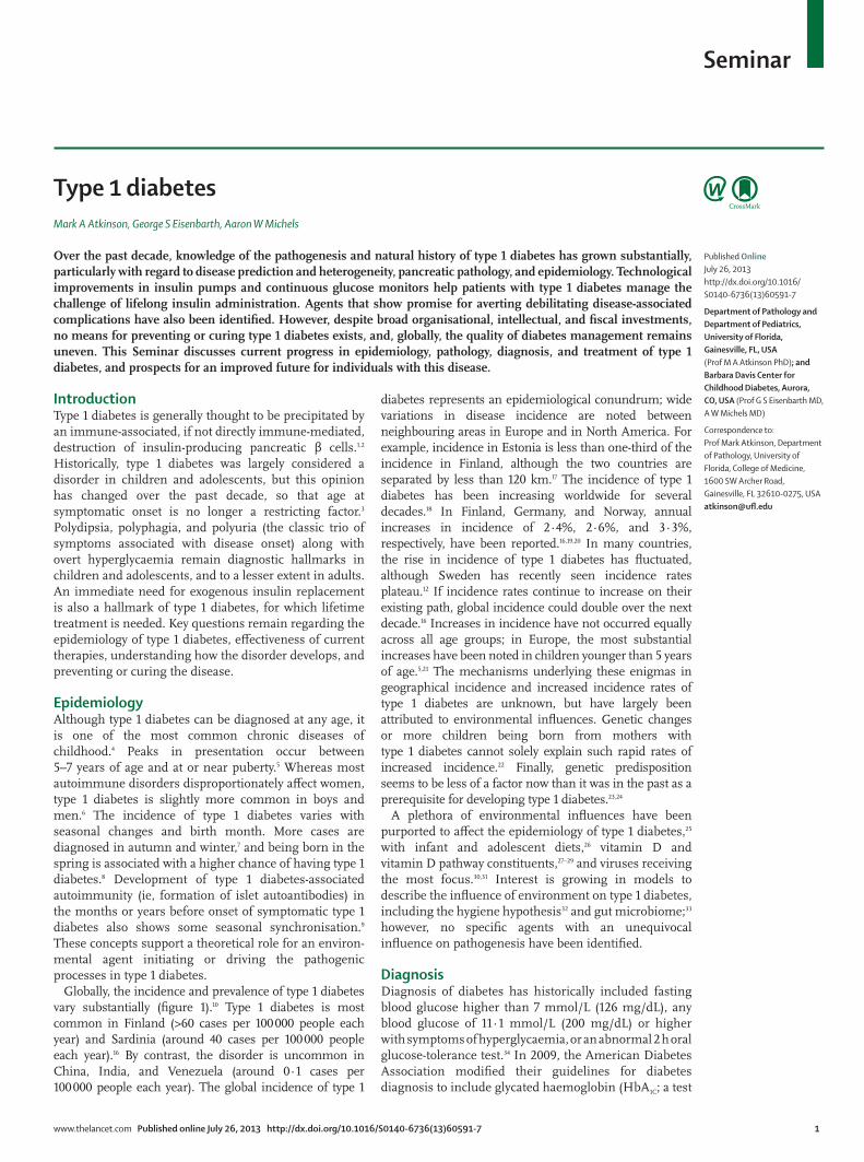
Globally, the incidence and prevalence of type 1 diabetes vary substantially (fi gure 1).10 Type 1 diabetes is most common in Finland (>60 cases per 100 000 people each year) and Sardinia (around 40 cases per 100 000 people each year).16 By contrast, the disorder is uncommon in China, India, and Venezuela (around 0∙1 cases per 100 000 people each year). The global incidence of type 1
diabetes represents an epidemiological conundrum; wide variations in disease incidence are noted between neighbouring areas in Europe and in North America. For example, incidence in Estonia is less than one-third of the incidence in Finland, although the two countries are separated by less than 120 km.17 The incidence of type 1 diabetes has been increasing worldwide for several decades.18 In Finland, Germany, and Norway, annual increases in incidence of 2∙4%, 2∙6%, and 3∙3%, respectively, have been reported.16,19,20 In many countries, the rise in incidence of type 1 diabetes has fl uctuated, although Sweden has recently seen incidence rates plateau.12 If incidence rates continue to increase on their existing path, global incidence could double over the next decade.16 Increases in incidence have not occurred equally across all age groups; in Europe, the most substantial increases have been noted in children younger than 5 years of age.5,21 The mechanisms underlying these enigmas in geographical incidence and increased incidence rates of type 1 diabetes are unknown, but have largely been attributed to environmental infl uences. Genetic changes or more children being born from mothers with type 1 diabetes cannot solely explain such rapid rates of increased incidence.22 Finally, genetic predisposition seems to be less of a factor now than it was in the past as a prerequisite for developing type 1 diabetes.23,24
A plethora of environmental infl uences have been purported to aff ect the epidemiology of type 1 diabetes,25 with infant and adolescent diets,26 vitamin D and vitamin D pathway constituents,27–29 and viruses receiving the most focus.30,31 Interest is growing in models to describe the infl uence of environment on type 1 diabetes, including the hygiene hypothesis32 and gut microbiome;33 however, no specifi c agents with an unequivocal infl uence on pathogenesis have been identifi ed.
DiagnosisDiagnosis of diabetes has historically included fasting blood glucose higher than 7 mmol/L (126 mg/dL), any blood glucose of 11∙1 mmol/L (200 mg/dL) or higher with symptoms of hyperglycaemia, or an abnormal 2 h oral glucose-tolerance test.34 In 2009, the American Diabetes Association modifi ed their guidelines for diabetes diagnosis to include glycated haemoglobin (HbA1C; a test
Published OnlineJuly 26, 2013http://dx.doi.org/10.1016/S0140-6736(13)60591-7
Department of Pathology and Department of Pediatrics, University of Florida, Gainesville, FL, USA (Prof M A Atkinson PhD); and Barbara Davis Center for Childhood Diabetes, Aurora, CO, USA (Prof G S Eisenbarth MD, A W Michels MD)
Correspondence to: Prof Mark Atkinson, Department of Pathology, University of Florida, College of Medicine, 1600 SW Archer Road, Gainesville, FL 32610-0275, USA atkinson@ufl .edu

Seminar
2 www.thelancet.com Published online July 26, 2013 http://dx.doi.org/10.1016/S0140-6736(13)60591-7
that averages blood glucose concentrations over 3 months) of 6∙5% or higher.35 Despite eff orts to standardise diagnosis of type 1 diagnosis, the causes and typology remain unclear. Particularly among adults, diagnosis of type 1 versus type 2 diabetes can be challenging. Around 5–15% of adults diagnosed with type 2 diabetes might actually have type 1 disease with islet autoantibodies present;36 if this is the case, perhaps as many as 50% of actual type 1 diabetes cases are misdiagnosed as type 2, meaning that the number of cases of type 1 disease is vastly underestimated. Accurate diagnosis of this disorder is crucial for optimum care and avoiding complications, and correctly noting diabetic ketoacidosis at diagnosis of type 1 disease represents a key window for survival.37
Attempts to distinguish adult cases of type 1 diabetes from those with type 2 disease have resulted in the proposal of new disease classifi cations, including latent autoimmune disease of adults (LADA) and ketosis-prone diabetes.38,39 The lack of fi rm diagnostic criteria for LADA,
including retrospective criteria and similarities between patients with type 1 diabetes and LADA, have stunted enthusiasm for adopting it as a novel category for diabetes.40
Disease heterogeneityMost cases of type 1 diabetes represent an immune, if not autoimmune-mediated disorder, meaning patients often show features of an immunological contribution to disease pathogenesis (eg, autoantibodies or genetic associations with genes controlling immune responses). However, not all patients with type 1 diabetes have these characteristics, leading to proposed classifi cations of type 1A (autoimmune) diabetes,41 for the 70–90% of patients with type 1 disease that have immunological, self-reactive autoantibodies, and type 1B (idiopathic) diabetes, representing the remainder whose specifi c pathogenesis remains unclear.42 A subset of individuals within this latter group have monogenic forms of diabetes, such as maturity onset diabetes of the young (MODY).43 Despite knowledge gains that could allow for adopting this new set of terminologies for subgrouping cases of type 1 diabetes, the terms type 1A and type 1B diabetes are not commonly used; similarly, subtypes of type 2 diabetes in children are poorly characterised, particularly in minority populations in the USA.44
Other factors that complicate diagnosis of type 1 diabetes include the growing problem of obesity (both childhood and adult), diffi culties in health-care provider recognition of disease, and increasingly diverse genetic admixtures due to migration and social changes.45–47 For example, a third to half of Hispanic and African American children seem to have a form of type 1 diabetes without islet autoantibodies, and with pancreatic histology showing a lack of islets and complete loss of β cells—ie, pseudoatrophic islets.42 A 2011 study of adult-onset type 1 diabetes suggested that autoimmune type 1 diabetes in children and adults diff ers by just a few age-dependent genetic eff ects;48 however, overall, type 1 diabetes seems to represent a heterogeneous disease whose pathogenic processes, genetics, and phenotypic characteristics show marked variation.
PathophysiologyMost research articles on the pathogenesis of type 1 diabetes begin by noting that the disorder results from an autoimmune destruction of insulin-secreting pancreatic β cells. The presence of a chronic infl ammatory infi ltrate that aff ects pancreatic islets at symptomatic onset of type 1 diabetes is the basis of this observation (fi gure 2).49 Another dogma is that in patients with longstanding disease, the pancreas is devoid of insulin-producing cells and the remaining β cells are incapable of regeneration. Both of these concepts of pathogenesis of type 1 diabetes have been debated.50,51 Recent data suggest that although most patients with longstanding type 1 diabetes have few β cells, if any, there is evidence for β-cell regeneration in infants and very young children (but not in adolescents or
0·1–9·49·5–18·618·7–31·431·5–43·143·2–57·6No data
A
B
1950 1960 1970 1980 1990 20000
10
20
30
40
50
60
70
New
case
s per
100
000
peo
ple
per y
ear
FinlandSwedenColorado, USAGermany
Figure 1: Incidence of type 1 diabetes in children aged 0–14 years, by geographical region and over time(A) Estimated global incidence of type 1 diabetes, by region, in 2011.11 (B) Time-based trends for the incidence of type 1 diabetes in children ages 0–14 years in areas with high or high-intermediate rates of disease.12–15

Seminar
www.thelancet.com Published online July 26, 2013 http://dx.doi.org/10.1016/S0140-6736(13)60591-7 3
adults).51,52 Much of what we understand about the pathogenesis of type 1 diabetes derives from analysis of pancreatic specimens, serum, and peripheral-blood lymphocytes obtained from patients with the disorder.53,54 Studies of these constituents suggest that a series of functional defects in the bone marrow and thymus, immune system, and β cells collectively contribute to the pathophysiology of type 1 diabetes (fi gure 3).
Pancreatic pathologyMost studies of pancreatic pathology of type 1 dia-betes involve retrospective, sample-based analysis of
pancreata obtained at autopsy from individuals who died at or near the time of diagnosis, revealing a range of islet cell and whole organ features (fi gure 2). To overcome limitations with investigations of autopsy tissue, and to extend studies of pancreatic pathology throughout the natural history of type 1 diabetes, eff orts are being made in Belgium, Finland, and the USA (Network for Pancreatic Organ Donors with Diabetes [nPOD]57) to collect tissues from cadaveric donors with serological evidence of anti-islet autoimmunity (ie, type 1 diabetes-associated autoantibodies)—a subset of whom would presumably have developed type 1 diabetes
Figure 2: Pathological characteristics of the pancreas in type 1 diabetes (A) Islet infi ltrate (ie, insulitis) seen in a patient with recent-onset type 1 diabetes. Immunohistochemistry shows the intra-islet presence of CD3-positive cells (brown) and glucagon-producing alpha cells (pink). Image courtesy of M Campbell Thompson, University of Florida, Gainsville, FL, USA. (B) Histological features of islets and (C) gross pathological characteristics of the pancreas associated with the natural history of type 1 diabetes (ie, preonset, onset, postonset).
• Insulitis (mixed mononuclear, adjacent or within islet)
• Loss of β cells (increases with disease duration)• Hyperexpression of class I MHC • β-cell necrosis or apoptosis possible • Diminished insulin in remaining β cells • β-cell expression of interferon alpha
Islet cells in type 1 diabetes
• Decreased overall weight • Atrophy of dorsal region • Exocrine atrophy • Hydrophic change (hypertrophy) possible • Comprised of pseudoatrophic (glucagon-staining)
islets in type 1A • Lobular loss of β cells • Heterogeneous lobular insulitis
Pancreas in type 1 diabetes
A B C
Figure 3: Physiological contributions to the pathogenic processes that underlie type 1 diabetes A series of defects emanating from (A) the bone marrow and thymus, (B) immune system, and (C) β cells collectively lead to loss of insulin production by autoimmune mechanisms. These actions are continuous throughout the natural history of type 1 diabetes.2,54–56 Teff =eff ector T cell. Treg=regulatory T cell. APC=anaphase-promoting complex.
Monocyte/macrophage Natural killer cell
Regulatory T lymphocyte
Dendritic cell
B lymphocyte
Effector T lymphocyte
Autoantibodies
β cell
Capillaries α cell
δ cell
Bone marrow and thymus• Defective thymic selection (positive or negative)• Potential for self-antigens presented in incorrect
register of MHC binding• Influence of Aire and VNTR expression in thymus• Mobilopathy • Intrinsic defects in lymphocyte precursors • Inherited genetic susceptibility • Niche for persistent autoreactive lymphocytes
Immune system• Defective immune regulation (eg, Teff
resistance to Treg, Treg abnormalities, etc)• Chronic APC activation • Autoantibody production • Self-antigens with low-affinity epitopes
recognised by low-avidity autoreactive T-cell receptors
• Failure to resolve autoreactiveimmune memory
• Abnormal cytokine production and regulation • Cellular trafficking and adhesion defects
β cells• Expression of class I MHC • Production of cytokines and chemokines • Free-radical sensitivity • Sensitivity to stress-protein response • Potential to present high quantities of
self-antigen via class II MHC • Susceptibility to viral tropism and inability to
resolve inflammation • Limited replication potential • Rate of immune destruction influenced by
metabolic activity
A B C

Seminar
4 www.thelancet.com Published online July 26, 2013 http://dx.doi.org/10.1016/S0140-6736(13)60591-7
if they had survived. Additionally, the nPOD eff ort attempts to extend investigations to the entire pancreas, rather than be limited by use of a biopsy sample. Through these and other studies, analyses of pancreata from individuals with recent-onset type 1 diabetes suggest that around 70% of islets display complete insulin absence;51,52 nearly 20% of insulin-containing islets, as opposed to only 1% of insulin-defi cient islets, are infl amed (ie, insulitis), and many pancreata have non-infl amed insulin-containing islets that seem to be normal.58,59 In patients with type 1 diabetes with surviving β cells, insulitic lesions are usually lobular, analogous to the lobular loss of melanocytes in vitiligo.60 Although it is often stated that symptoms occur when 90–95% of β cells are lost, diagnosis of type 1 diabetes can occur when roughly two-thirds of the islets are devoid of insulin-producing cells.61,62 Among individuals who have had type 1 diabetes for more than 5 years, most of the remaining islets are insulin defi cient, containing a normal complement of other hormone-secreting cells (ie, α cells that secrete glucagon, δ cells that secrete somatostatin, and PP cells that secrete pancreatic polypeptide).62 Thus, type 1 diabetes involves a selective loss of β cells. In terms of potential pathogenic mechanisms, CD8+ T cells are the most predominant population within the insulitis lesion, followed by (in declining order) macrophages (CD68+), CD4+ T cells, B lymphocytes (CD20+), and plasma cells (CD138+).62 Surprisingly, FOXP3+ cells (ie, regulatory T cells; a population of intense research interest2) and natural killer cells are rare in this lesion. Although much focus has been directed at infl ammatory-cell com-position, other pancreatic features in type 1 diabetes could have pathogenic signifi cance (fi gure 3). One of the most underappreciated aspects of disease might be pancreatic size. Recent eff orts suggest that at the time of diagnosis of type 1 diabetes, and in the period before disease onset (ie, autoantibodies are present), aff ected individuals have a smaller pancreas compared with age-matched, BMI-matched, and age-plus-BMI-matched individuals.63,64 This feature, combined with the absence of insulitis, suggests that multiple mechanisms lead to the loss of β cells in the pathogenesis of type 1 diabetes.
SerologicalA key distinguishing feature between type 1 and type 2 diabetes is the presence of autoantibodies against β-cell autoantigens. More than 90% of individuals with newly diagnosed type 1 diabetes have one or more of the following autoantibodies at disease onset:53 those reactive to insulin (IAA), glutamic acid decarboxylase (GADA), insulinoma-associated autoantigen 2 (IA2A), and zinc transporter 8 (ZnT8A).65 These autoantibodies can appear as early as 6 months of age, with a peak incidence before 2 years of age in genetically susceptible individuals;66 thus, they are present months to years before sympto-matic onset. In addition to having diagnostic value in
type 1 diabetes, autoantibodies can help identify people with an increased risk for developing the disease, through detection in fi rst-degree relatives or in the general population. IAA concentration correlates with the rate of progression to overt type 1 diabetes in children followed from birth.67,68 This fi nding, combined with an extensive series of independent investigations in humans and in rodent models of type 1 diabetes, support the growing notion that proinsulin is a key autoantigen in the disease;69 a concept that might partly explain the selective β-cell loss in type 1 diabetes.
Lipid and metabolite profi les can also serve as markers for impending type 1 diabetes; these markers include decreased phosphatidylcholine at birth, and reduced triglycerides and antioxidant ether phospholipids followed by increased proinfl ammatory lysophosphatidyl-choline several months before seroconversion to auto-antibody positivity.70 Another study found higher con cen trations of odd-chain triglycerides and poly-unsaturated fatty acid-containing phospholipids, and lower concentrations of methionine, in those who developed type 1 diabetes-associated autoantibodies.71
GeneticsType 1 diabetes is clearly a polygenic disorder, with nearly 40 loci (so far) known to aff ect disease susceptibility.72 The HLA region on chromosome 6 (ie, the IDDM1 locus) provides perhaps one-half of the genetic susceptibility that leads to risk of type 1 diabetes.73 Of the many HLA types, HLA class II show the strongest association with type 1 diabetes, where haplotypes DRB1*0401-DQB1*0302 and DRB1*0301-DQB1*0201 confer the greatest suscept-ibility, and DRB1*1501 and DQA1*0102-DQB1*0602 provide disease resistance.74 Class I MHCs also seem to infl uence risk for type 1 diabetes, indepen dent of class II molecules.73 Of the remaining loci, only those for the insulin VNTR, PTPN22, CTLA4, and IL2RA are asso-ciated with odds ratios greater than 1∙1.75 Most of the loci associated with risk of type 1 diabetes are thought to involve immune responses,72 supporting the notion that the genetic infl uences involve mechanisms that collec-tively contribute to aberrant immune responsive ness, includ ing the development and maintenance of toler-ance. This mechanism might help explain the diff ering rates of progression to type 1 diabetes in adults versus children, where only minor variations in genetic suscept-ibility have been noted.48 Genetic susceptibility might also infl uence responses to environmental stimuli or physiological pathways (eg, vitamin D and interferon-induced helicase).29,76
Natural historyA model originally posed in 1986,77 updated in our 2001 article,78 and modifi ed subsequently, poses that indiv-iduals are born with various degrees of genetic susceptibility for type 1 diabetes. Although this model has stood the test of time, some modifi cations should be

Seminar
www.thelancet.com Published online July 26, 2013 http://dx.doi.org/10.1016/S0140-6736(13)60591-7 5
considered due to knowledge gains (fi gure 4). For example, environmental infl uences might occur as early as in utero and probably continue during the fi rst months to years of life, thereby aff ecting the onset and continuance of β-cell auto immunity. Physiological events, including immune-system develop ment and normal turnover of β cells, might also contribute to these pathogenic processes.55 Inherent immune dys-regulation, probably facilitated by genetic susceptibility, results in early serological evidence of β-cell destruction—ie, altered aminoacids and auto antibodies associated with type 1 diabetes. In most individuals, changes in insulin secretion and glucose tolerance occur months to decades after multiple islet autoantibodies are detected.79 Not all individuals with anti-β-cell autoimmunity progress to overt disease (less than 5% who express a single type 1 diabetes-associated autoantibody progress41), for reasons unknown. Metabolic changes in the natural history of type 1 diabetes are marked by decreased early C-peptide response at least 2 years before onset,80 increased glucose fl uctuations as an individual approaches onset,81 and an overall linear rise, with a last-minute surge, in plasma glucose in the months before onset.82 Once a critical mass (not well defi ned) of β cells is destroyed, symptomatic onset occurs, and the need for exogenous insulin replacement begins. This symptomatic onset
happens after a silent phase that lasts for months to many years, that could, in genetically susceptible individuals with multiple autoantibodies, be considered asymptomatic type 1 diabetes. This classifi cation seems appropriate in view of the ongoing disease processes and the near certainty that such individuals will eventually become symptomatic (insulin dependent). The loss of β-cell mass probably aff ects the performance of remaining β cells and other islet cell types, as shown by functional (and structural) studies. This disease feature will probably have implications for detecting and defi ning the stage of decline and the eff ect of therapeutic interventions.83 After diag nosis, the ability to retain residual β-cell function (assessed by production of C-peptide) is heterogeneous, in terms of the time it takes to reach an undetectable stage and the number of patients who, despite decades with type 1 diabetes, retain the ability to produce C-peptide.84 Thus, disease hetero geneity is an important aspect of type 1 diabetes, and suggests a role for genetics, age at disease onset, and intensity of disease management on the ability to retain β-cell function.
Management of type 1 diabetesThe discovery of insulin in 1921–22 was clearly the most signifi cant therapeutic event in the history of type 1 diabetes; however, exogenous insulin replacement
β-ce
ll m
ass
Age (years)
8. β-cell mass not always zero in longstanding patients
7. Some patients produce low concentrations of C-peptide long after onset
No C-peptide
6. Increasing glucose excursionsas individual approachessymptomatic onset
Glucosenormal
Progressiveloss of insulin release
Overt diabetes
C-peptidepresent
Overt immunological abnormalities
Normal insulin release
5. Presence of two or more islet autoantibodies might representasymptomatic type 1 diabetes
3. Beyond precipitating,environment might influenceentire natural history
2. Genetic predisposition probablythe key driver or linkage to immune abnormalities
(Precipitating event)
1. Precipitatingevents might occur in utero
Genetic predisposition
4. Although overall loss of β cells ispotentially linear, it could show a relapsing or remitting pattern
Figure 4: The natural history of type 1 diabetes—a 25-year-old concept revisited A re-creation of the model of type 1 diabetes, originally proposed in 1986, is shown in black.77 Additions and conjectures based on recent knowledge gains are shown in purple.

Seminar
6 www.thelancet.com Published online July 26, 2013 http://dx.doi.org/10.1016/S0140-6736(13)60591-7
does not always provide the metabolic regulation necessary to avoid one or more disease associated-complications (eg, retinopathy, neuropathy, cardio-vascular disease, and hypoglycaemia). As a result, diabetes management in modern countries often includes use of insulin analogues and mechanical tech-nologies (eg, insulin pumps and continuous glucose monitors) for improved treatment of type 1 disease.85 In the future, therapies that closer emulate the physiological role of the endocrine pancreas will, hopefully, improve lifestyles in addition to preventing complications. As a fi rst step, global disparities in insulin access and diabetes management must be addressed.
Present careAfter initial diagnosis and metabolic stabilisation, some patients with type 1 diabetes retain the ability to pro-duce endogenous insulin. Although this endogenous secretion is typically low, maintenance is important since it is associated with less retinopathy and less-severe hypoglycaemia at later stages of the disease.86 Therefore, preserving insulin secretion after disease onset is increasingly a therapeutic goal, and can involve intensive insulin therapy, mechanical technologies, or, as in several trials, immune intervention to disrupt β-cell destruction. C-peptide is secreted from β cells at a one-to-one ratio with insulin, and analysis of C-peptide concentration after disease onset shows that loss is more rapid in the fi rst year after diagnosis than in the second year.83 Furthermore, children and adolescents lose endogenous insulin production at a greater rate than do adults with type 1 diabetes.
Several methods exist for metabolic optimisation via insulin therapy. With multiple daily injections, a long-acting insulin analogue provides basal insulin and a rapid-acting insulin is administered before meals, based on grams of carbohydrate consumed (ie, basal-bolus therapy). Over the past decade, use of continuous sub-cutaneous insulin infusions (CSII; insulin pumps) has increased substantially.87 A randomised controlled trial in adults with type 1 diabetes reported lower HbA1C concentrations with sensor-augmented pump therapy than with injection therapy, and a greater proportion of patients reaching the targeted levels of HbA1C.88 A meta-analysis has also shown that insulin pumps lower HbA1C concentrations more than multiple daily injections in adults with type 1 diabetes, with similar rates of hypo-glycaemia.89 However, whether CSII is better, overall, than multiple daily injections for management of type 1 diabetes is debated, since outcomes reported in studies have varied substantially.90
In addition to improved insulin preparations and delivery systems, advancements to enhance glycaemic control and lessened hypoglycaemia include point-of-care HbA1C measurements, self-monitoring blood-glucose reports, and real-time continuous glucose monitors. Tamborlane and colleagues91 reported that a
real-time continuous glucose monitoring system decreased the amount of time spent in hypoglycaemia (<4 mmol/L [70 mg/dL]) and lowered HbA1C when used by patients an average of 6 days a week. In this study, the degree of HbA1C reduction directly correlated with higher HbA1C concentrations before beginning continuous glucose monitoring. In a second study, continuous glucose monitoring lowered nocturnal hypoglycaemia in children (<18 years) with type 1 diabetes, compared with self-monitored blood glucose.92 Therefore, continuous glucose monitoring is most appropriate for highly motivated patients with type 1 diabetes who are willing to wear the monitoring device, and those with continuous poor control during intensive insulin therapy.93
With insulin pumps and continuous glucose monitor-ing improving diabetes care, these two technologies are now being used together as sensor-augmented pump therapy. A trial comparing a sensor-augmented pump with multiple daily injection therapy showed signifi cant improvement in HbA1C reduction with less hypoglycaemia in the sensor-augmented pump cohort.88,94 Although current sensor-augmented pump therapy uses each device independently, integration of both systems is being investigated. A key element for such eff orts involves low-glucose suspend systems that monitor blood glucose with a continuous glucose monitor and suspend insulin delivery when glucose falls below a preset threshold for up to 2 h, to prevent hypoglycaemic episodes.95 Low-glucose suspend systems are currently available for clinical use in Europe, but remain in clinic trial testing in the USA.
Future careInsulin pumps and continuous glucose monitors are making substantial progress in diabetes care, with additional improvements on the horizon. Eff orts are underway to combine insulin pumps and continuous glucose monitors with a computer algorithm—ie, an integrated closed-loop system, or artifi cial pancreas (fi gure 5). The integrated closed-loop systems tested so far have reported favourable results;97 when comparing the safety and effi cacy of overnight closed-loop delivery of insulin with conventional insulin-pump therapy in adults with type 1 diabetes, closed-loop delivery improved overnight control of glycaemia and reduced the risk of nocturnal hypoglycaemia.98,99 It is hoped that newer generations of continuous glucose monitors will have improved signal transmission and accuracy, and avoid the need for fi nger-stick glucose calibration.
New insulin analogues, incretins, and other hor-mones are being investigated for their ability to improve management of type 1 diabetes. Examples include insulin degludec (recently approved for use in the EU, although approval declined by the US Food and Drug Administration), an analogue that might improve basal insulin administration in patients with type 1 diabetes, since it provides eff ective glycaemic control and reduces

Seminar
www.thelancet.com Published online July 26, 2013 http://dx.doi.org/10.1016/S0140-6736(13)60591-7 7
the risk of nocturnal hypoglycaemia.100 GLP-1 might also prove benefi cial, with studies noting that this incretin decreased peak postprandial glucose by 45% regardless of residual β-cell function.101 The hormone pramlintide has been shown to reduce postprandial hyperglycaemia, bodyweight, insulin dosage, and HbA1C concentrations, and to reduce postprandial glucagon and glucose excur sions and slow gastric emptying.102 Leptin, the adipocyte hormone, might also benefi t type 1 diabetes therapy via its ability to reverse a catabolic state through suppression of hyper gluca-gonaemia.103 Amidst the optimism sur rounding poten-tial benefi ts with these new therapies, the need for long-term studies validating their safety in large popu-lations remains.
Burden of type 1 diabetes: complications, excess mortality, and insulin accessThe physical, social, and economic costs of type 1 diabetes are diffi cult to calculate, and attempts to quantify these variables typically do not distinguish between type 1 and type 2 disease. However, two studies have provided cost estimates specifi cally for type 1 diabetes, proposing an annual fi gure of $14∙4–14∙9 billion in the USA.104,105 Regardless of the fi nancial costs, achieving normo-glycaemia is an important therapeutic goal for patients with type 1 diabetes, especially for avoiding complications.
Complications associated with type 1 diabetesComplications in type 1 (and type 2) diabetes are classi-fi ed as macrovascular or microvascular. Cardio vascular
disease is becoming a more common macro vasular complication as individuals with in type 1 diabetes live longer.106 Individuals with type 1 diabetes have a ten-times higher risk for cardiovascular events (eg, myocardial infarction, stroke, angina, and the need for coronary-artery revascularisation) than age-matched non-diabetic pop ul ations.107 The Pittsburgh Epidemiology of Diabetes Complications study108 of type 1 diabetes reported cardiovascular events in adult patients younger than 40 years of age to be 1% per year, and three times higher in individuals older than 55 years. The Epi demiology of Diabetes Interventions and Compli cations (EDIC) study,109 which followed participants with type 1 diabetes for long-term complications, found intensive diabetes treatment reduced the risk of cardiovascular events by 42% compared with conventional treatment. Patients with type 1 diabetes have less favourable out comes than non-diabetic patients after an acute coronary event,110 a fi nding that might be explained by a recent report that, after myocardial infarction, patients with type 1 diabetes express antibodies to cardiac proteins, whereas patients with type 2 diabetes do not.111 The risk for microvascular complications, including retinopathy, nephropathy, and neuropathy, decreases with intensive insulin therapy. Over the past 5 years, several large clinical trials have advanced the prediction and preven tion of microvascular complications (table 1).
Access to insulinDespite the progress made for treatment of type 1 diabetes, individuals in many parts of the world die
Glucosesensor
Insulinpump
Individual
Algorithmcontroller
Meal
Delay (30–100 min) in insulin absorption
Need for improved insulin kinetics
Adjust insulin dosing based on glucose fluctuations
• Compensation for delays, errors, and noise • Safeguards against insulin overdose and underdose• Appreciate patient characteristics (eg, exercise, behaviours)
Delay in insulin action 20 min periphery 100 min liver
Correct for: • Time lag • Errors and noise • Blood versus
interstitial values
Delay (5–15 min) in interstitial plasma sensing
BA
Glucose sensor
Insulin pump
Algorithm
Figure 5: Closed-loop system for type 1 diabetes therapy (artifi cial pancreas)(A) Prototype of a closed-loop system.96 (B) Components of a closed-loop system. Three potential delays in the system include glucose sensing in interstitial fl uid, insulin absorption (depends on use of rapid vs regular insulin), and insulin action in peripheral tissues and liver.

Seminar
8 www.thelancet.com Published online July 26, 2013 http://dx.doi.org/10.1016/S0140-6736(13)60591-7
because of lack of access to insulin.122 For example, in Mozambique, the life expectancy for a newly diagnosed child with type 1 diabetes is 7 months.123 Inequalities in the availability of technologies to reduce complications, improve quality of life, and improve diabetes manage-ment (eg, HbA1C testing and blood-glucose monitoring) also raise ethical concerns. Much public debate has centred on why the global community accepts this treatment disparity.124 Fortunately, organisations such as the International Diabetes Federation, Life for a Child, Insulin for Life, and others are developing means to alleviate this disparity.
Prevention and cureNearly three decades have passed since the fi rst immune-based therapies, using ciclosporin, were attempted to reverse type 1 diabetes.125 Many practical and intellectual advances have been made since then, including improved metabolic testing, better understanding of disease patho-genesis, and availability of immune markers.126 Eff orts to prevent or cure type 1 diabetes are now done via large collaborative networks (eg, NIH TrialNet, Immune Tolerance Network, and Islet Cell Transplantation Consortium), with rigorous mechanistic assays and uni-form protocols. Finally, although contro versial, thera peutic interventions have clearly benefi ted from studies in animal models of type 1 diabetes, particularly the NOD mouse.127
Primary and secondary preventionSince type 1 diabetes is now a predictable disease, several large trials are investigating methods to prevent or delay
the onset of disease. Primary prevention studies, in individuals with a genetic risk for type 1 diabetes but without islet autoantibodies, have largely focused on dietary modifi cations early in infancy. A study in Finland128 identifi ed 230 infants with a fi rst-degree relative with type 1 diabetes, and randomly assigned infants to receive a hydrolysed infant formula or con-ventional formula whenever breast milk was not available during the fi rst 6–8 months of life. Children who received the hydrolysed formula were less likely to develop two or more islet autoantibodies compared with those who received the conventional formula, with an unadjusted hazard ratio of 0∙52.128 Another trial removed bovine insulin from infant formula and reported less progression (compared with infants who received normal cow’s milk formula) to the development of one islet autoantibody after 3 years of follow-up.129
Studies of secondary prevention, to delay onset of type 1 diabetes, are done in individuals with multiple islet autoantibodies but without overt hyperglycaemia. In one trial, individuals with at least two islet autoantibodies (one being an antibody against insulin) who had a fi rst-degree relative with type 1 diabetes, received oral insulin.130 Overall, administration of oral insulin did not delay progression to overt diabetes, but a post-hoc analysis suggested that individuals with high titre insulin autoantibodies benefi ted from treatment—it was esti-mated that diabetes onset was delayed as much as 5 years.131 Other agents used for secondary prevention, nicotinamide and intranasal insulin, have not been shown to delay or prevent diabetes onset.132,133
Complications assessed
Main fi ndings
Diabetes Control and Complications Trial (DCCT)/Pittsburgh Epidemiology of Diabetes Complications study (2009)112
Cardiovascular disease, nephropathy, retinopathy
The frequencies of serious complications in patients with type 1 diabetes, especially when treated intensively, are lower than those reported historically
Finnish Diabetic Nephropathy (FinnDiane) Study (2009)113
Cardiovascular disease, nephropathy
In patients with type 1 diabetes, variations in glycated haemoglobin concentration predicted the incidence of microalbuminuria and progression to renal disease, and incidence of cardiovascular disease
DCCT/ Epidemiology of Diabetes Interventions and Complications (EDIC) study (2011)114
Nephropathy In patients with type 1 diabetes and persistent microalbuminuria, intensive glycaemic control, blood pressure control, and favourable lipid panels lead to fewer long-term renal complications
FinnDiane (2009)115 Nephropathy An independent and graded association exists between the presence and severity of kidney disease and premature mortality in type 1 diabetes
Genetics of Diabetes in Kidney Collection (2009)116
Nephropathy Identifi ed genes associated with susceptibility to diabetic nephropathy, near the FRMD3 and CARS loci
Swedish Renal Registry (2010)117 Nephropathy Substantial diff erences in risk for nephropathy in male versus female patients with type 1 diabetes, with age at diagnosis an important factor (early diagnosis lowers risk)
DCCT/EDIC (2009)118 Autonomic neuropathy
Patients given intensive insulin therapy had less cardiac autonomic neuropathy than those who received conventional treatment
Acetyl-L-carnitine Clinical Trials (2009)119 Neuropathy Raised triglycerides correlate with progression of diabetic neuropathy
DCCT/EDIC (2008)120 Retinopathy Intensive insulin therapy (vs conventional therapy) reduces development and progression of diabetic retinopathy, with a treatment-related diff erence (metabolic memory) continuing for at least 10 years
DIabetic REtinopathy Candesartan Trials (DIRECT; 2008)121
Retinopathy The angiotensin receptor blocker, candesartan, reduces retinopathy development but does not stop retinopathy progression
Table 1: Large-scale studies on prediction and prevention of complications associated with type 1 diabetes

Seminar
www.thelancet.com Published online July 26, 2013 http://dx.doi.org/10.1016/S0140-6736(13)60591-7 9
ReversalCurrently, there are no approved agents to stop the autoimmune destruction of β cells after diagnosis of type 1 diabetes. In the past 5 years, interest in reversal of type 1 diabetes has grown.134 In addition to preserving production of C-peptide, a key goal is to induce immune tolerance against β cells and thereby halt autoimmune destruction. Most approaches involve provision of self-antigen (eg, vaccination with specifi c islet-cell proteins, such as insulin or GAD) or immune suppression (table 2). Disappointingly, after promising phase 1–2 trials in patients with recent-onset type 1 diabetes and detectable endogenous insulin production, phase 3 trials of anti-CD3 antibodies (otelixi-zumab and teplizumab), and the Diamyd vaccine (GAD-alum immunotherapy) did not meet primary endpoints.135–141 Administration of DiaPep277, a synthetic immunomodulator, at 3-month intervals resulted in less of a decline in stimulated C-peptide concentrations at 1 year in adults with type 1 diabetes than in the cohort that received placebo.142,143 Other phase 2 studies of immune modulators showed evidence of therapeutic effi cacy in settings of recent-onset type 1 diabetes; however, even with continued use, most did not show durable eff ects. For example, the fusion protein CTLA4-Ig (abatacept) preserved stimulated C-peptide concentration for only 9 months despite continuous intravenous adminis tra-tion for 2 years.138 These results imply that single-agent immuno suppression alone might be insuffi cient to completely control the autoimmune destruction of β cells, or that more specifi c and targeted therapies are needed. Combination therapies that target several pathogenic pathways and improve β-cell viability might be needed to preserve endogenous insulin production in patients with type 1 diabetes. A 2007 trial of autologous haematopoietic stem-cell transplantation combined with high-dose immunosuppression (ie, cytoxan and thymoglobulin) reported increased C-peptide production and insulin independence in most patients who received treatment at disease onset.144 However, the eff ects of this invasive treat-ment waned over time, with loss of insulin independence
in most patients after 5 years.145 It is crucial to re-examine the design and metabolic and immuno logical outcomes of these phase 2–3 trials, and to consider disease hetero-geneity, to better understand how to approach reversal of type 1 diabetes.146,147 Additionally, testing agents that target infl ammation (eg, anakinra [interleukin-1 receptor antagon ist] and canakinumab [anti-interleukin-1b com-pound]), alone or in combination could prove benefi cial.148
Islet-cell transplantationIn 2000, a breakthrough protocol was developed for islet transplantation without the use of glucocorticoids for immune suppression;149 the initially promising results deteriorated so that at 5 years, only 10% of patients remained independent of exogenous insulin.150 Therefore, islet transplantation remains an experimental procedure, with ongoing research focusing on new methods using biomaterials (eg, encapsulation), immune modulation, site of delivery, improved vascularisation, and more.151 Many of the limitations for islet transplantation hold true for another promising area, the use of stem cells as insulin-producing surrogates for β cells. It remains hopeful that an insulin-producing cell (stem cell, cadaveric islet, xenogeneic islet, etc), combined with an immuno protective barrier (ie, encapsulation) will provide a therapeutically meaningful advance.
Unanswered questionsThis is a season of change with respect to understanding of the epidemiology, pathogenesis, treatment, and prospects for curing type 1 diabetes. In hindsight, many long-held goals once thought readily achievable have been diffi cult to realise, and concepts regarded as dogmas have proven to be fl awed.
Lessons learnedDespite the advances in type 1 diabetes research and therapy, some researchers and clinicians are disappointed by a perceived lack of progress. Large investments in terms of time, fi nances (foundation, government, and
Study phase and year Main fi ndings
Insulin APL(NBI-6042)
Phase 2; 2009 No change in metabolic response (ie, C-peptide preservation)135
Anti-CD20(rituximab)
Phase 2; 2011 Preservation of C-peptide concentrations at 1 year, but no diff erence from placebo at 2 years136
Anti-CD3(teplizumab)
Phase 3; 2011 Although phase 2 studies showed preservation of C-peptide concentrations , phase I trials (Protégé study)137 showed no change in metabolic respons and the study stopped early
CTLA4—immunoglobulin fusion protein (abatacept)
Phase 2; 2011 T-cell co-stimulatory modulation slowed reduction in β-cell function over 2 years, although preservation of C-peptide was seen for 9·6 months138
Anti-CD3(otelixizumab)
Phase 3; 2011 Although phase 2 studies showed preservation of C-peptide concentrations, a phase 3 trial showed no change in metabolic response139
GAD65 protein(Diamyd)
Phase 3; 2012 Phase 2 studies reported preserved C-peptide concentration, with no improvements in insulin needs. Two phase 3 trials did not meet endpoints140,141
HSP60(DiaPep277)
Phase 3; 2012 Phase 2 trials suggested increased C-peptide concentrations; a phase 3 trial noted C-peptide preservation at 1 year, but only in adults (age 16-45 years) with type 1 diabetes142
Table 2: Agents assessed as immunomodulatory therapy to reverse type 1 diabetes

Seminar
10 www.thelancet.com Published online July 26, 2013 http://dx.doi.org/10.1016/S0140-6736(13)60591-7
industry-based), and patient resources have been directed to several promising areas—ie, islet-cell transplantation, stem cells, genetics, primary and secondary disease prevention, and reversal of type 1 diabetes—with results that are often deemed to have limited benefi t.134
Type 1 diabetes has proven to be much more resistant than initially expected to therapeutic interventions with conventional or experimental agents, whether the goal is disease prevention or reversal.152 Inability to overcome the autoimmune nature of this disease, perhaps the result of robust immunological memory combined with failure to attenuate deleterious immune responses that are not subject to normal regulation, is a hurdle that needs to be addressed with intense research. Similarly, islet-cell transplantation depends on overcoming recurrent autoimmunity and averting alloimmunity.153 Additional hurdles for islet-cell transplantation include a limited donor pool and the need for chronic immuno-suppression (or a method to induce long-term immuno-logical tolerance) to allow for functional engraftment. To achieve progress with islet-cell trans plantation, investi-gators are focusing on xeno transplantation, encapsu-lation, novel sites for cell delivery (eg, eye), and develop ment of surrogate insulin-producing cells.151
Investigations into the genetic basis of type 1 diabetes have been criticised for making little headway into understanding the pathogenesis of this disease. The polygenic nature of type 1 diabetes (more than 40 loci have been associated with disease susceptibility or resistance72) combined with environ mental associations mean that disease pathogenesis can be unpredictable. An additional complication arises from the fact that although many genotypic associations with the disease exist, the specifi c phenotypes resulting from these genetic infl uences are largely unknown. Eff orts are underway to assign specifi c phenotypes to genotypes, and to improve understanding of the genetic risk for type 1 diabetes by genotyping at multiple susceptibility loci.154
Diffi culty understanding the genetic complexity of type 1 diabetes is compounded by a lack of knowledge regarding the immune response in this disorder. Despite decades of investigation, the mechanisms by which β cells are eliminated or selectively destroyed (apart from antigen-specifi c immune responses) remain unclear.55 Over the past decade, investigators have devoted much eff ort to describing the putative role for adaptive, rather than innate immune responses, in terms of their pathogenic contributions to type 1 diabetes. Understanding the innate and adaptive immune response, and the role of β cells in the pathogenesis of type 1 diabetes, will be crucial for development of improved therapies. Fortunately, well organised trial networks (eg, NIH TrialNet and Immune Tolerance Network) and registries (eg, T1D Exchange) can test agents capable of providing therapeutic benefi t, improve patient recruitment, and increase the precision of disease prediction.155 Additional modifi cations that
could improve the applicability of type 1 diabetes research include changes in clinical trial design (eg, adaptive trial design),156 identifying more practical therapies (in terms of fi nance and delivery of public health care), better defi ning disease heterogeneity,147 utilising animal models of type 1 diabetes more eff ectively,127 and applying the concept that type 1 diabetes begins long before symptomatic onset. Redefi ning type 1 diabetes as having a silent or asymptomatic state (eg, multiple auto antibodies, genetic risk, with varying degrees of dysglycaemia) could allow therapeutic interventions to be given earlier in the natural history of disease when they might be more eff ective. This concept is based on studies in animal models of type 1 diabetes, where earlier interventions seem to be more effi cacious, and the belief that intervention before a critical threshold of remnant β-cell mass is lost would avoid several sequelae that are often present at symptomatic onset (eg, glucose toxicity, stress response, etc).
Where do we go from here? Knowledge voids that have long existed for type 1 diabetes, unfortunately, remain today. The most pressing questions are: what environmental constituents un-equivocally contribute to the formation of type 1 diabetes? In what way does genetic susceptibility contribute to disease development? Can a safe and eff ective closed-loop therapy system be developed? What drugs should be used in attempts to prevent or reverse type 1 diabetes? Are agents capable of instilling long-term immunological tolerance available? Can improved markers for predicting disease development be obtained? Can β-cell replication and neogenesis be safely induced in humans? Finally, why are pancreatic β cells specifi cally targeted for destruction, and do inherent processes contribute to their demise? These questions form a roadmap for the next generation of investigations, and if properly addressed, should result in substantial improve ments in the lives of individuals burdened with type 1 diabetes.ContributorsAll authors planned the outline, contributed to fi gures and tables, and wrote and approved the manuscript. MAA and AWM did the literature search.
Confl icts of interest MAA serves, or has served, as an adviser to Genzyme, Diamyd, GlaxoSmithKline, Takeda, Tolerx, Coronado Biosciences, Sanofi , Exsulin, Grifols, and Amylin. GSE served as an adviser to Sanofi . GSE and AWM received research support from Novartis.
Search strategy and selection criteriaWe searched Medline from Jan 1, 2008, to Feb 1, 2013, with the terms “type 1 diabetes”, with restrictions for studies in humans and articles published in English. Earlier articles were derived from our longstanding reference collections.
AcknowledgmentsThis work was partly supported by grants from the National Institutes of Health, the Juvenile Diabetes Research Foundation, the American Diabetes Association, the Brehm Coalition for Type 1 Diabetes research, the Children’s Diabetes Foundation, the Helmsley Trust, and the Jeff rey Keene Family Professorsh ip.

Seminar
www.thelancet.com Published online July 26, 2013 http://dx.doi.org/10.1016/S0140-6736(13)60591-7 11
References1 Todd JA. Etiology of type 1 diabetes. Immunity 20 10; 32: 457–67.2 Bluestone JA, Herold K, Eisenbarth G. Genetics, pathogenesis
and clinical interventions in type 1 diabetes. Nature 2010; 464: 1293–300.
3 Leslie RD. Predicting adult-onset autoimmune diabetes: clarity from complexity. Diabetes 20 10; 59: 330–31.
4 Gale EA. Type 1 diabetes in the young: the harvest of sorrow goes on. Diabetologia 200 5; 48: 1435–38.
5 Harjutsalo V, Sjoberg L, Tuomilehto J. Time trends in the incidence of type 1 diabetes in Finnish children: a cohort study. Lancet 2008 ; 371: 1777–82.
6 Ostman J, Lonnberg G, Arnqvist HJ, et al. Gender diff erences and temporal variation in the incidence of type 1 diabetes: results of 8012 cases in the nationwide Diabetes Incidence Study in Sweden 1983–2002. J Intern Med 2008; 263: 386–94.
7 Moltchanova EV, Schreier N, Lammi N, Karvonen M. Seasonal variation of diagnosis of type 1 diabetes mellitus in children worldwide. Diabet Med 20 09; 26: 673–78.
8 Kahn HS, Morgan TM, Case LD, et al. Association of type 1 diabetes with month of birth among US youth: the SEARCH for Diabetes in Youth study. Diabetes Care 200 9; 32: 2010–15.
9 Kukko M, Kimpimaki T, Korhonen S, et al. Dynamics of diabetes-associated autoantibodies in young children with human leukocyte antigen-conferred risk of type 1 diabetes recruited from the general population. J Clin Endocrinol Metab 200 5; 90: 2712–17.
10 Maahs DM, West NA, Lawrence JM, Mayer-Davis EJ. Epidemiology of type 1 diabetes. Endocrinol Metab Clin North Am 2010; 39: 481–97.
11 Whiting DR, Guariguata L, Weil C, Shaw J. IDF diabetes atlas: global estimates of the prevalence of diabetes for 2011 and 2030. Diabetes Res Clin Pract 2011; 94: 311–21.
12 Berhan Y, Waernbaum I, Lind T, Mollsten A, Dahlquist G. Thirty years of prospective nationwide incidence of childhood type 1 diabetes: the accelerating increase by time tends to level off in Sweden. Diabetes 2011; 60: 577–81.
13 Ehehalt S, Dietz K, Willasch AM, Neu A. Epidemiological perspectives on type 1 diabetes in childhood and adolescence in Germany: 20 years of the Baden-Wurttemberg Diabetes Incidence Registry (DIARY). Diabetes Care 2010; 33: 338–40.
14 Knip M. Pathogenesis of type 1 diabetes: implications for incidence trends. Horm Res Paediatr 2011; 76 (s uppl 1): 57–64.
15 TEDDY Study Group. The Environmental Determinants of Diabetes in the Young (TEDDY) Study. Ann NY Acad Sci 2008; 1150: 1–13.
16 Patterson CC, Dahlquist GG, Gyürüs E, Green A, Soltész G; EURODIAB Study Group. Incidence trends for childhood type 1 diabetes in Europe during 1989–2003 and predicted new cases 2005–20: a multicentre prospective registration study. Lancet 2009 ; 373: 2027–33.
17 Podar T, Solntsev A, Karvonen M, et al. Increasing incidence of childhood-onset type I diabetes in 3 Baltic countries and Finland 1983–1998. Diabetologia 2001; 44 (s uppl 3): 17–20.
18 Dabelea D. The accelerating epidemic of childhood diabetes. Lancet 2009; 373: 1999–2000.
19 Thunander M, Petersson C, Jonzon K, et al. Incidence of type 1 and type 2 diabetes in adults and children in Kronoberg, Sweden. Diabetes Res Clin Pract 20 08; 82: 247–55.
20 Ehehalt S, Dietz K, Willasch AM, Neu A. Prediction model for the incidence and prevalence of type 1 diabetes in childhood and adolescence: evidence for a cohort-dependent increase within the next two decades in Germany. Pediatr Diabetes 2 012; 13: 15–20.
21 DIAMOND Project Group. Incidence and trends of childhood type 1 diabetes worldwide 1990–1999. Diabet Med 20 06; 23: 857–66.
22 Soltesz G, Patterson CC, Dahlquist G. Worldwide childhood type 1 diabetes incidence—what can we learn from epidemiology? Pediatr Diabetes 2007; 8 ( suppl 6): 6–14.
23 Gillespie KM, Bain SC, Barnett AH, et al. The rising incidence of childhood type 1 diabetes and reduced contribution of high-risk HLA haplotypes. Lancet 2004; 364: 1699–700.
24 Steck AK, Armstrong TK, Babu SR, Eisenbarth GS. Stepwise or linear decrease in penetrance of type 1 diabetes with lower-risk HLA genotypes over the past 40 years. Diabetes 201 1; 60: 1045–49.
25 Maclaren N, Atkinson M. Is insulin-dependent diabetes mellitus environmentally induced? N Engl J Med 199 2; 327: 348–49.
26 Knip M, Virtanen SM, Akerblom HK. Infant feeding and the risk of type 1 diabetes. Am J Clin Nutr 201 0; 91: 1506–13.
27 Svoren BM, Volkening LK, Wood JR, Laff el LM. Signifi cant vitamin D defi ciency in youth with type 1 diabetes mellitus. J Pediatr 200 9; 154: 132–34.
28 Blanton D, Han Z, Bierschenk L, et al. Reduced serum vitamin D-binding protein levels are associated with type 1 diabetes. Diabetes 201 1; 60: 2566–70.
29 Cooper JD, Smyth DJ, Walker NM, et al. Inherited variation in vitamin D genes is associated with predisposition to autoimmune disease type 1 diabetes. Diabetes 201 1; 60: 1624–31.
30 Yeung WC, Rawlinson WD, Craig ME. Enterovirus infection and type 1 diabetes mellitus: systematic review and meta-analysis of observational molecular studies. BMJ 2011; 342: 35.
31 Stene LC, Rewers M. Immunology in the clinic review series; focus on type 1 diabetes and viruses: the enterovirus link to type 1 diabetes: critical review of human studies. Clin Exp Immunol 20 12; 168: 12–23.
32 Bach JF, Chatenoud L. The hygiene hypothesis: an explanation for the increased frequency of insulin-dependent diabetes. Cold Spring Harb Perspect Med 20 12; 2: a007799.
33 Boerner BP, Sarvetnick NE. Type 1 diabetes: role of intestinal microbiome in humans and mice. Ann NY Acad Sci 2011 ; 1243: 103–18.
34 American Diabetes Association. Diagnosis and classifi cation of diabetes mellitus. Diabetes Care 2012; 35 (s uppl 1): 64–71.
35 International Expert Committee. International Expert Committee report on the role of the A1C assay in the diagnosis of diabetes. Diabetes Care 200 9; 32: 1327–34.
36 Tuomi T. Type 1 and type 2 diabetes: what do they have in common? Diabetes 2005; 54 (s uppl 2): 40–45.
37 Usher-Smith JA, Thompson MJ, Sharp SJ, Walter FM. Factors associated with the presence of diabetic ketoacidosis at diagnosis of diabetes in children and young adults: a systematic review. BMJ 2 011; 343: 4092.
38 Leslie RD, Kolb H, Schloot NC, et al. Diabetes classifi cation: grey zones, sound and smoke: Action LADA 1. Diabetes Metab Res Rev 20 08; 24: 511–19.
39 Naik RG, Brooks-Worrell BM, Palmer JP. Latent autoimmune diabetes in adults. J Clin Endocrinol Metab 200 9; 94: 4635–44.
40 Gale EA. Latent autoimmune diabetes in adults: a guide for the perplexed. Diabetologia 200 5; 48: 2195–99.
41 Eisenbarth GS. Update in type 1 diabetes. J Clin Endocrinol Metab 20 07; 92: 2403–7.
42 Gianani R, Campbell-Thompson M, Sarkar SA, et al. Dimorphic histopathology of long-standing childhood-onset diabetes. Diabetologia 20 10; 53: 690–98.
43 Hattersley A, Bruining J, Shield J, Njolstad P, Donaghue KC. The diagnosis and management of monogenic diabetes in children and adolescents. Pediatr Diabetes 2009; 10 (su ppl 12): 33–42.
44 Dabelea D, Bell RA, D’Agostino RB Jr, et al. Incidence of diabetes in youth in the United States. JAMA 2007 ; 297: 2716–24.
45 Wilkin TJ. The accelerator hypothesis: a review of the evidence for insulin resistance as the basis for type I as well as type II diabetes. Int J Obes (Lond) 20 09; 33: 716–26.
46 Soderstrom U, Aman J, Hjern A. Being born in Sweden increases the risk for type 1 diabetes—a study of migration of children to Sweden as a natural experiment. Acta Paediatr 20 12; 101: 73–77.
47 Puett RC, Lamichhane AP, Nichols MD, et al. Neighborhood context and incidence of type 1 diabetes: the SEARCH for Diabetes in Youth study. Health Place 20 12; 18: 911–16.
48 Howson JM, Rosinger S, Smyth DJ, Boehm BO, Todd JA. Genetic analysis of adult-onset autoimmune diabetes. Diabetes 201 1; 60: 2645–53.
49 In’t Veld P. Insulitis in human type 1 diabetes: the quest for an elusive lesion. Islets 2 011; 3: 131–38.
50 Butler PC, Meier JJ, Butler AE, Bhushan A. The replication of beta cells in normal physiology, in disease and for therapy. Nat Clin Pract Endocrinol Metab 2 007; 3: 758–68.
51 Gregg BE, Moore PC, Demozay D, et al. Formation of a human beta-cell population within pancreatic islets is set early in life. J Clin Endocrinol Metab 2012; 97: 3197–206.
Seminar
12 www.thelancet.com Published online July 26, 2013 http://dx.doi.org/10.1016/S0140-6736(13)60591-7
52 Keenan HA, Sun JK, Levine J, et al. Residual insulin production and pancreatic ß-cell turnover after 50 years of diabetes: Joslin Medalist Study. Diabetes 2010; 59: 2846–53.
53 Bingley PJ. Clinical applications of diabetes antibody testing. J Clin Endocrinol Metab 2 010; 95: 25–33.
54 Roep BO, Peakman M. Diabetogenic T lymphocytes in human type 1 diabetes. Curr Opin Immunol 2011; 23: 746–53.
55 Atkinson MA, Bluestone JA, Eisenbarth GS, et al. How does type 1 diabetes develop?: the notion of homicide or beta-cell suicide revisited. Diabetes 2011; 60: 1370–79.
56 Lehuen A, Diana J, Zaccone P, Cooke A. Immune cell crosstalk in type 1 diabetes. Nat Rev Immunol 20 10; 10: 501–13.
57 Campbell-Thompson M, Wasserfall C, Kaddis J, et al. Network for Pancreatic Organ Donors with Diabetes (nPOD): developing a tissue biobank for type 1 diabetes. Diabetes Metab Res Rev 20 12; 28: 608–17.
58 Gepts W. Pathologic anatomy of the pancreas in juvenile diabetes mellitus. Diabetes 1965; 14: 619–33.
59 Foulis AK, Liddle CN, Farquharson MA, Richmond JA, Weir RS. The histopathology of the pancreas in type 1 (insulin-dependent) diabetes mellitus: a 25-year review of deaths in patients under 20 years of age in the United Kingdom. Diabetologia 1986; 29: 267–74.
60 Michels AW, Eisenbarth GS. Immune intervention in type 1 diabetes. Semin Immunol 2011; 23: 214–19.
61 Foulis AK, Stewart JA. The pancreas in recent-onset type 1 (insulin-dependent) diabetes mellitus: insulin content of islets, insulitis and associated changes in the exocrine acinar tissue. Diabetologia 19 84; 26: 456–61.
62 Willcox A, Richardson SJ, Bone AJ, Foulis AK, Morgan NG. Analysis of islet infl ammation in human type 1 diabetes. Clin Exp Immunol 200 9; 155: 173–81.
63 Gaglia JL, Guimaraes AR, Harisinghani M, et al. Noninvasive imaging of pancreatic islet infl ammation in type 1A diabetes patients. J Clin Invest 2011; 121: 442–45.
64 Campbell-Thompson M, Wasserfall C, Montgomery EL, Atkinson MA, Kaddis JS. Pancreas organ weight in individuals with disease-associated autoantibodies at risk for type 1 diabetes. JAMA 2012; 308: 2337–39.
65 Ziegler AG, Nepom GT. Prediction and pathogenesis in type 1 diabetes. Immunity 20 10; 32: 468–78.
66 Ziegler AG, Bonifacio E. Age-related islet autoantibody incidence in off spring of patients with type 1 diabetes. Diabetologia 201 2; 55: 1937–43.
67 Steck AK, Johnson K, Barriga KJ, et al. Age of islet autoantibody appearance and mean levels of insulin, but not GAD or IA-2 autoantibodies, predict age of diagnosis of type 1 diabetes: diabetes autoimmunity study in the young. Diabetes Care 201 1; 34: 1397–99.
68 Parikka V, Nanto-Salonen K, Saarinen M, et al. Early seroconversion and rapidly increasing autoantibody concentrations predict prepubertal manifestation of type 1 diabetes in children at genetic risk. Diabetologia 201 2; 55: 1926–36.
69 Kent SC, Chen Y, Bregoli L, et al. Expanded T cells from pancreatic lymph nodes of type 1 diabetic subjects recognize an insulin epitope. Nature 200 5; 435: 224–28.
70 Oresic M, Simell S, Sysi-Aho M, et al. Dysregulation of lipid and amino acid metabolism precedes islet autoimmunity in children who later progress to type 1 diabetes. J Exp Med 2008 ; 205: 2975–84.
71 Pfl ueger M, Seppanen-Laakso T, Suortti T, et al. Age- and islet autoimmunity-associated diff erences in amino acid and lipid metabolites in children at risk for type 1 diabetes. Diabetes 201 1; 60: 2740–47.
72 Concannon P, Rich SS, Nepom GT. Genetics of type 1A diabetes. N Engl J Med 2009 ; 360: 1646–54.
73 Noble JA, Valdes AM, Varney MD, et al. HLA class I and genetic susceptibility to type 1 diabetes: results from the type 1 Diabetes Genetics Consortium. Diabetes 201 0; 59: 2972–79.
74 Erlich H, Valdes AM, Noble J, et al. HLA DR-DQ haplotypes and genotypes and type 1 diabetes risk: analysis of the type 1 diabetes genetics consortium families. Diabetes 200 8; 57: 1084–92.
75 Polychronakos C, Li Q. Understanding type 1 diabetes through genetics: advances and prospects. Nat Rev Genet 20 11; 12: 781–92.
76 Winkler C, Lauber C, Adler K, et al. An interferon-induced helicase (IFIH1) gene polymorphism associates with diff erent rates of progression from autoimmunity to type 1 diabetes. Diabetes 20 11; 60: 685–90.
77 Eisenbarth GS. Type I diabetes mellitus. A chronic autoimmune disease. N Engl J Med 1986 ; 314: 1360–68.
78 Atkinson MA, Eisenbarth GS. Type 1 diabetes: new perspectives on disease pathogenesis and treatment. Lancet 200 1; 358: 221–29.
79 Bonifacio E, Ziegler AG. Advances in the prediction and natural history of type 1 diabetes. Endocrinol Metab Clin North Am 20 10; 39: 513–25.
80 Sosenko JM, Palmer JP, Rafkin LE, et al. Trends of earlier and later responses of C-peptide to oral glucose challenges with progression to type 1 diabetes in diabetes prevention trial-type 1 participants. Diabetes Care 20 10; 33: 620–25.
81 Sosenko JM, Skyler JS, Krischer JP, et al. Glucose excursions between states of glycemia with progression to type 1 diabetes in the diabetes prevention trial-type 1 (DPT-1). Diabetes 201 0; 59: 2386–89.
82 Ferrannini E, Mari A, Nofrate V, Sosenko JM, Skyler JS. Progression to diabetes in relatives of type 1 diabetic patients: mechanisms and mode of onset. Diabetes 20 10; 59: 679–85.
83 Greenbaum CJ, Beam CA, Boulware D, et al. Fall in C-peptide during fi rst 2 years from diagnosis: evidence of at least two distinct phases from composite type 1 diabetes TrialNet data. Diabetes 201 2; 61: 2066–73.
84 Keenan HA, Sun JK, Levine J, et al. Residual insulin production and pancreatic ss-cell turnover after 50 years of diabetes: Joslin Medalist Study. Diabetes 201 0; 59: 2846–53.
85 Hirsch IB. Clinical review: realistic expectations and practical use of continuous glucose monitoring for the endocrinologist. J Clin Endocrinol Metab 2009; 94: 2232–38.
86 Steff es MW, Sibley S, Jackson M, Thomas W. Beta-cell function and the development of diabetes-related complications in the diabetes control and complications trial. Diabetes Care 20 03; 26: 832–36.
87 Pickup JC. Insulin-pump therapy for type 1 diabetes mellitus. N Engl J Med 2012; 366: 1616–24.
88 Bergenstal RM, Tamborlane WV, Ahmann A, et al. Eff ectiveness of sensor-augmented insulin-pump therapy in type 1 diabetes. N Engl J Med 2010; 363: 311–20.
89 Yeh HC, Brown TT, Maruthur N, et al. Comparative eff ectiveness and safety of methods of insulin delivery and glucose monitoring for diabetes mellitus: a systematic review and meta-analysis. Ann Intern Med 201 2; 157: 336–47.
90 Pickup JC, Freeman SC, Sutton AJ. Glycaemic control in type 1 diabetes during real time continuous glucose monitoring compared with self monitoring of blood glucose: meta-analysis of randomised controlled trials using individual patient data. BMJ 2011; 343: 3805.
91 Juvenile Diabetes Research Foundation Continuous Glucose Monitoring Study Group, Tamborlane WV, Beck RW, et al. Continuous glucose monitoring and intensive treatment of type 1 diabetes. N Engl J Med 2008; 359: 1464–76.
92 Juvenile Diabetes Research Foundation Continuous Glucose Monitoring Study Group. Prolonged nocturnal hypoglycemia is common during 12 months of continuous glucose monitoring in children and adults with type 1 diabetes. Diabetes Care 2010; 33: 1004–8.
93 Ahmet A, Dagenais S, Barrowman NJ, Collins CJ, Lawson ML. Prevalence of nocturnal hypoglycemia in pediatric type 1 diabetes: a pilot study using continuous glucose monitoring. J Pediatr 2011; 159: 297–302.
94 STAR 3 Study Group. Sensor-augmented pump therapy for A1C reduction (STAR 3) study: results from the 6-month continuation phase. Diabetes Care 2011; 34: 2403–5.
95 Hirsch IB. Low glucose suspend: ready for prime time? Diabetes Technol Ther 2012; 14: 201–2.
96 Cobelli C, Renard E, Kovatchev B. Artifi cial pancreas: past, present, future. Diabetes 201 1; 60: 2672–82.
97 Breton M, Farret A, Bruttomesso D, et al. Fully integrated artifi cial pancreas in type 1 diabetes: modular closed-loop glucose control maintains near normoglycemia. Diabetes 201 2; 61: 2230–37.
98 Garg S, Brazg RL, Bailey TS, et al. Reduction in duration of hypoglycemia by automatic suspension of insulin delivery: the in-clinic ASPIRE study. Diabetes Technol Ther 2012; 14: 205–9.

Seminar
www.thelancet.com Published online July 26, 2013 http://dx.doi.org/10.1016/S0140-6736(13)60591-7 13
99 Buckingham B, Chase HP, Dassau E, et al. Prevention of nocturnal hypoglycemia using predictive alarm algorithms and insulin pump suspension. Diabetes Care 2010; 33: 1013–17.
100 Heller S, Buse J, Fisher M, et al. Insulin degludec, an ultra-longacting basal insulin, versus insulin glargine in basal-bolus treatment with mealtime insulin aspart in type 1 diabetes (BEGIN Basal-Bolus Type 1): a phase 3, randomised, open-label, treat-to-target non-inferiority trial. Lancet 2012; 379: 1489–97.
101 Kielgast U, Holst JJ, Madsbad S. Antidiabetic actions of endogenous and exogenous GLP-1 in type 1 diabetic patients with and without residual beta-cell function. Diabetes 2011 ; 60: 1599–607.
102 Ryan G, Briscoe TA, Jobe L. Review of pramlintide as adjunctive therapy in treatment of type 1 and type 2 diabetes. Drug Des Devel Ther 2 009; 2: 203–14.
103 Oral EA. Leptin for type 1 diabetes: coming onto stage to be (or not?). Pediatr Diabetes 2012; 13: 68–73.
104 Dall TM, Mann SE, Zhang Y, et al. Distinguishing the economic costs associated with type 1 and type 2 diabetes. Popul Health Manag 20 09; 12: 103–10.
105 Tao B, Pietropaolo M, Atkinson M, Schatz D, Taylor D. Estimating the cost of type 1 diabetes in the US: a propensity score matching method. PLoS One 2010; 5: 11501.
106 Melendez-Ramirez LY, Richards RJ, Cefalu WT. Complications of type 1 diabetes. Endocrinol Metab Clin North Am 20 10; 39: 625–40.
107 Orchard TJ, Costacou T, Kretowski A, Nesto RW. Type 1 diabetes and coronary artery disease. Diabetes Care 200 6; 29: 2528–38.
108 Maser RE, Wolfson SK Jr, Ellis D, et al. Cardiovascular disease and arterial calcifi cation in insulin-dependent diabetes mellitus: interrelations and risk factor profi les. Pittsburgh Epidemiology of Diabetes Complications Study-V. Arterioscler Thromb 1991; 11: 958–65.
109 Nathan DM, Cleary PA, Backlund JY, et al. Intensive diabetes treatment and cardiovascular disease in patients with type 1 diabetes. N Engl J Med 2005 ; 353: 2643–53.
110 Eckel RH, Eisenbarth GS. Autoimmune diabetes infl ames the heart. Sci Transl Med 20 12; 4: 138fs18.
111 Gottumukkala RV, Lv H, Cornivelli L, et al. Myocardial infarction triggers chronic cardiac autoimmunity in type 1 diabetes. Sci Transl Med 2012; 4: 138ra80.
112 Nathan DM, Zinman B, Cleary PA, et al. Modern-day clinical course of type 1 diabetes mellitus after 30 years’ duration: the diabetes control and complications trial/epidemiology of diabetes interventions and complications and Pittsburgh epidemiology of diabetes complications experience (1983-2005). Arch Intern Med 2009; 169: 1307–16.
113 Waden J, Forsblom C, Thorn LM, Gordin D, Saraheimo M, Groop PH. A1C variability predicts incident cardiovascular events, microalbuminuria, and overt diabetic nephropathy in patients with type 1 diabetes. Diabetes 2009; 58: 2649–55.
114 de Boer IH, Rue TC, Cleary PA, et al. Long-term renal outcomes of patients with type 1 diabetes mellitus and microalbuminuria: an analysis of the Diabetes Control and Complications Trial/Epidemiology of Diabetes Interventions and Complications cohort. Arch Intern Med 2011; 171: 412–20.
115 Groop PH, Thomas MC, Moran JL, et al. The presence and severity of chronic kidney disease predicts all-cause mortality in type 1 diabetes. Diabetes 2009; 58: 1651–58.
116 Pezzolesi MG, Poznik GD, Mychaleckyj JC, et al. Genome-wide association scan for diabetic nephropathy susceptibility genes in type 1 diabetes. Diabetes 2009; 58: 1403–10.
117 Mollsten A, Svensson M, Waernbaum I, et al. Cumulative risk, age at onset, and sex-specifi c diff erences for developing end-stage renal disease in young patients with type 1 diabetes: a nationwide population-based cohort study. Diabetes 2010; 59: 1803–8.
118 Pop-Busui R, Low PA, Waberski BH, et al. Eff ects of prior intensive insulin therapy on cardiac autonomic nervous system function in type 1 diabetes mellitus: the Diabetes Control and Complications Trial/Epidemiology of Diabetes Interventions and Complications study (DCCT/EDIC). Circulation 2009; 119: 2886–93.
119 Wiggin TD, Sullivan KA, Pop-Busui R, Amato A, Sima AA, Feldman EL. Elevated triglycerides correlate with progression of diabetic neuropathy. Diabetes 2009; 58: 1634–40.
120 White NH, Sun W, Cleary PA, et al. Prolonged eff ect of intensive therapy on the risk of retinopathy complications in patients with type 1 diabetes mellitus: 10 years after the Diabetes Control and Complications Trial. Arch Ophthalmol 2008; 126: 1707–15.
121 Chaturvedi N, Porta M, Klein R, et al. Eff ect of candesartan on prevention (DIRECT-Prevent 1) and progression (DIRECT-Protect 1) of retinopathy in type 1 diabetes: randomised, placebo-controlled trials. Lancet 2008; 372: 1394–402.
122 Gale EA. Dying of diabetes. Lancet 2006 ; 368: 1626–28.123 Beran D, Yudkin JS, de Courten M. Access to care for patients with
insulin-requiring diabetes in developing countries: case studies of Mozambique and Zambia. Diabetes Care 200 5; 28: 2136–40.
124 Beran D, Basey M, Wirtz V, Kaplan W, Atkinson M, Yudkin JS. On the road to the insulin centenary. Lancet 2 012; 380: 1648.
125 Bach JF, Chatenoud L. A historical view from thirty eventful years of immunotherapy in autoimmune diabetes. Semin Immunol 20 11; 23: 174–81.
126 van Belle TL, Coppieters KT, von Herrath MG. Type 1 diabetes: etiology, immunology, and therapeutic strategies. Physiol Rev 20 11; 91: 79–118.
127 Atkinson MA. Evaluating preclinical effi cacy. Sci Transl Med 2 011; 3: 96cm22.
128 Knip M, Virtanen SM, Seppä K, et al; Finnish TRIGR Study Group. Dietary intervention in infancy and later signs of beta-cell autoimmunity. N Engl J Med 2010; 363: 1900–8.
129 Vaarala O, Ilonen J, Ruohtula T, et al. Removal of bovine insulin from cow’s milk formula and early initiation of beta-cell autoimmunity in the FINDIA pilot study. Arch Pediatr Adolesc Med 2012; 166: 608–14.
130 Skyler JS; Type 1 Diabetes TrialNet Study Group. Update on worldwide eff orts to prevent type 1 diabetes. Ann NY Acad Sci 2008; 1150: 190–96.
131 Skyler JS, Krischer JP, Wolfsdorf J, et al. Eff ects of oral insulin in relatives of patients with type 1 diabetes: the Diabetes Prevention Trial—Type 1. Diabetes Care 2005; 28: 1068–76.
132 Gale EA, Bingley PJ, Emmett CL, et al. European Nicotinamide Diabetes Intervention Trial (ENDIT): a randomised controlled trial of intervention before the onset of type 1 diabetes. Lancet 2004; 363: 925–31.
133 Näntö-Salonen K, Kupila A, Simell S, et al. Nasal insulin to prevent type 1 diabetes in children with HLA genotypes and autoantibodies conferring increased risk of disease: a double-blind, randomised controlled trial. Lancet 2008; 372: 1746–55.
134 Greenbaum C, Atkinson MA. Persistence is the twin sister of excellence: an important lesson for attempts to prevent and reverse type 1 diabetes. Diabetes 2011; 60: 693–94.
135 Walter M, Philotheou A, Bonnici F, Ziegler AG, Jimenez R. No eff ect of the altered peptide ligand NBI-6024 on beta-cell residual function and insulin needs in new-onset type 1 diabetes. Diabetes Care 2009; 32: 2036–40.
136 Pescovitz MD, Greenbaum CJ, Krause-Steinrauf H, et al. Rituximab, B-lymphocyte depletion, and preservation of beta-cell function. N Engl J Med 2009; 361: 2143–52.
137 Sherry N, Hagopian W, Ludvigsson J, et al. Teplizumab for treatment of type 1 diabetes (Protege study): 1-year results from a randomised, placebo-controlled trial. Lancet 2011; 378: 487–97.
138 Orban T, Bundy B, Becker DJ, et al. Co-stimulation modulation with abatacept in patients with recent-onset type 1 diabetes: a randomised, double-blind, placebo-controlled trial. Lancet 2011; 378: 412–19.
139 Bach JF. Anti-CD3 antibodies for type 1 diabetes: beyond expectations. Lancet 2011; 378: 459–60.
140 Ludvigsson J, Krisky D, Casas R, et al. GAD65 antigen therapy in recently diagnosed type 1 diabetes mellitus. N Engl J Med 2012; 366: 433–42.
141 Wherrett DK, Bundy B, Becker DJ, et al. Antigen-based therapy with glutamic acid decarboxylase (GAD) vaccine in patients with recent-onset type 1 diabetes: a randomised double-blind trial. Lancet 2011; 378: 319–27.
142 Buzzetti R, Cernea S, Petrone A, et al. C-peptide response and HLA genotypes in subjects with recent-onset type 1 diabetes after immunotherapy with DiaPep277: an exploratory study. Diabetes 201 1; 60: 3067–72.
143 Schloot NC, Meierhoff G, Lengyel C, et al. Eff ect of heat shock protein peptide DiaPep277 on beta-cell function in paediatric and adult patients with recent-onset diabetes mellitus type 1: two prospective, randomized, double-blind phase II trials. Diabetes Metab Res Rev 2007; 23: 276–85.

Seminar
14 www.thelancet.com Published online July 26, 2013 http://dx.doi.org/10.1016/S0140-6736(13)60591-7
144 Voltarelli JC, Couri CE, Stracieri AB, et al. Autologous nonmyeloablative hematopoietic stem cell transplantation in newly diagnosed type 1 diabetes mellitus. JAMA 2007 ; 297: 1568–76.
145 Couri CE, Oliveira MC, Stracieri AB, et al. C-peptide levels and insulin independence following autologous nonmyeloablative hematopoietic stem cell transplantation in newly diagnosed type 1 diabetes mellitus. JAMA 2009 ; 301: 1573–79.
146 Herold KC, Bluestone JA. Type 1 diabetes immunotherapy: is the glass half empty or half full? Sci Transl Med 2011; 3: 95fs1.
147 Pozzilli P. Type 1 diabetes mellitus in 2011: heterogeneity of T1DM raises questions for therapy. Nat Rev Endocrinol 2012; 8: 78–80.
148 Moran A, Bundy B, Becker DJ, et al. Interleukin-1 antagonism in type 1 diabetes of recent onset: two multicentre, randomised, double-blind, placebo-controlled trials. Lancet 2013; 381: 1905–15.
149 Shapiro AM, Lakey JR, Ryan EA, et al. Islet transplantation in seven patients with type 1 diabetes mellitus using a glucocorticoid-free immunosuppressive regimen. N Engl J Med 200 0; 343: 230–38.
150 Ryan EA, Paty BW, Senior PA, et al. Five-year follow-up after clinical islet transplantation. Diabetes 200 5; 54: 2060–69.
151 Gibly RF, Graham JG, Luo X, Lowe WL Jr, Hering BJ, Shea LD. Advancing islet transplantation: from engraftment to the immune response. Diabetologia 2011 ; 54: 2494–505.
152 Staeva TP, Chatenoud L, Insel R, Atkinson MA. Recent lessons learned from prevention and recent-onset type 1 diabetes immunotherapy trials. Diabetes 2013; 62: 9–17.
153 Vendrame F, Pileggi A, Laughlin E, et al. Recurrence of type 1 diabetes after simultaneous pancreas-kidney transplantation, despite immunosuppression, is associated with autoantibodies and pathogenic autoreactive CD4 T-cells. Diabetes 20 10; 59: 947–57.
154 Winkler C, Krumsiek J, Lempainen J, et al. A strategy for combining minor genetic susceptibility genes to improve prediction of disease in type 1 diabetes. Genes Immun 20 12; 13: 549–55.
155 Sosenko JM, Skyler JS, Mahon J, et al. The application of the diabetes prevention trial-type 1 risk score for identifying a preclinical state of type 1 diabetes. Diabetes Care 201 2; 35: 1552–55.
156 Atkinson MA. It’s time to consider changing the rules: the rationale for rethinking control groups in clinical trials aimed at reversing type 1 diabetes. Diabetes 2011; 60: 361–63.