ubiquitous fibrous antigorite veins from the lanzo...
TRANSCRIPT

Per. Mineral. (2007), 76, 169-181 doi:10.2451/2007PM0014SPECIAL ISSUE: From Petrogenesis to Orogenesis http://go.to/permin
PERIODICO di MINERALOGIAestablished in 1930
An International Journal ofMINERALOGY, CRYSTALLOGRAPHY, GEOCHEMISTRY,ORE DEPOSITS, PETROLOGY, VOLCANOLOGYand applied topics on Environment, Archaeometry and Cultural Heritage
l
AbstrAct. — Discontinuous veins, 1 to 20 cm thick and from few centimetres to several decimetres long, of fibrous antigorite are widespread in the serpentinites of the Lanzo Ultramafic Massif, Western Alps. The cross fibre antigorite veins consist of rigid and brittle bundles of fibres and typically show a banded structure parallel to the vein selvages. The fibrous antigorite has been characterised by optical microscopy, electron microscopy (SEM-EDS, EMPA and TEM), and vibrational spectroscopy (FTIR and µ-Raman). The determined value of m = 17 (m = number of tetrahedra along an entire wavelength) and thermobarometric data on the associated mafic rocks and rodingites, suggest that fibrous antigorite veins formed during exhumation at T = 350-400 °C and P = 0.3-0.6 GPa, i.e. under greenschist facies conditions. The fibrous habit of antigorite may be explained by two different mechanisms, i.e. the crack-seal process and the dissolution-precipitation creep mechanism. Both these processes are compatible with the banded structure of the studied veins and the estimated P-T conditions of formation.
riAssunto. — Nelle serpentiniti del Massiccio Ultrabasico di Lanzo (Alpi Occidentali) sono comuni vene di antigorite fibrosa potenti da ca.
1 a ca. 20 cm e lunghe da alcuni cm a svariati dm. Le vene antigoritiche consistono di fasci di fibre rigide e fragili di tipo “cross” che mostrano una caratteristica struttura a bande parallele alle salbande della vena. L’antigorite fibrosa è stata caratterizzata mediante microscopia ottica in luce polarizzata, microscopia elettronica (SEM-EDS, EMPA e TEM) e spettroscopia vibrazionale (FTIR e µ-Raman). In base al valore sperimentale di m = 17 (m = numero di tetraedri lungo un’intera lunghezza d’onda della struttura dell’antigorite) e ai dati termobarometrici delle metabasiti e rodingiti associate è possibile ipotizzare che le vene ad antigorite fibrosa si siano sviluppate durante l’esumazione in condizioni di facies Scisti Verdi a T = 350-400 °C e P = 0,3-0,6 GPa. L’abito fibroso dell’antigorite può essere spiegato mediante due diversi meccanismi: riempimento di fratture (crack-seal) oppure scorrimento viscoso con dissoluzione e precipitazione (dissolution-precipitation creep). Entrambi i processi sono in grado di spiegare la struttura a bande delle vene studiate e sono compatibili con le condizioni termobariche stimate per la loro genesi.
Key-words: fibrous antigorite, optical and electron microscopy, spectroscopy, P-T estimates, vein growth
Ubiquitous fibrous antigorite veins from the Lanzo Ultramafic Massif, Internal western Alps (Italy): characterisation and genetic conditions
chiArA GroPPo* and roberto comPAGnoni
Centro interdipartimentale “G. Scansetti” per lo studio sull’asbesto ed altri particolati tossici,Universita’ degli Studi di Torino, 7 via Pietro Giuria, 10125, Torino, Italy
Dipartimento di Scienze Mineralogiche e Petrologiche, Università di Torino, 35 via Valperga Caluso, 10125, Torino, Italy
Accepted, April 2007
* Corresponding author, E-mail: [email protected]

170 c. GroPPo and r. comPAGnoni
introduction
Since the discovery of the close association between the exposure to chrysotile and the development of several diseases (asbestosis, lung cancer and pleural mesothelioma) (Nicholson et al., 1978; Mossman, 1993), a great stimulus has been given to the study of serpentinites and asbestos-bearing veins. Though only chrysotile and tremolite are officially considered as asbestos by most European Countries [the other four fibrous minerals – actinolite, anthophyllite, riebeckite (crocidolite), and grunerite (amosite) – considered as asbestos and used industrially - do not occur in Italy], other minerals with fibrous habit may occur in serpentinite veins, such as polygonal serpentine, diopside, carlosturanite, balangeroite, and antigorite (for a review, see: Belluso et al., 1995; O’Hanley, 1996; Alberico et al., 1997). In the geologic literature, fibrous antigorite is reported as “asbestiform antigorite”, “picrolite” or “splintery antigorite” (Hess et al., 1952; Riordon, 1955; Udovkina et al., 1987; Belluso et al., 1995; Viti, 1995; Viti and Mellini, 1996).
In the Lanzo Ultramafic Massif (Western Alps), different generations of metamorphic veins have been recognised (Compagnoni et al., 1980; Groppo and Compagnoni, 2003, 2005), most of them containing fibrous minerals. A careful examination of about one hundred vein specimens from several localities of the Massif (Fig. 1) by means of conventional (optical microscopy, X-ray diffraction, electron microscopy, and IR spectroscopy) and non-conventional (µ-Raman spectroscopy: cf. Groppo et al., 2006) techniques has shown that the commonest type is the antigorite ± diopside-bearing vein (in the following referred to as antigorite vein), which is the subject of this paper.
GeoLoGicAL settinG
The Lanzo Ultramafic Massif is located in the innermost part of the Piemonte Zone of Calcschists with meta-ophiolites, Western Alps. It is a large body of fresh tectonitic spinel-plagioclase lherzolite with subordinate harzburgite and rare dunite, converted to antigorite-serpentinite at its rim and along shear zones (Nicolas, 1966; Boudier, 1976; Compagnoni et al., 1980; Pognante et al., 1985;
Bodinier, 1988) (Fig. 1). According to Pognante et al. (1985), Rampone and Piccardo (2000), Müntener et al. (2004) and Piccardo et al. (2004), the Lanzo Massif is a portion of sub-continental lithosphere emplaced at shallow levels during the opening of the Mesozoic Piemonte-Liguria basin and subsequently involved in a subduction event. The polyphase Alpine metamorphic evolution, well recorded in the serpentinised portion of the Massif, consists of a first oceanic stage followed by an eclogite-facies peak (Compagnoni et al., 1980; Bodinier, 1988; Pognante, 1991). During the post-climax exhumation, the Lanzo Massif experienced a clockwise P-T path from eclogite- to greenschist-facies conditions (Compagnoni et al., 1980; Pognante, 1991).
Alpine metamorphic veins in the Lanzo Massif
Four generations of Alpine metamorphic veins have been recognised in the serpentinised portion of the Lanzo Massif (Compagnoni et al., 1980; Groppo and Compagnoni, 2003, 2005). The first vein generation consists of balangeroite, chrysotile, FeNi-alloys (mainly awaruite and taenite) and magnetite: it probably developed during an early stage of the peridotite serpentinisation under prograde high-pressure metamorphic conditions (Compagnoni et al., 1983; Groppo et al., 2004). The second vein generation, related to the high-pressure metamorphic peak at about T = 500-600 °C and P = 1.2 – 2.0 GPa (Sandrone and Compagnoni, 1983; Pognante, 1991; Pelletier and Müntener, 2006), consists of lamellar antigorite, olivine, Ti-clinohumite, diopside and magnetite. The third vein generation consists of antigorite ± diopside crystallised under greenschist facies conditions (Castelli et al., 1995). The fourth vein generation, filled with short fibre chrysotile, forms local stockwork-type mineralisations, such as that formerly exploited in the Balangero asbestos mine (Compagnoni et al., 1980).
Samples
The studied samples were collected from 10 localities from the serpentinised rim of the Lanzo Ultramafic Massif (Fig. 1). The discontinuous antigorite-bearing veins, typically 1 to 20 cm thick and from few centimetres to several decimetres long, cut across massive serpentinite consisting

Ubiquitous fibrous antigorite veins from the Lanzo Ultramafic Massif, Internal Western Alps (Italy)... 171
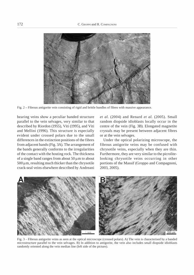
of an antigorite matrix with interlocking or interpenetrating microstructure (Wicks and Whittaker, 1977) and rare mineralogical relicts (mainly clinopyroxene) of the peridotitic protolith. The veins consist of rigid and brittle bundles of fibres with massive appearance in the hand specimen, light greyish-whitish to pale green in colour and with a splintery fracture (Fig. 2). The contact with the host rock is sharp and the fibres, up to 15-20 cm in length, are generally oriented perpendicularly to the vein selvages. It is noteworthy that the antigorite, in spite of its
massive appearance in the fresh specimen, on weathered surfaces or after grinding exhibits a fibrous habit.
resuLts: fibrous AntiGorite chArActerisAtion
Optical microscopy
More than one hundred specimens of antigorite veins have been observed under the polarizing microscope. In thin section, the fibrous antigorite-
Fig. 1 – Simplified tectonic map of the Lanzo Ultramafic massif (modified from Nicolas, 1966): sample localities are shown as circled numbers. The inset shows location of the Lanzo Massif within a simplified tectonic sketch-map of the Western Alps. Mont Blanc-Aiguilles Rouges (MB) of the Helvetic-Dauphinois Domain; SB: Grand St. Bernard Zone of the external Penninic Domain; Internal Crystalline Massifs of the Penninic Domain: Monte Rosa (MR), Gran Paradiso (GP), Dora-Maira (DM); DB: Dent Blanche nappe of the Austroalpine Domain. SA: Southern Alps. TO: Town of Torino.
172 c. GroPPo and r. comPAGnoni
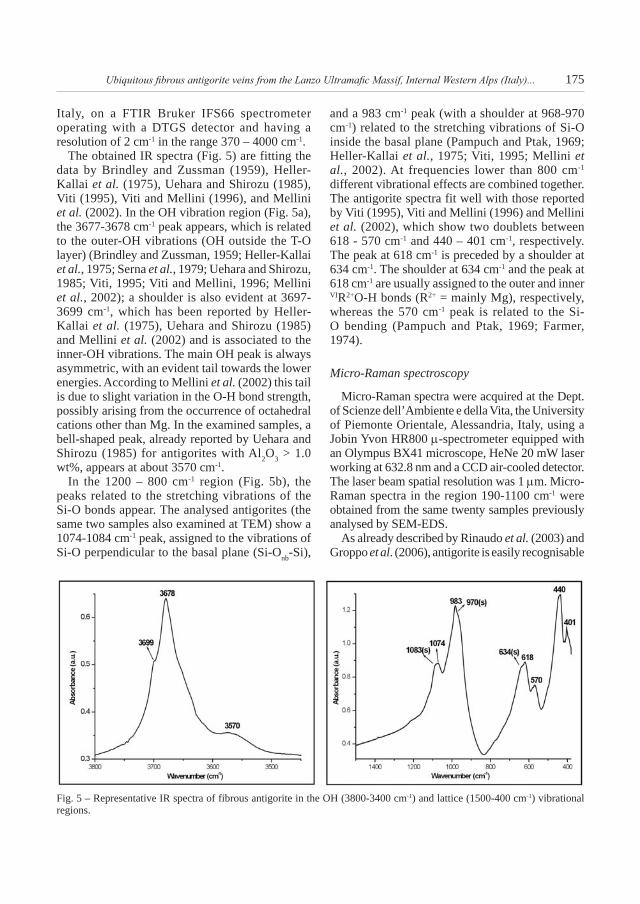
bearing veins show a peculiar banded structure parallel to the vein selvages, very similar to that described by Riordon (1955), Viti (1995), and Viti and Mellini (1996). This structure is especially evident under crossed polars due to the small differences in the extinction positions of the fibres from adjacent bands (Fig. 3A). The arrangement of the bands generally conforms to the irregularities of the contact with the hosting rock. The thickness of a single band ranges from about 50 µm to about 500 µm, resulting much thicker than the chrysotile crack-seal veins elsewhere described by Andreani
et al. (2004) and Renard et al. (2005). Small random diopside idioblasts locally occur in the centre of the vein (Fig. 3B). Elongated magnetite crystals may be present between adjacent fibres or at the vein selvages.
Under the optical polarizing microscope, the fibrous antigorite veins may be confused with chrysotile veins, especially when they are thin. Furthermore, they are very similar to the picrolite-looking chrysotile veins occurring in other portions of the Massif (Groppo and Compagnoni, 2003, 2005).
Fig. 3 – Fibrous antigorite veins as seen at the optical microscope (crossed polars). A) The vein is characterised by a banded microstructure parallel to the vein selvages. B) In addition to antigorite, the vein also includes small diopside idioblasts randomly oriented along the vein median line (left side of the picture).
Fig. 2 – Fibrous antigorite vein consisting of rigid and brittle bundles of fibres with massive appearance.

Ubiquitous fibrous antigorite veins from the Lanzo Ultramafic Massif, Internal Western Alps (Italy)... 173
TEM
Two representative antigorite samples (OF 2831 from Balangero and OF 2956 from Traves – Fig. 1) were observed by Transmission Electron Microscopy (TEM) using Selected Area Electron Diffraction (SAED), in order to recognise the possible existence of sub-microscopic intergrowths not visible with SEM. TEM observations were carried out at Dept. of Mineralogical and Petrological Sciences, the University of Torino, Italy, using a Philips CM12 instrument operating at 120 kV. The samples were prepared by suspending the powder in isopropylic alcohol; fibres aggregation was minimised by means of an ultrasound treatment, and several drops of the suspension were deposited on carbon supported Cu grid.
Morphological observations confirmed the fibrous nature of the studied samples: antigorite fibres show a fibre diameter << 1 µm and a length/diameter ratio significantly higher than the 3:1 ratio commonly used to define a fibre (American OSHA guidelines).
SAED patterns were obtained from powdered specimens (Fig. 4). The average supercell periodicities of the two studied specimens, measured on different SAED patterns, resulted to be 43.8 and 43.0 Å, respectively, which correspond to m values (m = number of tetrahedra along an entire wavelength – Kunze, 1961) of 17 on the base
of the data from Mellini et al. (1987). This m value is the most frequent value reported by Mellini et al. (1987), Wunder et al. (2001), Bromiley and Powley (2003) and Evans (2004) for antigorite formed under greenschist facies conditions.
SEM-EDSandEMPA-WDS
Twenty fibrous antigorite samples were analysed with a Cambridge Stereoscan 360 SEM equipped with an EDS Energy 200 (Oxford Instruments) at Dept. of Mineralogical and Petrological Sciences, the University of Torino, Italy. The operating conditions were: 50 s counting time and 15 kV accelerating voltage. Check analyses of the two antigorites previously observed at TEM were carried out also with an electron microprobe (EMPA) Jeol JXA-8600 equipped with wavelength dispersion spectrometers (WDS) at C.N.R. – Institute of Geosciences and Georesources, Section of Florence (Italy). The operating conditions were 15 kV accelerating voltage and 10 nA beam current. The raw data were calibrated on natural mineral standards and corrected using the B-A method (Bence and Albee, 1968). Si, Fe, Al, Mg and Ti counts were collected for 15 s and Mn, Ca, Cr and Ni counts for 40 s, respectively. SEM-EDS and EPMA-WDS analyses have been obtained on bundles of fibres rather than on a single fibre, being the fibre diameter lower than 1 µm. However, the close similarity of the spot analyses - supported
Fig. 4 – Selected Area Electron Diffraction (SAED) patterns of fibrous antigorite from samples OF2831 and OF2956 (a*c* plane).

174 c. GroPPo and r. comPAGnoni
by the TEM observations, which indicate the exclusive presence in the veins of the antigorite - allows considering our SEM-EDS analyses as quantitative.
Antigorite chemical analyses were normalised on the base of 6.824 anhydrous oxygens (as resulting from the ideal formula for m = 17 antigorite), in order to obtain 2 a.p.f.u. in the tetrahedral site and an octahedral cation sum of 2.824. Al has been allocated into both octahedral and tetrahedral sites to obtain the full occupancy of the tetrahedral site.
Chemical analyses obtained from different samples of fibrous antigorite (Table 1) were quite similar and consistent with literature data (Wicks and Whittaker, 1975, 1977; Wicks and Plant, 1979; Dungan, 1979; Mellini et al., 1987; Viti, 1995; Viti and Mellini, 1996). The Al content in the tetrahedral sites is very low (AlIV = 0.05-0.08
a.p.f.u.) and Mg in the octahedral sites is partly replaced by Al (0.04-0.08 a.p.f.u.) and Fe (0.09-0.17 a.p.f.u.). Cr is rarely present, whereas Ni has been detected only by means of WDS, being below the EDS detection limit (NiO = 0.14-0.15 wt%).
The peculiar banded structure parallel to the vein selvages is difficult to be observed in BSE images; SEM-EDS analyses confirm that the different bands are compositionally homogeneous.
IR spectroscopy
Each pellet for the IR analysis was prepared by mixing 3 mg of powdered antigorite with 200 mg of KBr; the adsorbed water was eliminated by heating the sample at 120 °C for one night. IR spectra from the two samples already analysed at TEM, SEM-EDS, EPMA-WDS and µ-Raman were recorded at Dept. of Chemistry IFM, the University of Torino,
Sample D4 OF2831 OF2831 OF2833 OF1645 OF2855 OF2864 OF2956 OF2956
Analyses 4.1 4.2 1.10* 8.2 8.3 7.3 6.2 2.1 2.1*
SiO242.56 42.62 42.56 42.80 42.53 42.69 42.09 42.47 42.53
Al2O31.90 2.02 1.94 2.66 2.50 3.04 1.98 1.94 2.03
Cr2O30.00 0.00 0.00 0.00 0.00 0.00 0.38 0.00 0.00
FeO 2.90 3.26 3.32 2.42 2.27 2.69 2.53 4.39 3.88
MnO 0.00 0.00 0.03 0.00 0.00 0.00 0.00 0.00 0.07
MgO 39.57 39.24 38.67 39.21 39.54 39.27 39.42 38.51 38.21
NiO 0.00 0.00 0.14 0.00 0.00 0.00 0.00 0.00 0.15
Total 86.92 87.13 86.67 87.09 86.84 87.69 86.40 87.31 86.90
Si 1.94 1.94 1.95 1.94 1.93 1.92 1.93 1.94 1.95
AlIV 0.06 0.06 0.05 0.06 0.07 0.08 0.07 0.06 0.05
AlVI 0.04 0.05 0.05 0.08 0.07 0.08 0.04 0.04 0.06
Cr 0.00 0.00 0.00 0.00 0.00 0.00 0.01 0.00 0.00
Fe2+ 0.11 0.12 0.13 0.09 0.09 0.10 0.10 0.17 0.15
Mn 0.00 0.00 0.00 0.00 0.00 0.00 0.00 0.00 0.00
Mg 2.68 2.66 2.64 2.64 2.67 2.63 2.69 2.62 2.61
Ni 0.00 0.00 0.01 0.00 0.00 0.00 0.00 0.00 0.01
Cations VI 2.83 2.83 2.82 2.82 2.83 2.82 2.84 2.83 2.82
tAbLe 1SEM-EDS and WDS analyses of fibrous antigorite from the Lanzo massif
The analyses have been normalized on the basis of 6.824 anhydrous oxygens* WDS analyses
Ubiquitous fibrous antigorite veins from the Lanzo Ultramafic Massif, Internal Western Alps (Italy)... 175
Italy, on a FTIR Bruker IFS66 spectrometer operating with a DTGS detector and having a resolution of 2 cm-1 in the range 370 – 4000 cm-1.
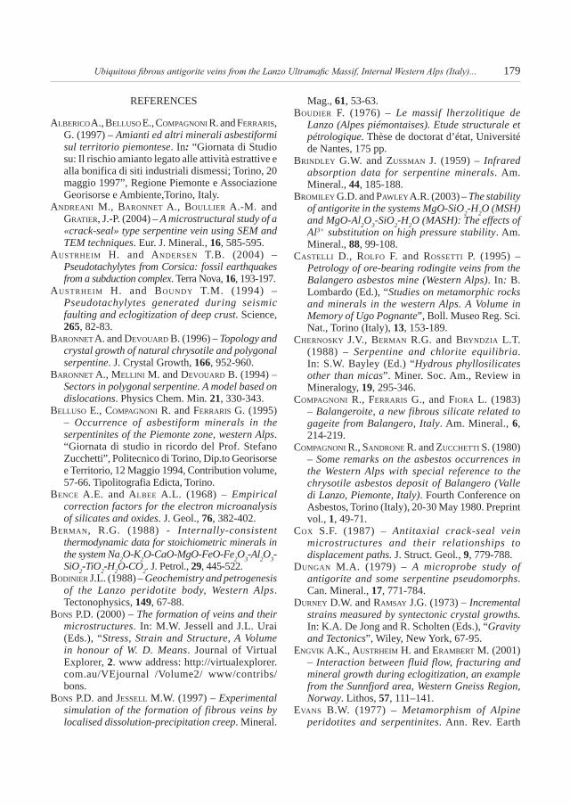
The obtained IR spectra (Fig. 5) are fitting the data by Brindley and Zussman (1959), Heller-Kallai et al. (1975), Uehara and Shirozu (1985), Viti (1995), Viti and Mellini (1996), and Mellini et al. (2002). In the OH vibration region (Fig. 5a), the 3677-3678 cm-1 peak appears, which is related to the outer-OH vibrations (OH outside the T-O layer) (Brindley and Zussman, 1959; Heller-Kallai et al., 1975; Serna et al., 1979; Uehara and Shirozu, 1985; Viti, 1995; Viti and Mellini, 1996; Mellini et al., 2002); a shoulder is also evident at 3697-3699 cm-1, which has been reported by Heller-Kallai et al. (1975), Uehara and Shirozu (1985) and Mellini et al. (2002) and is associated to the inner-OH vibrations. The main OH peak is always asymmetric, with an evident tail towards the lower energies. According to Mellini et al. (2002) this tail is due to slight variation in the O-H bond strength, possibly arising from the occurrence of octahedral cations other than Mg. In the examined samples, a bell-shaped peak, already reported by Uehara and Shirozu (1985) for antigorites with Al2O3 > 1.0 wt%, appears at about 3570 cm-1.
In the 1200 – 800 cm-1 region (Fig. 5b), the peaks related to the stretching vibrations of the Si-O bonds appear. The analysed antigorites (the same two samples also examined at TEM) show a 1074-1084 cm-1 peak, assigned to the vibrations of Si-O perpendicular to the basal plane (Si-Onb-Si),
and a 983 cm-1 peak (with a shoulder at 968-970 cm-1) related to the stretching vibrations of Si-O inside the basal plane (Pampuch and Ptak, 1969; Heller-Kallai et al., 1975; Viti, 1995; Mellini et al., 2002). At frequencies lower than 800 cm-1 different vibrational effects are combined together. The antigorite spectra fit well with those reported by Viti (1995), Viti and Mellini (1996) and Mellini et al. (2002), which show two doublets between 618 - 570 cm-1 and 440 – 401 cm-1, respectively. The peak at 618 cm-1 is preceded by a shoulder at 634 cm-1. The shoulder at 634 cm-1 and the peak at 618 cm-1 are usually assigned to the outer and inner VIR2+O-H bonds (R2+ = mainly Mg), respectively, whereas the 570 cm-1 peak is related to the Si-O bending (Pampuch and Ptak, 1969; Farmer, 1974).
Micro-Raman spectroscopy
Micro-Raman spectra were acquired at the Dept. of Scienze dell’Ambiente e della Vita, the University of Piemonte Orientale, Alessandria, Italy, using a Jobin Yvon HR800 µ-spectrometer equipped with an Olympus BX41 microscope, HeNe 20 mW laser working at 632.8 nm and a CCD air-cooled detector. The laser beam spatial resolution was 1 µm. Micro-Raman spectra in the region 190-1100 cm-1 were obtained from the same twenty samples previously analysed by SEM-EDS.
As already described by Rinaudo et al. (2003) and Groppo et al. (2006), antigorite is easily recognisable
Fig. 5 – Representative IR spectra of fibrous antigorite in the OH (3800-3400 cm-1) and lattice (1500-400 cm-1) vibrational regions.

176 c. GroPPo and r. comPAGnoni
from the other serpentine minerals on the basis of its Raman spectrum. The analysed samples show four intense peaks at about 230, 379, 684 and 1045 cm-1 (Fig. 6), corresponding to O-H-O groups vibrations, symmetric bending ν5(e) of the SiO4 tetrahedra and symmetric and antisymmetric Si-Ob-Si stretching vibrations (Kloprogge et al., 1999; Rinaudo et al., 2003), respectively. Two less intense peaks at 202 and 640 cm-1 are also present (Fig. 6): the first one may be ascribed to Mg(O,OH)6 group vibrations, while the second one is probably related to the epoxy resin used for thin section preparation, even if a possible small contribution from the OH-Mg-OH translation modes cannot be excluded (Groppo et al., 2006).
Because the analysed antigorites are chemically quite homogeneous, no significant shifts in the peak positions have been observed for different samples. However, it must be pointed out that the relative peak intensities - especially for the 230 cm-1 peak - are strongly dependent on the mineral crystallographic orientation.
discussion
Antigorite stability field and variation in its m-value
According to Evans (2004 with ref. therein), field, isotopic and experimental data suggest that antigorite is the high temperature form of Mg-serpentine, developing at temperatures from ~250 °C to more
than 500 °C (Wenner and Taylor, 1971; Evans et al., 1976; Chernosky et al., 1988; O’Hanley, 1996).
The low temperature stability limit of antigorite is constrained by the reaction Lz/Ctl = Atg + Brc (brucite), which is widely observed during prograde metamorphism (Evans, 1977; Wicks and Whittaker, 1977; Wicks and Plant, 1979; O’Hanley et al., 1989). The position of this reaction curve, which is strongly dependent on both the internally consistent database used to model the reaction (e.g. Berman, 1988; Holland and Powell, 1998) and the considered antigorite m-value, ranges from a minimum of 170 °C (at 1 bar) (using the Holland and Powell, 1998 database) to a maximum of 310 °C (at 1 bar) (using the Berman, 1988 database) (Fig. 7).
The upper stability limit of antigorite was studied in detail by numerous petrologists (Evans et al., 1976; Ulmer and Trommsdorff, 1995; Wunder and Schreyer, 1997; Wunder et al., 2001; Bromiley and Powley, 2003). At pressure below 1.5 GPa, antigorite breaks down to form forsterite + talc + water, the reaction approximately occurring at T ≈ 600 °C for P ≈ 1.0 GPa. Above 1.5 GPa, antigorite dehydrates to form forsterite + enstatite + water (Fig. 7).
Mellini et al. (1987) and Wunder et al. (2001) demonstrated that the m-values of antigorite decrease with increasing temperature and decreasing pressure. Wunder et al. (2001) experimentally obtained m-values in the range 18-14 for temperatures increasing from 350 to 600 °C, the antigorite with m = 17 being stable in the 350-450 °C temperature interval at P ≈ 0.5 GPa (Fig. 7). Mellini et al. (1987) assume that antigorite with m = 17 is stable at 435 ± 30 °C and 0.1-0.2 GPa.
P-T estimates
Thermobarometric calculations in the serpentine system are generally difficult to perform because most reactions involving serpentine minerals are only T-dependent (Evans et al., 1976); hence the stability fields of serpentine minerals are extremely wide and poorly constrained at low temperature (Evans, 2004). In addition, serpentine minerals easily recrystallise, obliterating the previous history.
For these reasons, the P-T-t path of the Lanzo Ultramafic Massif has been mainly constrained on the basis of thermobarometric data derived from the associated metamorphic mafic rocks and rodingites. T = 550-620 °C, P ≥ 2.0 GPa represent
Fig. 6 – Representative µ-Raman spectrum of fibrous antigorite in the 1100-190 cm-1 frequency region.

Ubiquitous fibrous antigorite veins from the Lanzo Ultramafic Massif, Internal Western Alps (Italy)... 177
peak conditions (Sandrone and Compagnoni, 1983; Pognante, 1991; Müntener, 2006). On the other hand, the post-climactic history is poorly constrained not only in the Lanzo Massif, but also all over the Piemonte Zone; 400 °C and pressures lower than 0.6 GPa were estimated by Pognante (1991) for the exhumation trajectory (Fig. 7). Castelli et al. (1995), studying rodingitic veins cutting across the serpentinite of the Balangero mine, estimated T = 300-400 °C and P = 0.3-0.4 GPa (Fig. 7).
Genetic conditions
Microstructural relationships with the other vein generations suggest that fibrous antigorite has formed during the decompressional evolution of the Lanzo Ultramafic Massif, prior to the development of the chrysotile asbestos veins once exploited in the Balangero mine (Compagnoni et al., 1980; Groppo and Compagnoni, 2003, 2005). Fibrous antigorite veins, in fact, are often crosscut by slip and/or
cross fibre chrysotile veins, which probably formed during later vein opening events in a very shallow environment.
The antigorite a parameter of 43.0-43.8 Å, determined by TEM-SAED, corresponds to m = 17 on the base of the data from Mellini et al. (1987). Taking into consideration the results of Mellini et al. (1987) and Wunder et al. (2001) and the thermobarometric estimates of Pognante (1991) and Castelli et al. (1995), it may be suggested that the studied fibrous antigorite formed at T = 350-400 °C and P = 0.3-0.6 GPa (Fig. 7). This result must be considered with care, since the m-value of antigorite is not only dependent on P and T but also on additional factors, such as mineral assemblage, water activity, and chemical composition, especially the Al and Fe contents (Wunder et al., 2001).
Why did antigorite grow with a fibrous habit?
Antigorite veins occurring in the Lanzo Ultramafic Massif frequently exhibit a banded structure and a fibrous morphology in spite of the usual lath-like habit. Which is the mechanism responsible for the fibrous growth of antigorite? Two different mechanisms are generally envisaged to explain the fibrous vein formation: i) mineral growth after the opening of a fracture (Ramsay, 1980); and ii) the development of fibrous veins in the absence of fracturing (Taber, 1918; Mügge, 1928; Durney and Ramsey, 1973; Fisher and Brantley, 1992). A third mechanism (iii) may be also considered, which envisages the epitaxial replacement of antigorite after a former fibrous mineral, most likely chrysotile.
(i) The growth of fibrous minerals inside a fracture is also known as “crack-seal” mechanism, first suggested by Ramsay (1980) and later supported by Cox (1987), Fisher and Byrne (1990), Urai et al. (1991), Jessell et al. (1994) and Renard et al. (2000). According to this hypothesis, the veins form by cracking and incremental opening events. After each cracking event, the narrow fracture is sealed with the vein-forming mineral. The crack-seal cycle can be repeated hundred of times (Fisher and Brantley, 1992): this mechanism can explain the banded structures, marked either by wall rock inclusion trails parallel to the fracture selvages or by slight differences in the orientation of the minerals in the vein.
Fig. 7 – Antigorite stability field (modified from Wunder et al., 2001), limited at high temperature by the experimentally determined reactions of Ulmer and Trommsdorff (1995) (U. and T., 95) and Wunder and Schreyer (1997) (W. and S., 97). The low temperature stability limit is based on the data of Evans et al. (1976). Dotted lines represent the P-T-dependence of the most frequent m-value in antigorite, as reported in Wunder et al. (2001). The P-T estimates of Pognante (1991) and Castelli et al. (1995) for mafic rocks and rodingites from the Lanzo Ultramafic Massif are also reported.

178 c. GroPPo and r. comPAGnoni
(ii) The essence of the “vein growth without fracturing” is the dissolution of matter at certain sites and its precipitation elsewhere (Bons and Jessel, 1997). This process is also called “dissolution-precipitation creep” and involves transport of material from one site to another one by diffusional mass transfer in a grain boundary fluid. The driving force of this process is the presence of chemical concentration gradients, which in turn arise from local variations in the stress conditions (Bons, 2000). Recent experiments by Means and Li (1995, 2001), Li and Means (1997) and Bon and Jessel (1997) have supported this mechanism. The obtained fibrous veins show several similarities with the crack-seal veins and are characterized by a banded structure and by a medial plane, across which fibres that nucleate separately at the two walls abut. The banded structure is only a morphological feature, which is not related to compositional variations due to fluctuations in the chemical potential of the pore fluid from which fibres have grown.
The banded structure of the studied veins and the fibrous habit of the filling antigorite may be explained through both growth mechanisms (i) and (ii), though microstructures clearly related to cracking events (i.e. wall rock inclusions) have not been observed. In addition, the brittle regime necessary to explain the crack-seal process is generally not compatible with the estimated pressure and temperature conditions of formation: nevertheless, it has been demonstrated that the crack-seal process is possible also under much higher temperatures and pressures (cf. Austrheim and Boundy, 1994; Engvik et al., 2001; Lund and Austrheim, 2003; Austrheim and Andersen, 2004).
About the third possibility, i.e. the epitaxial replacement of former chrysotile veins (Baronnet et al., 1994; Baronnet and Devouard, 1996), this mechanism can be excluded for the studied fibrous antigorite. In fact, microstructural observations clearly show that: fibrous antigorite veins developed during the exhumation history of the serpentinites, since they crosscut the peak metamorphic veins consisting of olivine + Ti-clinohumite + diopside + antigorite (Groppo and Compagnoni, 2003, 2005). Moreover, antigorite veins did develop prior to the chrysotile vein formation. The antigorite for chrysotile replacement implies a prograde evolution, which is exactly the opposite of the P-T path estimated for the studied serpentinites.
Then, the fibrous habit of antigorite is not due to the replacement of a former fibrous mineral, but to the mechanisms of vein formation by crack-seal or dissolution-precipitation creep previously discussed.
concLusions
On the basis of the reported mineralogical and microstructural data, and the above discussion, it may be concluded that:
i) In the Lanzo Ultramafic Massif, antigorite-bearing veins formed during different stages of the metamorphic evolution: at peak metamorphic conditions antigorite grew with a lath habit, in association with olivine, Ti-clinohumite and diopside; in the later exhumation history, antigorite developed as a fibrous mineral in 1 to 20 cm-thick ubiquitous veins.
ii) The identification and characterisation of fibrous antigorite may be easily and rapidly performed by combining several analytical techniques, such as optical microscopy, SEM-EDS and micro-Raman vibrational spectroscopy.
iii) The experimentally determined m = 17 value of the Lanzo fibrous antigorite and the thermobarometric estimates get from the associated basic and rodingitic rocks, suggest that the veins formed under greenschist facies conditions, at T = 350-400 °C and P = 0.3-0.6 GPa.
The fibrous habit of antigorite may be explained by two different mechanisms, i.e. the crack-seal process and the dissolution-precipitation creep mechanism. These processes are equally able to explain the banded structure of the studied veins and are compatible with the estimated P-T conditions of formation.
AcKnowLedGements
This work is part of a multidisciplinary research project entitled “Asbestos Hazard in the Western Alps”, supported by Regione Piemonte (Italy) and coordinated by the Interdepartmental Centre “Giovanni Scansetti” for Studies on Asbestos and Other Toxic Particulates at the University of Turin. Authors are grateful to A. Baronnet and B.W. Evans for careful and constructive reviews of a former version. The reviews by Pietro Marescotti and Vittorio Scribano are greatly appreciated.
Ubiquitous fibrous antigorite veins from the Lanzo Ultramafic Massif, Internal Western Alps (Italy)... 179
REFERENCES
ALberico A., beLLuso e., comPAGnoni r. and ferrAris, G. (1997) – Amianti ed altri minerali asbestiformi sul territorio piemontese. In: “Giornata di Studio su: Il rischio amianto legato alle attività estrattive e alla bonifica di siti industriali dismessi; Torino, 20 maggio 1997”, Regione Piemonte e Associazione Georisorse e Ambiente,Torino, Italy.
AndreAni m., bAronnet A., bouLLier A.-m. and GrAtier, J.-P. (2004) – A microstructural study of a «crack-seal» type serpentine vein using SEM and TEM techniques. Eur. J. Mineral., 16, 585-595.
Austrheim h. and Andersen t.b. (2004) – Pseudotachylytes from Corsica: fossil earthquakes from a subduction complex. Terra Nova, 16, 193-197.
Austrheim h. and boundy t.m. (1994) – Pseudotachylytes generated during seismic faulting and eclogitization of deep crust. Science, 26�, 82-83.
bAronnet A. and devouArd b. (1996) – Topology and crystal growth of natural chrysotile and polygonal serpentine. J. Crystal Growth, 166, 952-960.
bAronnet A., meLLini m. and devouArd b. (1994) – Sectors in polygonal serpentine. A model based on dislocations. Physics Chem. Min. 21, 330-343.
beLLuso e., comPAGnoni r. and ferrAris G. (1995) – Occurrence of asbestiform minerals in the serpentinites of the Piemonte zone, western Alps. “Giornata di studio in ricordo del Prof. Stefano Zucchetti”, Politecnico di Torino, Dip.to Georisorse e Territorio, 12 Maggio 1994, Contribution volume, 57-66. Tipolitografia Edicta, Torino.
bence A.e. and ALbee A.L. (1968) – Empirical correction factors for the electron microanalysis of silicates and oxides. J. Geol., 76, 382-402.
bermAn, r.G. (1988) - Internally-consistent thermodynamic data for stoichiometric minerals in the system Na2O-K2O-CaO-MgO-FeO-Fe2O3-Al2O3-SiO2-TiO2-H2O-CO2. J. Petrol., 29, 445-522.
bodinier J.L. (1988) – Geochemistry and petrogenesis of the Lanzo peridotite body, Western Alps. Tectonophysics, 149, 67-88.
bons P.d. (2000) – The formation of veins and their microstructures. In: M.W. Jessell and J.L. Urai (Eds.), “Stress, Strain and Structure, A Volume in honour of W. D. Means. Journal of Virtual Explorer, 2. www address: http://virtualexplorer.com.au/VEjournal /Volume2/ www/contribs/bons.
bons P.d. and JesseLL m.w. (1997) – Experimental simulation of the formation of fibrous veins by localised dissolution-precipitation creep. Mineral.
Mag., 61, 53-63.boudier f. (1976) – Le massif lherzolitique de
Lanzo (Alpes piémontaises). Etude structurale et pétrologique. Thèse de doctorat d’état, Université de Nantes, 175 pp.
brindLey G.w. and zussmAn J. (1959) – Infrared absorption data for serpentine minerals. Am. Mineral., 44, 185-188.
bromiLey G.d. and PAwLey A.r. (2003) – The stability of antigorite in the systems MgO-SiO2-H2O (MSH) and MgO-Al2O3-SiO2-H2O (MASH): The effects of Al3+ substitution on high pressure stability. Am. Mineral., 88, 99-108.
cAsteLLi d., roLfo f. and rossetti P. (1995) – Petrology of ore-bearing rodingite veins from the Balangero asbestos mine (Western Alps). In: B. Lombardo (Ed.), “Studies on metamorphic rocks and minerals in the western Alps. A Volume in Memory of Ugo Pognante”, Boll. Museo Reg. Sci. Nat., Torino (Italy), 1�, 153-189.
chernosKy J.v., bermAn r.G. and bryndziA L.t. (1988) – Serpentine and chlorite equilibria. In: S.W. Bayley (Ed.) “Hydrous phyllosilicates other than micas”. Miner. Soc. Am., Review in Mineralogy, 19, 295-346.
comPAGnoni r., ferrAris G., and fiorA L. (1983) – Balangeroite, a new fibrous silicate related to gageite from Balangero, Italy. Am. Mineral., 6, 214-219.
comPAGnoni r., sAndrone r. and zucchetti s. (1980) – Some remarks on the asbestos occurrences in the Western Alps with special reference to the chrysotile asbestos deposit of Balangero (Valle di Lanzo, Piemonte, Italy). Fourth Conference on Asbestos, Torino (Italy), 20-30 May 1980. Preprint vol., 1, 49-71.
cox s.f. (1987) – Antitaxial crack-seal vein microstructures and their relationships to displacement paths. J. Struct. Geol., 9, 779-788.
dunGAn m.A. (1979) – A microprobe study of antigorite and some serpentine pseudomorphs. Can. Mineral., 17, 771-784.
durney d.w. and rAmsAy J.G. (1973) – Incremental strains measured by syntectonic crystal growths. In: K.A. De Jong and R. Scholten (Eds.), “Gravity and Tectonics”, Wiley, New York, 67-95.
enGviK A.K., Austrheim h. and erAmbert m. (2001) – Interaction between fluid flow, fracturing and mineral growth during eclogitization, an example from the Sunnfjord area, Western Gneiss Region, Norway. Lithos, �7, 111–141.
evAns b.w. (1977) – Metamorphism of Alpine peridotites and serpentinites. Ann. Rev. Earth

180 c. GroPPo and r. comPAGnoni
Planet. Sci., �, 397-448.evAns b.w. (2004) – The serpentinite multisystem
revisited: chrysotile is metastable. Int. Geol. Rev., 46, 479-506.
evAns b.w., JohAnnes w., oterdoom h. and trommsdorff v. (1976) – Stability of chrysotile and antigorite in the serpentinite multisystem. Schweiz. Mineral. Petrogr. Mitt., �6, 79-93.
fArmer v.c. (1974): The infrared spectra of minerals. Mineralogical Society Ed., London, 539 pp.
fisher d. and brAntLey s.L. (1992) – Models of quartz overgrowth and vein formation: deformation and fluid flow in an ancient subduction zone. J. Geophys. Res., 97, 20043-20061.
fisher d. and byrne t. (1990) – The character and distribution of mineralized fractures in the Kodiak Formation, Alaska: implications for fluid flow in an underthrust sequence. J. Geophys. Res., 9�, 9069-9080.
GroPPo c. and comPAGnoni r. (2003) – Polyphase metamorphic veins in the antigorite serpentinite of the Ultramafic Lanzo Massif, Western Italian Alps. Journées Tématiques “Serpentines” (SGF) – Paris, 20-21/11/2003 – Abstract Vol., 19.
GroPPo c. and comPAGnoni r. (2005) – Different generations of metamorphic asbestos veins in the serpentinites of the Piemonte Zone (Western Alps): hypothesis on their genetic conditions. ISPET - International Seminars of Petrology - Fourth Seminar - “Advanced analytical and experimental techniques in petrology” - Canberra (Australia), 5-12 February 2005.
GroPPo c., GuLA A., comPAGnoni r. and ferrAris G. (2004) – Balangeroite from the Lanzo Massif (Western Alps) revisited: fibrous vs. prismatic morphology and genetic considerations. 32nd Int. Geol. Congress – Florence, 20-28 August 2004, Abstract Vol., 249.
GroPPo c., rinAudo c., cAiro s., GAstALdi d. and comPAGnoni r. (2006) – Raman Spectroscopy as a rapid method for the identification of serpentine minerals from serpentinized ultramafics. Eur. J. Mineral., 18, 319-329.
heLLer-KALLAi L., yAriv s. and Gross s. (1975) – Hydroxyl-stretching frequencies of serpentine minerals. Mineral. Mag., 40, 197-200.
hess h.h., smith J.r. and denGo G. (1952) – Antigorite from the vicinity of Caracas, Venezuela. Am. Mineral., �7, 68-75.
hoLLAnd, t. J. b. h. & PoweLL, r. (1998) - An internally consistent thermodynamic data set for phases of petrological interest. J. Metam. Geol., 16, 309–343.
KLoProGGe J.t., frost r.L. and rintouL L. (1999) – Single crystal Raman microscopic study of the asbestos mineral chrysotile. Phys. Chem. Chem. Phys., 1, 2559-2564.
Kunze G. (1961) - Antigorit. Strukturtheoretische Grundlagen und ihre praktische Bedeutung für die weitere Serpentin-Forschung. Fortschr. Mineral., �9, 206-324.
JesseL m.v., wiLLiAm c.e. and GrAy d.r. (1994) – Bedding parallel veins and their relationship to folding. J. Struct. Geol., 16, 753-767.
Li t. and meAns w.d. (1997) – Taber growth of fibrous veins. Geol. Soc. Am., Abs. with Programs, 29, A-161.
Lund m.G. and Austrheim h. (2003) – High-pressure metamorphism and deep-crustal seismicity: evidence from contemporaneous formation of pseudotachylytes and eclogite facies coronas. Tectonophysics, �72, 59-83.
meAns w.d. and Li t. (2001) – A laboratory simulation of fibrous veins: some first observations. J. Struct. Geol., 2�, 857-863.
meAns, w.d. and Li, t. (1995) – Experimental antitaxial growth of fibrous crystals, II: internal structures. Geol. Soc. Am., Abs. with Programs, 27, A-70.
meLLini m., fuchs y., viti c., LemAire c., and LinAres J. (2002) – Insights into the antigorite structure from Mössbauer and FTIR spectroscopies. Eur. J. Mineral., 14, 97-104.
meLLini m., trommsdorff v. and comPAGnoni r. (1987) – Antigorite polysomatism: behaviour during progressive metamorphism. Contrib. Mineral. Petrol., 97, 147-155.
mossmAn b.t. (1993) – Cellular and molecular mechanisms of disease. In: G.D. Jr. Guthrie and B. T. Mossman (Eds.), “Health Effects of Mineral Dusts”, Mineral. Soc. Am., Washington, 28, 513-521.
müGGe o. (1928) – Über die Entstellung faseriger Minerale und ihrer Aggregationsformen. N. Jahrb. Min. Geol. Paläont., �8, 303-348.
müntener o., PettKe t., desmurs L., meier m. and schALteGGer u. (2004) – Refertilization of mantle peridotite in embryonic ocean basins: trace element and Nd isotopic evidence and implications for crust-mantle relationship. Earth Planet. Sci. Lett, 221, 293-308.
nichoLson w.J., LAnGer A.m. and seLiKoff i.J. (1978) – Epidemiological evidence on asbestos. In: “Proceedings of the workshop on asbestos: definitions and measurement methods”, N.B.S. Special Publ., �06, 71-84.

Ubiquitous fibrous antigorite veins from the Lanzo Ultramafic Massif, Internal Western Alps (Italy)... 181
nicoLAs A. (1966) – Etude pétrochimique des Roches vertes et de leurs minéraux entre Dora Maira et Grand Paradis (Alpes piémontaises); le complexe ophiolite-schistes lustrés. Thèse, Fac. Sc. Nantes, 299 p.
o’hAnLey d.s. (1996) - Serpentinites. Records of tectonic and petrological history. Oxford Univ. Press, 277 pp.
o’hAnLey d.s., chernosKy J.v. and wicKs f.J. (1989) – The stability of lizardite and chrysotile. Can. Mineral., 27, 483-493.
PAmPuch r. and PtAK w. (1969) – Infrared spectra and structure of 1:1 layer lattice silicates. Part 1: the vibrations of the tetrahedral layer. Pol. Akad. Nauk., Oddizal Krakowie, Prace Mineral., 1�, 7-39.
PeLLetier L. and müntener o. (2006) – High pressure metamorphism of the Lanzo peridotite and its oceanic cover, and some consequences for the Sesia-Lanzo zone (northwestern Italian Alps). Lithos, 90, 111-130.
PiccArdo G.b., müntener o., zAnetti A. and PettKe t. (2004) – Ophiolitic peridotites of the Alpine-Apennine system: mantle processes and geodynamic relevance. Int. Geol. Rev., 46, 1119-1159.
PoGnAnte u. (1991) – Petrological constraints on the eclogite and blueschist-facies metamorphism and P-T-t path in the Western Alps. J. Metam. Geol., 9, 5-17.
PoGnAnte u., rösLi u. and toscAni L. (1985) – Petrology of ultramafic and mafic rocks from the Lanzo peridotite body (Western Alps). Lithos, 18, 201-214.
rAmPone e. and PiccArdo G.b. (2000) – The ophiolite-oceanic lithosphere analogue: new insights from the Northern Apennines (Italy). In: Y. Dilek, E.M. Moores, D. Elthon and A. Nicolas (Eds.), “Ophiolites and oceanic crust: new insights from field studies and the oceanic drilling program”, Geol. Soc. Am., Special Paper, Boulder, Colorado, �49, 21-34.
rAmsAy J.G. (1980) – The crack-seal mechanism of rock deformation. Nature, 284, 135-139.
renArd f. GrAtier J.P. JAmveit b. (2000) Kinetics of crack-sealing, intergranular pressure solution and compaction around active faults. J. Struct. Geol. 22, 1395-1407.
renArd f., AndreAni m., bouLLier A.m. and LAbAume P. (2005) – Crack-seal patterns: records of uncorrelated stress release variations in crustal rocks. In: D. Gapais, J.P. Brun and P.R. Cobbold (Eds.), “Deformation mechanisms, rheology and tectonics: from minerals to lithosphere”. Geol. Soc. London, Special Publ., 24�, 67-79.
rinAudo c., GAstALdi d. and beLLuso e. (2003) – Characterization of chrysotile, antigorite and lizardite by FT-Raman spectroscopy. Can. Mineral., 41, 883-890.
riordon P.h. (1955) – The genesis of asbestos in ultrabasic rocks. Econ. Geol., �0, 67-83.
sAndrone r. and comPAGnoni r. (1983) – Relics of pargasite-bearing peridotite in the antigorite serpentinite of Balangero, near Lanzo (Western Alps). Ofioliti, 11, 35-47.
sernA c.J., white J.L. and veLde b.d. (1979) – The effect of aluminium on the infrared spectra of 7 Å trioctahedral minerals. Mineral. Mag., 4�, 141-148.
udovKinA n.G., GorshKov A.i. and shKuroPAt b.A. (1987) – Asbestiform antigorite in the serpentinite from the eclogite-bearing Aktyuz block, northern Tien Shan. Doklady Akademii Nauk., SSSR, 29�, 963-968.
uehArA s. and shirozu h. (1985) – Variations in chemical composition and structural properties of antigorites. Mineral. J., 12, 299-318.
uLmer P. and trommsdorff v. (1995) – Serpentine stability to mantle depths and subduction related magmatism. Science, 268, 858-861.
urAi, J. L., wiLLiAms, P.f. and vAn roermund, h.L.m. (1991) – Kinematics of crystal growth in syntectonic fibrous veins. J. Struct. Geol., 1�, 823-836.
viti c. (1995) – I serpentini dell’Isola d’Elba. PhD Thesis, Università degli Studi di Siena, 59 p.
viti c. and meLLini m. (1996) – Vein antigorites from Elba Island, Italy. Eur. J. Mineral., 8, 423-434.
wenner d.b. and tAyLor h.P. (1971) – Temperatures of serpentinization of ultramafic rocks based on 18O/16O fractionation between coexisting serpentine and magnetite. Contrib. Miner. Petrol., �2, 165-185.
wicKs f.J. and PLAnt A.G. (1979) – Electron-microprobe and X-ray-microbeam studies of serpentine textures. Can. Mineral., 17, 785-830.
wicKs f.J. and whittAKer e.J.w. (1975) – A reappraisal of the structures of the serpentine minerals. Can. Mineral., 1�, 227-243.
wicKs f.J. and whittAKer e.J.w. (1977) – Serpentine textures and serpentinization. Can. Mineral., 1�, 449-488.
wunder b. and schreyer w. (1997) – Antigorite: high pressure stability in the system MgO-SiO2-H2O (MSH). Lithos, 41: 213-227.
wunder b., wirth r. and GottschALK m. (2001) – Antigorite: pressure and temperature dependence of polysomatism and water content. Eur. J. Mineral., 1�, 485-495.