ucsf - core.ac.uk
TRANSCRIPT

UCSFUC San Francisco Previously Published Works
TitleCorrelation of clinical neuromusculoskeletal and central somatosensory performance: variability in controls and patients with severe and mild focal hand dystonia.
Permalinkhttps://escholarship.org/uc/item/6183t8xg
JournalNeural plasticity, 9(3)
ISSN2090-5904
AuthorsByl, Nancy NNagarajan, Srikantan SMerzenich, Michael Met al.
Publication Date2002
DOI10.1155/NP.2002.177 Peer reviewed
eScholarship.org Powered by the California Digital LibraryUniversity of California

NEURAL PLASTICITY VOLUME 9, NO. 3, 2002
Correlation of Clinical Neuromusculoskeletal and CentralSomatosensory Performance: Variability in Controls and Patients
with Severe and Mild Focal Hand Dystonia
Nancy N. Byl, Srikantan S. Nagarajan, Michael M. Merzenich, Tim Roberts2 and Alison McKenzie4
1Dept. ofPhysical Therapy & Rehabilitation Science, School ofMedicine; 2Dept. ofRadiology,Bioimaging Laboratory," 3Dept. ofOtolaryngology & Dept. ofPhysiology, Coleman Laboratory & Keck
Centerfor Neuroscience Research University ofCalifornia, San Francisco, California, USA,"aDept, ofPhysical Therapy, Chapman University, Orange, California, USA,"
SUMMARY
Focal hand dystonia (FHd) is a recalcitrant,disabling movement disorder, characterized byinvoluntary co-contractions of agonists andantagonists, that can develop in patients whooveruse or misuse their hands. The aim of this
study was to document clinical neuromusculo-skeletal performance and somatosensoryresponses (magnetoencephalography) in healthycontrols and in FHd subjects with mild versussevere hand dystonia. The performance ofhealthy subjects (n 17) was significantly betterthan that of FHd subjects (n 17) on all clinical
parameters. Those with mild dystonia (n 10)demonstrated better musculoskeletal skills,task-specific motor performance, and sensorydiscrimination, but the performance of sensoryand fine motor tasks was slower than that ofpatients with severe dystonia. In terms ofsomatosensory evoked field responses (SEFs),FHd subjects demonstrated a significantdifference in the location of the handrepresentation on the x and y axes, loweramplitude of SEFs integrated across latency,
Reprint requests to: Nancy N. Byl, PhD, MPH, PT,FAPTA, 1360 9th Ave. Box 0736, Dept. of Physical Therapy& Rehabilitation Science, University of California, SanFrancisco, CA 94143-0736, USA [email protected]
and a higher ratio of mean SEF amplitude tolatency than the controls. Bilaterally, those withFHd (mild and severe) lacked progressivesequencing of the digits from inferior tosuperior. On the affected digits, subjects withsevere dystonia had a significantly higher ratioof SEF amplitude to latency and a significantlysmaller mean volume of the cortical handrepresentation than those with mild dystonia.Severity of dystonia positively correlated withthe ratio of SEF mean amplitude to latency(0.9029 affected, 0.8477 unaffected; p < 0.01).The results of the present study strengthen theevidence that patients with FHd demonstratesigns of somatosensory degradation of the handthat correlates with clinical sensorimotordysfunction, with characteristics of the de-differentiation varying by the severity of handdystonia. If these findings represent aberrantlearning, then effective rehabilitation mustincorporate the principles of neuroplasticity.Training must be individualized to each patientto rebalance the sensorimotor feedback loopand to restore normal fine motor control.
KEYWORDS
somatosensory evoked potentials, neuroplasticity,focal hand dystonia, magnetoencephalography
(C) 2002 Freund & Pettman, U.K. 177

178 N.N. BYL ET AL.
INTRODUCTION
Most tasks performed with the hands requiredelicate, complex, individuated, fine motor move-ments (Gerloff et al., 1996; Johansson, 1996; Sher-rington, 1906). The hand has a large, orderly,somatotopic, highly differentiated representationin the sensory and motor areas of the brain(Iwamura et al., 1983; Iwamura et al., 1992;Jenkins et al., 1988; Kass et al., 1986; Penfield,1950; Yang et al., 1994). These topographicalrepresentations are complemented by functionalrepresentations of well- learned tasks (e.g. writing)(Rijintjes et al., 1999).
Somatotopic and functional representations aremodified by injury, development, learning,environmental enrichment, deprivation, disease,and practice. Representational changes can bemodified over a lifetime by attended, goal-directed, rewarded, nonstereotypic, progressivespaced practice (Jenkins et al., 1990; Wang et al.,1994). Selective changes in cell assemblies can bedriven by behaviors that selectively specializehand representations in parallel with theemergence of more efficient, accurate, anddifferentiated behaviors. This neural adaptationhas been extensively documented in terms ofmodulation in neural transmitters, myelination,synaptic and dendritic complexity, as well asfunction in studies involving animals (Allard et al.,1991; Hebb, 1949; Jenkins et al., 1990; Jenkins etal., 1990; Kass et al., 1983; Merzenich et al., 1983a-b, 1984, 1991; Nudo et al., 1996 a-b, 1999, 2000;Penfield, 1950; Recanzone et al., 1992a-c; Wang etal., 1994, 1995; Yang et al., 1994; Zerri et al.,1996; 1999) and humans (Merzenich et al., 1996 a-
e, 1998; Elbert et al., 1998, Nagarajan et al., 1997;Sanger & Merzenich, 2000; Spengler et al., 1997;Wright et al., 1997).
Despite the infinite degrees of freedom andpermutations of a wide variety of movements,much ofwhat we do becomes repetitive, stereotypic,
and automatic. Given the limits of neural adaptation,stressful, attended, stereotypic, near-coincident,repetitions can have negative consequences onmotor performance, e.g. focal hand dystonia (FHd)(Bara-Jimenez et al., 2000; Byl et al., 1996 a-b;2000 a-b; Chen et al., 1998; Mc Kenzie et al., 2000).
Focal hand dystonia, also referred to asoccupational hand cramps, is one type of focal limbdystonia (Altenmueller, 1997). Involuntary co-contractions of agonists and antagonists causewrithing and twisting movements of the hand andwrist that interfere with controlled, target specificvoluntary fine motor movements (Altenmueller etal., 1997; Bell, 1883; Cohen & Hallet, 1988; Cole etal., 1995; Fry, 1986; Hochberg et al., 1990; Jankovic& Shale, 1989; Marsden et al., 1990; Newark et al.,1987; Rothwell et al., 1983; Tubiana, 1983; Utti etal., 1995). Etiologic factors for focal dystonia rangefrom genetics (Gasser et al., 1996; Illarioshkin et al.,1988; Leube et al., 1996; Ozelius et al., 1997),imbalance between the inhibitory and excitatorypathways in the globus pallidus (Black et al., 1998;DeLong et al., 1985, 1990; Perlmutter et al., 1997),cortical dysfunction (Chase et al., 1988; Defendini& Fahn, 1988; Deuschl et al., 1995; Gilman et al.,1988; Tempel, 1993; Toro, 2000), degradation ofthesomatotopic maps in the thalamus (Lenz et al.,1996; Utti, 1995), disruption of reciprocal inhibitionat the level of the spinal cord, (Chen et al., 1995,Kaji et al., 1995; Nakashima et al., 1989; Naumannet al., 1997; Panizza et al., 1989, 1990), tosecondary problems related to chronic pain, trauma,nerve root irritation, peripheral nerve entrapment, oranatomic restrictions (Chamess et al., 1992; 1993;Katz et al., 1990; Leijinse. 1996; Quartarone et al.,1998; Topp & Byl, 1999; Wilson et al., 1991; 1993).
Initial evidence from animal and human studiesimplies that FHd could be a consequence of aberrantlearning. Repetitive, near simultaneous, alternating,reciprocal digital movements, performed understressful conditions, can lead to de-differentiationof the somatosensory hand representation that

NEUROMUSCULOSKELETAL AND CENTRAL SOMATOSENSORY PERFORMANCE 179
disrupts sensory discrimination, sensorimotor feedback, and fine motor control (sensorimotorlearning hypothesis) (Bara-Jimenez et al., 1998; Bylet al., 1996 a-c; 1997; 2000 a-c; Ikeda et al., 1999;Odergren et al., 1996; Sanger & Merzenich, 2000;Sanger et al., 2001; Tinassi et al., 1999). In onenonhuman primate model designed to test thesensorimotor learning hypothesis, owl monkeysperformed attended, repetitive hand opening andclosing (1.5 h/d) until they were unable to performthe task in a controlled way (simulating a targetspecific clinical hand dystonia) (Byl et al., 1996 c;1997). Electrophysiological mapping revealedshrinkage of the somatosensory hand representationon the trained side, unusually large overlappingreceptive fields bilaterally, and persistence of thesame digital representations across broad columnardistances on the cortex bilaterally (Blake et al.,2002; Byl et al., 1996; 1997). Two of the primatesdid not work intensely and used the whole arm andthe trunk rather than the hand alone to close thehand piece. These two monkeys did not develop adisorder in fine motor control (Byl et al., 1997).Magnetoencephalography studies of human subjectswith FHd revealed similar somatosensorydegradation (Bara-Jimenez et al., 1998; Chen &Hallet, 1998; McKenzie et al., 2000). The authorsreported abnormalities in the area of thesomatosensory hand representation on the trainedside (e.g., reduction of in the spread of the digitsand the overall hand area on the affected comparedwith the unaffected side) and the sequentialordering of the digits (e.g., loss of sequentialdigital ordering from inferior to superiorbilaterally).
No intervention strategy has been one-hundredpercent effective for restoring normal motorcontrol in all FHd patients. Although botulinumtoxin injections or baclophen can decrease dystoniccramping (Brin et al., 1987; Ceballos-Baumann et
al., 1995; Cole et al., 1995; Fahn et al., 1987; Karp et
al., 1994; Pullman et al., 1996; Tsui et al., 1993;
Van Hilten et al., 2000), the medications do notimprove somatosensory differentiation and rarelyenable musicians to return to their previous highlevels of performance. Conservative interventionstrategies based on the principles of neuro-plasticity, including constraint induced therapy(Candia et al., 1999), sensitivity training (Tubianaet al., 1998), kinematic training (Mai et al., 1994),conditioning techniques (Liversedge et al., 1955,1960), immobilization (Priori et al., 2001), andcomprehensive sensory discrimination training(Byl et al., 2000 c) are being explored as alternateintervention strategies.
Although all FHd patients complain ofuncontrolled, involuntary movements, primarilywhen performing a specific task, the clinicalpresentation varies from patient to patient. Forexample, certain patients have trouble inperforming a single target task and others havedifficulty with a variety of tasks, includinguncontrolled movements with the involved digitssimply with contact of the glabrous palm or digitswith any surface. Some patients complain of a"dullness or numbness", whereas others complainof "increased sensitivity or jumpiness" of thedystonic digits. Possibly such clinical differencesare related to the underlying somatosensorydegradation. If this variability in response weremore clearly understood, then intervention andprevention strategies could be tailored to theindividual and potentially be more effective. Thepresent study contributes to this gap inunderstanding.
We hypothesized that if FHd represented alearned de-differentiation of the cortical somato-sensory organization of the hand, then clinicalproblems in sensory processing and motor controlshould be measurable. We also hypothesized thatvariations in clinical performance and somato-sensory evoked responses should differentiatebetween subjects with severe versus mild handdystonia.
18o N.N. BYL ET AI,.
EXPERt-MENTAL
Subjects
The subjects were recruited ?om the PeterOstwald Health Progra for Performing Artistsand the Physical Therapy Facalty Practice at theUniversity of Califi?mia, San Francisco (UCSF).Male and female subjects between 20 and 60 yearsof age were eligible to participate in the study.Each suect (l) had been diagnosed with aunilateral focal hand dyatonia (FHd) based on anevaluation by a aerologist; (2) had demonstratedobservable involntau twisting movements of thedigits and wrist when performing the target orsimilar tasks; (3) had normal reflexes and noevidence of a periphera netropathy or centralnervous system pathology; (4) had a histo ofFHd br more than 6 too; (5) worked in anoccupation demanding epetitive se of the hands(e.g., computer use, card dealing, playing amusical instmment, writing, court reporting); and(6) had not received an injected or systemic drugto control the dystonia tbr more than 6 mo beforeadmission to the study.
The sbjects lived in the San Francisco Bayarea or were willing to stay there for several daysto complete the testing. From a previous databaseof healthy controls, two groups of healthy subjectswere age and sex matched and hen randomlyselected as historic ret?rence controls. The puoseand procedures of the study were explained to eachsubject, and signed consent was obtained beforetesting The study was approved by the UCSFCommittee on Hmna Research.
Procedures
.A broad, battery of standardized clinical testswere administered to each FHd subject (n 17)and to all historic controls (n 17). The testprocedures and the reliability ef testing described
in prior studies are summarized in Table (Byl et
al., 1996 a-b; 2000 a.-c; 2002). Specific subtestswere smmed into seven dependent variables:1. physical musculoskeletal performance (selected
range of motion, strength, neural tension);2. sensory discrimination (graphesthesia, locali-
zation, kinesthesia, stereognosis)3. fine motor efficiency (Purdue Test-time), fine
motor skill (line tracing accuracy and time)and digital reaction ti.me (averaged across the5 digits for each hand);
4. motor control at the target task;5. posture and balance;6. functional independence; and7o pain.
The subtests allowed for the comprehensivemeasurement of" clinical perfomance. Bycombining the scores .into seven dependentvariables, we could control for the experiment-wise error in the study. Low intercorrelationsbetween each summated dependent variable
(r < 0.1) suggested that the dependent variableswere measuring a unique characteristic (Byl et al.,2000 a). All subjects with FHd were classified as
having simple (dystonia limited to one target task)or dystonic dystonia (dystonic movements occurredwith the target task, tasks similar to the target task,or surthce contact of the hand). All subjects withFHd were first rated in terms of severity accordingto the Tubiana Dystonia Scale for Musicians(Tubiana et al., 1998):
0 unable to do the target task;able to do limited aspects of the task orperform the task for very short periods;
2 able to do the task with modification oftechnique;
3 able to do the task but not efficiently orwith normal quality).
Then, for purposes of correlational analysis, allsubjects with FHd were classified into one of two

NEUROMUSCULOSKEI.,ETAI_, AND CENTRAL SOMATOSENSORY PERFORMAN(’}:,

182 N.N. BYL ET AL.

NEUROMUSCULOSKELETAL AND CENTRAL SOMATOSENSORY PERFORMANCE 183
categories: mild or severe. Those with simpledystonia rated as 0 or were classified as severe
dystonia and those rated 2 or 3 as mild dystonia.All subjects with dystonic dystonia were rated as 0or and classified as severe dystonia.
Somatosensory testing was carried outaccording to standard protocols by trained researchassistants. All testing in the Biomagnetic ImagingLaboratory at UCSF was performed by trained staff(Roberts et al., 1998; Rowley et al., 1995). Thetest-retest values for the magnetic source imagingtesting established in this lab are high (> 0.9)(Spengler et al., 1997). A 37-channel biomagneto-meter (Magnes II, 4D Neuroimaging, 1.5 fI’, SanDiego, Califomia) placed in a magneticallyshielded room with two circular sensors (14.4 cm)was used to create a magnetic source image of thehand. Two hundred and fifty air puffs weredelivered within 1 cm2 sacs, for 30 msec, at 17 to20 pounds psi, with a pseudorandom interstimulusinterval 450 to 500 msec. The stimulus served as asuper-threshold force designed to indent the skin400 microns. Each digit was stimulated on thedistal pad, middle and proximal segment on eachdigit on each hand. In addition, a similar stimuluswas delivered to each side of the upper lip.Preliminary studies demonstrated that a psi of 15was adequate to evoke a somatosensory fieldresponse, and that the latency and amplitude ofthis response were not significantly enhanced by astimulus of 20 or 25 psi for normal subjects or forsubjects with dystonia (See Fig. 1).
A normal somatosensory-evoked field response(SEF) elicited by the pneumatic cutaneous responseis characterized by a peak amplitude at a latencybetween 30 and 70 msec, subject to a signal tonoise ratio greater than 4, goodness of fit (model/data) > 0.95, with a minimal confidence volumeless than 3000 mm3 (Roberts, et al., 1998; Rowleyet al., 1995). The dependent variables, recorded foreach SEF response, included latency (msec), rootmean square (rms) amplitude across sensor channels
(IT), and location of the digits on the x, y, and z axes(cm). The following variables were determined:(1) amplitude of the SEF was integrated over time;(2) ratio of mean SEF amplitude to latency wascalculated; (3)order of the digits on the z axis,plotted inferior to superior from D1 to D5;(4) spread of digits, calculated by subtracting themaximum centimeter distance between the mostwidely separated digits; and (5)volume of thedigital representation, calculated based on theformula for an ellipsoid (4/3 n times the radius ofthe spread on x, y, and z axes).
Research design
This was a descriptive study including threegroups of subjects. All dependent variables weredescribed by mean and standard deviation (SD).Line graphs and scatter plots were created forvisual analysis of the relation of SEF amplitudeacross latency. Given the low correlation betweenthe dependent variables, each dependent variablefor each limb was considered independent. Thus,we tested each dependent variable for significanceat p < 0.05 (two-tailed). Where multiple subtestswere combined or multiple trials were combined tocreate a dependent variable, the number ofmeasurements was based on the number of subjectstimes the number of test components/trials.
Data analysis
Based on the somatosensory and clinicaldependent variables, we analyzed the differencesbetween controls and FHd subjects and withinsubjects with FHd using the Student t test or ananalysis of variance (ANOVA) for the dependentvariables measured with ratio data or the WilcoxonRanked Sum or Wilcoxon Two-Sample Test for thedependent variables measured on an ordinal scaleThe severity groupings for the FHd subjects werecorrelated with clinical performance parameters and

184 N.N. BYL ET AL.
msec60
20
Latency
5 10 15 20 25
FHd=two subject.s, 3 trials, 3 digitsNormal=four subjects, 3 trials, 3 digits
FHd AftFHd UnaffNormal LtNormal Rt
Amplitude
6O
5O
40
30
20
10
05 10 15
psi20 25
Fig. 1: Latency and amplitude of somatosensory evoked field response (SEF).by stimulus force. Controls and subjectswith mild versus severe Fltd. There were no consistent differences in the latency or the amplitude of thesomatosensory evoked response between 5 and 20 psi for the healthy control subjects (4 subjects, 3 trials, 3digits) or those with focal hand dystonia (2 subjects, 3 trials, 3 digits). The amplitude did increase slightlywhe, the stimulus force was increased to 25 psi.
somatoserv;ory variables, with miM dystonia codedas and severe hand dyslonia coded as 2 in thecorrelation matrices. The correlations were testedfor significance using the Z-test fbr CorrelationCoefficients based on preliminary studies, with aneffect size of 1.0, alpha at .05 and beta at .20, a totalof 15 subjects was needed to achieve a power of80% [:or’ finding a significant difference.
RESUI.,TS
There were 9 males and 8 females with FHdbetween the ages of 23 and 55 years [with a meanof 39.9 :t: 11.1 years). All worked in jobs requiring
repetitive hand movements. Ten were musicians.Eleven of the FHd subjects had simple dystoniaand six had dystonic dystonia. Ten subjects couldno longer practically perform the task and wereclassified with severe dysonia and seven couldperform the task for short periods of time withmodification of technique and were classified withmild dystonia (Table 2). The 15 subjects in thecontrol group for somatosensory measurementsranged in age from 23 to 57 years with a mean ageof 37.4 years (+ 9.7 years). There were 8 malesand 7 females. Two were musicians and the othersubjects worked in jobs requiring repetitive handuse on a computer keyboard (Table 3). Of the 17reference controls selected for comparing clinical
NEUROMUSCUI_,OSKELETAI_, AND CENTRAL SOMA’FOSENS()RY PERFOI,MANCE 185
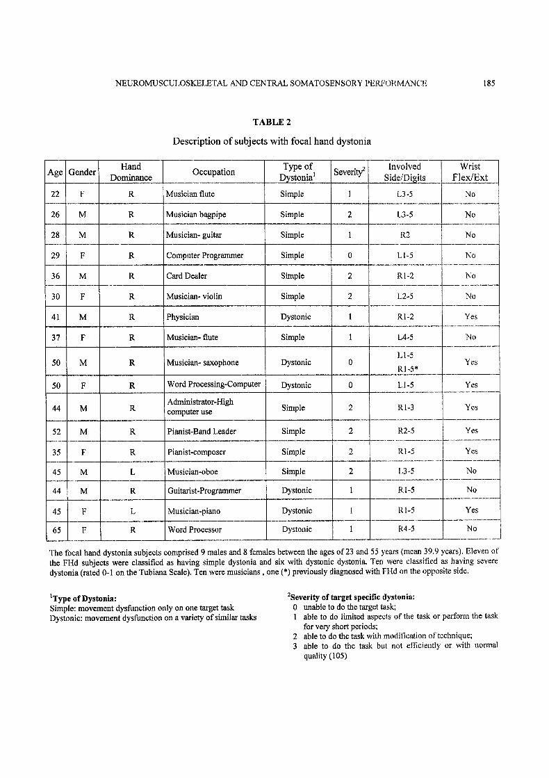
TABLE 2
Description of subjects with focal hand dystonia
Ae Gender
22 F
26 M
28 M
29 F
36 M
30 F
41 M
37 F
50 M
50 F
44 M
52 M
35 F
45 M
44 M
45 F
65 F
HandDominance
Occupation
Musician flute
Musician bagpipe
Musician- guitar
Computer Programmer
Card Dealer
Musician- violin
Physician
Type of
Simple
Simple
Simple
Simple
Simple
Dystonic
SeverityInvolved
L3-5
L3-5
R2
WristFlex/Ext
No
No
No
L1-5 No
RIo2 No
L25 No
Rl2 Yes
Musician- flute
Musician- saxophone
Word Processing-Computer
Administrator-Highcomputer use
Pianist-Band Leader
Pianist..composer
Musician,ooboe
Simple
Dystonic
Dystonic
Simple
Simple
Simple
Simple
No
Guitarist-Programmer
Musician-piano
Word Processor
Dystonic
Dystonic
Dystonic
L4-5
0
0
YesR1 -5’
LI ,,.,5 Yes
RI-3 Yes
R2-5 Yes
R1-5 Yes
I,,3o.,5 No
R.l5 No
RI,5 Yes
R4..o5 No
The bcal hand dystonia subjects comprised 9 males and 8 fernales between the ages of 23 trod 55 years (mean 39,9 years), Eleven of
the Fttd subjects were classified as having simple dystonia and six with dystonic dystonia. Ten were classified as having severe
dystonia (rated 0.-1 on the ’Fubiana Scale). Ten were musicians, one (*) previously diagnosed with FHd on the opposite side,
Type of Dystonia:Simple: movement dysfinction only on one target taskDystonic: movement dysthnction on a variety of similar tasks
2Severity of target specific dystonia:0 unable to do the target task;
able to do limited aspects of the task or perform the taskfor very short periods;
2 able to do the task with modification of technique;3 able to do the task but not efficiently or with normal
quality (105)

186 N.N. BYL ET AL.
TABLE 3
Description of healthy control subjects
HandAge Gender
DominanceRepetitive hand use
22 F R High level of computer use and manual therapy
28 M
28 M
28 F
32 M
R
R
R
R
35 F
38 M
40 F
50 M
50 F
R
R
R
R
R
30 M
30 F
50 M
42 M
58 F
R
R
R
High level of computer use and manual therapy
High level of computer use and manual therapy
High levels of computer use-Data analysis
High levels of computer use-Data analysis
High repetitive hand use on Musical instrument
High level of computer use-data analysis
High levels of computer use-data analysis
High levels of computer use
High repetitive hand use on musical instrument
High levels of writing
High levels of writing
High level of computer use-data analysis
Occupation
Graduate Student
Graduate Student
Graduate Student
Graduate Student
PhD Researcher
Musician
PhD Researcher
Post Doc Researcher
Administrator
Musician
Graduate student
Graduate student
Research Scientist
R
R
High levels of computer use
High level of computer use
Business administrator
Educator
The control group comprised 7 females and 8 males between the ages of 22 and 58 years (mean age 37.4 years).All healthy control subjects were involved in jobs demanding high levels of repetition; two were musicians.
performance, there were 5 males and 12 femaleswith an average age of 30.2 years (+3.6 years).The majority of control subjects who volunteeredfor the clinical measurements were graduatestudents, faculty, or friends of students or facultywho had a history of repetitive hand use (e.g.intensive note taking and computer use).
Table 4 summarizes the results of the clinicalperformance parameters for patients with FHd andhealthy controls. Compared with healthy controls,patients with FHd performed significantly worsewhen using either the affected or unaffected side interms of musculoskeletal skills, fine motor control,and sensory discrimination. Scores for posture and
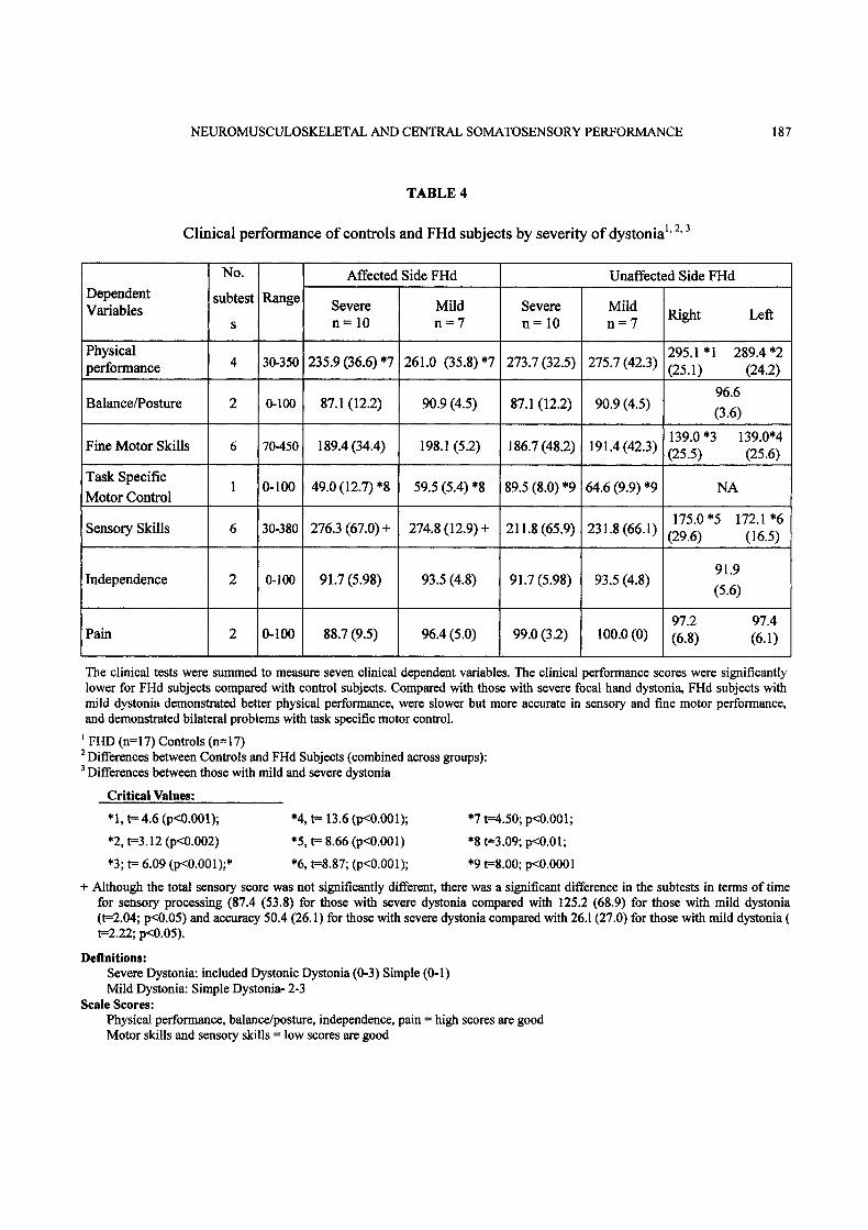
NEUROMUSCULOSKELETAL AND CENTRAL SOMATOSENSORY PERFORMANCE 187
TABLE 4
Clinical performance of controls and FHd subjects by severity of dystonia’’
DependentVariables
performance
NO.subtest Range
Affected Side FHd
Severen=10
30-350 235.9 (36.6) *7
Balance/Posture 2 0-100 87.1 (12.2)
Fine Motor Skills 6 70-450 189.4 (34.4)
Task Specific0-100 49.0(12.7)’8
Motor Control
Sensory Skills 6 30-380 276.3 (67.0) +
Independence 2 0-100 91.7 (5.98)
Pain 2 0-100 88.7 (9.5)
Mildn=7
261.0 (35.8)*7
90.9 (4.5)
198.1 (5.2)
59.5 (5.4) *8
274.8 (12.9) +
93.5 (4.8)
96.4 (5.0)
Severen=10
273.7 (32.5)
87.1 (12.2)
186.7 (48.2)
89.5 (8.0) *9
91.7 (5.98)
99.0 (3.2)
Unaffected Side FHd
Mildn 7 Right
295.1 *1275.7 (42.3) (25.1)
90.9 (4.5)
139.0"3191.4 (42.3) (25.5)
64.6 (9.9) *9
175.0"5231.8 (66.1) (29.6)
93.5 (4.8)
oo.o (o)
Left
289.4 *2(24.2)
96.6
(3.6)
139.0"4(25.6)
NA
172.1 *6(16.5)
91.9
(5.6)
97.2(6.8)
97.4(6.1)
The clinical tests were summed to measure seven clinical dependent variables. The clinical performance scores were significantlylower for FHd subjects compared with control subjects. Compared with those with severe focal hand dystonia, FHd subjects withmild dystonia demonstrated better physical performance, were slower but more accurate in sensory and fine motor performance,and demonstrated bilateral problems with task specific motor control.
FHD (n 17) Controls (n 17)Differences between Controls and FHd Subjects (combined across groups):Differences between those with mild and severe dystonia
Critical Values:
* 1, 4.6 (p<0.001);
*2, t=3.12 (p<0.002)
*3; 6.09 (p<0.001);*
*4, 13.6 (p<0.001);
’5, 8.66 (p<0.001)
*6, t=8.87; (p<0.001);
*7 t=4.50; p<0.001;
*8 t=3.09; p<0.01;
*9 t=8.00; p<0.0001
Definitions:Severe Dystonia: included Dystonic Dystonia (0-3) Simple (0-1)Mild Dystonia: Simple Dystonia- 2-3
Scale Scores:Physical performance, balance/posture, independence, pain high scores are goodMotor skills and sensory skills low scores are good
+ Although the total sensory score was not significantly different, there was a significant difference in the subtests in terms of timefor sensory processing (87.4 (53.8) for those with severe dystonia compared with 125.2 (68.9) for those with mild dystonia(t=2.04; p<0.05) and accuracy 50.4 (26.1) for those with severe dystonia compared with 26.1 (27.0) for those with mild dystoniat=2.22; p<0.05).

188 N.N. BYL ET AL.
balance were lower for FHd subjects than controls,but the differences were not significant. Therewere no differences between the groups in terms ofpain or functional independence. On the affectedlimb, those with severe dystonia had reducedperformance measurements in musculoskeletalskills and target specific motor control comparedwith those with mild dystonia. Although theoverall sensory discrimination accuracy was lowfor all FHd subjects (with no significantdifferences by severity), for tasks that includedspeed and accuracy, those with severe dystoniaperformed the tasks faster than those with mild
dystonia. On the unaffected side, those with milddystonia demonstrated greater inaccuracy whenperforming the target specific task.
There were no significant differences betweenmean SEF latency or mean SEF amplitude for FHdsubjects and reference controls, but the location ofthe digits on the x (bilateral) and y axes (affected)were significantly different (p<0.0001, respectively)and the ratio of SEF mean amplitude to latency washigher for FHd subjects compared with the controls(p < 0.05). On the unaffected side, the volume of thehand representation was significantly larger for FHdsubjects than for controls (p < 0.05) (see Table 5).
TABLE 5
Differences in somatosensory responses for controls and subjects with FHd
Affected Side Unaffected Side
Controls Subjects Controls Subjects
Latency (msec) 45.9 (7.4) 43.6 (4.0) 45.1 (7.8) 44.1 (4.3)Amplitude (Ft) 45.2 (20.6) 54.4 (12.1) 48.7 (18.8) 46.7 (7.4)Ratio (Amplitudeto Latency)
1.08 (0.6) *4 1.36 (0.7)*4 1.20 (0.6) 1.31 (0.8)
Location (cm)Axis x 1.25 (0.3)’1 1.7 (0.3)’1 1.21 (0.2)*2 2.00 (0.4)*2Axis y 3.84 (0.5)*3 4.3 (0.3)*3 3.95 (0.3) 3.90 (0.2)Axis z 9.32 (0.3) 9.5 (0.9) 9.58 (0.3) 9.50 (0.4)
Spread (cm)Axis x 0.63 (0.2) 1.0 (0.4) 0.60 (0.3) 0.84 (0.4)Axis y 1.04 (0.4) 1.3 (0.8) 0.91 (0.5) 1.07 (0.5)
Axis z 0.94 (0.4) 1.1 (0.4) 0.89 (0.3) 1.10 (0.5)
0.58 (0.5)0.39 (0.2)._____ 0.26 (0.3)*5Volume (cm3) 0.67 (0.6)*5
As measured by magnetoencephalography, there were no significant differences in the mean amplitude and latency of the somato-sensory evoked field responses for controls and those with FHd. However, compared with controls, the FHd subjects had a higherratio of the mean amplitude to mean latency on the affected side, there was a significant difference in location of the digits on thex and y axes, and the area of the hand representation was significantly greater on the unaffected side.
Controls: n=15; Subjects: n 17*significant t 7.62; p<O.O001; t2=7.60; p<O.O001; t3 =5.93; p<O.O001; t4=2.05; p<O.05; ts=2.12; p<O.05

NEUROMUSCULOSKELETAL AND CENTRAL SOMATOSENSORY PERFORMANCE 189
TABLE 6
Somatosensory evoked field response by severity of dystonia: digit spread by axes and volume
Spread of digits
Axis x
Axis y
Axis z
Volume (cm3)
Controls
1.05 (0.61)
1.32 (0.67)
0.81 (0.98)
0.71 (0.69) *
Affected Side
Subjects
0.82 (0.31)
1.01 (0.52)
0.90 (0.42)
0.45 (0.38) *1
Controls
1.18 (0.56)
1.08 (0.57)
1.11 (0.41)
0.82 (0.76) *2
Unaffected Side
Subjects
0.822 (0.30)
0.761 (0.32)
0.782(0.52)
0.524 (0.52) *2
* t=2.15 (<0.05); *2 t=2.00 (<0.05)
On both the affected and unaffected side, the volume of the representation of the hand was significantly larger for those with mildcompared with those with severe hand dystonia.
Table 6 summarizes the parameters of thesomatosensory evoked field responses (SEFs) forthose with mild versus severe dystonia. Bilaterally,for those with mild dystonia, the volume of therepresentation of the hand was significantly largerthan in those with severe dystonia.
Figure 2 illustrates the different patterns of thesomatosensory evoked field responses (SEFs) forthe digits for one control and two subjects withFHd. The patterns of the SEFs for controls andthose with FHd were similar on the lip (uninvolvedbody part), but the SEF patterns for the digits weredifferent for healthy controls when compared withthose of subjects with FHd. The pattern of the SEFfor the control subject (A and D) was characterizedwith a primary burst of activity at 40 msec at an
amplitude between 70 and 100 fI’, quieting at 120msec. For the subject with severe dystonia (B and
E) on both sides, the first burst of activity was
<(at) 40 msec with an amplitude of > 150 fI’ andeither a second burst of activity or continued
activity (60 IT) after 120 msec. On the unaffectedside for the subject with mild dystonia, the SEF(C)
had the first burst of activity at 30 msec (amplitudeof > 150 fT) with continued firing at an amplitudeof 70 fT even at 120 msec. On the affected side
(F), the activity appeared poorly organized with
the most activity at 80 msec (60 fI’) withcontinued activity beyond 120 msec.
The distribution of amplitude by latency for thesomatosensory evoked field responses for FHdsubjects and controls is presented in Fig. 3. Whenamplitude was integrated across the somatosensoryevoked field response time, the amplitude was
significantly higher for controls compared with FHd
subjects on the affected side, affected digits (A, Fig.3) but not unaffected digits (B, Fig. 3). On theunaffected side (digits matched to the dystonicdigits), however, the amplitude of the somato-
sensory evoked field response was significantlygreater for FHd subjects than for controls (C, Fig. 3).
The scatter plots in Fig. 4 illustrate the distribu-tion of SEF amplitude by latency (by side) forcontrol and FHd subjects. For the FHd subjects, thedistribution of amplitude by latency was not linear
on the affected side. On scatter plot A, there was a

190 N.N. BYL ET AL.
A. Control- Unaffected digit
2001
-2001.100 0 100 200
200D. Control- Affected digit
100
0
-100
-200-100 0 100 200
B. FHd(Severe)- Unaffected digit200 200
1 O0 .I /.c;.’.., O0
0 0
-100 "/ -100
-200 ....200-100 0 100 200
E. FHd(Severe)- Affected digit
-100 0 100
C. FHd(Mild)- Unaffected digit200 200
100
["
0
-100
-200-100 0 100
100
-100
F. FHd(Mild)- Affected digit
-200200 -100 100 2)0
Fig. 2" A visual representation ofthe differences in amplitude and volume over time for FHd subjects and controls

NEUROMUSCULOSKELETAL AND CENTRAL SOMATOSENSORY PERFORMANCE 191
A120
100
80
60
40
20
FHd-Affected side-Affected digitControl
p<O.01
-20 0 20 40 60 80 100 120
B120
100
80
FHd-Affected side-Unaffected digitControl
60-
40-
20-
0-20 0 20 40 60 80 100 120
C120
Control100
60-
40-
20
FHd-Unaffected side-Affected digit
p<O.01
0-20 0 20 40 60 80 100 120
Time (ms)
Fig. 3: Somatosensory evoked field responses: amplitude integrated by latency for FHd and control subjects.

192 N.N. BYL ET AL.

NEUROMUSCULOSKELETAL AND CENTRAL SOMATOSENSORY PERFORMANCE 193
A
100
Severe Dystonics: AffectedMild Dystonics: Affected
8O
60
4O
2O
p<O.05
-20 0 20 40 60 80 100 120 140
120
100
8O
6O
4O
2O
Severe Dystonics: UnaffectedMild Dystonics: Unaffected ’
-20 0 2 4 80 100 120 140
Fig. 5 Somatosensory evoked field responses: Amplitude integrated by latency for FHd subjects (severe versus milddystonia).
bimodai distribution of mean SEF amplitudeplotted by mean latency (mean latency rangingfrom 30 to 60 milliseconds and the meanamplitude ranging from 20 to 119 f’F). There was anegative linear trend of amplitude by latency forthe digits on the unaffected side for FHd subjectsand controls (B, C); as the latency increased, the
amplitude decreased. Figure 5 provides a visualrepresentation of the differences in amplitudeintegrated by latency for those with severe versusmild dystonia. For subjects with focal handdystonia, on the affected side, the amplitude of thesomatosensory evoked field response wassignificantly lower for those with mild dystonia.

194 N.N. BYL ET AL.
TABLE 7
Somatosensory evoked field responses (SEF)" correlation of the ratio of amplitude to latencywith severity ofFHd and clinical performance criteria
Dependent VariablesAffected Side Unaffected Side
SEF Ratio Severity ofFHd SEF Ratio Severity ofFHd
Physical Performance -0.3910"1 -0.3755*2 -0.1071 -0.401"3
Fine Motor Skills -0.4670 *4 -0.1076 -0.1667 -0.1851
Motor Control Target Task -0. 6116*5 -0.5881’6 -0.7867*7 0.7929*8
Sensory Skills -0.28271 -0.0062 -0.1077 -0.0402
Pain -0.19841 -0.3896 -0.2090 -0.0190
Posture/Balance (n=34)Independence (n 17)
SEF Ratio Severity of FHd
-0.2911 -0.362
-0.1425 -0.328
Note: Correlated with severe dystonia coded as "2" and mild dystonia coded as "1"
Critical Values*1 z=3.29; p<0.0006* *3 z=3.53; p<0.0004 *5 z=2.38; p<0.0094*2 z=3.31; p<0.0005 *4 z=3.67; p<0.0004 *6 z=3.47; p<0.0004
*7 z=3.02; p<0.0013*8 z=3.13; p<0.001
when compared with subjects with severe dystoniaSuch a discrepancy in amplitude across latency forthose with mild compared with those with severe
dystonia was also measured on the unaffected side.
The digits for the control subjects were orderlysequenced from inferior to superior on the z axis
as expected. The digits of FHd subjects were not
orderly (see Fig. 6). For example, on the affectedside of the FHd subjects, the digits were all
represented at the same location.
High, significant correlations (0.9029f,td and
0.8477unaffected; P < 0.001) were found between
dystonia severity and the ratio of SEF amplitude to
latency, respectively (see Table 7). On the affected
side, the SEF ratio and dystonia severity were
significantly, negatively correlated (moderate to
moderately high) with musculoskeletal perfor-mance and motor control on the target task; FHd
subjects with mild dystonia and those with a low
SEF ratio demonstrated higher performance than
those with severe dystonia. There was also a
significantly negative correlation between fine
motor skills and the SEF ratio on the affected side;those with a high ratio of SEF amplitude to latencydemonstrated greater inaccuracy. Similar to the
affected side, on the unaffected side, there was a
significant, moderately negative correlation
between the severity of dystonia and musculo-
skeletal performance and a significantly negativecorrelation between the SEF ratio and motor
control at the target task. However, on the
unaffected side, there was a significant, positivecorrelation between the severity of dystonia and
motor control on the target task; those with mild
dystonia had lower scores on the target task than
those with severe dystonia.

NEUROMUSCULOSKELETAL AND CENTRAL SOMATOSENSORY PERFORMANCE 195

196 N.N. BYL ET AL.
DI.$CUSSION
The findings from this study contribute newevidence supporting a strong relationship betweenclinical sensory and motor performance withsomatosensory degradation in patients with FHdthat distinguishes them from healthy controls anddifferentiates them by severity of dystonia. Theresults presented here are consistent with theproposed sensorimotor hypothesis of aberrantleaming. The evidence also suggests, however, thatFHd subjects do not present as a homogeneousgroup.
On the affected side, subjects with severedystonia had more musculoskeletal limitations andmore impaired task specific motor controlcompared with those with miM dystonia, with theSEP characterized by a short latency, highamplitude, and a reduced area of somatosensoryhand representation.
Previous investigators have reported differencesin somatosensory evoked responses in FHdsubjects (digit clumping, loss of orderly digitsequencing, differences in the size of the area ofthe somato-sensory hand representation), but nosignificant difference in mean amplitude (Bara-Jimenez et al., 2000; Byl et al., 1996 a-c; Elbert etal., 1998; Juliano et al., 1991). The findings fromthe current study indicate that the SEF patterns inthose with severe dystonia and those with miMdystonia are complementary. Thus, when amplitudeand latency are averaged across all subjects, thedifferences are cancelled.
The SEF ratio of amplitude and latency couldbe viewed as a neural slew factor, projecting alinear relation between the clinical severity of FHdand the severity of somatosensory degradation. Ahigh neural slew factor is consistent with a highgain in the sensorimotor feedback loop, whereas alow neural slew factor is consistent with a lowgain in the sensorimotor feedback loop (Sanger &Merzenieh, 2000). Such feedback errors could
explain the differences noted between the groupsin terms of clinical performance. Although previouinvestigators (Bara-Jimenez et al., 2000; Byl et al.,1996 b-e; Sanger et al., 2000; Tinazzi et al., 1999),reported problems in sensory spatial discriminationin patients with FHd, information on clinicalperfomaanee and somatosensory structure, asmeasured by magnetoencephalography, was notreported nor were clinical skills correlated byseverity of dystonia. In the current study, subjectswith focal dystonia demonstrated compromisedclinical sensorimotor performance compared withcontrols with subjects with severe dystoniaworking faster but making more errors than thosewith miM dystonia.
Restrictions in musculoskeletal structure havebeen reported in patients with FHd (Wilson et al.1991; Wilson et al. 1993; Lejinse, 1996). The currentstudy confirms that subjects with FHd haverestrictions in musculoskeletal performance whencompared with controls, but the finding thatsubjects with severe dystonia had greater limi-tations in flexibility, strength, and neural tensionthan those with miM dystonia was totallyunexpected. Many individuals with tight musclesand joint restrictions develop compensatorymovements, but for such movements to resembledystonia is unusual. Under conditions of stress andrepetitive overuse, conceivably mtsculoskeletallimitations could increase the risk for developingdystonia (Wilson et al., 1991; 1993; Lej inse, 1996).Factors of causality would have to be confirmedby prospective, longitudinal, cohort studiesdocumenting the occupational, physical, sensory,and fine motor characteristics of individuals whoperform complex, repetitive hand tasks understressful environmental or psychological conditions.
In the current study, for subjects with FHd, thefingers were not normally sequenced on theprimary sensory cortex on either the affected or theunaffected side, and the size of the handrepresentation on the affected side was reduced in
NEUROMUSCULOSKELETAL AND CENTRAL SOMATOSENSORY PERFORMANCE 19’7
comparison with that on the unaffected side. Theobservation that the hand representation wasreduced in those with severe dystonia would beconsistent with a more serious degradation,including more extensive overlap of receptivefields across the columnar representation of thedigits on the cortex. Compared with healthycontrols, the size of the area of the handrepresentation for those with FHd was not
significantly different on the affected side; on theunaffected side, however, the area of representationwas larger for FHd subjects than for controls. Thisfinding might represent an artifact of the studysamples. Compared with control subjects, moreFHd subjects were musicians (58.8% versus 17.8%).Musicians reportedly have a larger somatosensoryhand representation as a result of complex,repetitive hand use (Elbert et al., 1998). In Elbert’sstudy of violinists, the representation for the lefthand (used for complex fingering) was larger thanthat for the bowing hand. Even though computerkeyboard users use their hands intensely, computerkeyboarding does not necessitate the samecomplex movements that are required to play amusical instrument. For example, when playingadvanced music on the piano, both hands andmultiple digits must play simultaneously andindividually with the arms moving concurrently tocover the length of the keyboard. Although somepaired digital movements are required on thecomputer keyboard (e.g. the use of the shift orfunction key with a letter or number key), most
frequently the user depresses one finger at a time.If computer users are applying stress-free handstrategies, then they can depress a single key usingthe intrinsic muscles (e.g. lumbrieals and interosseiinstead of the long finger flexors) and then simplyrelease the pressure down without requiring areciprocal digital extension movement. In addition,they can use the mouse at a close distance, movingfrom the shoulder and elbow to position the mouseand then rotate the forearm to click instead of
using rapid alternating digital movemerts to clickthe mouse.
The ’integration times’ are primarily dictated bytemporal requirements needed to recover from theinhibition that gates movement and dominates po,;t-stimulus excitability. With practice, possibly someindividuals can improve performance until theyexceed the limits of their integration time. Thosewho intensively use their hands should be encouragedto achieve precise sensory discrimination, to useintrinsic hand muscles and to avoid excessiveoveruse ofthe extrinsic flexors and extensors.
The sensorimotor learning hypothesis isconsistent with the principles of neuroplasticity,the finite nature of neural adaptation, and theimportance of accurate sensorimotor feedback fbrfine motor control (Byl et al., 1996; Sanger &Merzenich, 2000). Neuroplasticity is also consistentwith the hypothesis that FHd represents a case ofaberrant learning. If FHd develops as a conse-quence of abnormal learning, then patients retestfirst stop the repetition of the abnormal movementand then re-differentiate the somatosensory handrepresentation by performing attended, progressive,rewarded, sensorimotor behavioral activities
(Juliano et al., 1991; Kaas et al., 1983; Merzenichet al., 1982; Merzenich et al., 1996 a-b; Nagarajanet al., 1997; Nakashima et al., 1989).
Our study has several limitations. The datawere gathered from a small sample of patients withfocal hand dystonia. Two different control groupswere used to obtain somatosensory data andclinical performance. The stimulus (air puff) didnot represent the minimum threshold stimulus foreach individual subject. In the UCSF BiomagneticImaging Laboratory, a cutaneous stimulus of 17 to20 pounds per square inch (psi) was considered an
adequate stimulus to indent the skin 400 microns.This stimulus possibly represented a minimum
stimulus for those whose evoked field responsewas characterized by a long latency and lowamplitude but a maximum stimulus for those
198 N.N. BYL ET AL.
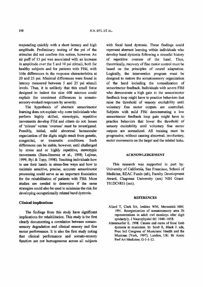
responding quickly with a short latency and highamplitude. Preliminary testing of the psi of thestimulus did not confirm this notion, however. Anair puff of 15 psi was associated with an increasein amplitude over the 5 and 10 psi stimuli, both forhealthy subjects and for patients with FHd, withlittle differences in the response characteristics at20 and 25 psi. Minimal differences were found inlatency measured between 5 and 25 psi stimulilevels. Thus, it is unlikely that this small forcedesigned to indent the skin 400 microns couldexplain the consistent differences in somato-sensory-evoked responses by severity.
The hypothesis of aberrant sensorimotorlearning does not explain why some individuals whoperform highly skilled, stereotypic, repetitivemovements develop FHd and others do not. Issuesof ’misuse’ versus ’overuse’ must be investigated.Possibly, initial, mild abnormal homuncularorganization of the digits might result from genetic,congenital, or traumatic conditions. Suchdifferences can be stable, however, until challengedby stress and or highly repetitive, stereotypiemovements (Bara-Jimenez et al., 1998; Lejinse,1999; Byl & Topp, 1998). Teaching individuals howto use their hands in stress-free ways and how tomaintain sensitive, precise, accurate sensorimotorprocessing could serve as an important foundationfor the rehabilitation of patients with FHd. Morestudies are needed to determine if the samestrategies could also be used to minimize the risk fordeveloping occupationally related hand dystonia.
Clinical implications
The findings from this study have significantimplications for rehabilitation. This study is the firstclearly documenting a correlation between somato-sensory degradation and clinical sensory and finemotor performance. It is also the first study notingthat clinical performance and somato-sensoryfunction are not homogeneous across all subjects
with focal hand dystonia. These findings couldrepresent aberrant learning within individuals whodevelop hand dystonia following a stressful historyof repetitive overuse of the hand. Thus,theoretically, recovery of fine motor control must bebased on the principles of neural adaptation.Logically, the intervention program must bedesigned to restore the somatosensory organizationof the hand including the normalization ofsensorimotor feedback. Individuals with severe FHdwho demonstrate a high gain in the sensorimotorfeedback loop might have to practice behaviors thatraise the threshold of sensory excitability untilvoluntary fine motor outputs are controlled.Subjects with mild FHd demonstrating a lowsensorimotor feedback loop gain might have topractice behaviors that lower the threshold ofsensory excitability until voluntary fine motoroutputs are normalized. All training must beprogressive, without causing abnormal, involuntary,motor movements on the target and the related tasks.
ACKNOWLEDGEMENT
This research was supported in part by"University of Califomia, San Francisco, School ofMedicine, REAC Funds (nb), Faculty DevelopmentAward, Chapman University (am) NIH Grant:T01DC4855 (ssn).
REFERENCES
Allard T, Clark SA, Jenkins WM, Merzenich MM.1991. Reorganization of somatosensory area 3brepresentations in adult owl monkeys after digitsyndactyly. J Neurophysiol 66:1048-1058.
Altenmueller E. 1998. Causes and cures of focal limbdystonia in musicians. In: Scott R, Black J. eds,Proc Intl Congress of Musicians: Health and theMusician (York, 1997). London, UK: Br AssocPerfArt Medicine; G-I-l-12.

NEUROMUSCULOSKELETAL AND CENTRAL SOMATOSENSORY PERFORMANCE 199
Ayres AF. 1989. Sensory Integration and Praxis Tests(SIPT). Los Angeles, California, USA: WesternPsychological Services.
Bara-Jiminez W, Catalan M, Hallett M. 1998. Abnormalsomatosensory homunculus in dystonia of thehand. Ann Neurol 44:828-831.
Bara-Jimenez W, Shelton P, Sanger TD, Hallett M.2000. Sensory discrimination capabilities in patientswith focal hand dystonia. Ann Neuro147: 377-380.
Bell C. 1883. The Hand: Its Mechanism and VitalEndowments as Evincing Design. London, UK:William Pickering; 23.
Black KJ, Ongur D, Perlmutter JS. 1998. Putamenvolume in idiopathic focal dystonia. Neurology51: 819-824.
Blake DT, Byl NN, Cheung S, Bedenbaugh P,Nagarajan S, Lamb M, Merzenich M. 2002.Sensory representation abnormalities that parallelfocal hand dystonia in a primate model.Somatosens Mot Res 19: 347-357.
Bohannon RW. 1995. Stopwatch for measuring thumb-movement time. Percept Mot Skills 81’ 211-216.
Brin MF, Fahn S, Moskowitz C, Friedman A, ShaleHM, Greene PE, et al. 1987. Localized injectionsof botulinum toxin for the treatment of focaldystonia and hemifacial spasm. Mov Disord 2:237-254.
Butler D. Mobilization of the nervous system. Mel-bourne, Australia: Churchill Livingston, 1991.
Byl N, Wilson F, Merzenich M, Melnick M, Scott P,Oakes A, McKenzie A. 1996a. Sensory dysfunctionassociated with repetitive strain injuries oftendinitis and focal hand dystonia: a comparativestudy. J Orthop Sports Phys Ther 23:234-244.
Byl N, Hamati D, Melnick M, Wilson F, McKenzie A.1996b. The sensory consequences of repetitivestrain injury in musicians: Focal dystonia of thehand. J Back Musculoskel Rehab 7: 27-39.
Byl N, Merzenich M, Jenkins W. 1996c. A primategenesis model of focal dystonia and repetitivestrain injury: I. Learning-induced dedifferentiationof the representation of the hand in the primarysomatosensory cortex in adult monkeys. Neurology47: 508-520.
Byl NN, Merzenich MM, Cheung S, Bedenbaugh P,Nagarajan SS, Jenkins WM. 1997. A primatemodel for studying focal dystonia and repetitivestrain injury: effects on the primary somatosensorycortex. Phys Ther 77: 269-284.
Byl N, Topp KS. 1998. Focal hand dystonia. Phys
Ther Case Rep 1: 39-.-52.Byl NN, Nagarajan SS, Newton N, McKenzie AL.
2000. Effect of sensory discrimination training onstructure and function in a musician with focalhand dystonia. Phys Ther Case Rep 3:94-113.
Byl N, McKenzie A, Nagarajan SS. 2000a. Differencesin somatosensory hand organization: healthy flutistand flutist with focal hand dystonia. J Hand Ther13: 301-309.
Byl N, Nagarajan SS, McKenzie A. 2000b. Effective-ness of sensory retraining: three case studies ofpatients with focal hand dystonia. [Abstract]Society for Neuroscience Meeting, New Orleans,Louisiana, USA.
Byl N, McKenzie A. 2000c. Treatment effectiveness ofpatients with a history of repetitive hand use andfocal hand dystonia: A planned prospective followup study. J Hand Ther 13:289-301.
Byl N, Leano J, Cheney LK. 2002. The Byl-Cheney-Boczai Sensory Discriminator: reliability, validity,and responsiveness for testing stereognosis. J HandTher 15:315-330.
Candia V, Elbert T, Altenmuller E, Rau H, Schafer T,Taub E. 1999. Constraint-induced movementtherapy for focal hand dystonia in musicians.[Letter] Lancet 353: 42--43.
Carlsson AM. 1983. Assessment of chronic pain. I.Aspects of the reliability and validity of the visualanalogue scale. Pain 16" 87-101.
Ceballos-Baumann AO, Sheean G, Pasingham RE,Marsden CD, Brooks DJ. 1995. Cerebral activationwith stereotyped writing in patients with writer’scramp before and atter botulhmm toxin treatment: aPET study. Neurology Suppl 45: 393-396.
Charness ME. 1992. Unique upper extremity disordersof musicians. In: Millender LH, Louis DS,Simmons BP, eds, Occupational Disorders of theUpper Extremity. New York, NY, USA: ChurchillLivingstone; 117-151.
Chamess M. 1993. The relationship between peripheralnerve injury and focal dystonia in musicians. AmAcad Neurol 162: 21-27.
Chase T, Tamminga C. and Burrows H. 1988. Positronemission tomographic studies of regional cerebralglucose metabolism in idiopathic dystonia. AdvNeurol 50:237--241.
Chen RS, Tsai CH, Lu CS. 1995. Reciprocal inhibitionin writer’s cramp. Mov Disord 10" 556-561.
Chen R, Hallett M. 1998. Focal dystonia and repetitivemotion disorders. Clin Ortho 351: 102-106.

200 N.N. BYL ET AL.
Cohen LG, Hallett M. 1988. Hand cramps’ clinicalfeatures and electromyographic patterns in a focaldystonia. Neurology 38:1005-1012.
Cole R, Hallett M, Cohen LG. 1995. Double-blind trialof botulinum toxin for treatment of focal handdystonia. Mov Disord 10: 466--471.
Defendini R, Falm S. 1988. Magnetic resonance imagingof dystonic states. Adv Neurol 50: 265-275.
DeLong MR, Crutcher MD, Georgopoulis AP. 1985.Primate globus pallidus and subthalamic nucleus:fimctional organization. J Neurophysiol 53" 530-543.
DeLong MR. 1990. Primate models of movementdisorders of basal ganglia origin. Trends Neurosci13: 281-285.
Deuschl G, Toro C, Matsumoto J, Hallett M. 1995.Movement-related cortical potentials in writer’scramp. Ann Neurol 38: 862-868.
Edgelow PI. 1992. Thoracic Outlet Home Program Treat-ment Kit. Edgelow, Union City, California, USA.
Elbert T, Candia V, Altenmuller E, Rau H, Sterr A,Rockstroh B, et al. 1998. Alteration of digitalrepresentations in somatosensory cortex in focalhand dystonia. Neuroreport 9: 3571-3575.
Fahn S, Marsden CD, Calne D. 1987. The treatment ofdystonia. In: Marsden CD, Fahn S, eds, MovementDisorders. London, UK: Butterworths; 359-382.
Faure C, Keuss PJG, Lorette G, Vinter A, eds,Advances in Handwriting and Drawing: A Multi-disciplinary Approach. Paris, France: Europia;445-.t61.
Fung S, Byl N, Melnick M, Callahan P, Selinger A,Ischi K, et al. 1997. Functional outcomes: thedevelopment of a new instrument to monitoreffectiveness of therapy. Eur J Phys Med Rehabil7:31-41.
Fry H. 1986. Overuse syndromes in musicians 100years ago: An historical review. Med J Aust 146:620-625.
Gasser T, Bove C, Ozelius L, Hallett M, Chamess M,Hochberg F, et al. 1996. Haplotype analysis at theDYTI locus in Ashkenazi Jewish patients withoccupational hand dystonia. Mov Disord 11" 163-166.
Gerloff Ch, Corwell B, Chen R, Hallett M, Cohen LG.1998. The role of the human motor cortex in thecontrol of complex and simple finger movementsequences. Brain 121" 1695-1709.
Gilman S unck L, Young A. 1988. Cerebral metabolicactivity in idiopathic dystonia studied with positron
emission tomography. Adv Neurol 50:231-236.Grunewald RA, Yoneda Y, Shipman JM, Sagar HJ. 1997.
Idiopathic focal dystonia: a disorder of musclespindle afferent processing? Brain 120:2179-2185.
Hallett M. 1998. Physiology of dystonia. Adv .Neurol78:11-18.
Hebb DO. 1949. The Organization of Behavior. NewYork, NY, USA: Wiley.
Hochberg F, Harris S, Blartert T. 1990. Occupationalhand cramps: Professional disorders ofmotor control.Hand Injury Sports Perform Arts 6: 427-428.
Ikeda A, Shibasaki H, Kaji R, Terado K, Nugamine T,Honda M, et al. 1999. Abnormal sensorimotorintegration in writer’s cramp: Study of contingentnegative variation. Mov Disord 17: 683-690.
Illarioshkin SN, Markova ED, Slominsky PA, MiklenaNI, Popovov SN, Limborska SA, et al. 1988. TheGTP cyclohydrolase gene in Russian familieswith dopa responsive dystonia. Arch Neurol 55"789-792.
Iwamura Y, Tanaka M, Sakamoto M, Hikosaka O. 1983.Functional subdivisions representing different fingerregions in area 3 of the first somatosensory cortexof the conscious monkey. Exp Brain Res 51:315-326.
Iwamura Y. 1992. Dynamic and hierarchical processingin the monkey somatosensory cortex. Biomed Res14:107-111.
Jankovic J, Shale H. 1989. Dystonia in musicians. SemNeurol 9: 131-135.
Jenkins W, Allard T, Nudo R. 1988. Cortical repre-sentational plasticity. In: Raskic P, Singer w, eds,Neurobiology of the Neocortex. New York, NY,USA: John Wiley and Sons; 41-67.
Jenkins WM, Merzenich MM, Ochs MT, Allard T,Guic-Robles E. 1990. Functional reorganization ofprimary somatosensory coretex in adult owlmonkeys after behaviorally controlled tactilestimulation. J Neurophysiol 53" 82-104.
Johansson RS. 1996. Sensory control of dexterousmanipulation in humans. In: Wing AM, HaggardP, Flanagan JR, eds, Hand and Brain. The Neuro-physiology and Psychology of Hand Movements.San Diego, California, USA: Academic Press;381-414.
Juliano SL, Ma W, Eslin D. 1991. Cholinergic depletionprevents expansion of topographic maps in soma-tosensory cortex. Proc Natl Acad Sci 88: 780-784.
Kaas JH, Merzenich MM, Killackey HP. 1983. Thereorganization of somatosensory cortex following

NEUROMUSCULOSKELETAL AND CENTRAL SOMATOSENSORY PERFORMANCE 201
peripheral nerve damage in adult and developingmarnmalso Annu Rev Neurosci 6: 325-356.
Kaji R. Rothwell J, Katayama M, Ikeda T, Kubori,Kohara N, et al. 1995. Tonic vibration reflex andmuscle afferent block in writer’s cramp. AnnNeuro138: 155-162.
Karp B, Cole R, Cohen L, Grill L, Lou J, Hallet M.1994. Long-term botulinurn toxin treatment of focalhand dystoniao Neurology 44: 70-76.
Katz R, Williams C. 1990. Focal dystonia followingsoft tissue injury: three case reports with long-termoutcome. Arch Phys Med Rehab 71: 345--349.
Kendall F, McCreary E. 1983. Muscles Testing andFunction, 3ra Ed. Baltimore, Maryland, USA:Williams Watkins.
Leijnse JNAL. 1997. Anatomical factors predisposingto focal dystonia in the musician’s handprinciples, theoretical examples, clinical signifi-cance. J Biomeeh 30: 659-669.
Lenz FA, Byl NN. 1999. Reorganization in the cutaneouscore of the human thalamic principal somaticsensory nucleus (Ventral caudal) in patients withdystonia. J Neurophysiol 82:3204-3212.
Leube B, Rudnieki D, Ratzlaff T, Kessler K, BeneekeR, Auburger G. 1996. Idiopathic torsion dystonia:Assignment of a gene to chromosome 18p in aGerman family with. adult onset, autosomaldominant inheritance and purely focal distribution.Hum Molec Genet 5: 1673-1677.
Liversedge LA, Sylvester JD. Conditioning techniquesin the treatment of writer’s cramp. Lancet 1955; 1"1147-1149.
Liversedge LA, Sylvester JD. 1960. Conditioningtechniques in the treatment of writer’s cramp. In:Eysenck HJ, ed, Behaviour Therapy and theNeuroses. Oxford, UK: Pergamon Press.
Mai N, Marguardt C. 1994. Treatment of writer’scramp: Kinematic measures as assessment toolsfor planning and evaluating handwriting trainingprocedures. In: Faure C, Keuss PJG, Lorette G,Vnter A, eds, Advances in Handwriting andDrawing: A Multidisciplinary Approach. Paris,France: Europia.
Mailloux Z. 1990. An overview of the SensoryIntegration and Praxs Tests. Am J Occup Ther 44:589-594.
Marsden CD, Sheehy MP. 1990. Writer’s cramp.Trends Neurosei. 13’ 148-153.
McKenzie AM. 1997. A new test to measure stereog-nosis: Key Test. Annual meeting, APTA
Califomia Chapter, Sacramento, Caliibmia, USA.Abstract.
McKenzie AM, Nagajaran SR, Merzenich MM, BylNN. 2000. Somatosensory differences in somato-sensory evoked responses using magnetic sourceimaging in patients with focal hand dystonia.Society of Neuroscienee Meeting, New Orleans,Louisiana, USA. Abstract.
Merzenieh MM, Kaas JH, Wall J, Nelson RJ, Sur M,Felleman D. 1983a. Topographic reorganizationof somatosensory cortical areas 3b and in adultmonkeys following restricted deafferentation.Neuroscienee 8’ 33-55.
Merzenich MM, Kaas JH, Wall JT, Sur M, Nelson RJ,Felleman DJ. 1983b. Progression of changefollowing median nerve section in the corticalrepresentation of the hand in areas 3b and inadult owl and squirrel monkeys. Neuroscience 10:639-665.
Merzenich MM, Nelson RJ, Stryker MP, Cynader MS,Sehoppmann A, Zook JM. 1984. Somatosensorycortical map changes following digit amputation inadult monkeys. J Comp Neurol 224:591-605.
Merzenich MM. 1990. Development and maintenanceof cortical somatosensoo" representations: Functional"maps" and neuroanatomical repertoires. InBamard KE, Brazelton TB, eds, Touch: TheFoundation of Experience. Madison, Connecticut,USA’ International Universities Press; 47-71.
Merzenich MM, Jenkins WM. 1995. Cortical plasticity,learning, and learning dysfunction. In: Julesz B,Kovaes I, eds, Maturational Windows and AdultCortical Plasticity. New York, NY, USA’Addison-Wesley; 247-272.
Merzenich M, Wright B, Jenkins W, Xerri C, Byl N,Miller S, Tallal P. 1996a. Cortical plasticityunderlying perceptual, motor, and cognitive skilldevelopment: implications for neurorehabilitation"Implications for neurorehabilitation. Cold SpringHarbor Syrup Quant Biol 61: 1-8.
Merzenieh M, deCharms, C. 1996b. Neural representa-tions, experience, and change. In: Llinas, R,Churchland PS, eds, Mind Brain Continuum.Cambridge, Massachusetts, USA: MIT Press; 61-81.
Merzenich MM, Tallal P, Peterson B, Miller SL, JenkinsWM. 1999. Some neurological principles relevantto the origins of---and the cortical plasticity basedremediation of--language learning impairments. In:Grafrnan J, Christen Y, eds, Neuroplasticity:Building a Bridge from the Laboratory to the Clinic.

202 N.N. BYL ET AL.
Berlin, Germany: Springer Verlag; 169-187.Mulligan S. 1996. An analysis of score patterns of
children with attention disorders on the SensoryIntegration and Praxis Tests. Am J Occup Ther 50:647-654.
Nagarajan SS, Blake DT, Wright BA, Byl N, MerzenichMM. 1998. Practice-related improvements insomatosensory interval discrimination are temporallyspecific but generalize across skin location, hemi-sphere, and modality. J Neurosci 18: 1559-1570.
Nakashima K, Rothwell JC, Day BL, Thompson PD,Shannon K. 1989. Reciprocal inhibition betweenforearm muscles in patients with writer’s crampand other occupational cramps, symptomatichemidystonia and hemiparesis due to stroke. Brain112: 681-697.
Naumann M, Reiners K. 1997. Long-latency reflexesof hand muscles in idiopathic focal dystonia andtheir modification by botulinum toxin. Brain 120:409-416.
Newmark J, Hochberg F. 1987. Isolated painlessmanual coordination in 57 musicians. Brit JNeurol Neurosurg Psychiatry 50:291-295.
Norkin C, Levangie P. 1992. Joint Structure and Function,A Comprehensive Analysis, 2nd ed. Philadelphia,Pennsylvania, USA: Davis FA; 276.
Nudo RJ, Wise BM, SiFuentes F, Milliken GW. 1996.Neural substrates for the effects of rehabilitativetraining on motor recovery after ischemic infarct.Science 272(5269): 1791-1794.
Nudo RJ, Millikin GW. 1996. Reorganization ofmovement representations in primary motor cortexfollowing focal ischemic infarcts in adult squirrelmonkeys. J Neurophysiol 75:2144-2149.
Nudo RJ. 1999. Recovery after damage to motorcortical areas. Curr Opin Neurobiol 9" 740-747.
Nudo ILl, Friel KM, Delia SW. 2000. Role of sensorydeficits in motor impairments aider injury to primarymotor cortex. Neuropharmacology 39: 733-742.
Odergren T, Iwasaki N, Borg J, Forssberg H. 1996.Impaired sensory-motor integration during graspingin writer’s cramp. Brain 119: 569-583.
Ozelius LJ, Hewett JW, Page CE, Bressman SB,Kramer PL, Shalish C, et al. 1997. The early-onsettorsion dystonia gene (DYT1) encodes an ATP-binding protein. Nat Genet 17" 40-48.
Panizza ME, Hallett M, Nilsson J. 1989. Reciprocalinhibition in patients with hand cramps. Neurology39: 85-89.
Panizza M, Lelli S, Nilsson J, Hallett M. 1990. H-
reflex recovery curve and reciprocal inhibition ofH-reflex in different kinds of dystonia. Neurology40: 824-828.
Penfield W, Rasmussen, T. 1950. The cerebral cortexof man: a clinical study of localization of function.New York, NY, USA: MacMillan Publishing.
Perlmutter JS, Stambuk MK, Markham J, Black KJ,McGee-Minnich L, Jankovic J, Moerlein SM.1997. Decreased [SF]spiperone binding in putamenin idiopathic focal dystonia. J Neurosci 17: 843-850.
Priori A, Pesenti A, Cappellari A, Scarlato G, Barbieri,S. 2001. Limb irrtmobilization for the treatment offocal occupational dystonia. Neurology 57: 405-409.
Pullman SL, Greene P, Fahn S, Pedersen SF. 1996.Approach to the treatment of limb disorders withbotulinum toxin A. Experience with 187 patients.Arch Neurol 53:617-624.
Quartarone A, Girlanda P, Risitano G, Picciolo G,Sinicropi S, Nicolosi C, et al. 1998. Focal handdystonia in a patient with thoracic outlet syndrome.J Neurol Neurosurg Psychiatry 65: 272-274.
Recanzone GH, Jenkins WM, Hradek GT, MerzenichMM. 1992a. Progressive improvement in discrimi-native abilities in adult owl monkeys performing atactile frequency discrimination task. JNeurophysiol 67:1015-1030.
Recanzone GH, Merzenich MM, Jenkins WM, GrajskiKA, Dinse HR. 1992b. Topographic reorganizationof the hand representation in cortical area 3b of owlmonkeys trained in a frequency-discrimination task.J Neurophysiol 67:1031-1056.
Recanzone GH, Merzenich MM, Jenkins WM. 1992c.Frequency discrimination training engaging arestricted skin surface results in an emergence of acutaneous response zone in cortical area 3a. JNeurophysiol 67:1057-1070.
Ridding MC, Sheean G, Rothwell JC, Inzelberg R,Kujirai T. 1995. Changes in the balance betweenmotor cortical excitation and inhibition in focal,task specific dystonia. J Neurol Neurosurg Psychiatry59: 493-498.
Rijntjes M, Dettmers C, Buchel C, Kiebel S, FrackowiakRS, Weiller C. 1999. A blueprint for movement:functional and anatomical representations in thehuman motor system. J Neurosci 19: 8043-8048.
Roberts TPL, Poeppel D, Rowley HA. 1998. Magneto-encephalography and magnetic source imaging.Neuropsychiatry Neuropsychol Behav Neurol 11:49-64.
Rothwell JC, Obeso JA, Day BL, Marsden CD. 1983.

NEUROMUSCULOSKELETAL AND CENTRAL SOMATOSENSORY PERFORMANCE 203
Pathophysiology of dystonias. Adv Neurol 39:851-863.
Rowley HA, Roberts TPL. Functional localization bymagnetoencephalography. 1995. Neuroimaging ClinNorth Am 5:695-710.
Sanger TD, Merzenich MM. 2000. Computationalmodel of the role of sensory disorganization infocal task-specific dystonia. J Neurophysiol 84:2458-2464.
Sanger TD, Ttroy DM, Pascual-Leone A. 2001. Abnor-malities of spatial and temporal sensory discrimi-nation in writer’s cramp. Mov Disord 16: 94-99.
Sherrington CS. 1906. The Integrative Action of theNervous System, 2nd, 1947 Edition. New Haven,Connecticut, USA: Yale University Press.
Spengler F, Roberts TP, Poeppel D, Byl N, Wang X,Rowley HA, Merzenich MM. 1997. Learningtransfer and neuronal plasticity in humans trainedin tactile discrimination. Neurosci Lett 232:151-154.
Tempel LW, Perlmutter JS. 1993. Abnormal corticalresponses in patients with writer’s cramp.Neurology 43: 2252-2257.
Tinazzi M, Frasson E, Bertolasi L, Fiaschi A, AgliotiS. 1999 Temporal discrimination of somestheticstimuli is impaired in dystonic patients. Neuro-report 10: 1547-1550.
Topp KS, Byl NN. 1999. Movement dysfunctionfollowing repetitive hand opening and closing:anatomical analysis in Owl monkeys. Mov Disord14: 295-306.
Toro C, Deuschl G, Hallett M. 2000. Movement-related electroencephalographic desynchronizationin patients with hand cramps" evidence for motorcortical involvement in focal dystonia. Ann Neurol47:456-461.
Tsui J, Bhatt M, Calne S, Calne DB. 1993. Botulinumtoxin in the treatment of writer’s cramp: a double-blind study. Neurology 43:183-185.
Tubiana R, Chamagne P. 1983. [Occupational "cramps"ofthe upper limb.] Ann Chir Main 2: 134-142.
Tubiana R. 1998. Focal dystonia. Incidence: classi-
fication of severity and results of therapy. In:Winspur I, Wynn Parry CB, eds, The Musician’sHand. A Clinical Guide. London, UK: MartinDunitz LTD; 164-167.
Uitti RJ, Vingerhoets FJ, Tsui JK. 1995. Limb dystonia.In: Tsui JK, Calne D, eds, Handbook of Dystonia.New York, NY, USA: Marcel Dekker; 143-158.
van Hilten BJ, van de Beek WJT, Hoff JI, VoormolenJHC, Delhaas EM. 2000. Intrathecal baclofen forthe treatment of dystonia in patients with reflexsympathetic dystrophy. N Engl J Med. 343" 625-630.
Wang X, Merzenich MM, Sameshima K, Jenkins WM.1995. Remodelling of hand representation in adultcortex determined by timing of tactile stimulation.Nature 378(6552)" 71-75.
Wang X, Merzenich MM, Sameshima K, Jenkins WM.1994. Afferent input integration and segregation inlearning are input timing dependent. NeuroscienceAbstr 20: 1427.
Wilson FR, Wagner C, H6mberg V. 1993. Bio-mechanical abnormalities in musicians withoccupational cramp/focal dystonia. J Hand Ther 6:298-307.
Wilson FR, Wagner C, H6mberg V. Noth J. 1991.Interaction of biomechanical and training factorsin musicians with occupational cramp/focal dystonia.Neurology Suppl 41" 291.
Yang TT, Gallen C, Schwartz B, Bloom FE, Ramac-handran VS, Cobb S. 1994. Sensory maps in thehuman brain [letter] Nature 368: 592-593.
Xerri C, Coq JO, Merzenich MM, Jenkins WM. 1996.Experience-induced plasticity of cutaneous mapsin the primary somatosensory cortex of adultmonkeys and rats. J Physiol Paris 90: 277-287.
Xerri C, Merzenich MM, Jenkins W, Santucci S. 1999.Representational plasticity in cortical area 3bparalleling tactual-motor skill acquisition in adultmonkeys. Cereb Cortex 9: 264-276.
Zirh TA, Reich SG, Perry V, Lenz FA. 1998. Thalamicsingle neuron and electromyographic activities inpatients with dystonia. Adv Neurol 78: 27-32.