udo hoffmann, m.d., m.p.h. nih public access quynh … · emergency department with chest pain ......
TRANSCRIPT

Coronary CT Angiography versus Standard Evaluation in AcuteChest Pain
Udo Hoffmann, M.D., M.P.H., Quynh A. Truong, M.D., M.P.H., David A. Schoenfeld, Ph.D.,Eric T. Chou, M.D., Pamela K. Woodard, M.D., John T. Nagurney, M.D., M.P.H., J. HectorPope, M.D., Thomas H. Hauser, M.D., M.P.H., Charles S. White, M.D., Scott G. Weiner, M.D.,M.P.H., Shant Kalanjian, M.D., Michael E. Mullins, M.D., Issam Mikati, M.D., W. FrankPeacock, M.D., Pearl Zakroysky, B.A., Douglas Hayden, Ph.D., Alexander Goehler, M.D.,Ph.D., Hang Lee, Ph.D., G. Scott Gazelle, M.D., M.P.H., Ph.D., Stephen D. Wiviott, M.D.,Jerome L. Fleg, M.D., and James E. Udelson, M.D. for the ROMICAT-II Investigators
AbstractBackground—It is unclear whether an evaluation incorporating coronary computed tomographicangiography (CCTA) is more effective than standard evaluation in the emergency department inpatients with symptoms suggestive of acute coronary syndromes.
Methods—In this multicenter trial, we randomly assigned patients 40 to 74 years of age withsymptoms suggestive of acute coronary syndromes but without ischemic electrocardiographicchanges or an initial positive troponin test to early CCTA or to standard evaluation in theemergency department on weekdays during daylight hours between April 2010 and January 2012.The primary end point was length of stay in the hospital. Secondary end points included rates ofdischarge from the emergency department, major adverse cardiovascular events at 28 days, andcumulative costs. Safety end points were undetected acute coronary syndromes.
Results—The rate of acute coronary syndromes among 1000 patients with a mean (±SD) age of54±8 years (47% women) was 8%. After early CCTA, as compared with standard evaluation, themean length of stay in the hospital was reduced by 7.6 hours (P<0.001) and more patients weredischarged directly from the emergency department (47% vs. 12%, P<0.001). There were noundetected acute coronary syndromes and no significant differences in major adversecardiovascular events at 28 days. After CCTA, there was more downstream testing and higherradiation exposure. The cumulative mean cost of care was similar in the CCTA group and thestandard-evaluation group ($4,289 and $4,060, respectively; P=0.65).
Conclusions—In patients in the emergency department with symptoms suggestive of acutecoronary syndromes, incorporating CCTA into a triage strategy improved the efficiency of clinicaldecision making, as compared with a standard evaluation in the emergency department, but itresulted in an increase in downstream testing and radiation exposure with no decrease in theoverall costs of care. (Funded by the National Heart, Lung, and Blood Institute; ROMICAT-IIClinicalTrials.gov number, NCT01084239.)
Treatment of Patients with Acute chest pain but an inconclusive initial evaluation with theuse of biomarkers and electrocardiographic (ECG) testing is often diagnostically challenging
Copyright © 2012 Massachusetts Medical Society.
Address reprint requests to Dr. Hoffmann at Massachusetts General Hospital, Cardiac MR PET CT Program, 165 Cambridge St., Suite400, Boston, MA 02114, or at [email protected].
No other potential conflict of interest relevant to this article was reported.
Disclosure forms provided by the authors are available with the full text of this article at NEJM.org.
NIH Public AccessAuthor ManuscriptN Engl J Med. Author manuscript; available in PMC 2013 May 23.
Published in final edited form as:N Engl J Med. 2012 July 26; 367(4): 299–308. doi:10.1056/NEJMoa1201161.
NIH
-PA Author Manuscript
NIH
-PA Author Manuscript
NIH
-PA Author Manuscript

and inefficient. The majority of patients with acute coronary syndromes have underlyingcoronary artery disease.1 Contrast-enhanced coronary computed tomographic angiography(CCTA) has high sensitivity and specificity for the detection of clinically significantcoronary artery disease, as compared with invasive coronary angiography, in patients instable condition with suspected or known coronary artery disease.2-5
Rule Out Myocardial Infarction/Ischemia Using Computer Assisted Tomography(ROMICAT-I),6 a blinded observational study involving patients in the emergencydepartment with suspected acute coronary syndromes, and other studies7,8 have shown thatnormal findings on CCTA have a very high negative predictive value for ruling out acutecoronary syndromes during the index hospitalization and the occurrence of major adversecardiovascular events over the next 2 years.7,9 The results of two previous randomized,multicenter trials10,11 suggest that CCTA may facilitate safe and earlier triage of low-riskpatients and that CCTA can rule out coronary artery disease faster than stress myocardial-perfusion imaging. However, imaging the coronary anatomy with CCTA can involve moreprocedures and greater costs than functional testing.12 Thus, equipoise exists regarding theeffectiveness of incorporating CCTA into an evaluation strategy in the emergencydepartment.
The objectives of this study were to compare the effectiveness of a CCTA-based evaluationstrategy with that of standard evaluation in the emergency department for patients withsymptoms suggestive of an acute coronary syndrome and to evaluate the downstreamtesting, cost, and radiation exposure associated with CCTA.
MethodsStudy Design and Oversight
ROMICAT-II was designed as a randomized, controlled, multicenter trial in which anevaluation and management strategy that included CCTA as a first diagnostic test performedas early as possible was compared with a standard emergency department evaluation forpatients with acute chest pain suggestive of an acute coronary syndrome. After randomassignments had been made to initial CCTA or standard evaluation without CCTA, patientcare in both groups was not mandated by the study protocol but instead was at the discretionof local physicians. The design of ROMICAT-II has been described in detail previously,13
and the study protocol is available with the full text of this article at NEJM.org.
Study PopulationPatient enrollment began on April 23, 2010, and ended on January 30, 2012, at ninehospitals in the United States. All patients provided written informed consent to participatein the study. Eligibility criteria were chosen according to the ROMICAT-I study,6 with thegoal of enrolling a population with a similar prevalence of acute coronary syndromes(approximately 8%). Eligible patients were 40 to 74 years of age, presented to theemergency department with chest pain (or the anginal equivalent) of at least 5 minutes'duration within 24 hours before presentation in the emergency department, were in sinusrhythm, and warranted further risk stratification to rule out acute coronary syndromes, asdetermined by an attending physician in the emergency department. Major exclusion criteriawere a history of known coronary artery disease, new diagnostic ischemic changes on theinitial ECG, an initial troponin level in excess of the 99th percentile of the local assay,impaired renal function (creatinine level, >1.5 mg per deciliter [132.6 μmol per liter]),hemodynamic or clinical instability, known allergy to an iodinated contrast agent, a body-mass index (the weight in kilograms divided by the square of the height in meters) greaterthan 40, or currently symptomatic asthma.
Hoffmann et al. Page 2
N Engl J Med. Author manuscript; available in PMC 2013 May 23.
NIH
-PA Author Manuscript
NIH
-PA Author Manuscript
NIH
-PA Author Manuscript
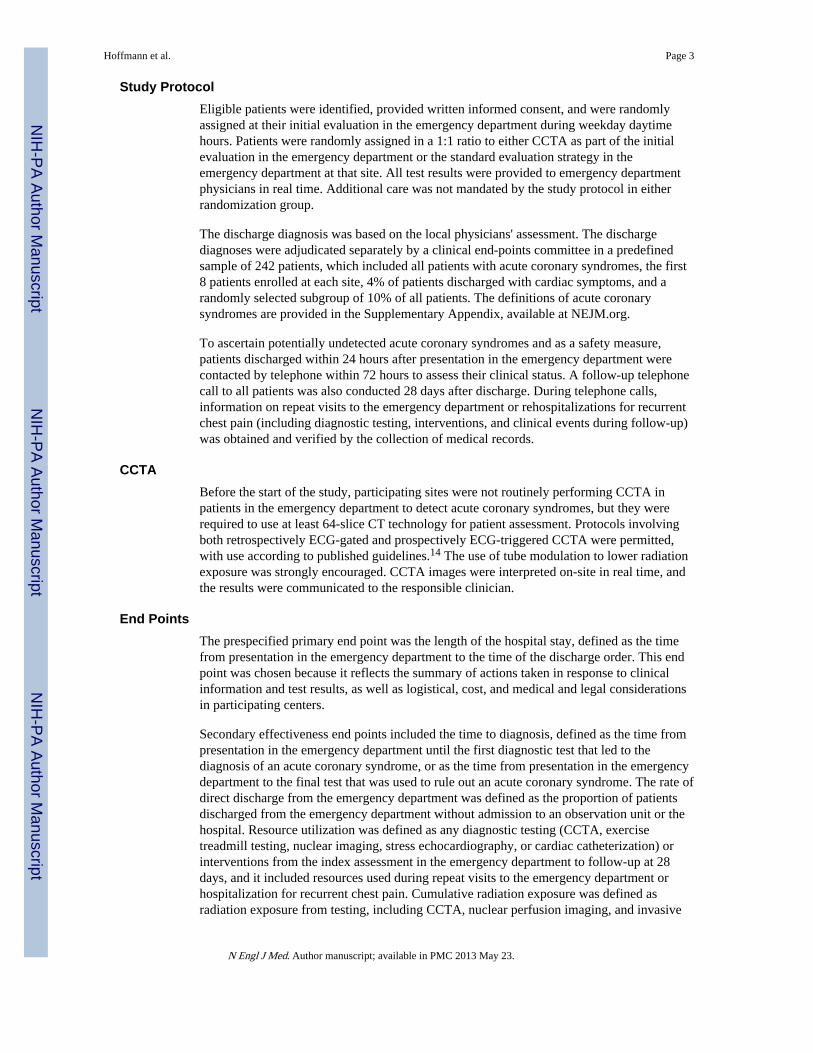
Study ProtocolEligible patients were identified, provided written informed consent, and were randomlyassigned at their initial evaluation in the emergency department during weekday daytimehours. Patients were randomly assigned in a 1:1 ratio to either CCTA as part of the initialevaluation in the emergency department or the standard evaluation strategy in theemergency department at that site. All test results were provided to emergency departmentphysicians in real time. Additional care was not mandated by the study protocol in eitherrandomization group.
The discharge diagnosis was based on the local physicians' assessment. The dischargediagnoses were adjudicated separately by a clinical end-points committee in a predefinedsample of 242 patients, which included all patients with acute coronary syndromes, the first8 patients enrolled at each site, 4% of patients discharged with cardiac symptoms, and arandomly selected subgroup of 10% of all patients. The definitions of acute coronarysyndromes are provided in the Supplementary Appendix, available at NEJM.org.
To ascertain potentially undetected acute coronary syndromes and as a safety measure,patients discharged within 24 hours after presentation in the emergency department werecontacted by telephone within 72 hours to assess their clinical status. A follow-up telephonecall to all patients was also conducted 28 days after discharge. During telephone calls,information on repeat visits to the emergency department or rehospitalizations for recurrentchest pain (including diagnostic testing, interventions, and clinical events during follow-up)was obtained and verified by the collection of medical records.
CCTABefore the start of the study, participating sites were not routinely performing CCTA inpatients in the emergency department to detect acute coronary syndromes, but they wererequired to use at least 64-slice CT technology for patient assessment. Protocols involvingboth retrospectively ECG-gated and prospectively ECG-triggered CCTA were permitted,with use according to published guidelines.14 The use of tube modulation to lower radiationexposure was strongly encouraged. CCTA images were interpreted on-site in real time, andthe results were communicated to the responsible clinician.
End PointsThe prespecified primary end point was the length of the hospital stay, defined as the timefrom presentation in the emergency department to the time of the discharge order. This endpoint was chosen because it reflects the summary of actions taken in response to clinicalinformation and test results, as well as logistical, cost, and medical and legal considerationsin participating centers.
Secondary effectiveness end points included the time to diagnosis, defined as the time frompresentation in the emergency department until the first diagnostic test that led to thediagnosis of an acute coronary syndrome, or as the time from presentation in the emergencydepartment to the final test that was used to rule out an acute coronary syndrome. The rate ofdirect discharge from the emergency department was defined as the proportion of patientsdischarged from the emergency department without admission to an observation unit or thehospital. Resource utilization was defined as any diagnostic testing (CCTA, exercisetreadmill testing, nuclear imaging, stress echocardiography, or cardiac catheterization) orinterventions from the index assessment in the emergency department to follow-up at 28days, and it included resources used during repeat visits to the emergency department orhospitalization for recurrent chest pain. Cumulative radiation exposure was defined asradiation exposure from testing, including CCTA, nuclear perfusion imaging, and invasive
Hoffmann et al. Page 3
N Engl J Med. Author manuscript; available in PMC 2013 May 23.
NIH
-PA Author Manuscript
NIH
-PA Author Manuscript
NIH
-PA Author Manuscript

coronary angiography, measured in millisieverts and calculated with the use of standardmethods15 during the index care episode (the visit to the emergency department andhospitalization) and follow-up. Health care costs during the index care episode wereassessed from reports from hospital cost-accounting systems and physician billing recordsand were adjusted to 2011 dollars. Mean costs for patient care, diagnostic testing, andinterventions during the index care episode were used to estimate the costs during follow-up.
Safety variables prespecified as secondary end points included an undetected acute coronarysyndrome (defined as an unexpected cardiovascular event within 72 hours after hospitaldischarge in patients with a hospital stay of <24 hours), to ensure that potentially earlierdischarge in the CCTA group was not associated with increased adverse events, majoradverse cardiovascular events (defined as death, myocardial infarction, unstable angina, orurgent coronary revascularization within 28 days), and periprocedural complications (stroke,bleeding, anaphylaxis, or renal failure). These predefined safety variables were adjudicatedby an external, independent clinical-events committee.
Statistical AnalysesAll statistical analyses were performed by an independent data coordinating center on thebasis of an intention-to-treat analysis. Continuous data are presented as means ±SD andmedians with interquartile ranges. Comparisons between groups were performed with theuse of an independent-sample t-test for continuous variables, Fisher's exact test forcategorical variables, and the Wilcoxon rank-sum test for ordinal variables. A two-sided Pvalue of less than 0.05 was considered to indicate statistical significance. Concordancebetween the discharge diagnosis made at the study site and the independently adjudicateddiagnosis in a selected subpopulation was assessed with the use of the kappa statistic.
The study was designed to have greater than 83% power with the use of a t-test at a two-sided 5% significance level if the true between-group difference in the length of stay in thehospital was at least 8.3 hours. Details of the simulation are described elsewhere.13
The study did not have predefined stopping rules or boundaries with respect to the primaryend point or safety end points. Rather, the data and safety monitoring board was responsiblefor assessing every case in which an acute coronary syndrome might have been undetected.
ResultsStudy Population
Of 1000 enrolled patients, 501 were randomly assigned to CCTA and 499 were randomlyassigned to a standard evaluation in the emergency department. All patients were included inthe intention-to-treat analysis (Fig. 1). CCTA was not performed in 28 patients (6%) becauseof the patient's decision to decline CCTA (9 patients), safety concerns (5 patients),unavailability of CCTA (5 patients), or technical difficulties (9 patients). Overall, 987 of1000 randomly assigned patients (99%) had complete follow-up at 28 days. The originalmedical records for repeat visits to the emergency department or hospitalizations wereavailable in all cases.
Baseline characteristics of the study population are shown in Table 1. After a completeevaluation, 75 patients (8%) had a final diagnosis of an acute coronary syndrome.Agreement between the site and independent adjudication for the discharge diagnosis wasvery high (concordance, 98% [236 of 242 patients]; kappa, 0.94).
Hoffmann et al. Page 4
N Engl J Med. Author manuscript; available in PMC 2013 May 23.
NIH
-PA Author Manuscript
NIH
-PA Author Manuscript
NIH
-PA Author Manuscript
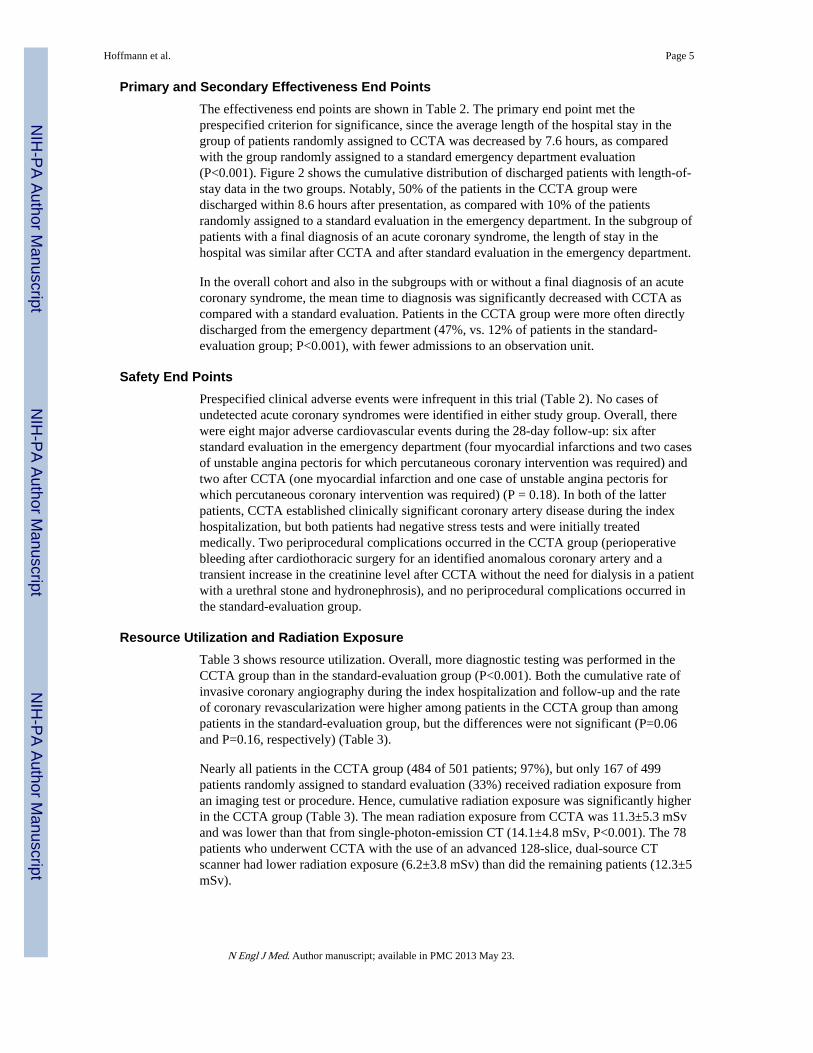
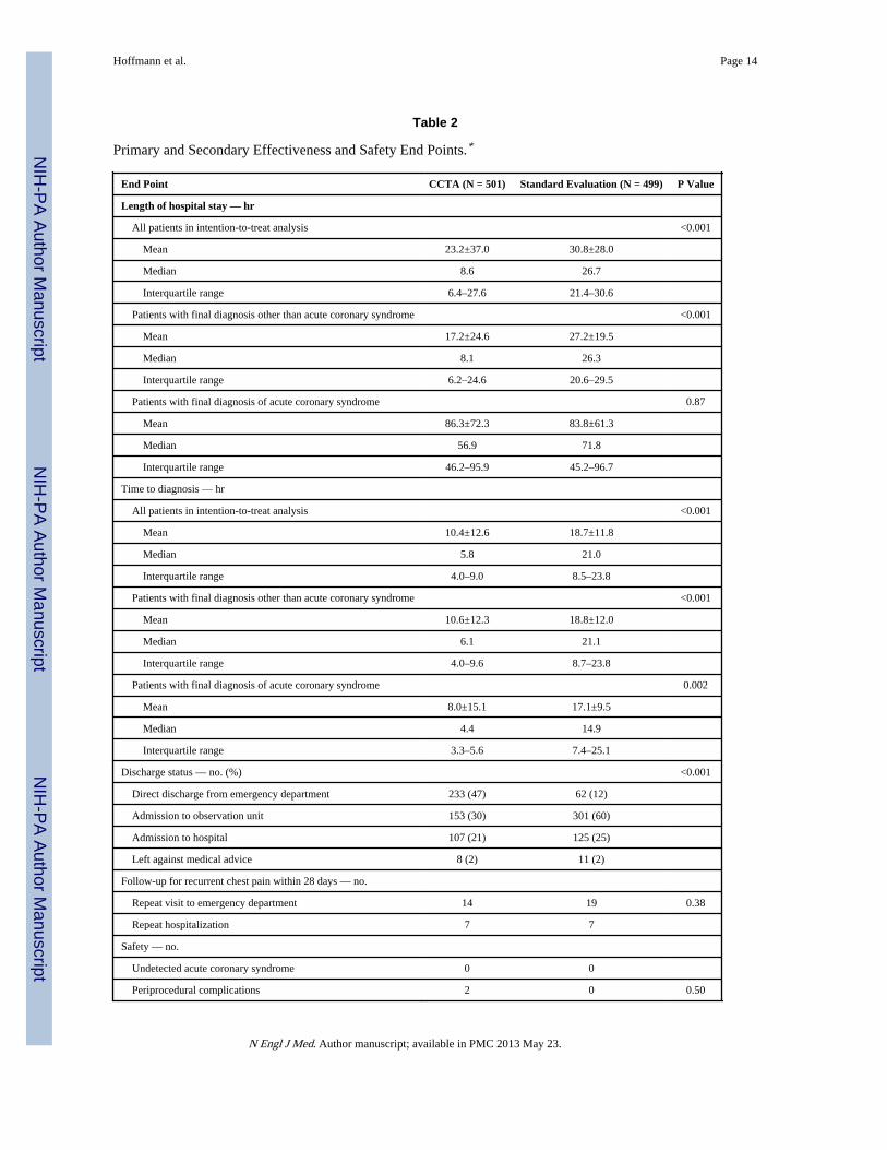
Primary and Secondary Effectiveness End PointsThe effectiveness end points are shown in Table 2. The primary end point met theprespecified criterion for significance, since the average length of the hospital stay in thegroup of patients randomly assigned to CCTA was decreased by 7.6 hours, as comparedwith the group randomly assigned to a standard emergency department evaluation(P<0.001). Figure 2 shows the cumulative distribution of discharged patients with length-of-stay data in the two groups. Notably, 50% of the patients in the CCTA group weredischarged within 8.6 hours after presentation, as compared with 10% of the patientsrandomly assigned to a standard evaluation in the emergency department. In the subgroup ofpatients with a final diagnosis of an acute coronary syndrome, the length of stay in thehospital was similar after CCTA and after standard evaluation in the emergency department.
In the overall cohort and also in the subgroups with or without a final diagnosis of an acutecoronary syndrome, the mean time to diagnosis was significantly decreased with CCTA ascompared with a standard evaluation. Patients in the CCTA group were more often directlydischarged from the emergency department (47%, vs. 12% of patients in the standard-evaluation group; P<0.001), with fewer admissions to an observation unit.
Safety End PointsPrespecified clinical adverse events were infrequent in this trial (Table 2). No cases ofundetected acute coronary syndromes were identified in either study group. Overall, therewere eight major adverse cardiovascular events during the 28-day follow-up: six afterstandard evaluation in the emergency department (four myocardial infarctions and two casesof unstable angina pectoris for which percutaneous coronary intervention was required) andtwo after CCTA (one myocardial infarction and one case of unstable angina pectoris forwhich percutaneous coronary intervention was required) (P = 0.18). In both of the latterpatients, CCTA established clinically significant coronary artery disease during the indexhospitalization, but both patients had negative stress tests and were initially treatedmedically. Two periprocedural complications occurred in the CCTA group (perioperativebleeding after cardiothoracic surgery for an identified anomalous coronary artery and atransient increase in the creatinine level after CCTA without the need for dialysis in a patientwith a urethral stone and hydronephrosis), and no periprocedural complications occurred inthe standard-evaluation group.
Resource Utilization and Radiation ExposureTable 3 shows resource utilization. Overall, more diagnostic testing was performed in theCCTA group than in the standard-evaluation group (P<0.001). Both the cumulative rate ofinvasive coronary angiography during the index hospitalization and follow-up and the rateof coronary revascularization were higher among patients in the CCTA group than amongpatients in the standard-evaluation group, but the differences were not significant (P=0.06and P=0.16, respectively) (Table 3).
Nearly all patients in the CCTA group (484 of 501 patients; 97%), but only 167 of 499patients randomly assigned to standard evaluation (33%) received radiation exposure froman imaging test or procedure. Hence, cumulative radiation exposure was significantly higherin the CCTA group (Table 3). The mean radiation exposure from CCTA was 11.3±5.3 mSvand was lower than that from single-photon-emission CT (14.1±4.8 mSv, P<0.001). The 78patients who underwent CCTA with the use of an advanced 128-slice, dual-source CTscanner had lower radiation exposure (6.2±3.8 mSv) than did the remaining patients (12.3±5mSv).
Hoffmann et al. Page 5
N Engl J Med. Author manuscript; available in PMC 2013 May 23.
NIH
-PA Author Manuscript
NIH
-PA Author Manuscript
NIH
-PA Author Manuscript
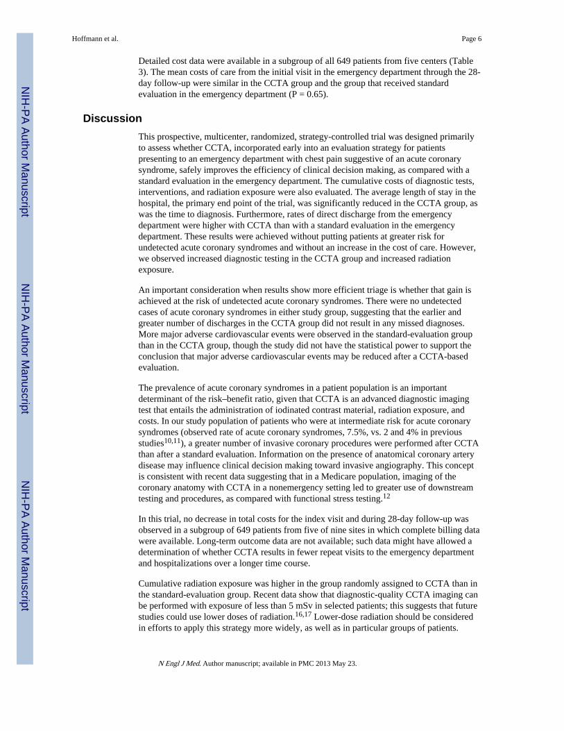
Detailed cost data were available in a subgroup of all 649 patients from five centers (Table3). The mean costs of care from the initial visit in the emergency department through the 28-day follow-up were similar in the CCTA group and the group that received standardevaluation in the emergency department (P = 0.65).
DiscussionThis prospective, multicenter, randomized, strategy-controlled trial was designed primarilyto assess whether CCTA, incorporated early into an evaluation strategy for patientspresenting to an emergency department with chest pain suggestive of an acute coronarysyndrome, safely improves the efficiency of clinical decision making, as compared with astandard evaluation in the emergency department. The cumulative costs of diagnostic tests,interventions, and radiation exposure were also evaluated. The average length of stay in thehospital, the primary end point of the trial, was significantly reduced in the CCTA group, aswas the time to diagnosis. Furthermore, rates of direct discharge from the emergencydepartment were higher with CCTA than with a standard evaluation in the emergencydepartment. These results were achieved without putting patients at greater risk forundetected acute coronary syndromes and without an increase in the cost of care. However,we observed increased diagnostic testing in the CCTA group and increased radiationexposure.
An important consideration when results show more efficient triage is whether that gain isachieved at the risk of undetected acute coronary syndromes. There were no undetectedcases of acute coronary syndromes in either study group, suggesting that the earlier andgreater number of discharges in the CCTA group did not result in any missed diagnoses.More major adverse cardiovascular events were observed in the standard-evaluation groupthan in the CCTA group, though the study did not have the statistical power to support theconclusion that major adverse cardiovascular events may be reduced after a CCTA-basedevaluation.
The prevalence of acute coronary syndromes in a patient population is an importantdeterminant of the risk–benefit ratio, given that CCTA is an advanced diagnostic imagingtest that entails the administration of iodinated contrast material, radiation exposure, andcosts. In our study population of patients who were at intermediate risk for acute coronarysyndromes (observed rate of acute coronary syndromes, 7.5%, vs. 2 and 4% in previousstudies10,11), a greater number of invasive coronary procedures were performed after CCTAthan after a standard evaluation. Information on the presence of anatomical coronary arterydisease may influence clinical decision making toward invasive angiography. This conceptis consistent with recent data suggesting that in a Medicare population, imaging of thecoronary anatomy with CCTA in a nonemergency setting led to greater use of downstreamtesting and procedures, as compared with functional stress testing.12
In this trial, no decrease in total costs for the index visit and during 28-day follow-up wasobserved in a subgroup of 649 patients from five of nine sites in which complete billing datawere available. Long-term outcome data are not available; such data might have allowed adetermination of whether CCTA results in fewer repeat visits to the emergency departmentand hospitalizations over a longer time course.
Cumulative radiation exposure was higher in the group randomly assigned to CCTA than inthe standard-evaluation group. Recent data show that diagnostic-quality CCTA imaging canbe performed with exposure of less than 5 mSv in selected patients; this suggests that futurestudies could use lower doses of radiation.16,17 Lower-dose radiation should be consideredin efforts to apply this strategy more widely, as well as in particular groups of patients.
Hoffmann et al. Page 6
N Engl J Med. Author manuscript; available in PMC 2013 May 23.
NIH
-PA Author Manuscript
NIH
-PA Author Manuscript
NIH
-PA Author Manuscript

There are several limitations of the present study and analysis. Enrollment occurred onlyduring weekday hours when all imaging testing was available with technologists and readerson site. However, the results of triage decision making and particularly the timing ofdecisions to discharge or hospitalize patients would probably be different if the imagingstudies were carried out during the night, when testing and interpretation are not asaccessible. Similarly, the results cannot be generalized to clinical sites that perform adedicated accelerated diagnostic protocol18 in the standard evaluation.
Inherent in the design of any randomized, comparative-effectiveness trial assessing a testingprocedure is the lack of blinding to the intervention. We acknowledge that there may havebeen a bias in decision making toward earlier discharge in the CCTA group. For both groupsof patients, however, the decision making was left to a large number of clinicians at the ninesites who were not directly associated with the study and whose decisions were subject tothe same imperatives to provide high-quality clinical care and to take into account medicaland legal considerations. Finally, the results of this study may not be applicable topopulations that we did not study, including patients younger than 40 years of age and thoseolder than 74 years of age.
In conclusion, in this trial involving patients with suspected acute coronary syndromes, anevaluation strategy incorporating early CCTA, as compared with a standard evaluationstrategy, improved the efficiency of clinical decision making for triage in the emergencydepartment, with a shorter length of stay in the hospital and more direct discharges from theemergency department. This improvement appeared to be accomplished safely, withoutputting patients at greater risk for undetected acute coronary syndromes. There wasincreased diagnostic testing and higher radiation exposure in the CCTA group, with nooverall reduction in the cost of care. These data should allow providers and patients to makeinformed decisions about the use of this technology as an option for evaluation whensymptoms are suggestive of an acute coronary syndrome.
Supplementary MaterialRefer to Web version on PubMed Central for supplementary material.
AcknowledgmentsSupported by grants from the National Heart, Lung, and Blood Institute (U01HL092040 and U01HL092022) andthe National Institutes of Health (UL1RR025758, K23HL098370, and L30HL093896, to Dr. Truong).
Dr. Gazelle reports receiving consulting fees from GE Healthcare; Dr. Hauser, receiving consulting fees fromAstellas and the Harvard Clinical Research Institute; Dr. Hoffmann, receiving grant support from the AmericanCollege of Radiology Imaging Network, Bracco Diagnostics, Genentech, and Siemens Healthcare on behalf of hisinstitution; Dr. Nagurney, receiving grant support from Alere (Biosite), Brahms Diagnostica (Fischer), andNanosphere on behalf of his institution; Dr. Truong, receiving grant support from St. Jude Medical and Qi Imagingon behalf of her institution and travel support from Medconvent and the Society of Cardiac ComputedTomography; Dr. Wiviott, receiving consulting fees from Arena Pharmaceuticals, AstraZeneca, Bayer, Bristol-Myers Squibb, and Ortho-McNeil, grant support from AstraZeneca, Daiichi Sankyo, Eli Lilly, and Merck andSchering-Plough on behalf of his institution, and lecture fees from Astra-Zeneca, Daiichi Sankyo, Eli Lilly,Novartis, and Schering-Plough; and Dr. Udelson, being on the scientific advisory board of Lantheus MedicalImaging.
Appendix
Appendix 1:The author's affiliations are as follows: the Department of Radiology (U.H., P.Z., A.G.,G.S.G.), the Cardiac MR PET CT Program (U.H., Q.A.T., P.Z., A.G.), the Division of
Hoffmann et al. Page 7
N Engl J Med. Author manuscript; available in PMC 2013 May 23.
NIH
-PA Author Manuscript
NIH
-PA Author Manuscript
NIH
-PA Author Manuscript

Cardiology (U.H., Q.A.T.), the Department of Emergency Medicine (J.T.N.), theBiostatistics Center (D.A.S., D.H., H.L.), and the Institute of Technology Assessment (A.G.,G.S.G.), Massachusetts General Hospital, Boston; Harvard Medical School, Boston (U.H.,Q.A.T., D.A.S., T.H.H., A.G., H.L., G.S.G., S.D.W.); the Department of EmergencyMedicine, Baystate Medical Center, Springfield, MA (J.H.P.); the Cardiovascular Institute,Beth Israel Deaconess Medical Center, Boston (T.H.H.); the Department of EmergencyMedicine (S.G.W.) and the Division of Cardiology and the CardioVascular Center (J.E.U.),Tufts Medical Center, Boston; and the Cardiovascular Division, Brigham and Women'sHospital, Boston (S.D.W.); Kaiser Permanente Fontana Medical Center, Fontana, CA(E.T.C., S.K.); Mallinckrodt Institute of Radiology (P.K.W.) and the Division of EmergencyMedicine (M.E.M.), Washington University School of Medicine, St. Louis; University ofMaryland School of Medicine, Baltimore (C.S.W.); the Division of Cardiovascular Sciences,National Heart, Lung, and Blood Institute, Bethesda, MD (J.L.F.); the Department ofEmergency Medicine, Department of Medicine, Center for Cardiovascular Innovation,Northwestern University Feinberg School of Medicine, Chicago (I.M.); the Department ofEmergency Medicine, Baylor College of Medicine, Houston (W.F.P.); and the Thrombolysisin Myocardial Infarction (TIMI) Study Group, Boston (S.D.W.).
References1. Roe MT, Harrington RA, Prosper DM, et al. Clinical and therapeutic profile of patients presenting
with acute coronary syndromes who do not have significant coronary artery disease. Circulation.2000; 102:1101–6. [PubMed: 10973837]
2. Miller JM, Rochitte CE, Dewey M, et al. Diagnostic performance of coronary angiography by 64-row CT. N Engl J Med. 2008; 359:2324–36. [PubMed: 19038879]
3. Budoff MJ, Dowe D, Jollis JG, et al. Diagnostic performance of 64-multidetector row coronarycomputed tomographic angiography for evaluation of coronary artery stenosis in individualswithout known coronary artery disease: results from the prospective multicenter ACCURACY(Assessment by Coronary Computed Tomographic Angiography of Individuals UndergoingInvasive Coronary Angiography) trial. J Am Coll Cardiol. 2008; 52:1724–32. [PubMed: 19007693]
4. Marano R, De Cobelli F, Floriani I, et al. Italian multicenter, prospective study to evaluate thenegative predictive value of 16- and 64-slice MDCT imaging in patients scheduled for coronaryangiography (NIMISCAD-Non Invasive Multicenter Italian Study for Coronary Artery Disease).Eur Radiol. 2009; 19:1114–23. [PubMed: 19089430]
5. Meijboom WB, Meijs MF, Schuijf JD, et al. Diagnostic accuracy of 64-slice computed tomographycoronary angiography: a prospective, multicenter, multivendor study. J Am Coll Cardiol. 2008;52:2135–44. [PubMed: 19095130]
6. Hoffmann U, Bamberg F, Chae CU, et al. Coronary computed tomography angiography for earlytriage of patients with acute chest pain: the ROMICAT (Rule Out Myocardial Infarction usingComputer Assisted Tomography) trial. J Am Coll Cardiol. 2009; 53:1642–50. [PubMed: 19406338]
7. Hollander JE, Chang AM, Shofer FS, et al. One-year outcomes following coronary computerizedtomographic angiography for evaluation of emergency department patients with potential acutecoronary syndrome. Acad Emerg Med. 2009; 16:693–8. [PubMed: 19594460]
8. Rubinshtein R, Halon DA, Gaspar T, et al. Usefulness of 64-slice cardiac computed tomographicangiography for diagnosing acute coronary syndromes and predicting clinical outcome inemergency department patients with chest pain of uncertain origin. Circulation. 2007; 115:1762–8.[PubMed: 17372178]
9. Schlett CL, Banerji D, Siegel E, et al. Prognostic value of CT angiography for major adverse cardiacevents in patients with acute chest pain from the emergency department: 2-year outcomes of theROMICAT trial. JACC Cardiovasc Imaging. 2011; 4:481–91. [PubMed: 21565735]
10. Goldstein JA, Chinnaiyan KM, Abidov A, et al. The CT-STAT (Coronary Computed TomographicAngiography for Systematic Triage of Acute Chest Pain Patients to Treatment) trial. J Am CollCardiol. 2011; 58:1414–22. [PubMed: 21939822]
Hoffmann et al. Page 8
N Engl J Med. Author manuscript; available in PMC 2013 May 23.
NIH
-PA Author Manuscript
NIH
-PA Author Manuscript
NIH
-PA Author Manuscript

11. Litt HI, Gatsonis C, Snyder B, et al. CT angiography for safe discharge of patients with possibleacute coronary syndromes. N Engl J Med. 2012; 366:1393–403. [PubMed: 22449295]
12. Shreibati JB, Baker LC, Hlatky MA. Association of coronary CT angiography or stress testing withsubsequent utilization and spending among Medicare beneficiaries. JAMA. 2011; 306:2128–36.[PubMed: 22089720]
13. Hoffmann U, Truong QA, Fleg JL, et al. Design of the Rule Out Myocardial Ischemia/InfarctionUsing Computer Assisted Tomography: a multicenter randomized comparative effectiveness trialof cardiac computed tomography versus alternative triage strategies in patients with acute chestpain in the emergency department. Am Heart J. 2012; 163:330–8. [PubMed: 22424002]
14. Abbara S, Arbab-Zadeh A, Callister TQ, et al. SCCT guidelines for performance of coronarycomputed tomographic angiography: a report of the Society of Cardiovascular ComputedTomography Guidelines Committee. J Cardiovasc Comput Tomogr. 2009; 3:190–204. [PubMed:19409872]
15. Gerber TC, Carr JJ, Arai AE, et al. Ionizing radiation in cardiac imaging: a science advisory fromthe American Heart Association Committee on Cardiac Imaging of the Council on ClinicalCardiology and Committee on Cardiovascular Imaging and Intervention of the Council onCardiovascular Radiology and Intervention. Circulation. 2009; 119:1056–65. [PubMed:19188512]
16. von Ballmoos MW, Haring B, Juillerat P, Alkadhi H. Meta-analysis: diagnostic performance oflow-radiation-dose coronary computed tomography angiography. Ann Intern Med. 2011; 154:413–20. Erratum, Ann Intern Med 2011;154:848. [PubMed: 21403076]
17. Achenbach S, Marwan M, Ropers D, et al. Coronary computed tomography angiography with aconsistent dose below 1 mSv using prospectively electrocardiogram-triggered high-pitch spiralacquisition. Eur Heart J. 2010; 31:340–6. [PubMed: 19897497]
18. Than M, Cullen L, Reid CM, et al. A 2-h diagnostic protocol to assess patients with chest painsymptoms in the Asia-Pacific region (ASPECT): a prospective observational validation study.Lancet. 2011; 377:1077–84. [PubMed: 21435709]
Hoffmann et al. Page 9
N Engl J Med. Author manuscript; available in PMC 2013 May 23.
NIH
-PA Author Manuscript
NIH
-PA Author Manuscript
NIH
-PA Author Manuscript
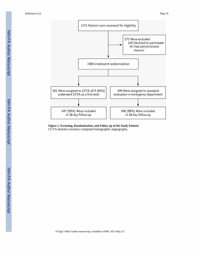
Figure 1. Screening, Randomization, and Follow-up of the Study PatientsCCTA denotes coronary computed tomographic angiography.
Hoffmann et al. Page 10
N Engl J Med. Author manuscript; available in PMC 2013 May 23.
NIH
-PA Author Manuscript
NIH
-PA Author Manuscript
NIH
-PA Author Manuscript

Figure 2. Length of Stay in the Hospital and Proportion of Patients DischargedThe cumulative frequency of discharge from the index visit according to the length of stay isshown. The horizontal line indicates the median length of stay in the two study groups,which was significantly different (8.6 hours in the CCTA group vs. 26.7 hours in thestandard-evaluation group, P<0.001).
Hoffmann et al. Page 11
N Engl J Med. Author manuscript; available in PMC 2013 May 23.
NIH
-PA Author Manuscript
NIH
-PA Author Manuscript
NIH
-PA Author Manuscript

NIH
-PA Author Manuscript
NIH
-PA Author Manuscript
NIH
-PA Author Manuscript
Hoffmann et al. Page 12
Table 1
Baseline Demographic and Clinical Characteristics of the Patients.*
Variable CCTA (N = 501)Standard Evaluation (N
= 499) P Value
Mean age — yr 54±8 54±8 0.44
Female sex — % 48 46 0.57
Race or ethnic group — no. (%)
Black 141 (28) 141 (28) 1.00
White 330 (66) 330 (66) 0.95
Asian 18 (4) 13 (23) 0.47
Other 12 (2) 18 (4) 0.27
Non-Hispanic 435 (87) 422 (85) 0.57
Cardiovascular risk factors — no. (%)
Hypertension 269 (54) 272 (54) 0.80
Diabetes mellitus 86 (17) 87 (17) 0.93
Dyslipidemia 230 (46) 224 (45) 0.75
Former or current smoker 249 (50) 243 (49) 0.75
Family history of premature coronary artery disease 135 (27) 136 (27) 0.94
No. of cardiovascular risk factors — % 0.68
0 or 1 36 38
2 or 3 54 52
≥4 10 10
Relevant prior medication — no. (%)
Aspirin 115 (23) 113 (23) 0.94
Beta-blocker 88 (18) 82 (16) 0.67
Statin 143 (28) 151 (30) 0.58
Initial presentation in emergency department
Chief symptom — no. (%) 0.47
Radiating or nonradiating chest pain or anginal equivalent 444 (89) 452 (91)
Arm, jaw, shoulder, or epigastric pain 21 (4) 16 (3)
Shortness of breath 7 (1) 10 (2)
Other 29 (6) 21 (4)
Heart rate — beats/min 78±14 77±14 0.58
Blood pressure — mm Hg
Systolic 144±23 144±23 0.80
Diastolic 83±13 83±13 0.94
BMI 29.4±5.3 29.1±4.8 0.41
Discharge diagnosis after index emergency department visit or hospitalization —no. (%) - 0.16
Noncardiac chest pain 426 (85) 445 (89)
Noncoronary cardiac pain 7 (1) 8 (2)
N Engl J Med. Author manuscript; available in PMC 2013 May 23.

NIH
-PA Author Manuscript
NIH
-PA Author Manuscript
NIH
-PA Author Manuscript
Hoffmann et al. Page 13
Variable CCTA (N = 501)Standard Evaluation (N
= 499) P Value
Coronary chest pain not associated with acute coronary syndrome 25 (5) 14 (3)
Acute coronary syndrome 43 (9) 32 (6)
Unstable angina pectoris 35 (7) 17 (3)
Myocardial infarction 8 (2) 15 (3)
*Plus–minus values are means ±SD. BMI denotes body-mass index (the weight in kilograms divided by the square of the height in meters), and
CCTA coronary computed tomographic angiography.
N Engl J Med. Author manuscript; available in PMC 2013 May 23.
NIH
-PA Author Manuscript
NIH
-PA Author Manuscript
NIH
-PA Author Manuscript
Hoffmann et al. Page 14
Table 2
Primary and Secondary Effectiveness and Safety End Points.*
End Point CCTA (N = 501) Standard Evaluation (N = 499) P Value
Length of hospital stay — hr
All patients in intention-to-treat analysis <0.001
Mean 23.2±37.0 30.8±28.0
Median 8.6 26.7
Interquartile range 6.4–27.6 21.4–30.6
Patients with final diagnosis other than acute coronary syndrome <0.001
Mean 17.2±24.6 27.2±19.5
Median 8.1 26.3
Interquartile range 6.2–24.6 20.6–29.5
Patients with final diagnosis of acute coronary syndrome 0.87
Mean 86.3±72.3 83.8±61.3
Median 56.9 71.8
Interquartile range 46.2–95.9 45.2–96.7
Time to diagnosis — hr
All patients in intention-to-treat analysis <0.001
Mean 10.4±12.6 18.7±11.8
Median 5.8 21.0
Interquartile range 4.0–9.0 8.5–23.8
Patients with final diagnosis other than acute coronary syndrome <0.001
Mean 10.6±12.3 18.8±12.0
Median 6.1 21.1
Interquartile range 4.0–9.6 8.7–23.8
Patients with final diagnosis of acute coronary syndrome 0.002
Mean 8.0±15.1 17.1±9.5
Median 4.4 14.9
Interquartile range 3.3–5.6 7.4–25.1
Discharge status — no. (%) <0.001
Direct discharge from emergency department 233 (47) 62 (12)
Admission to observation unit 153 (30) 301 (60)
Admission to hospital 107 (21) 125 (25)
Left against medical advice 8 (2) 11 (2)
Follow-up for recurrent chest pain within 28 days — no.
Repeat visit to emergency department 14 19 0.38
Repeat hospitalization 7 7
Safety — no.
Undetected acute coronary syndrome 0 0
Periprocedural complications 2 0 0.50
N Engl J Med. Author manuscript; available in PMC 2013 May 23.

NIH
-PA Author Manuscript
NIH
-PA Author Manuscript
NIH
-PA Author Manuscript
Hoffmann et al. Page 15
End Point CCTA (N = 501) Standard Evaluation (N = 499) P Value
Length of hospital stay — hr
Major adverse cardiovascular events at 28 days — no. 2 6 0.18
*Plus–minus values are means ±SD.
N Engl J Med. Author manuscript; available in PMC 2013 May 23.

NIH
-PA Author Manuscript
NIH
-PA Author Manuscript
NIH
-PA Author Manuscript
Hoffmann et al. Page 16
Tabl
e 3
Res
ourc
e U
tiliz
atio
n, R
adia
tion
Exp
osur
e, a
nd C
osts
of
Car
e.*
Var
iabl
eIn
dex
Vis
itIn
dex
Plu
s F
ollo
w-u
p V
isit
CC
TA
(N
= 5
01)
Stan
dard
Eva
luat
ion
(N =
499
)P
Val
ueC
CT
A (
N =
501
)St
anda
rd E
valu
atio
n (N
= 4
99)
P V
alue
Dia
gnos
tic te
stin
g —
no.
of
patie
nts
(%)†
<0.
001
<0.
001
N
o te
stin
g‡9
(2)
109
(22)
9 (2
)89
(18
)
1
test
376
(75)
337
(68)
359
(72)
350
(70)
≥2
test
s11
6 (2
3)53
(11
)13
3 (2
7)60
(12
)
Func
tiona
l tes
ting
— n
o. (
%)§
<0.
001
<0.
001
SP
EC
T50
(10
)12
4 (2
5)58
(12
)13
3 (2
7)
St
ress
ech
ocar
diog
raph
y20
(4)
102
(20)
20 (
4)10
2 (2
0)
E
TT
12 (
2)14
7 (2
9)22
(4)
162
(32)
Inva
sive
cor
onar
y an
giog
raph
y —
no.
(%
)54
(11
)36
(7)
0.06
59 (
12)
40 (
8)0.
06
Inte
rven
tion
— n
o. (
%)
PC
I24
(5)
14 (
3)0.
1427
(5)
17 (
3)0.
16
C
AB
G5
(1)
4 (1
)0.
995
(1)
4 (1
)0.
99
Cum
ulat
ive
radi
atio
n ex
posu
re —
mSv
/pat
ient
¶13
.9±
10.4
4.7±
8.4
<0.
001
14.3
±10
.95.
3±9.
6<
0.00
1
Cos
ts o
f ca
re —
U.S
. dol
lars∥
<0.
001
E
mer
genc
y de
part
men
t
Mea
n2,
101±
1,07
02,
566±
1,32
3
Med
ian
1,77
02,
293
In
terq
uart
ile r
ange
1,43
5–2,
161
1,59
2–3,
583
H
ospi
tal
0.19
Mea
n1,
925±
6,69
71,
308±
5,33
3
T
otal
0.75
0.65
Mea
n4,
026±
6,79
23,
874±
5,29
84,
289±
7,11
04,
060±
5,45
2
Med
ian
1,93
72,
742
1,94
62,
809
Inte
rqua
rtile
ran
ge1,
504–
4,05
71,
755–
3,83
21,
514–
4,16
41,
822–
4,06
0
N Engl J Med. Author manuscript; available in PMC 2013 May 23.

NIH
-PA Author Manuscript
NIH
-PA Author Manuscript
NIH
-PA Author Manuscript
Hoffmann et al. Page 17* Pl
us–m
inus
val
ues
are
mea
ns ±
SD. P
erce
ntag
es m
ay n
ot s
um to
100
bec
ause
of
roun
ding
. CA
BG
den
otes
cor
onar
y-ar
tery
byp
ass
graf
ting,
ET
T e
xerc
ise
trea
dmill
test
ing,
PC
I pe
rcut
aneo
us c
oron
ary
inte
rven
tion,
and
SPE
CT
sin
gle-
phot
on-e
mis
sion
com
pute
d to
mog
raph
y.
† Dia
gnos
tic te
stin
g in
clud
ed C
CT
A, E
TT
, SPE
CT
, str
ess
echo
card
iogr
aphy
, and
inva
sive
cor
onar
y an
giog
raph
y.
‡ Seri
al m
easu
rem
ent o
f bi
omar
kers
and
ele
ctro
card
iogr
aphi
c te
stin
g w
ere
not c
onsi
dere
d as
dia
gnos
tic te
sts
in th
is ta
ble.
§ At t
he in
dex
visi
t, fu
nctio
nal t
estin
g w
as th
e se
cond
test
in th
e C
CT
A g
roup
and
the
firs
t tes
t in
the
stan
dard
-eva
luat
ion
grou
p.
¶ Rad
iatio
n ex
posu
re in
clud
ed e
xpos
ure
from
CC
TA
, SPE
CT
, and
inva
sive
cor
onar
y an
giog
raph
y.
∥ Cos
ts in
clud
ed th
ose
for
patie
nts
disc
harg
ed d
irec
tly f
rom
the
emer
genc
y de
part
men
t and
thos
e di
scha
rged
fro
m a
n ob
serv
atio
n un
it.
N Engl J Med. Author manuscript; available in PMC 2013 May 23.