ulcerative-vegetative locally advanced breast carcinoma
TRANSCRIPT
1Mastology 2021;31:e20200079
Ulcerative-vegetative Locally Advanced Breast Carcinoma Mimicking Flower Image
René Aloisio da Costa Vieira1,2,3 , Idam de Oliveira-Junior1,4
1Postgraduate Program in Tocogynecology, Faculdade de Medicina de Botucatu – Botucatu (SP), Brazil.2Postgraduate Program in Oncology, Barretos Cancer Hospital – Barretos (SP), Brazil.3Department of Surgery, Division of Mastology, Cancer Hospital of Muriaé – Muriaé (MG), Brazil.4Department of Mastology and Breast Reconstruction, Barretos Cancer Hospital – Barretos (SP), Brazil.*Corresponding author: [email protected] of interests: nothing to declare. Received on: 04/10/2021. Accepted on: 06/09/2021.
IMAGES IN MASTOLOGYhttps://doi.org/10.29289/2594539420200079
Ulcerated locally advanced breast carcinoma (LABC)1,2 is an uncommon topic in the literature. Several factors contribute to delayed diagnosis, such as the health system, factors related to the patient (lack of knowledge, fear and denial of the disease), in addition to rapid tumor growth.
Ulcerated tumors can occur in any breast location, includ-ing in areolar Paget’s disease3. They are usually high histologic-grade tumors, high Ki67 index and triple negative breast can-cer (TNBC) molecular subtype with lymph node involvement2. There are disagreements about the simple presence of ulceration determining worsening of patient’s prognosis1,2.
An ulcerated lesion leads to bleeding and may be the gate-way to secondary infection. In this context, surgery can be4-6:• up-front hygiene (avoids bleeding and infection, but is
associated with the need for local flaps); • elective, after neoadjuvant chemotherapy treatment (leaves
the patient vulnerable to infection and sepsis7, regardless of neutropenia);
• elective, after radiotherapy, in extreme situations8 (attempt to increase resectability).
Prior to surgery, a surgical wound culture can be performed to guide the choice of antibiotic therapy. During surgery, special care must be taken (covering the exposed area with compresses and administration of broad-spectrum antibiotic for therapeu-tic purposes)9.
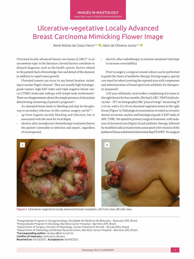
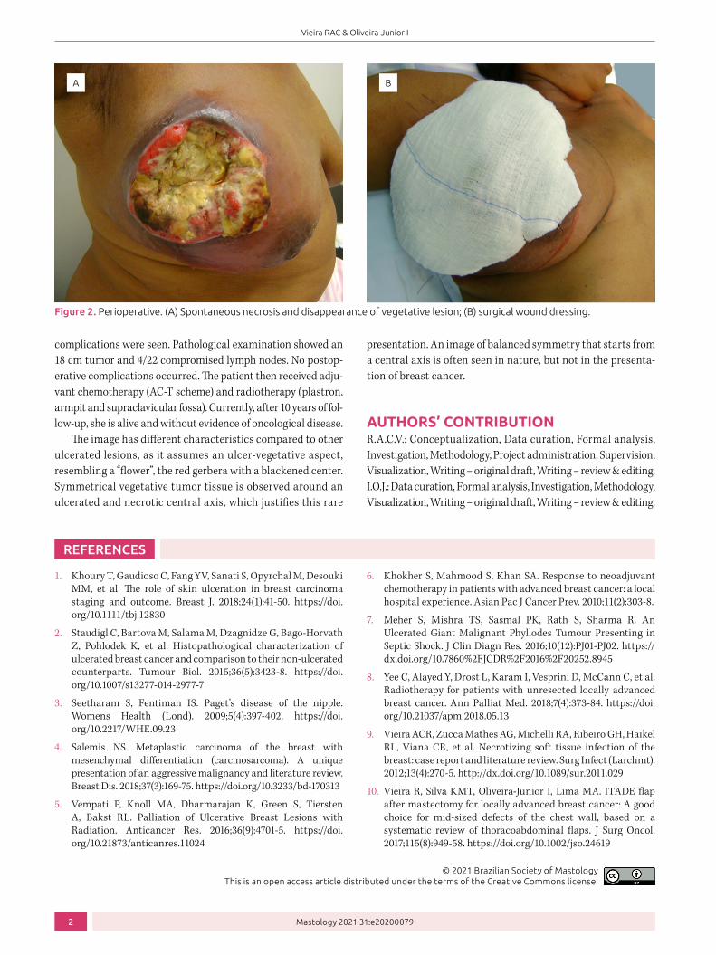
A 52-year-old female, rural worker, complaining of a tumor in the right breast for four months. She had LABC, T4bN3 (infracla-vicular – IFV on tomography) M0, “peau d’orange” measuring 19 x 15 cm, with a 15 x 10 cm ulcerated vegetative lesion in the right breast (Figure 1). Pathological examination revealed an invasive ductal carcinoma, nuclear and histological grade 3, Ki67 index of 50%, TNBC. We opted for primary surgical treatment, with isola-tion of ulcerated area (Figure 2) and antibiotic therapy, followed by modified radical mastectomy associated with rotation of the ipsilateral thoracoabdominal dermomat flap (ITADE)10. No surgical
Figure 1. Ulcerative-vegetative locally advanced breast neoplasm. (A) front view; (B) side view.
A B
2
Vieira RAC & Oliveira-Junior I
Mastology 2021;31:e20200079
© 2021 Brazilian Society of Mastology This is an open access article distributed under the terms of the Creative Commons license.
1. Khoury T, Gaudioso C, Fang YV, Sanati S, Opyrchal M, Desouki MM, et al. The role of skin ulceration in breast carcinoma staging and outcome. Breast J. 2018;24(1):41-50. https://doi.org/10.1111/tbj.12830
2. Staudigl C, Bartova M, Salama M, Dzagnidze G, Bago-Horvath Z, Pohlodek K, et al. Histopathological characterization of ulcerated breast cancer and comparison to their non-ulcerated counterparts. Tumour Biol. 2015;36(5):3423-8. https://doi.org/10.1007/s13277-014-2977-7
3. Seetharam S, Fentiman IS. Paget’s disease of the nipple. Womens Health (Lond). 2009;5(4):397-402. https://doi.org/10.2217/WHE.09.23
4. Salemis NS. Metaplastic carcinoma of the breast with mesenchymal differentiation (carcinosarcoma). A unique presentation of an aggressive malignancy and literature review. Breast Dis. 2018;37(3):169-75. https://doi.org/10.3233/bd-170313
5. Vempati P, Knoll MA, Dharmarajan K, Green S, Tiersten A, Bakst RL. Palliation of Ulcerative Breast Lesions with Radiation. Anticancer Res. 2016;36(9):4701-5. https://doi.org/10.21873/anticanres.11024
REFERENCES
6. Khokher S, Mahmood S, Khan SA. Response to neoadjuvant chemotherapy in patients with advanced breast cancer: a local hospital experience. Asian Pac J Cancer Prev. 2010;11(2):303-8.
7. Meher S, Mishra TS, Sasmal PK, Rath S, Sharma R. An Ulcerated Giant Malignant Phyllodes Tumour Presenting in Septic Shock. J Clin Diagn Res. 2016;10(12):PJ01-PJ02. https://dx.doi.org/10.7860%2FJCDR%2F2016%2F20252.8945
8. Yee C, Alayed Y, Drost L, Karam I, Vesprini D, McCann C, et al. Radiotherapy for patients with unresected locally advanced breast cancer. Ann Palliat Med. 2018;7(4):373-84. https://doi.org/10.21037/apm.2018.05.13
9. Vieira ACR, Zucca Mathes AG, Michelli RA, Ribeiro GH, Haikel RL, Viana CR, et al. Necrotizing soft tissue infection of the breast: case report and literature review. Surg Infect (Larchmt). 2012;13(4):270-5. http://dx.doi.org/10.1089/sur.2011.029
10. Vieira R, Silva KMT, Oliveira-Junior I, Lima MA. ITADE flap after mastectomy for locally advanced breast cancer: A good choice for mid-sized defects of the chest wall, based on a systematic review of thoracoabdominal flaps. J Surg Oncol. 2017;115(8):949-58. https://doi.org/10.1002/jso.24619
Figure 2. Perioperative. (A) Spontaneous necrosis and disappearance of vegetative lesion; (B) surgical wound dressing.
A B
complications were seen. Pathological examination showed an 18 cm tumor and 4/22 compromised lymph nodes. No postop-erative complications occurred. The patient then received adju-vant chemotherapy (AC-T scheme) and radiotherapy (plastron, armpit and supraclavicular fossa). Currently, after 10 years of fol-low-up, she is alive and without evidence of oncological disease.
The image has different characteristics compared to other ulcerated lesions, as it assumes an ulcer-vegetative aspect, resembling a “flower”, the red gerbera with a blackened center. Symmetrical vegetative tumor tissue is observed around an ulcerated and necrotic central axis, which justifies this rare
presentation. An image of balanced symmetry that starts from a central axis is often seen in nature, but not in the presenta-tion of breast cancer.
AUTHORS’ CONTRIBUTIONR.A.C.V.: Conceptualization, Data curation, Formal analysis, Investigation, Methodology, Project administration, Supervision, Visualization, Writing – original draft, Writing – review & editing.I.O.J.: Data curation, Formal analysis, Investigation, Methodology, Visualization, Writing – original draft, Writing – review & editing.