ultrasound clinical news magazine from philips · ultrasound clinical news magazine from philips...
TRANSCRIPT

Ultrasound Clinical News Magazine from Philips
November 2018 USCAN
Overview:
The targeted fetal anomaly scan involves detail evaluation of the fetus, ascertaining the presence or absence of any fetal abnormality. While conventional curvilinear transducers satisfy the basic requirements, they fail to provide a high resolution that is needed to visualise the fetal anatomy in detail. Linear transducers overcome this impediment by delivering a high resolution image, but at the cost of penetration. The newly introduced, Philips eL18-4 linear transducer, by virtue of its high frequency, PureWave technology, elevation focusing capability and the high number of elements (1920), is an excellent solution that offers both - better resolution and penetration.
The following cases depicts the utility of this transducer in diagnosing anomalies and delivering unprecedented clinical information
2 U SCAN Clinical News Magazine November 2018
Case study 1:
Clinical history: A 21year old primigravida had come for a TIFFA scan. She had no previous bad obstetric history. No ultrasound scanning was done prior to this scan.
Ultrasound findings:
A detailed ultrasound evaluation of the fetus was done. The ultrasound evaluation revealed a single live intrauterine pregnancy of
Evaluation of Fetal Anomalies with
eL18-4 PureWave linear array transducer
Dr. T. L. N. Praveen
About the author:
Dr Praveen is currently Director and Consultant, Abhishek's Institute Of
Imageology, Hyderabad, India. His main clinical and research interests are in
ultrasound - both diagnostic and interventional, with special interest in
Obstetrics and Gynaecology. He has written a Chapter on Ultrasonography
in Obstetrics & Gynaecology in Text Book of Diagnostic Radiology and
Imaging, 1997, and also has various publications in National and International
journals. His work has been published in the American Journal of Ultrasound
and Journal of Clinical Ultrasound as well.
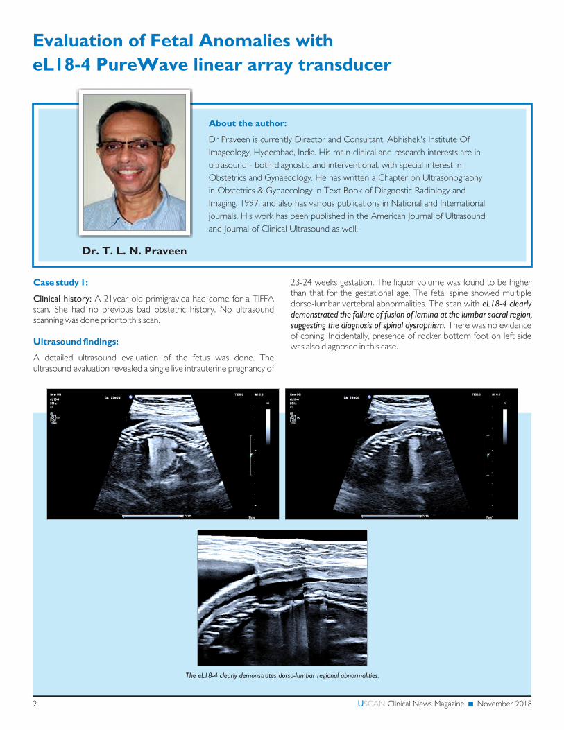
23-24 weeks gestation. The liquor volume was found to be higher than that for the gestational age. The fetal spine showed multiple dorso-lumbar vertebral abnormalities. The scan with eL18-4 clearly demonstrated the failure of fusion of lamina at the lumbar sacral region, suggesting the diagnosis of spinal dysraphism. There was no evidence of coning. Incidentally, presence of rocker bottom foot on left side was also diagnosed in this case.
The eL18-4 clearly demonstrates dorso-lumbar regional abnormalities.
Case study 2:
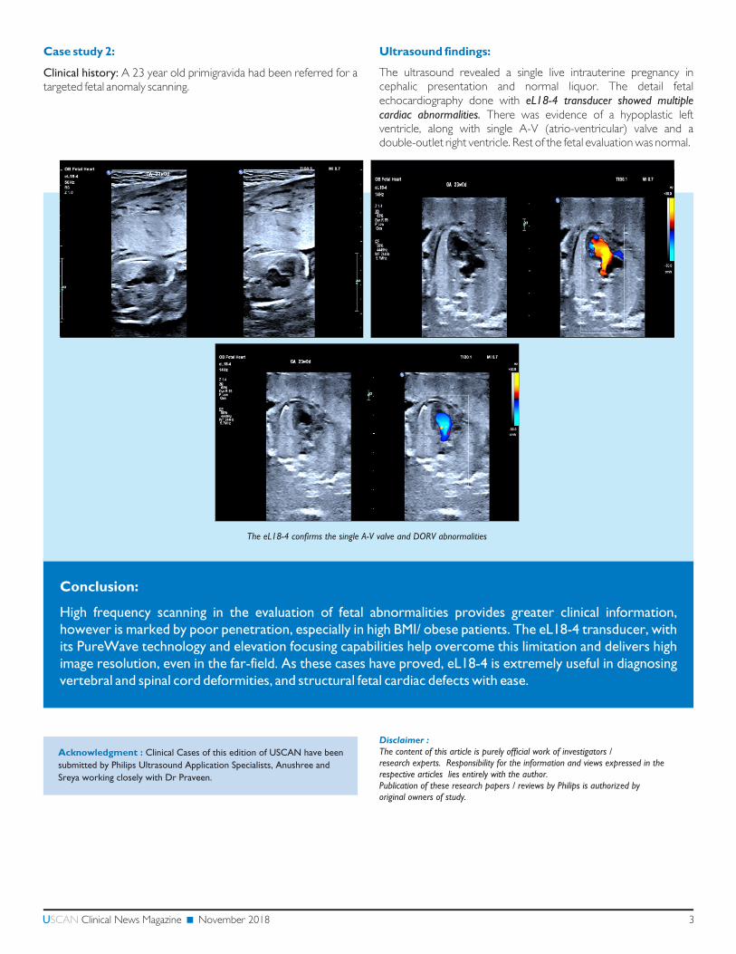
Clinical history: A 23 year old primigravida had been referred for a targeted fetal anomaly scanning.
The eL18-4 confirms the single A-V valve and DORV abnormalities
3U SCAN Clinical News Magazine November 2018
Ultrasound findings:
The ultrasound revealed a single live intrauterine pregnancy in cephalic presentation and normal liquor. The detail fetal echocardiography done with eL18-4 transducer showed multiple cardiac abnormalities. There was evidence of a hypoplastic left ventricle, along with single A-V (atrio-ventricular) valve and a double-outlet right ventricle. Rest of the fetal evaluation was normal.
Conclusion:
High frequency scanning in the evaluation of fetal abnormalities provides greater clinical information, however is marked by poor penetration, especially in high BMI/ obese patients. The eL18-4 transducer, with its PureWave technology and elevation focusing capabilities help overcome this limitation and delivers high image resolution, even in the far-field. As these cases have proved, eL18-4 is extremely useful in diagnosing vertebral and spinal cord deformities, and structural fetal cardiac defects with ease.
Acknowledgment : Clinical Cases of this edition of USCAN have been
submitted by Philips Ultrasound Application Specialists, Anushree and
Sreya working closely with Dr Praveen.
Disclaimer :The content of this article is purely official work of investigators / research experts. Responsibility for the information and views expressed in the respective articles lies entirely with the author.Publication of these research papers / reviews by Philips is authorized by original owners of study.

Your inputs are important to us.
Kindly send your feedback and comments to
Printed in India. USCAN 1018
Gurgaon (Corporate Office and Northern Regional Office)
9th Floor, 9B DLF Cyber City, Phase 3, Sector 25, Gurgaon - 122 002. • Tel : +91 124 460 6000
Chennai (South Regional Office)
Sunny Side, 3rd Floor, C Block, No. 8/17, Shafee Mohammed Road, (Off Greams Road), Chennai - 600006. • Tel : +91 44 6650 1000
Kolkata (Eastern Regional Office)
3rd Floor, Tower A, DLF IT Park, 08 Block AF, Major Arterial Road, New Town (Rajarhat) Kolkata, West Bengal - 700156. • Tel : +91 33 2455 9770
Mumbai (Western Regional Office)
Boomerang, B2 Wing, 5th Floor, Unit No. 506, Chandivali Farm Road, Near Chandivali Studio, Andheri (East), Mumbai - 400 072. • Tel : +91 6691 2000
The eL18-4 transducer leverages a combination of Philips innovations in order to support a diverse range of clinical applications while delivering extraordinary imaging and depth-of-field performance
Unique features of eL18-4 transducer:
High-frequency, Broadband Linear Transducer (Generates frequencies from 2 to 22 MHz)
PureWave technology with Fine-Elevation focusing
Superb 2D resolution with penetration
Micro Flow Imaging – for low flow, low volume flow in fetal, placental, uterine and ovarian vasculature.
Trapezoid view for extending FOV
A.I.Breast Imaging (Anatomical Intelligence based)
Philips eL18-4 – PureWave High Frequency Linear
Transducer with MicroFlow imagingSuperb imaging details for ObGyn scanning!
MicroFlow Imaging
Philips MicroFlow Imaging (MFI), found on the eL18-4, is a proprietary imaging mode designed to detect low volume, low velocity blood flow found in fetal, placental, uterine and ovarian vasculature. New 2D image subtraction, 2D blending and side-by-side display options offer excellent visualization versatility