ultrasound detection of colonic polyps dr. muhammad bin zulfiqar
TRANSCRIPT
Ultrasound Detection of Colonic Polyps: Perspective
DR MUHAMMAD BIN ZULFIQARPGR III FCPS Services institute of Medical
Sciences/ Services Hospital Lahore

Objectives
• Sonographic detection of colonic polyp.• Characterization of colonic polyps
S KUZMICH, J HARVEY, KUZMICH and K L TAN. Ultrasound detection of colonic polyps: perspective. The British Journal of Radiology, 85 (2012), 1155-64.
INTRODUCTION
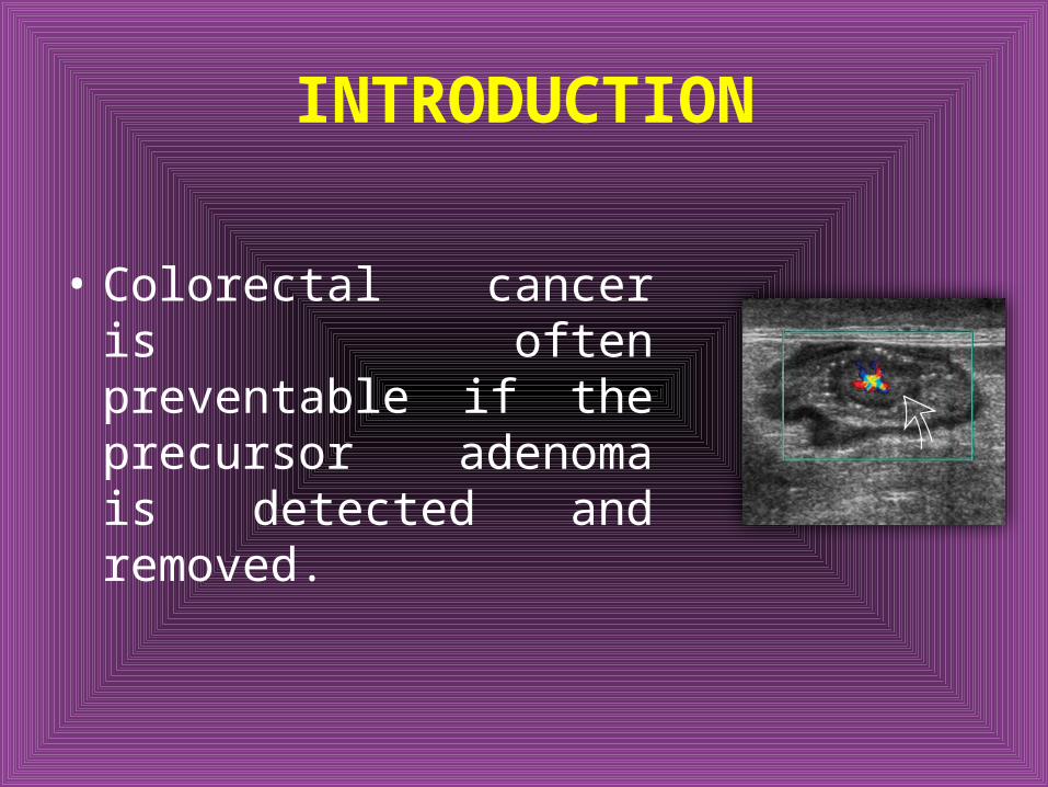
• Colorectal cancer is often preventable if the precursor adenoma is detected and removed.

INTRODUCTION
• Although ultrasound is clearly not one of the widely accepted screening techniques, this non-invasive and radiation-free modality is also capable of detecting colonic polyps, both benign and malignant.

INTRODUCTION• Such colon lesions may be encountered
when not expected, usually during general abdominal sonography.
• The discovery of large colonic polyps is important and can potentially help reduce the incidence of a common cancer, whereas detection of a malignant polyp at an early stage may result in a curative intervention.

METHODOLOGY
• This pictorial review will highlights sonographic detection of colonic polyps and characterize the nature of polyp

METHODOLOGY
• Polyps can be discovered during a routine abdominal or pelvic examination, particularly when scanning is supplemented by a brief focused sonographic inspection of the colon with a 6–10MHz linear transducer.

ULTRASOUND TECHNIQUE
• Much of the large bowel can be imaged with conventional ultrasound.
• Small-footprint curvilinear and linear 5–10MHz transducers are preferred because they can be pressed to the maximum depth, bringing the cross-section of the colon into focus and displacing the fecal residue from the area of interest.

ULTRASOUND TECHNIQUE
• The distal descending colon is recognised due to its fixed position in the left iliac fossa just above the iliac vessels.

ULTRASOUND TECHNIQUE
• It is then traced downwards where it continues as a sigmoid colon looping into or away from the pelvis, depending on the sigmoid configuration.

ULTRASOUND TECHNIQUE
• The descending colon wall is then followed upwards to the splenic flexure, and further along the transverse and ascending colon, through to the caecum.

ULTRASOUND TECHNIQUE
• The normal colonic wall is usually 3–4mm in thickness.
• Visualisation of five wall layers can be achieved under optimal scanning conditions, although three main layers of mucosa, submucosa and muscularis propria are usually seen.

• Transverse colour Doppler view of left iliac fossa shows cross-section of empty distal descending colon with minimal air outlining the lumen (open arrow).
• Three sonographic layers, from outer to inner are – muscularis propria (1), – submucosa (2) – mucosa (3).
• The taeniae coli are seen as focal thickening in muscularis propria (arrowheads), as in this individual.

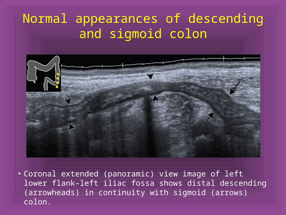
Normal appearances of descending and sigmoid colon
• Cross-section of semi-filled distal descending colon allows visualization of anterior wall, whereas posterior wall (open arrow) is obscured.

Normal appearances of descending and sigmoid colon
• Oblique view of the left iliac fossa shows sigmoid colon (arrows) containing some echogenic but non-shadowing residue hence posterior wall is visible.
Normal appearances of descending and sigmoid colon
• Coronal extended (panoramic) view image of left lower flank–left iliac fossa shows distal descending (arrowheads) in continuity with sigmoid (arrows) colon.

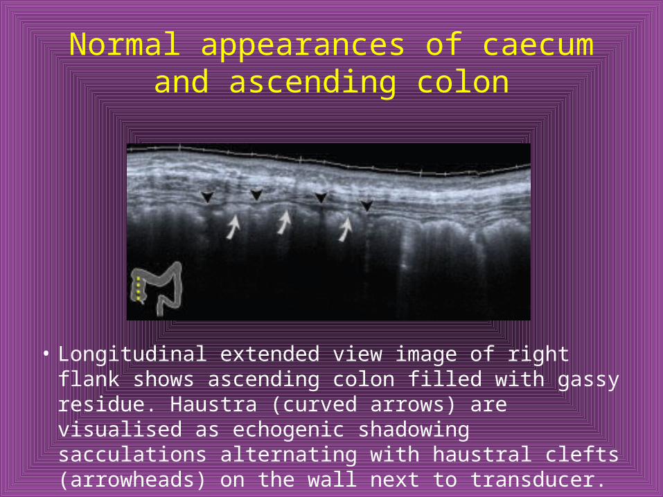
Normal appearances of caecum and ascending colon
• Cross-section of caecum filled with gassy residue allows visualisation of anterior wall (arrowheads); posterior wall is mostly obscured. Note normal appendix (arrow).

Normal appearances of caecum and ascending colon
• Transverse view of the right iliac fossa with transducer compression shows nearly empty caecum in cross-section. Taeniae coli are seen as focal thickening in muscularis propria (arrowheads).
Normal appearances of caecum and ascending colon
• Longitudinal extended view image of right flank shows ascending colon filled with gassy residue. Haustra (curved arrows) are visualised as echogenic shadowing sacculations alternating with haustral clefts (arrowheads) on the wall next to transducer.

A 70-year-old male with colonic diverticulosis.
• Sonogram of left iliac fossa shows cross-section of sigmoid colon with mildly thickened hypoechoic wall (arrowhead) and diverticulum (arrow).

Appearances of adenomatous sigmoid polyp with high-grade dysplasia proven histologically.
• Colour Doppler view of left iliac fossa in 61-year-old male shows a cross-section of sigmoid colon (arrowheads) and 18mm polyp (curved arrow).

Appearances of adenomatous sigmoid polyp with high-grade dysplasia proven histologically.
• Cross-section view of sigmoid colon in 60-year-old male reveals polyp (curved arrow) with convoluted surface outline, which measured 15mm.

A 55-year-old male with two confirmed benign adenomas in sigmoid colon.
• Coronal extended-view image of left flank–iliac fossa shows distal descending (black arrowheads) and proximal sigmoid colon (white arrowheads) with polyp consisting of head (curved arrow), which measured 20mm, and pedicle (black arrow).

A 55-year-old male with two confirmed benign adenomas in sigmoid colon.
• The same polyp (curved arrow) seen in cross-section of colon (arrowheads).

A 55-year-old male with two confirmed benign adenomas in sigmoid colon.
• 19mm second polyp in the sigmoid colon with internal vascularity demonstrated by colour flow Doppler.

A 55-year-old male with two confirmed benign adenomas in sigmoid colon.
• Close inspection of the second polyp discloses head with convoluted contour (curved arrow) and pedicle (open arrowheads), which is seen as prolongation of echogenic submucosa with a hypoechoic stripe of submucosal muscularis extending from colonic wall (arrowheads) to polyp head. Note feeding vessels in pedicle.
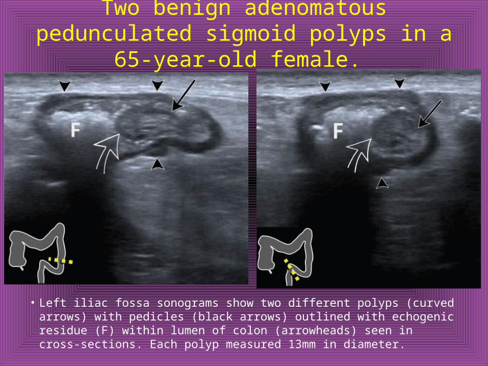
Two benign adenomatous pedunculated sigmoid polyps in a 65-year-old female.
• Left iliac fossa sonograms show two different polyps (curved arrows) with pedicles (black arrows) outlined with echogenic residue (F) within lumen of colon (arrowheads) seen in cross-sections. Each polyp measured 13mm in diameter.

Appearances of 23mm sigmoid polyp that proved to be villous adenoma harboring intramucosal carcinoma in an asymptomatic 67-year-old female
• Oblique colour Doppler view of sigmoid colon (arrowheads) in left iliac fossa depicts intraluminal lesion (open arrow) with probable vascularity.
• Spectral Doppler analysis of colour signal in the same lesion (open arrow) confirms presence of arterial flow.

60-year-old male with adenomatous polyp in ascending colon.
• Longitudinal image of right flank shows 15-mm polyp as hypoechoic lesion with undulating outline (curved arrow) outlined by echogenic residue underneath anterior wall of colon (arrowheads). The lesion maintained constant position, shape and size throughout the examination, although no colour Doppler signal was detected.
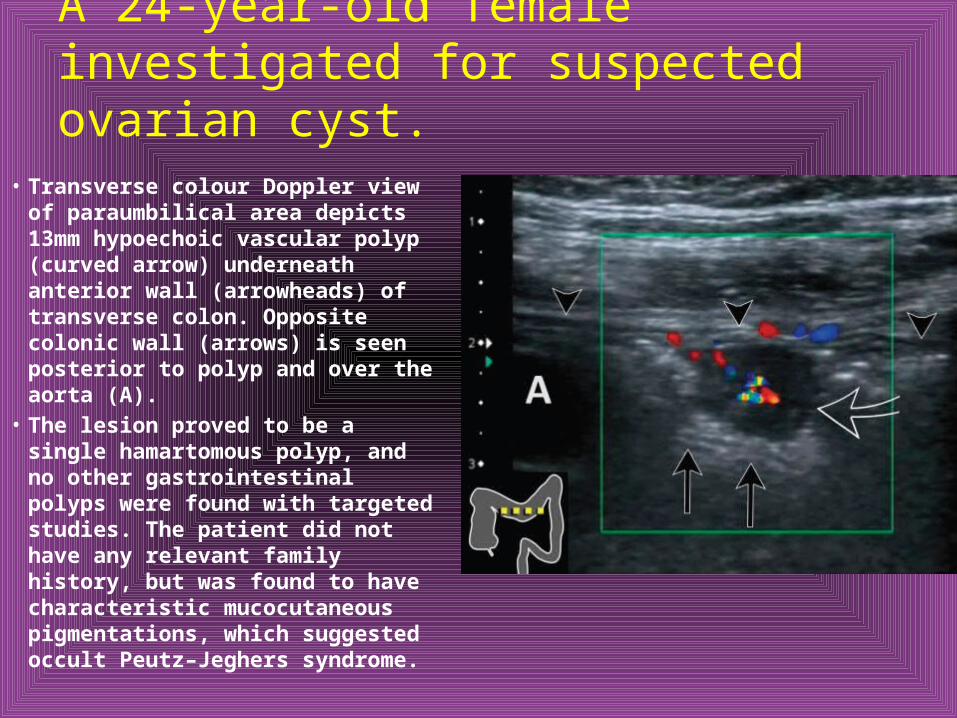
A 24-year-old female investigated for suspected ovarian cyst.
• Transverse colour Doppler view of paraumbilical area depicts 13mm hypoechoic vascular polyp (curved arrow) underneath anterior wall (arrowheads) of transverse colon. Opposite colonic wall (arrows) is seen posterior to polyp and over the aorta (A).
• The lesion proved to be a single hamartomous polyp, and no other gastrointestinal polyps were found with targeted studies. The patient did not have any relevant family history, but was found to have characteristic mucocutaneous pigmentations, which suggested occult Peutz–Jeghers syndrome.

A 55-year-old male with ulcerative colitis.
• Cross-section of distal descending colon shows thickened mucosa and prominent mucosal projection (curved arrow) suggesting inflammatory polyp.

A 55-year-old male with ulcerative colitis.
• Extended-view image of lower left flank shows longitudinal section of the same descending colon, with corresponding mucosal elevation (curved arrow). A useful differentiating feature is found by observing that echotexture of ‘‘pseudo polyp’’ is similar to that of thickened mucosa.

Sagittal sonogram of right upper quadrant in a 60-year-old asymptomatic male
• reveals 4.0 cm broad-based intraluminal mass (open arrow) in transverse colon shown in cross-section (arrowheads). Note loss of stratification and indistinct outline in colonic wall underlying base of polypoid mass (arrows), which suggests invasion through muscularis propria. Subsequent CT suggested T3 (modified Dukes B2) stage of the disease and surgical resection confirmed
• transverse colon adenocarcinoma penetrating into subserosal fat.

A 60-year-old male with unsuspected colonic carcinoma.
• (a) Sagittal epigastric view depicts a 3.0 cm polypoid lesion (asterisk) containing ulceration seen as a pocket of gas (open arrow) in transverse colon (arrowheads). The full circumference of the colonic wall muscularis propria is well visualised (arrowheads).
• (b) Analysis of the lesion with colour doppler reveals abundant vascularity reinforcing suspicion that the lesion is malignant. Subsequent CT suggested T2 (modified Dukes B1) stage of the local disease. Polypoid adenocarcinoma in transverse colon invading muscularis propria was confirmed at histological examination.

Potential pitfalls
• Cross-section of distal descending colon shows broad-based polypoid projection (arrow), which may be mistaken for plaque-like polypoid lesion.

Potential pitfalls
• Longitudinal view of the same colonic segment with the probe now rotated at the right angle demonstrates that the lesion in (a) is in fact not real but the result of tangential imaging of steep haustral fold (arrow). Cross-sectional image in (a) was made through the plane indicated by the arrow

Potential pitfalls
• Longitudinal coronal view of the distal descending colon shows prominent bulbous haustral fold (arrow), which may simulate small polyp.

Potential pitfalls
• Cross-section of proximal sigmoid colon shows focal bulge in colonic wall (arrow), which is due to prominent taeniae coli.

Potential pitfalls
• Cross-section of proximal sigmoid colon in a different patient shows two impacted diverticulae (arrows) containing echogenic shadowing stool.
CONCLUSION• When integrated into routine scanning, brief sonographic
examination of the accessible colon can reveal unsuspected large colonic polyps.
• Demonstration of vascularity within such lesions on Doppler is confirmatory.
• This maximizes the usefulness of conventional ultrasound and potentially help reduce the incidence of a common cancer since colonic polyps may harbor an early carcinoma or lead to malignancy.
