ultrasound microbubble contrast agents for diagnostic and therapeutic applications...
TRANSCRIPT

125Review Article
Ultrasound Microbubble Contrast Agents for Diagnostic andTherapeutic Applications: Current Status and Future Design
Shih-Tsung Kang, PhDc; Chih-Kuang Yeh, PhD
Ultrasound contrast agents are highly echogenicmicrobubbles with many unique properties. Microbubbles canbasically improve the sensitivity of conventional ultrasoundimaging to the microcirculation. The resonance of microbub-bles in response to an incident ultrasound pulse results in non-linear harmonic emission that serves as the signature ofmicrobubbles in microbubble-specific imaging. Inertial cavita-tion and destruction of microbubbles can produce a strongmechanical stress enhancing the permeability of the surround-ing tissues, and can further increase the extravasation of drugsfrom the blood into the cytoplasm or interstitium. Stable cavi-tation by high-frequency ultrasound can also mildly increasetissue permeability without causing any damage even at a highacoustic pressure. Microbubbles can carry drugs, release themupon ultrasound-mediated microbubble destruction, and simul-taneously enhance vascular permeability to increase drug deposition in tissues. Various tar-geting ligands can be conjugated to the surface of microbubbles to attain ligand-directed andsite-specific accumulation for targeted imaging. In addition to current developments inmicrobubble technology, this review introduces our studies of the applications of microbub-ble-specific imaging, ultrasound-aided drug delivery, and targeted imaging. These applica-tions are promising but may require further improvement for clinical use. (Chang Gung MedJ 2012;35:125-39)
Key words: microbubbles, contrast imaging, molecular imaging, targeted therapy, controlleddrug release, cavitation
From the Department of Biomedical Engineering and Environmental Sciences, National Tsing Hua University, Hsinchu, Taiwan.Received: Sep. 8, 2011; Accepted: Nov. 30, 2011Correspondence to: Prof. Chih-Kuang Yeh, Department of Biomedical Engineering and Environmental Sciences, National TsingHua University, Hsinchu, Taiwan. 101, Section 2, Kuang-Fu Road, Hsinchu 30013, Taiwan (R.O.C.) Tel: 886-3-5715131 ext. 34240; Fax: 886-3-5718649; E-mail: [email protected]
Ultrasound imaging has been one of the mostpopular medical diagnostic techniques because
of its superior safety, low cost, and easy accessibilitycompared with other imaging modalities such ascomputed tomography, positron emission tomogra-phy, and magnetic resonance imaging (MRI).Ultrasound imaging can provide real-time quantita-
tive information on the morphology and perfusion ofbiological tissues for evaluation of a variety of dis-eases in cardiology, radiology, and oncology.(1-5) Inrecent years, much attention has focused on observa-tion of the microcirculation in the different types ofdiseases or abnormalities in tissues. For example, thedependence of angiogenesis on tumor growth and
Prof. Chih-Kuang Yeh

Chang Gung Med J Vol. 35 No. 2March-April 2012
Shih-Tsung Kang and Chih-Kuang YehUltrasound contrast agents
126
progression has highlighted the importance of nonin-vasive visualization of the tumor microvasculatureto determine treatment strategies and assess thera-peutic efficacies.(6-9) However, the sensitivity andspecificity of ultrasound diagnoses are susceptible tolow contrast differences between blood and tissue.Conventional ultrasound Doppler imaging only per-mits flow assessment of vessels larger than hundredsof micrometers, such as arterioles and venules.These limitations have recently been overcome bythe development of ultrasound contrast agents(UCAs).
The first use of UCAs was reported in echocar-diography by Gramiak and Shah in 1968.(10) TheseUCAs were room air bubbles with no protectiveshell, so they disappeared within a few seconds afterintravenous administration. Currently-used UCAsare more stable microbubbles composed of low-dif-fusivity gases such as nitrogen or perfluorocarbonstabilized by a coating of biodegradable materialsuch as albumin, phospholipids, or polymers.(11)
Phospholipids are commonly used since lipid-coatedmicrobubbles are easier to fabricate and are muchmore echogenic than those made of other materi-als.(12) Microbubbles are highly echogenic in vivobecause of a mismatch in acoustic impedances (i.e.,the product of density and the speed of sound)between their gas cores and surrounding tissues.Once administered into a body cavity or the cardio-vascular system, microbubbles are capable ofincreasing the intensity of backscattered ultrasoundto up to 20–30 dB.(13) The contrast enhancement can
clarify delineation of the borders of body cavities(e.g., the left ventricular cavity) and increase theDoppler signals from intracranial vessels.(14-16) Theimproved sensitivity of ultrasound to microcircula-tion assists in characterization of possible lesions inthe liver and assessment of the vascular phenotype ofa tumor.(17-19) Several researchers have also reportedthe successful use of microbubbles in preclinicaldiagnoses of cardiovascular and renal diseases.(20-23)
In addition to contrast enhancing ability,microbubbles possess unique properties that can beexploited to improve both diagnoses and therapies.Taking advantage of these properties could lead toemerging applications such as microbubble-specificdetection and ultrasound-controlled targeted drugdelivery. These applications are promising for furtherimprovement in ultrasound imaging of the microcir-culation and treatment in the imaged regions. In thisarticle, we review current developments inmicrobubble technology and efforts by our laborato-ry to realize these emerging applications.
Commercial and homemade microbubblesAt present, 3 commercial agents, OptisonTM (GE
Healthcare, Milwaukee, WI, U.S.A.), Definity®
(Lantheus Medical Imaging, Billerica, MA, U.S.A.),and SonoVue® (Bracco SpA, Milano, Italy) arelicensed for cardiology applications. OptisonTM andDefinity® are available in both the United States andthe European Union, whereas SonoVue® is onlyavailable in the European Union. Detailed informa-tion on these agents is available in the Table.(11,24-26)
Table Specifications of Commercial and Homemade Microbubbles
Shell Mean sizePercentage
ConcentrationImaging
Name Manufacturermaterial
Gas(µm)
less than(bubbles/mL)
time10 µm (minutes)
OptisonTM GE Healthcare Albumin C3F8 2.0–4.5 95% 5.0–8.0 x 10⁸ 2.5–4.5
Definity® Lantheus Phospholipid C3F8 1.1–3.3 98% 1.2 x 10¹⁰ 2–10(Luminity® in the Medical European Union) Imaging
SonoVue® Bracco SpA Phospholipid SF6 2–8 99% (< 11 µm) 0.9–6 x 10⁹ 3–6
Homemade Yeh Group Phospholipid/ C3F8 0.2–0.7 > 99.9% 1.4–3.0 x 10¹⁰ 10–20Microbubbles at National Lipopolymer
Tsing Hua University
Chang Gung Med J Vol. 35 No. 2March-April 2012
Shih-Tsung Kang and Chih-Kuang YehUltrasound contrast agents
127
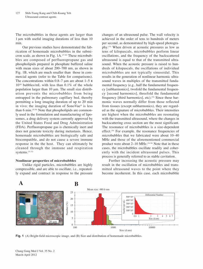
The microbubbles in these agents are larger than1 µm with useful imaging durations of less than 10min.
Our previous studies have demonstrated the fab-rication of homemade microbubbles in the submi-cron scale, as shown in Fig. 1A.(27,28) These microbub-bles are composed of perfluoropropane gas andphospholipids prepared in phosphate buffered salinewith mean sizes of about 200–700 nm, as shown inFig. 1B, which are much smaller than those in com-mercial agents (refer to the Table for comparisons).The concentrations within 0.8–7 µm are about 1–5 x10¹⁰ bubbles/mL with less than 0.1% of the wholepopulation larger than 10 µm. The small size distrib-ution prevents the microbubbles from beingentrapped in the pulmonary capillary bed, therebypermitting a long imaging duration of up to 20 minin vivo; the imaging duration of SonoVue® is lessthan 6 min.(29,30) Note that phospholipids are common-ly used in the formulation and manufacturing of lipo-somes, a drug delivery system currently approved bythe United States Food and Drug Administration(FDA). Perfluoropropane gas is chemically inert anddoes not generate toxicity during metastasis. Hence,homemade microbubbles are biologically safe andbiocompatible, and do not cause a severe immuneresponse in the the host. They can ultimately becleaned through the immune and respirationsystems.(31,32)
Nonlinear properties of microbubblesUnlike rigid particles, microbubbles are highly
compressible, and are able to oscillate, i.e., repeated-ly expand and contract in response to the pressure
changes of an ultrasound pulse. The wall velocity isachieved in the order of tens to hundreds of metersper second, as demonstrated by high-speed photogra-phy.(33) When driven at acoustic pressures as low astens of kilopascals, microbubbles perform linearoscillations, and the frequency of the backscatteredultrasound is equal to that of the transmitted ultra-sound. When the acoustic pressure is raised to hun-dreds of kilopascals, the oscillations of individualmicrobubbles are not typically sinusoidal. Thisresults in the generation of nonlinear harmonic ultra-sound waves in multiples of the transmitted funda-mental frequency (e.g., half the fundamental frequen-cy [subharmonics], twofold the fundamental frequen-cy [second harmonics], threefold the fundamentalfrequency [third harmonics], etc).(34) Since these har-monic waves normally differ from those reflectedfrom tissues (except subharmonics), they are regard-ed as the signature of microbubbles. Their intensitiesare highest when the microbubbles are resonatingwith the transmitted ultrasound, where the changes inbackscattering cross section are the most significant.The resonance of microbubbles is a size-dependenteffect.(34) For example, the resonance frequencies ofmicrobubbles that we fabricated were about 10–40MHz and those of the aforementioned commercialproduct were about 2–10 MHz.(34-36) Note that in thesecases, the microbubbles oscillate readily and coher-ently with the incident ultrasound pulses. Thisprocess is generally referred to as stable cavitation.
Further increasing the acoustic pressure mayresult in the oscillation of microbubbles and trans-mitted ultrasound waves to the point where theybecome incoherent. In this case, each microbubble
A B25
20
15
10
5
0
Num
ber
(%)
Num
ber (
per m
L)
500*106
400*106
300*106
200*106
100*106
00.8 1 2 3 4 5 6 7
Size (d.um)
Differential Number
100 1000 10000Size (d.um)
Mean size: 480.6 nm
20 µm
Fig. 1 (A) Bright-field microscopic image, and (B) Size and distribution of homemade microbubbles.

Chang Gung Med J Vol. 35 No. 2March-April 2012
Shih-Tsung Kang and Chih-Kuang YehUltrasound contrast agents
128
expands and contracts unstably for a few cycles andultimately collapses within a very short time. Whenthis occurs in a free field, shock waves can be gener-ated and give rise to microstreaming. When near awall, the collapse of each microbubble becomesasymmetrical, creating a liquid jet striking thewall.(37,38) Both phenomena are referred to as inertialcavitation. When exposed to ultrasound of appropri-ate frequency and sufficient pressure, microbubblesmay immediately be fragmented into small pieces inthe order of nanometers, and the encapsulated gasentirely dissolves in the surrounding medium.(39)
This process is referred to as the destruction of themicrobubbles, which may cause microstreaming orliquid jets at energy levels even higher than inertialcavitation.(40) Note that both inertial cavitation andmicrobubble destruction including stable cavitationcan cause mechanical stress to tissues in vivo.(41)
Many studies suggest the mechanical stress can alterthe permeability of the blood vessel and cell mem-brane to increase drug extravasation, which is intro-duced in later sections.
Linear contrast-enhanced ultrasound (CEUS)imaging
The strategies for contrast imaging are currentlybased on either the linear or nonlinear properties ofmicrobubbles. The linear scheme simply utilizes theecho enhancement of microbubbles. One example isfrom our pervious study of a colon cancer model in aBALB/c mouse. The imaging was conducted using ahomemade ultrasound imaging system with a single-element 25-MHz focused transducer.(42) After theadministration of microbubbles, the contrast oftumor tissue was clearly enhanced for up to 6 min, asshown in Fig. 2A–F. Owing to the entrances andexits of circulating microbubbles to the imagingplane, the regional brightness varied over a period oftime. These variations served as the signature of themicrobubbles, and they were extracted using a high-pass filter for interframe filtering. The obtainedinformation was demonstrated as a color-coded over-lay on a colocalized B-mode image, as shown in Fig.2G, which clearly showed the distribution of thetumor microcirculation. The color pixel values wereproportional to the microvascular blood flow volumeand velocity. Further, a destruction-replenishmenttechnique can be used to accurately assess the flowvelocity of the microcirculation. In this technique, a
destructive ultrasound pulse was first used todestroy most of the microbubbles in the region ofinterest. Blood flow velocity can be measured basedon the refill rate of microbubbles indicated by therecovery of contrast enhancement. The concept ofthis technique was first described by Wei et al. in1998 for use in echocardiology.(43) An adaptation hasbeen made by our group to improve the sensitivity ofthis technique for use in microperfusion.(44,45)
Nonlinear CEUS imaging (microbubble-specificcontrast imaging)
The performance of conventional CEUS imag-ing can deteriorate with low bubble concentration,tissue motion, and slow perfusion. Several studieshave shown the utilization of microbubble nonlinearemissions in microbubble-specific contrast imag-ing.(46,47) The second harmonics are not used becauseof interference from tissues at the same frequency.(48)
However, inducing the resonance of microbubblesrequires the application of sufficiently long ultra-sound pulses. Since the resonance frequencies ofcommercial agents are 2–10 MHz, imaging at a halfof these frequencies with long pulses can lead to lowspatial resolution. To overcome this limitation, aphase inversion technique that utilizes the sum of apair of images obtained from 2 inverted short ultra-sound pulses was proposed.(49) The oscillation ofmicrobubbles results in nonlinear distortion ofreflected echoes that cannot be clearly cancelled inpaired images, thereby leaving the signature ofmicrobubbles in the summed image. Nonetheless,this technique has to be operated at half the maxi-mum frame rate, and its performance may still besusceptible to movement. Our group has demonstrat-ed two techniques to perform nonlinear contrastimaging with improved spatial resolution. One isamplitude modulation chirp imaging, which utilizes2 different ultrasound pulses transmitted from sepa-rate ultrasound transducers.(50) A low-frequencypumping pulse is transmitted to induce resonance ofthe microbubbles. A high-frequency chirp pulse forimaging is then transmitted to simultaneously act onthe same group of microbubbles. Periodic changes inthe backscattering cross sections of the microbubblesin response to the pumping pulse can modulate theamplitude of the backscattered echoes of the chirppulse, producing modulated components in the fre-quency spectrum, as shown in Fig. 3A–D. After

Chang Gung Med J Vol. 35 No. 2March-April 2012
Shih-Tsung Kang and Chih-Kuang YehUltrasound contrast agents
129
extracting the derived signal components and apply-ing pulse compression, the contrast of microbubblescan be differentiated from that of tissues in a highspatial resolution. The performance was evaluated byimaging a mouse tail vein, as shown in Fig. 3E. Theother nonlinear imaging technique is a dual-frequen-cy (DF) chirp imaging, which utilizes a unique ultra-sound pulse that integrates DF difference excitationand chirp excitation techniques.(51) Our previous stud-ies have shown that the DF difference excitation effi-ciently induces a low-frequency driving force to res-onate microbubbles by using high-frequency ultra-sound.(52,53) High lateral resolution can be achieved byhigh-frequency carriers, and axial resolution can beimproved after applying pulse compression. Our pre-
vious study also showed that DF difference excita-tion can improve the ability of high-frequency ultra-sound to destroy microbubbles, which may be bene-ficial in the applications of commercial microbubblesin a high-frequency ultrasound system.(54,55) In thefuture, we aim to incorporate these 2 imaging strate-gies into clinical ultrasound systems.
Therapeutic usefulness of microbubble cavita-tion
The therapeutic usefulness of microbubbles hasgained much attention in recent years. Both inertialcavitation and destruction of microbubbles are capa-ble of producing strong mechanical stress to enhancethe permeability of the surrounding tissues and fur-
(A) Pre MBN (B) Post 1 min (C) Post 2 minD
epth
(m
m)
Dep
th (
mm
)
Dep
th (
mm
)
Dep
th (
mm
)
Dep
th (
mm
)
Dep
th (
mm
)
(D) Post 4 min (E) Post 5 min (F) Post 6 min
Width (mm) Width (mm) Width (mm)
Width (mm) Width (mm) Width (mm)
Dep
th (
mm
)
(G)
Width (mm)
Fig. 2 (A)–(F) B-mode imagesof a mouse colon tumor for 6 minafter the injection of homemademicrobubbles. (G) Contrast imageof circulating microbubblesobtained by a high-pass filter forinterframe filtering overlaid on acolocalized B-mode image.

Chang Gung Med J Vol. 35 No. 2March-April 2012
Shih-Tsung Kang and Chih-Kuang YehUltrasound contrast agents
130
ther increase the extravasation of drugs into the cyto-plasm or interstitium, as illustrated in Fig. 4A. Thismay involve several mechanisms. High-energymicrostreaming and liquid jets arising from the col-lapse of microbubbles can locally produce transientholes for direct passage of drugs.(41) They may alsocause a transient increase in temperature (reportedlyup to 5000 K) to alter the fluidity of the cell mem-brane.(56) Local deposition of such high energy mayresult in the production of free radicals, which proba-bly cause cell damage that enhances the permeabilityof endothelial cell layers.(57) In vivo applications havefocused, for instance, on disrupting the blood–brainbarrier (BBB), a layer of tightly-packed endothelialcells surrounding all capillaries in the brain.(58) Manygroups have reported the use of commercialmicrobubbles with low-frequency ultrasound (0.4–5MHz) to increase the permeability of the BBB,allowing the therapeutic or diagnostic agents to leakinto the affected regions.(59-61) The opening can betemporary and recoverable, and does not damage theneural cells.(62,63) However, microbubbles exposed tolow-frequency ultrasound have been shown to causerupture of microvessels with extravasation of redblood cells, even at a pressure under the FDA regula-tory limit for diagnostic ultrasound equipment.(64)
The clinical safety of BBB disruption with low-fre-quency ultrasound still carries great concern aboutthe risk of intracerebral hemorrhage.(65)
Given that concern, our group has developed ahigh-frequency-based technique (> 10 MHz) to dis-rupt the BBB by using stable cavitation of microbub-bles. It has been shown that stable cavitation mayalso mildly increase the tissue permeability byinduced acoustic streaming.(41) A noteworthy advancethat we have made in this technique was the use ofhomemade microbubbles that resonate at > 10 MHz.Sprague–Dawley rats were used in these experi-ments. The presence of BBB disruption was evaluat-ed by the extravasation of a model drug, Evans blue,into the brain tissue; the results are demonstrated inFig. 4B–D. Stable cavitation at a high frequencyenhanced by the resonance of small microbubbleswas able to produce effective BBB disruption, whichwas comparable to that with the low-frequency tech-nique but without any damage even at pressures ofup to 2.5 MPa. Interestingly, the amount of drugextravasation was found to highly correlate with theenhancement of subharmonic emission (i.e., the sig-nature of stable cavitation of microbubbles), asshown in Fig. 4E. Remarkable safety with the possi-bility of monitoring the extent of BBB disruption in
(A) (B)
(C) (D)
(E)
Fig. 3 Waveforms and frequency spectra of the backscattered echoes of chirp pulses, (A) and (B) before, and (C) and (D) afteramplitude modulation by a pumping pulse acting on the same group of microbubbles. (E) Contrast image of circulating microbub-bles in a mouse tail vein obtained by extracting the derived signal components and applying pulse compression.

Chang Gung Med J Vol. 35 No. 2March-April 2012
Shih-Tsung Kang and Chih-Kuang YehUltrasound contrast agents
131
real time suggests this technique has great promise inclinical use.
Drug-loaded microbubbles and ultrasound-con-trolled release
Microbubbles have been proposed as a newvehicle for carrying drugs and genes. Lipophilicchemotherapeutic drugs such as doxorubicin, pacli-taxel, and docetaxel can be incorporated into thelipid layer of microbubbles.(66-68) It has been shownthat the in vivo toxicity of paclitaxel-loadedmicrobubbles is about tenfold lower than that ofunencapsulated paclitaxel.(69) To increase the loadingcapacity, oil that dissolves lipophilic drugs can beintroduced into the microbubbles.(70-72) Drug-loadedparticles such as micelles or liposomes can be conju-gated to the surface of microbubbles usingligand–receptor interaction.(73) Genetic materials(e.g., plasmid DNA) can be electrostatically attachedto the surface of positively charged microbubblesthat bear cationic lipids.(74) Unlike liposomes, drug-loaded microbubbles are acoustically active and areable to exhibit stable or inertial cavitation in
response to ultrasound. The payload of drugs orDNA can be locally released by the destruction ofmicrobubbles within the ultrasound-treated region,with a simultaneous increase in the permeability ofthe tissues. This suggests the potential of microbub-ble technology in aiding drug or gene therapy, withreduced side effects to normal tissues.
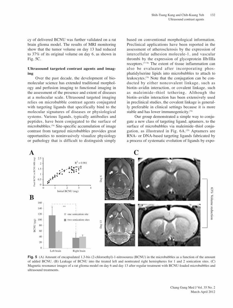
Our group has developed the loading ofmicrobubbles with 1,3-bis (2-chloroethyl)-1-nitrosourea (BCNU), a chemotherapeutic agent com-monly used in the treatment of brain tumors.(75) Theloading efficiency achieved was 75%, as shown inFig. 5A. Sprague–Dawley rats were used to test thedelivery of BCNU by these microbubbles into braintissues. The deposition of BCNU in the left hemi-spheres treated by ultrasound increased with thenumber of sonication sites, and was higher than thatin the right hemispheres which had no ultrasoundtreatment, as shown in Fig. 5B. This indicates thatthe destruction of BCNU-loaded microbubblesreleased the BCNU payload and simultaneouslyinduced the disruption of the BBB for passage ofBCNU into the brain tissues. The therapeutic effica-
(A) (B) (C)
(D) (E)
Fig. 4 (A) Illustration of vascular permeabi-lization by inertial cavitation of microbub-bles. Sections of rat brains treated by (B) 1-MHz ultrasound pulses with SonoVueTM and(C) 10-MHz ultrasound pulses with home-made microbubbles. (D) Leakage of Evansblue in the rat brain treated by the same 10-MHz ultrasound pulses but at differentacoustic pressures after the injection ofSonoVueTM (dash) and homemade microbub-bles (solid). (E) Leakage of Evans blue in therat brain as a function of the enhancement ofsubharmonic emission.
600
500
400
300
200
100
0
Am
ount
of
evan
s bl
ue (
ng)
160
140
120
100
80
60
40
20
0
Am
ount
of
evan
s bl
ue (
ng)
MBsSonoVue
160% R2 = 0.8455
0.5 1 1.5 2 2.5Acoustic pressure (MPa)
0 200 400 600 800Enhanced subharmonic
Emission (%)
Chang Gung Med J Vol. 35 No. 2March-April 2012
Shih-Tsung Kang and Chih-Kuang YehUltrasound contrast agents
132
cy of delivered BCNU was further validated on a ratbrain glioma model. The results of MRI monitoringshow that the tumor volume on day 13 had reducedto 37% of its original volume on day 6, as shown inFig. 5C.
Ultrasound targeted contrast agents and imag-ing
Over the past decade, the development of bio-molecular science has extended traditional morphol-ogy and perfusion imaging to functional imaging inthe assessment of the presence and extent of diseasesat a molecular scale. Ultrasound targeted imagingrelies on microbubble contrast agents conjugatedwith targeting ligands that specifically bind to themolecular signatures of diseases or physiologicalsystems. Various ligands, typically antibodies andpeptides, have been conjugated to the surface ofmicrobubbles.(76) Site-specific accumulation of imagecontrast from targeted microbubbles provides greatopportunities to noninvasively visualize physiologyor pathology that is difficult to distinguish simply
based on conventional morphological information.Preclinical applications have been reported in theassessment of atherosclerosis by the expression ofintercellular adhesion molecule-1, and vascularthrombi by the expression of glycoprotein IIb/IIIareceptors.(77,78) The extent of tissue inflammation canalso be evaluated after incorporating phos-phatidylserine lipids into microbubbles to attach toleukocytes.(79) Note that the conjugation can be con-ducted by either noncovalent linkage, such asbiotin–avidin interaction, or covalent linkage, suchas maleimide–thiol tethering. Although thebiotin–avidin interaction has been extensively usedin preclinical studies, the covalent linkage is general-ly preferable in clinical settings because it is morestable and has lower immunogenicity.(76)
Our group demonstrated a simple way to conju-gate a new class of targeting ligand, aptamers, to thesurface of microbubbles via maleimide–thiol conju-gation, as illustrated in Fig. 6A.(28) Aptamers areRNA- or DNA-based targeting ligands fabricated bya process of systematic evolution of ligands by expo-
Fina
l BC
NU
in B
CN
U-M
B (
mg)
BC
NU
(µ
g)
2.5
2
1.5
1
0.5
0
160
140
120
100
80
60
40
20
0
A
B
0 1 2 3 4Initial BCNU (mg)
R2 = 0.981
**
one sonication site
two sonication sites
Left brain Right brain
Day
13
Day
6
C
Fig. 5 (A) Amount of encapsulated 1,3-bis (2-chloroethyl)-1-nitrosourea (BCNU) in the microbubbles as a function of the amountof added BCNU. (B) Leakage of BCNU into the treated left and nontreated right hemispheres for 1 and 2 sonication sites. (C)Magnetic resonance images of a rat glioma model on day 6 and day 13 after regular treatment with BCNU-loaded microbubbles andultrasound treatments.
63% V
olume reduction

Chang Gung Med J Vol. 35 No. 2March-April 2012
Shih-Tsung Kang and Chih-Kuang YehUltrasound contrast agents
133
nential enrichment.(80) They are biologically stable,less immunogenic, and have low manufacturingcosts, but have comparable or even greater bindingaffinity and specificity than protein-based antibodies.The methods that we proposed are capable of com-pleting the conjugation prior to the formation ofmicrobubbles. Fewer steps will be required in clini-cal settings. The sgc8c aptamer (50-thiol-ATC TAACTG CTG CGC CGC CGG GAA AAT ACT GTACGG TTA GA) was used for the specific targeting ofCCRF-CEM cells (CCL-119 T-cell, human acutelymphoblastic leukemia). A flexible polyethyleneglycol spacer was designed between each aptamerand the surface of the microbubble to maximizeinteraction between each aptamer and receptors. Thein vitro results show that the aptamer-conjugatedmicrobubbles firmly bound to the surface of theCEM cells, and provided a great echo enhancementof up to 40 dB in the ultrasound image in comparisonwith the control, as shown in Fig. 6B–E.
In another in vivo study, we conjugated vascularendothelial growth factor receptor 2 (VEGFR2) anti-bodies onto microbubbles to assess tumor angiogene-sis. After administration into a mouse with a colontumor, the microbubbles gradually accumulated inthe tumor site during the wash-in and wash-out phas-
es of circulating microbubbles, as illustrated in Fig.7A. After the clearance of the circulating microbub-bles, the remaining contrast enhancement couldmostly be attributed to the adherent microbubbles.The extent of tumor angiogenesis correlating to theexpression of VEGFR2 was visualized by subtract-ing the images before and after microbubble accumu-lation, as shown in Fig. 7B. For comparison, noaccumulation was found for normal microbubbles inthe control test, as shown in Fig. 7C. The immunos-tained section of the tumor for DAPI (blue) andCD31 (red) in Fig. 7D and E, respectively, provedthat the accumulation of microbubbles (green) wasnear the tumor boundary where angiogenesis wassignificant.
Challenges and limitationsAs described earlier, microbubbles are able to
carry drugs or genes, and locally release the payloadby applying ultrasound in the intended regions. Thetargeting ability of bioconjugated microbubblesopens the door for targeted therapy. Combined withthe aforementioned microbubble technology,microbubbles can serve as a multifunction platformthat is not only useful in diagnoses but also in thera-pies, permitting simultaneous imaging and localiza-
A B C
D E
20 µm
20 µm
Fig. 6 (A) Scheme of an aptamer-conjugated microbubble binding to a CEM cell. (D) Bright-field microscopic images and ultra-sound C-mode scanning images of CEM cells treated by (B) and (C) the aptamer-conjugated microbubbles, and (D) and (E) normalmicrobubbles.

Chang Gung Med J Vol. 35 No. 2March-April 2012
Shih-Tsung Kang and Chih-Kuang YehUltrasound contrast agents
134
tion of the therapy. However, there are still somechallenges in targeted and drug-loaded microbubblesthat must be overcome before they are approved forclinical use. The adhesion efficiency of targetedmicrobubbles might be low under certain physiologi-cal flow conditions.(81) Although the microbubblesfabricated in our studies can exist in vivo for up to 20min, some situations may require them to remain invivo for several hours or more than one day to attainan accumulated contrast or therapeutic effect. Thechallenges may be overcome by acoustic radiationforce, which has been demonstrated to accelerate theadhesion and accumulation of targetedmicrobubbles.(82) The mechanism is to drive thesemicrobubbles toward the vessel wall by using low-energy ultrasound without causing microbubble dis-ruption. Another method is the use of polymer-shelled microbubbles, which are acoustically activein vivo for several hours.(83)
Our group is currently developing the precursorsof microbubbles–phase-change droplets–as ultra-sound contrast agents. These droplets are originallyin the liquid phase but can be vaporized into gaseousmicrobubbles upon ultrasound excitation.(84) Dropletsare much more stable in vivo for more than 1 day,and carry a larger drug payload than conventional
microbubbles.(85) Nanoscale agents are much easier tofabricate in the liquid phase than in the gaseousphase. The small size might allow the droplets toextravasate from the blood vessel to interact withreceptors that may be present within tissues, and mayenable passive targeting in tumors via an enhancedpermeability and retention effect.(86) Research isunderway to study whether the microbubbles derivedfrom phase-change droplets can also be applied inthe aforementioned microbubble technology.
ConclusionsIn this article, we reviewed current develop-
ments in microbubble technology, and efforts by ourlaboratory in emerging diagnostic and therapeuticapplications of microbubbles. Microbubbles wereoriginally designed as UCAs to improve the sensitiv-ity of conventional ultrasound imaging. The unique-ness of microbubbles, including bioconjugation, drugencapsulation, cavitation, and nonlinear emission,allow them to be used simultaneously as diagnosticand therapeutic agents. Applications includemicrobubble-specific imaging, ultrasound-aided drugdelivery, and targeted imaging and therapy. Ourgroup has developed two techniques of microbubble-specific imaging with high spatial resolution. Our
A B C
D E
Fig. 7 (A) Scheme of the concentration of adherent and circulating microbubbles during the wash-in and wash-out phases of vas-cular endothelial growth factor receptor 2-targeted microbubbles. (B) Contrast image obtained by subtracting images before andafter microbubble accumulation and overlaid on a colocalized B-mode image. (C) Control of (B) using normal microbubbles. (D),(E) Fluorescence microscopic images of an immunostained tumor section for DAPI (blue), CD31 (red), and adherent microbubbles(green).

Chang Gung Med J Vol. 35 No. 2March-April 2012
Shih-Tsung Kang and Chih-Kuang YehUltrasound contrast agents
135
group has also developed lipid-based microbubbleswith a drug payload (e.g., BCNU) and targeting lig-ands (e.g., VEGFR2 antibodies). In vivo studiesshowed the visualization of tumor angiogenesis in amouse colon tumor by VEGFR2-targeted microbub-bles, and the therapeutic effect of ultrasound-aidedBCNU delivery to a rat brain tumor. Stable cavita-tion at high frequency enhanced by the resonance ofsmall microbubbles seems to be a very safe modalityfor enhancing tissue permeability. These applicationsare promising but require further improvement forclinical use.
AcknowledgementsThe authors acknowledge the National Science
Council of Taiwan for grant support (NSC 98-2320-B-007-002-MY3 and 99-2218-E-182-002), and ourlaboratory staff, Jia-Jiun Chen, Chung-Hsin Wang,Ching-Hsiang Fan, Ting-Yu Huang, and Chien-YuTing, for their efforts in the experiments and prepara-tion of the figures.
REFERENCES
1. Jang IK, Bouma BE, Kang DH, Park SJ, Park SW, SeungKB, Choi KB, Shishkov M, Schlendorf K, PomerantsevE, Houser SL, Aretz HT, Tearney GJ. Visualization ofcoronary atherosclerotic plaques in patients using opticalcoherence tomography: comparison with intravascularultrasound. J Am Coll Cardiol 2002;39:604-9.
2. Joyner CR, Reid JM. Applications of ultrasound in cardi-ology and cardiovascular physiology. Prog CardiovascDis 1963;5:482-97.
3. Chen JJ, Chen JJ, Chiang CS, Hong JH, Yeh CK.Assessment of tumor vasculature for diagnostic and thera-peutic applications in a mouse model in vivo using 25-MHz power Doppler imaging. Ultrasonics 2011;51:925-31.
4. Yeh CK, Chen JJ, Li ML, Luh JJ, Chen JJ. In vivo imag-ing of blood flow in the mouse Achilles tendon usinghigh-frequency ultrasound. Ultrasonics 2009;49:226-30.
5. Liao YY, Tsui PH, Li CH, Chang KJ, Kuo WH, ChangCC, Yeh CK. Classification of scattering media withinbenign and malignant breast tumors based on ultrasoundtexture-feature-based and Nakagami-parameter images.Med Phys 2011;38:2198-207.
6. Schor AM, Schor SL. Tumour angiogenesis. J Pathol1983;141:385-413.
7. Folkman J. New perspectives in clinical oncology fromangiogenesis research. Eur J Cancer 1996;32A:2534-9.
8. Kerbel RS. Tumor angiogenesis: past, present and the
near future. Carcinogenesis 2000;21:505-15.9. Browder T, Butterfield CE, Kräling BM, Shi B, Marshall
B, O’Reilly MS, Folkman J. Antiangiogenic scheduling ofchemotherapy improves efficacy against experimentaldrug-resistant cancer. Cancer Res 2000;60:1878-86.
10. Gramiak R, Shah PM. Echocardiography of the aorticroot. Invest Radiol 1968;3:356-66.
11. Quaia E. Microbubble ultrasound contrast agents: anupdate. Eur Radiol 2007;17:1995-2008.
12. Dayton PA, Ferrara KW. Targeted imaging using ultra-sound. J Magn Reson Imaging 2002;16:362-77.
13. Foster FS, Burns PN, Simpson DH, Wilson SR,Christopher DA, Goertz DE. Ultrasound for the visualiza-tion and quantification of tumor microcirculation. CancerMetastasis Rev 2000;19:131-8.
14. Cohen JL, Cheirif J, Segar DS, Gillam LD, Gottdiener JS,Hausnerova E, Bruns DE. Improved left ventricular endo-cardial border delineation and opacification with OPTI-SON (FS069), a new echocardiographic contrast agent. JAm Coll Cardiol 1998;32:746-52.
15. Keller MW, Feinstein SB, Watson DD. Successful leftventricular opacification following peripheral venousinjection of sonicated contrast agent: An experimentalevaluation. Am Heart J 1987;114:570-5.
16. Ries F, Honisch C, Lambertz M, Schlief R. A transpul-monary contrast medium enhances the transcranialDoppler signal in humans. Stroke 1993;24:1903-9.
17. Harvey CJ, Blomley MJ, Eckersley RJ, Cosgrove DO,Patel N, Heckemann RA, Butler-Barnes J. Hepatic malig-nancies: improved detection with pulse-inversion US inlate phase of enhancement with SH U 508A-early experi-ence. Radiology 2000;216:903-8.
18. Hauff P, Fritzsch T, Reinhardt M, Weitschies W, Lüders F,Uhlendorf V, Heldmann D. Delineation of experimentalliver tumors in rabbits by a new ultrasound contrast agentand stimulated acoustic emission. Invest Radiol 1997;32:94-9.
19. Quaia E, Calliada F, Bertolotto M, Rossi S, Garioni L,Rosa L, Pozzi-Mucelli R. Characterization of focal liverlesions with contrast-specific US modes and a sulfurhexafluoride-filled microbubble contrast agent: diagnosticperformance and confidence. Radiology 2004;232:420-30.
20. Arnold JR, Karamitsos TD, Pegg TJ, Francis JM,Olszewski R, Searle N, Senior R, Neubauer S, Becher H,Selvanayagam JB. Adenosine stress myocardial contrastechocardiography for the detection of coronary artery dis-ease: a comparison with coronary angiography and car-diac magnetic resonance. JACC-Cardiovasc Imag2010;3:934-43.
21. Correas JM, Hélénon O, Moreau JF. Contrast-enhancedultrasonography of native and transplanted kidney dis-eases. Eur Radiol 1999;9:S394-400.
22. Eiberg JP, Hansen MA, Jensen F, Rasmussen JB,Schroeder TV. Ultrasound contrast-agent improves imag-
Chang Gung Med J Vol. 35 No. 2March-April 2012
Shih-Tsung Kang and Chih-Kuang YehUltrasound contrast agents
136
ing of lower limb occlusive disease. Eur J Vasc EndovascSurg 2003;25:23-8.
23. Hancock J, Dittrich H, Jewitt DE, Monaghan MJ.Evaluation of myocardial, hepatic, and renal perfusion ina variety of clinical conditions using an intravenous ultra-sound contrast agent (Optison) and second harmonicimaging. Heart 1999;81:636-41.
24. SonoVue: EPAR - Product Information. Available fromhttp://www.ema.europa.eu/docs/en_GB/document_library/EPAR_-_Product_Information/human/000303/WC500055380.pdf.Accessed September 2011.
25. Luminity: EPAR - Product Information. Available fromhttp://www.ema.europa.eu/docs/en_GB/document_library/EPAR_-_Product_Information/human/000654/WC500045020.pdf.Accessed September 2011.
26. Optison: EPAR - Product Information. Available fromhttp://www.ema.europa.eu/docs/en_GB/document_library/EPAR_-_Product_Information/human/000166/WC500059461.pdf.Accessed September 2011.
27. Kang ST, Yeh CK. A maleimide-based in-vitro model forultrasound targeted imaging. Ultrason Sonochem2011;18:327-33.
28. Wang CH, Huang YF, Yeh CK. Aptamer-conjugatednanobubbles for targeted ultrasound molecular imaging.Langmuir 2011;27:6971-6.
29. Meltzer RS, Glen Tickner E, Popp RL. Why do the lungsclear ultrasonic contrast? Ultrasound Med Biol1980;6:263-9.
30. Schneider M. Characteristics of SonoVueTM.Echocardiography 1999;16:743-6.
31. Klibanov AL. Targeted delivery of gas-filled micros-pheres, contrast agents for ultrasound imaging. Adv DrugDeliv Rev 1999;37:139-57.
32. Yanagisawa K, Moriyasu F, Miyahara T, Yuki M, IijimaH. Phagocytosis of ultrasound contrast agent microbub-bles by Kupffer cells. Ultrasound Med Biol 2007;33:318-25.
33. Morgan KE, Allen JS, Dayton PA, Chomas JE, KlibaovAL, Ferrara KW. Experimental and theoretical evaluationof microbubble behavior: effect of transmitted phase andbubble size. IEEE Trans Ultrason Ferroelectr Freq Control2000;47:1494-509.
34. Shi WT, Forsberg F. Ultrasonic characterization of thenonlinear properties of contrast microbubbles. UltrasoundMed Biol 2000;26:93-104.
35. Goertz DE, de Jong N, van der Steen AFW. Attenuationand size distribution measurements of Definity andmanipulated Definity populations. Ultrasound Med Biol2007;33:1376-88.
36. van der Meer SM, Versluis M, Lohse D, Chin CT,Bouakaz A, de Jong N. The resonance frequency ofsonovue as observed by high-speed optical imaging.Proceedings of IEEE Ultrasonics Symposium 2004;1:343-5.
37. Lauterborn W, Hentschel W. Cavitation bubble dynamics
studied by high speed photography and holography: partone. Ultrasonics 1985;23:260-8.
38. Lauterborn W, Hentschel W. Cavitation bubble dynamicsstudied by high speed photography and holography: parttwo. Ultrasonics 1986;24:59-65.
39. Chomas JE, Dayton PA, May D, Allen J, Klibanov A,Ferrara K. Optical observation of contrast agent destruc-tion. Appl Phys Lett 2000;77:1056.
40. Dijkmans PA, Juffermans LJM, Musters RJP, van WamelA, ten Cate FJ, van Gilst W, Visser CA, de Jong N, KampO. Microbubbles and ultrasound: from diagnosis to thera-py. Eur J Echocardiogr 2004;5:245-56.
41. Husseini GA, Diaz de la Rosa MA, Richardson ES,Christensen DA, Pitt WG. The role of cavitation inacoustically activated drug delivery. J Control Release2005;107:253-61.
42. Yeh CK, Chunh CH, Chen JJ, Chen JJ. High-frequencyultrasonic imaging system: a Preliminary small animalsstudy system. Int J Elec Eng 2008;15:195-202.
43. Wei K, Jayaweera AR, Firoozan S, Linka A, Skyba DM,Kaul S. Quantification of myocardial blood flow withultrasound-induced destruction of microbubbles adminis-tered as a constant venous infusion. Circulation1998;97:473-83.
44. Yeh CK, Ferrara KW, Kruse DE. High-resolution func-tional vascular assessment with ultrasound. IEEE TransUltrason Ferroelectr Freq Control 2004;23:1263-75.
45. Yeh CK, Lu SY, Chen YS. Microcirculation volumetricflow assessment using high-resolution, contrast-assistedimages. IEEE Trans Ultrason Ferroelectr Freq Control2008;55:74-83.
46. Forsberg F, Shi WT, Goldberg BB. Subharmonic imagingof contrast agents. Ultrasonics 2000;38:93-8.
47. Bouakaz A, Frigstad S, Ten Cate FJ, de Jong N. Superharmonic imaging: a new imaging technique for improvedcontrast detection. Ultrasound Med Biol 2002;28:59-68.
48. Ziegler L, O’Brien RT. Harmonic ultrasound: a review.Vet Radiol Ultrasound 2002;43:501-9.
49. Burns PN, Wilson SR, Simpson DH. Pulse inversionimaging of liver blood flow: improved method for charac-terizing focal masses with microbubble contrast. InvestRadiol 2000;35:58-71.
50. Li ML, Kuo YC, Yeh CK. Amplitude-modulation chirpimaging for contrast detection. Ultrasound Med Biol2010;36:1535-45.
51. Cheng CH, Shen CC, Yeh CK. Dual-frequency chirpimaging for contrast detection. Phys Med Biol 2011;56:2767-78.
52. Yeh CK, Su SY, Shen CC, Li ML. Dual high-frequencydifference excitation for contrast detection. IEEE TransUltrason Ferroelectr Freq Control 2008;55:2164-76.
53. Shen CC, Cheng CH, Yeh CK. Phase-dependent dual-fre-quency contrast imaging at sub-harmonic frequency. IEEETrans Ultrason Ferroelectr Freq Control 2011;58:379-88.
54. Shen CC, Su SY, Cheng CH, Yeh CK. Dual-high-frequen-

Chang Gung Med J Vol. 35 No. 2March-April 2012
Shih-Tsung Kang and Chih-Kuang YehUltrasound contrast agents
137
cy ultrasound excitation on microbubble destruction vol-ume. Ultrasonics 2010;50:698-703.
55. Yeh CK, Su SY, Shen CC. Microbubble destruction bydual-high-frequency ultrasound excitation. IEEE TransUltrason Ferroelectr Freq Control 2009;56:1113-8.
56. Holland CK, Apfel RE. An improved theory for the pre-diction of microcavitation thresholds. IEEE TransUltrason Ferroelectr Freq Control 1989;36:204-8.
57. Basta G, Venneri L, Lazzerini G, Pasanisi E, Pianelli M,Vesentini N, Del Turco S, Kusmic C, Picano E. In vitromodulation of intracellular oxidative stress of endothelialcells by diagnostic cardiac ultrasound. Cardiovasc Res2003;58:156-61.
58. Rubin LL, Staddon JM. The cell biology of the blood-brain barrier. Annu Rev Neurosci 1999;22:11-28.
59. Kinoshita M, McDannold N, Jolesz FA, Hynynen K.Noninvasive localized delivery of Herceptin to the mousebrain by MRI-guided focused ultrasound-induced blood-brain barrier disruption. Proc Natl Acad Sci USA2006;103:11719-23.
60. Liu HL, Hua MY, Chen PY, Chu PC, Pan CH, Yang HW,Huang CY, Wang JJ, Yen TC, Wei KC. Blood-brain barri-er disruption with focused ultrasound enhances deliveryof chemotherapeutic drugs for glioblastoma treatment.Radiology 2010;255:415-25.
61. McDannold N, Vykhodtseva N, Raymond S, Jolesz FA,Hynynen K. MRI-guided targeted blood-brain barrier dis-ruption with focused ultrasound: Histological findings inrabbits. Ultrasound Med Biol 2005;31:1527-37.
62. Hynynen K, McDannold N, Vykhodtseva N, Jolesz FA.Noninvasive MR imaging-guided focal opening of theblood-brain barrier in rabbits. Radiology 2001;220:640-6.
63. Hynynen K, McDannold N, Vykhodtseva N, Jolesz FA.Non-invasive opening of BBB by focused ultrasound.Acta Neurochir Suppl 2003;86:555-8.
64. Skyba DM, Price RJ, Linka AZ, Skalak TC, Kaul S.Direct in vivo visualization of intravascular destruction ofmicrobubbles by ultrasound and its local effects on tissue.Circulation 1998;98:290-3.
65. Reinhard M, Hetzel A, Krüger S, Kretzer S, Talazko J,Ziyeh S, Weber J, Els T. Blood-brain barrier disruption bylow-frequency ultrasound. Stroke 2006;37:1546-8.
66. Cochran MC, Eisenbrey J, Ouma RO, Soulen M,Wheatley MA. Doxorubicin and paclitaxel loadedmicrobubbles for ultrasound triggered drug delivery. Int JPharm 2011;414:161-70.
67. Tinkov S, Coester C, Serba S, Geis NA, Katus HA,Winter G, Bekeredjian R. New doxorubicin-loaded phos-pholipid microbubbles for targeted tumor therapy: in-vivocharacterization. J Control Release 2010;148:368-72.
68. Kang J, Wu X, Wang Z, Ran H, Xu C, Wu J, Wang Z,Zhang Y. Antitumor effect of docetaxel-loaded lipidmicrobubbles combined with ultrasound-targetedmicrobubble activation on VX2 rabbit liver tumors. JUltrasound Med 2010;29:61-70.
69. Tartis MS, McCallan J, Lum AFH, LaBell R, Stieger SM,Matsunaga TO, Ferrara KW. Therapeutic effects of pacli-taxel-containing ultrasound contrast agents. UltrasoundMed Biol 2006;32:1771-80.
70. Fang JY, Hung CF, Liao MH, Chien CC. A study of theformulation design of acoustically active lipospheres ascarriers for drug delivery. Eur J Pharm Biopharm2007;67:67-75.
71. Lentacker I, Geers B, Demeester J, De Smedt SC, SandersNN. Design and evaluation of doxorubicin-containingmicrobubbles for ultrasound-triggered doxorubicin deliv-ery: cytotoxicity and mechanisms involved. Mol Ther2010;18:101-8.
72. Shortencarier MJ, Dayton PA, Bloch SH, Schumann PA,Matsunaga TO, Ferrara KW. A method for radiation-forcelocalized drug delivery using gas-filled lipospheres. IEEETrans Ultrason Ferroelectr Freq Control 2004;51:822-31.
73. Lentacker I, De Smedt SC, Demeester J, Van Marck V,Bracke M, Sanders NN. Lipoplex-loaded microbubblesfor gene delivery: a trojan horse controlled by ultrasound.Adv Funct Mater 2007;17:1910-6.
74. Chen S, Ding JH, Bekeredjian R, Yang BZ, Shohet RV,Johnston SA, Hohmeier HE, Newgard CB, Grayburn PA.Efficient gene delivery to pancreatic islets with ultrasonicmicrobubble destruction technology. Proc Natl Acad SciUSA 2006;103:8469-74.
75. Barker M, Hoshino T, Gurcay O, Wilson CB, Nielsen SL,Downie R, Eliason J. Development of an animal braintumor model and its response to therapy with 1,3-bis(2-chloroethyl)-1-nitrosourea. Cancer Res 1973;33:976-86.
76. Lanza GM, Wickline SA. Targeted ultrasonic contrastagents for molecular imaging and therapy. Curr ProblCardiol 2003;28:625-53.
77. Schumann PA, Christiansen JP, Quigley RM, McCreeryTP, Sweitzer RH, Unger EC, Lindner JR, Matsunaga TO.Targeted-microbubble binding selectively to GPIIb IIIareceptors of platelet thrombi. Invest Radiol 2002;37:587-93.
78. Villanueva FS, Jankowski RJ, Klibanov S, Pina ML,Alber SM, Watkins SC, Brandenburger GH, Wagner WR.Microbubbles targeted to intercellular adhesion molecule-1 bind to activated coronary artery endothelial cells.Circulation 1998;98:1-5.
79. Lindner JR, Song J, Xu F, Klibanov AL, Singbartl K, LeyK, Kaul S. Noninvasive ultrasound imaging of inflamma-tion using microbubbles targeted to activated leukocytes.Circulation 2000;102:2745-50.
80. Nimjee SM, Rusconi CP, Sullenger BA. Aptamers: anemerging class of therapeutics. Annu Rev Med 2005;56:555-83.
81. Takalkar AM, Klibanov AL, Rychak JJ, Lindner JR, LeyK. Binding and detachment dynamics of microbubblestargeted to P-selectin under controlled shear flow. JControl Release 2004;96:473-82.
82. Dayton P, Klibanov A, Brandenburger G, Ferrara K.

Chang Gung Med J Vol. 35 No. 2March-April 2012
Shih-Tsung Kang and Chih-Kuang YehUltrasound contrast agents
138
Acoustic radiation force in vivo: a mechanism to assisttargeting of microbubbles. Ultrasound Med Biol1999;25:1195-201.
83. Lentacker I, De Geest BG, Vandenbroucke RE, Peeters L,Demeester J, De Smedt SC, Sanders NN. Ultrasound-responsive polymer-coated microbubbles that bind andprotect DNA. Langmuir 2006;22:7273-8.
84. Kripfgans OD, Fowlkes JB, Miller DL, Eldevik OP,Carson PL. Acoustic droplet vaporization for therapeuticand diagnostic applications. Ultrasound Med Biol
2000;26:1177-89.85. Dayton PA, Zhao S, Bloch SH, Schumann P, Penrose K,
Matsunaga TO, Zutshi R, Doinikov A, Ferrara KW.Application of ultrasound to selectively localize nan-odroplets for targeted imaging and therapy. Mol Imaging2006;5:160-74.
86. Hashizume H, Baluk P, Morikawa S, McLean JW,Thurston G, Roberge S, Jain RK, McDonald DM.Openings between defective endothelial cells explaintumor vessel leakiness. Am J Pathol 2000;156:1363-80.

139
(ultrasound contrast agents) (microbubbles)
(microbubble-specific imaging)(inertial cavitation)
(stable cavitation)
(ultrasound-aided drug delivery) (targeted imaging) (
2012;35:125-39)
100 9 8 100 11 3030013 101
Tel: (03)5715131 34240; Fax: (03)5718649; E-mail: [email protected]