ultrasound responsive macrophase-segregated microcomposite films...
TRANSCRIPT

Ultrasound Responsive Macrophase-Segregated MicrocompositeFilms for in Vivo BiosensingJian Yang,† James Wang,‡ Casey N. Ta,§ Erin Ward,∥ Christopher V. Barback,⊥ Tsai-Wen Sung,‡
Natalie Mendez,# Sarah L. Blair,*,∥ Andrew C. Kummel,*,† and William C. Trogler*,†
†Department of Chemistry and Biochemistry, ‡Department of Nanoengineering, §Department of Computer, ∥Department of Surgery,⊥Department of Radiology, and #Materials Science and Engineering Program, University of California, San Diego, 9500 GilmanDrive, La Jolla, California 92093, United States
*S Supporting Information
ABSTRACT: Ultrasound imaging is a safe, low-cost, and in situmethod for detecting in vivo medical devices. A poly(methyl-2-cyanoacrylate) film containing 2 μm boron-doped, calcined,porous silica microshells was developed as an ultrasound imagingmarker for multiple medical devices. A macrophase separationdrove the gas-filled porous silica microshells to the top surface ofthe polymer film by controlled curing of the cyanoacrylate glueand the amount of microshell loading. A thin film of polymerblocked the wall pores of the microshells to seal air in their hollowcore, which served as an ultrasound contrast agent. The ultrasoundactivity disappeared when curing conditions were modified toprevent the macrophase segregation. Phase segregated films wereattached to multiple surgical tools and needles and gave strongcolor Doppler signals in vitro and in vivo with the use of a clinicalultrasound imaging instrument. Postprocessing of the simultaneous color Doppler and B-mode images can be used forautonomous identification of implanted surgical items by correlating the two images. The thin films were also hydrophobic,thereby extending the lifetime of ultrasound signals to hours of imaging in tissues by preventing liquid penetration. Thistechnology can be used as a coating to guide the placement of implantable medical devices or used to image and help removeretained surgical items.
KEYWORDS: silica microshells, cyanoacrylate, macrophase segregated film, ultrasound imaging, in vivo medical devices
■ INTRODUCTION
Biomedical devices such as needles, catheters, biopsy markers,and guidewires are used widely in the health care field.1−3
Biomedical devices are often composed of materials such asstainless steel, titanium, silicon, and polymers.4 These devicesmay be implanted within complex physiological environmentssuch as the abdominal cavity, gastrointestinal lumen, or thecardiovascular system. At present, physicians rely on medicalimaging modalities, such as ultrasound, X-ray, and CT scans, tomonitor or detect implanted biomedical tools during and after aprocedure. Many procedures require precision; for example, tosuccessfully achieve a nerve block, a physician must accuratelyinject local anesthesia to the target nerve bundles whileavoiding surrounding blood vessels and other tissue structures.5
To minimize the chance of unintentionally damagingsurrounding nerves, blood vessels, and tissues while achievingthe goal of attaining a nerve block, the needle entry and paththrough the tissue must be precise. Successful injection of thetarget area depends greatly on the experience of the medicalprofessional.6 In order to improve the injection accuracy,ultrasound (US) guided needle injection technology wasdeveloped and is now widely used in a variety of medical and
surgical procedures. Ultrasound guidance is used for needlebiopsies and epidurals and to allow physicians to safely placecentral venous and arterial catheters. While conventional B-mode ultrasound imaging helps visualize a needle during itsapproach toward the target tissue,7 B-mode ultrasound hasseveral limitations. For instance, B-mode ultrasound exhibitsinterfering signals due to scattering by various tissues orimplants.8 In light of such limitations, it was postulated that itmay be beneficial to develop an ultrasound based platformbased on high contrast ultrasound imaging modes, such as colorDoppler or contrast-enhanced ultrasound, and apply it topositioning and monitoring implantable biomedical devices.Detection of retained surgical items (RSI) in the operating
room is another challenge for biomedical imaging technology.Small surgical items such as surgical needles, forceps, sutures,and blades can be accidentally left in patients’ bodies after anoperation. This may cause adverse consequences such as organdamage, bowel perforation, severe pain, sepsis, and even death.
Received: August 26, 2016Accepted: December 21, 2016Published: December 21, 2016
Research Article
www.acsami.org
© 2016 American Chemical Society 1719 DOI: 10.1021/acsami.6b10728ACS Appl. Mater. Interfaces 2017, 9, 1719−1727
It has been estimated that up to 2000 cases of RSI occur in theUnited States each year.9 A routine surgical tool count is themost commonly used method to prevent RSI from occurring.10
Radio-frequency (RF) tags attached to large surgical items, suchas sponges and gauges, can be detected by a RF reader and helpavoid RSI.7,8 Nevertheless, a RF tag cannot be attached to smallsurgical items such as needles. X-ray imaging has been acommonly used method to detect metal in tissues; however, anX-ray technician must be present during surgery, and aradiologist is required to review the film. Macilquham et al.used X-ray imaging to identify lost surgical needles and foundthat needles less than 20 mm in length were difficult toidentify.11 Additionally, X-ray imaging exposes the patient andmedical personnel to radiation and requires the patient toremain under anesthesia while the X-ray is completed and readby a radiologist.12 Currently, there is no viable universalplatform for real-time detection of various RSIs of differentsizes and materials during a surgical operation.A method was developed previously to synthesize calcined
porous silica nano- and microshells with a sol−gel reactionusing polystyrene beads as templates and tetramethylorthosilicate (TMOS) as the silica precursor.13 The techniquehas been employed to synthesize 500 nm iron-doped silicashells and 2 μm boron-doped silica shells encapsulatingperfluoropentane (PFP) as ultrasound contrast agents for anin vivo stationary tumor marker.14−17 Ultrasound tests showedthat PFP-filled silica nanoshells have superior performance tosoft microbubbles for imaging longevity (several weeks) bycolor Doppler imaging.In the present study, poly(methyl 2-cyanoacrylate) (PMCA)
thin films containing 2 μm boron-doped silica shells werefabricated. Commercial ultrasound sound imaging systemsdepict simple differences in acoustic index in B-mode, but theyhave additional nonlinear imaging modes such as contrast pulsesequencing (CPS) and color Doppler. In color Doppler, onlyreflected signals shifted in frequency from the incidentultrasound frequency are detected; this signal is sensitive tomotion in tissue such as blood flow and expanding gas. Whengas filled, the shells can be activated by widely used clinicalultrasound equipment to exhibit strong color Doppler signals.When the 2 μm particles are fractured during cavitation, theresulting increase in isotropic velocities and pressure results indetected Doppler frequency shifts in all directions. Because ofthe shell fragmentation, a mosaic Doppler signal is generated.The mosaic signal pattern reflects a fluid with a heterogeneousmixture of sound sources moving in different directions, due todifferent subpopulations of 2 μm particles being fragmented.
These ultrasound active films can be used to coat a variety ofsurgical tools. Synthetic cyanoacrylates have been traditionallyused as biodegradable tissue adhesives for efficient woundclosure or sealing vascular sutures.18 Initiated by surfacehydroxyl groups, cyanoacrylate monomers undergo anionicpolymerization rapidly in air at room temperature, and thepolymerization is catalyzed by moisture.19 Furthermore,cyanoacrylate polymers have low toxicity and a low rate ofinfection.20 Fast-curing poly(methyl 2-cyanoacrylate) can bindto a variety of materials such as metal, plastic, and glass.Polymerization of cyanoacrylate films was used to encapsulatesilica microshells while sealing ambient air within the hollowspace for ultrasound active coating properties.Cyanoacrylate monomer containing solvent and porous silica
microshells were coated on glass slides and surgical needles bydip coating to provide ultrasound active thin films. Umbilicaltapes and surgical clips were also coated with PMCA/microshells films for in vivo ultrasound testing. After curing,thin PMCA films were formed with a thickness ranging from 5to 100 μm. The thickness can be controlled by the coatingmethod. The PMCA film encapsulated the microshells whilesimultaneously adhering the composite material to the surfaceof glass slides or needles. The polymerization of methyl 2-cyanoacrylate monomer was initiated by the adsorbed water orthe hydroxyl groups on the surface of silica. In vitro and in vivoultrasound tests showed that the PMCA/microshells filmsproduced a strong color Doppler signal with a good persistence(>6 h), as required for potential applications to in vivo surgicaltool detection. The strong color Doppler signal is associatedwith a macrophase separation during film curing to form asurface layer consisting primarily of embedded microshells withcyanoacrylate acting as an adhesive matrix; however, the base ofthe film consists primarily of cyanoacrylate polymer tightlybound to the glass or metal substrate (Scheme 1). It is thoughtthat polarity differences between the polymer/solvent phaseand the dispersed silica phase cause segregation of the silicashells to the surface layer during film polymerization andsolvent evaporation at room temperature.
■ EXPERIMENTAL SECTIONMaterials. Tetramethyl orthosilicate (TMOS), N1-(3-trimethoxy-
silylpropyl)diethylenetriamine (DETA), and trimethyl borate (TMB)were purchased from Sigma-Aldrich (St. Louis, MO). The 2 μmpolystyrene beads were purchased from Polysciences (Warrington,PA). The commercial methyl-2-cyanoacrylate glue (Loctite 430 SuperBonder Instant Adhesive) was purchased from Henkel Corporation(Rocky Hill, CT). Hard stainless steel wires (0.031 in., 305 mmlength) were supplied by RF Surgical Systems, Inc. The wires were cut
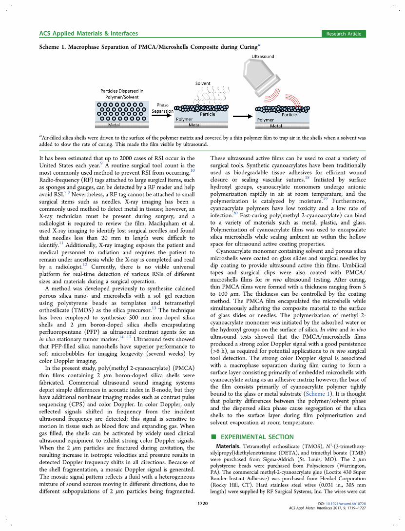
Scheme 1. Macrophase Separation of PMCA/Microshells Composite during Curinga
aAir-filled silica shells were driven to the surface of the polymer matrix and covered by a thin polymer film to trap air in the shells when a solvent wasadded to slow the rate of curing. This made the film visible by ultrasound.
ACS Applied Materials & Interfaces Research Article
DOI: 10.1021/acsami.6b10728ACS Appl. Mater. Interfaces 2017, 9, 1719−1727
1720
into short sections of 2 cm length and bent into curves to simulatesurgical needles. Surgical needles (3/8, taper point, 20 gauge) for thein vivo test were purchased from Santa Cruz Biotechnology, Inc.(Dallas, TX). The 11/2 in. 20 gauge hypodermic needles werepurchased from BD Medical (Franklin Lakes, NJ). Umbilical tape (1/8, cut into sections with length of 15 cm) was purchased fromJorgensen Laboratories Inc. (Loveland, CO). Titanium surgical clipswere purchased from Teleflex Medical (Research Triangle Park, NC).Silica Microshells Synthesis. The 2 μm calcined boron-doped
silica microshells were synthesized by modifying the literature15
method to improve templating by adding DETA. In brief, 100 mL of95% ethanol was added into a 500 mL cylindrical flask with a magneticstir bar, and 5 mL of 2.6% 2.0 μm polystyrene beads was added to theflask. The mixture was stirred at 1200 rpm at room temperature, while8 mL of 0.2% DETA in ethanol was added to the flask. The mixturewas stirred for 1 h before 310 μL of TMOS was added. After 2 h, 15μL of TMB was added, and the stirring continued for an additional 5h. The core−shell particles were centrifuged and washed with 95%ethanol and resuspended and washed two more times before drying inair overnight. The dried particles were calcined at 550 °C for 18 h andproduced 17.5 mg of 2 μm microshells.Fabrication of Silica Particle Containing PMCA Films. 10 mg
of 2 μm silica microshells was suspended in 1.0 mL of DCM bysonication and vortex mixed to disperse them before 0.5 mL of methyl2-cyanoacrylate glue was added. The glue/DCM/microshells mixturewas coated on glass slides (2 cm × 0.5 cm) by dipping the slides intothe liquid mixture. Four groups of glass slide samples were preparedwith 1, 2, 3, and 4 cycles of dip-coating, and each group contained fivesamples. The interval between cycles was 10 min. Needles were coatedwith the PMCA/microshells film by dipping the needles into themixture for eight cycles. The glue film was cured in air at roomtemperature for 24 h. The thickness of the films was measured by amicrometer. Before the ultrasound tests, the films on one side of theglass slides were removed to guarantee that sonographic propertiesrecorded are from a single film. In addition to DCM, ethyl acetate andacetonitrile were also used as solvents to study the relationshipbetween US performance and the dip-coating solvent. The method ofcoating of surgical clips is the same as coating of surgical needlesexcept that the clips were dip-coated for four cycles. For the coating ofumbilical tapes, 1 mL of DCM containing 2 mg of 2 μm silicamicroshells was added into 0.5 mL of methyl 2-cyanoacrylate glue. Theend of the umbilical tape was dipped into the mixture once. Thecoated section of the umbilical tape is 1.0 cm long from the end. Theglue was cured 24 h at room temperature in air.Optical and Electronic Microscopic Imaging and Contact
Angle Measurement. Optical microscopy was used to visualizePMCA/microshell films on needles. Transmission electron micros-copy (TEM) analysis of boron-doped microshells was performed withuse of a JEOL (JEOL, Tokyo, Japan) ARM200F operated at 200 kV.TEM samples were prepared by suspending calcined silica microshellsin ethanol and dropped onto a lacey carbon film grid substrate.Scanning electron microscopy (SEM) images of microshells and filmswere obtained using a FEI/Philips XL30 FEG ESEM microscope withan accelerating voltage ranging from 1.5 to 10 kV. SEM samples wereprepared by depositing microshells or film-coated needles on a carbontape substrate. Portions of film for analysis were exfoliated byscratching the film with a razor. Combined field emission SEM (FE-SEM) images were obtained using a Sigma 500 FE-SEM (Zeiss,Germany) with an accelerating voltage ranging from 0.8 to 20 kV. FE-SEM samples were prepared with the same procedure employed forthe TEM samples. The contact angles of the films on glass slides weremeasured by analyzing the photograph of the water drop on the filmswith ImageJ.In Vitro and in Vivo Ultrasound Testing. All in vitro and in vivo
US tests were performed using a Siemens Acuson Sequoia 512ultrasound machine with the Acuson 15L8 and 4C1 transducers withcenter frequencies of 7 and 3 MHz, respectively. Software used foranalysis of data included SanteDicom Viewer (Athens, Greece) andMicrosoft Excel (Redmond, WA). The tests of ultrasound responsivefilms on glass slides were performed with the samples in a water tank.
The 15L8 US transducer was clamped in the water tank with thesample film facing the transducer. The film was imaged with colorDoppler ultrasound with a mechanical index (MI) of 1.9, which is thehighest MI permitted by FDA for diagnostic ultrasound imaging. Theglass slides were kept in water and subjected to continuous ultrasoundradiation for 60 min. The color Doppler signals were recorded overseveral time periods. The attenuating rates of color Doppler signalswere studied by measuring the areas of the signals and comparing theareas with that of the initial signals.
In vitro ultrasound testing of glue/particles film coated needles wasperformed in a plastic box with a dimension of 20 cm × 15 cm × 6 cm.The box was filled with raw chicken livers to simulate organs in asurgical field. A PMCA/microshells film coated needle was placed inthe box. The distance between the needle and the top layer of chickenlivers was controlled between 0.5 and 6 cm. The ultrasound propertieswere studied with an ACUSON Sequoia ultrasound system with a15L8 and a 4C1 transducer.
In vivo US testing of PMCA/microshell film coated needles,umbilical tapes, and surgical clips was performed using female NewZealand white rabbits purchased from Western Oregon Rabbitry andhoused individually in a UCSD vivarium facility. They were kept on a12 h light/dark cycle and given water and Harlan Teklad commercialpellet diet ad libitum. All animal protocols were approved by theUCSD Institutional Animal Care and Use Program (IACUC).
Rabbits were anesthetized with isoflurane and placed on a warmedwater pad. The abdomen was shaved and depilated. Instruments andmaterials were cleaned and sanitized, but not sterilized. Gel was placedon the tip of the ultrasound transducer. Once anesthetized, heart rateand SpO2 were monitored via pulse oximetry, and jaw tone, mucousmembrane color, and pedal reflexes were also observed. A midlineincision was made from the xiphoid process to the groin; theabdominal wall was retracted. Sharp dissection was used to enter theperitoneal cavity; subsequently, needles were randomly placedthroughout to simulate a clinical situation in which a needle breaksoff a suture and needs to be retrieved. The surgeon used a 15L8transducer to explore and locate the needle, and ultrasound signalswere recorded. PMCA/microshell film coated umbilical tapes andsurgical needles were tested with the same method. Once all itemswere found, imaged, and removed, then retraction of the abdomen wasceased, and the animal was sacrificed immediately following thesurgeon’s search.
Postprocessing Detection of Film Signal. The films wereimaged using the color Doppler imaging mode which may displaycolor signals from the film, vascular blood flow, or movement from thetransducer or subject. Videos were saved along with simultaneous B-mode and color Doppler images in compressed DICOM format andwere postprocessed to selectively highlight signal from the film. Signalfrom the film was distinguishable by its persistent Doppler signal withhigh spatial and temporal heterogeneity collocated with a strong B-mode signal. Spatial heterogeneity was quantified by calculating themaximum magnitude of the spatial gradient across each of the red,green, and blue (RGB) color channels. Persistence was determined byfinding pixels with high spatial heterogeneity lasting at least threeframes (∼0.2 s). Temporal heterogeneity for each pixel was calculatedby integrating the difference in RGB colors from frame to frame. Pixelsmatching all criteria were shown using a green color overlay todistinguish from the Doppler color map. This processed signal (shownin green in the figures) will henceforth be referred to as microshellsignal (MSS). These methods were developed in MATLAB R2015a(The MathWorks Inc., Natick, MA).
Signal properties for detection of the film were compared betweenimaging in B-mode, color Doppler, and MSS. Sixteen video clips fromthe in vivo experiments of film coated needles were used for analysis.4% of the frames were randomly sampled (n = 69), and a user-definedregion of interest (ROI) was drawn around the needle if present. Allpixels outside of the ROI were considered background. B-mode signal-to-noise ratio (SNR) was calculated as the ratio of integrated B-modeimage intensity inside the ROI vs outside. Color Doppler and MSSSNR were calculated as the ratio of the area of detected signal insidethe ROI vs outside. Color Doppler and MSS sensitivity were calculated
ACS Applied Materials & Interfaces Research Article
DOI: 10.1021/acsami.6b10728ACS Appl. Mater. Interfaces 2017, 9, 1719−1727
1721
as the area of detected signal inside the ROI divided by the area of theROI; specificity was calculated as the area of undetected signal outsidethe ROI divided by the area outside the ROI. SNR was comparedbetween B-mode, Doppler, and MSS using the Kruskal−Wallis testwith multiple comparisons. Sensitivity and specificity were comparedbetween Doppler and MSS using the Mann−Whitney U-test.Statistical analyses were performed in MATLAB R2015a.
■ RESULTS AND DISCUSSIONSynthesis of 2 μm Boron-Doped Silica Microshells.
Figure 1 contains the TEM and SEM images of boron-doped
silica microshells. The size and size distribution of the hollowspherical particles were determined from SEM image analysis.The value of the average diameter and standard deviation was1.71 ± 0.03 μm (n = 20). The thickness of the silica shell wallwas determined by TEM image analysis as 30 ± 5 μm (n = 20).The silica shells have a dense, uniform wall with no resolvedpore structure, although the porous shell readily permits gases,solvents, and molecules to diffuse in and out. In TEM images,some colloidal silica particles were observed with a diameter ofless than 50 nm attached to the surface of the silica microshells.In the present study, nonmodified 2 μm PS beads were
employed as templates, and boron was doped into the silicamatrix during the sol−gel condensation to enhance themechanical strength of the microshells. DETA was used toserve as both the cationic electrolyte and a precursor of silica tobetter modify the surface of PS beads. The short, positivelycharged DETA is absorbed to the surface of anionic zeta-potential PS beads, and at the same time the silyl end of DETAwas cross-linked by the polycondensation reaction with itselfand templated a positively charged surface silica gel networkbefore adding the bulk of TMOS along with trimethyl borate tomake the thin silica shell more robust. Since the hydrolysis andpolycondensation of trimethy borate are faster than TMOS, itwas added 2 h after TMOS addition. The core−shell sol−gelcoated microparticles were obtained by centrifugation andcalcined at 550 °C to remove the PS cores. Dehydration of thesol gel during calcination resulted in a porous hollow silica gelshell with a diameter smaller than the 2 μm template.Cyanoacrylate glue was used to cross-link silica microshellsand bind them to metal or glass surfaces. Loctite 430 waschosen as the cyanoacrylate material because it has lowviscosity, which makes it easy to mix with silica shells. Theactive ingredient is methyl 2-cyanoacrylate which bondsstrongly with metals and cures rapidly compared to othersynthetic cyanoacrylate adhesives. Organic cosolvents wereused to further disperse silica microshells within the glue
solution, which lowered the viscosity and slowed curing. Themechanism of the curing of cyanoacrylate glue is by anionicpolymerization of cyanoacrylate monomer. The curing ofcommercial cyanoacrylate glue is initiated when water, a weakbase, neutralizes the strong acid inhibitor added tocyanoacrylate glue (Figure 2). For the PMCA/microshellsfilms, the water is likely supplied from the trace amount ofwater absorbed on the calcined porous silica gel shells.
Experiments suggest that water, which initiates thepolycondensation of cyanoacrylate, is already adsorbed on the2 μm silica microshells dispersed in acetonitrile before mixing.For example, two sealed tubes were prepared that wereidentical, except one omitted the silica microshells. Bothsamples were cured at room temperature. The viscosity of theglue mixture containing silica shells increased rapidly andturned into a white solid within 24 h. The sample withoutadded silica shells remained liquid for at least 7 days. In anotherexperiment, silica microshells were dried in a glovebox for 24 hat room temperature and then suspended in a glue/acetonitrilesolution in a sealed tube. After 24 h, the viscosity of thesuspension increased but did not solidify. The same test wasperformed without drying the silica shells in the glovebox, andthe suspension solidified within 24 h. This suggests the water isadsorbed on the silica microshells that were not dried. Thehydroxyl groups on the silica shells may play a minor role, butthe water absorbed on the shells dominates the curing process.When the acetonitrile solvent was replaced with DCM, the
curing time of glue containing silica microshells was prolongedto 3 days in sealed tubes. This can be attributed to the solubilityof water in acetonitrile being much higher than in DCM.Acetonitrile facilitates dissolution of adsorbed water from thesilica shells and disperses it into the bulk acetonitrile/cyanoacrylate solution. This initiated the polycondensationmuch more rapidly than when the water primarily remainsadsorbed on the surface of the silica shells in DCM solvent.The thickness of the PMCA/microshells film can be
controlled by varying the concentration of the particles inglue and the number of dip-coating repetition. To test the effectof the number of coating cycles, 20 mg/mL particles in methyl2-cyanoacrylate were coated onto glass slides with differentnumbers of repetitions. The film thicknesses on glass slides are15 ± 3, 31 ± 5, 59 ± 11, and 98 ± 13 μm (five samples each)by dip-coating the slides 1, 2, 3, and 4 times, respectively.Multiple coatings were needed to form a uniform PMCA/microshells film on the needles. A film with a thickness of 18 ±5 μm was obtained by dip-coating surgical needles eight timeseach in microshells/glue/DCM mixture; this was replicated for10 needles. The data are consistent with the surface tension ofthe round shape of the needle and the surface energy of metalrequiring more coatings for the needles. High particle
Figure 1. Electron microscopy images of calcined 2 μm boron-dopedsilica microshells. (a) Scanning electron microscopy (SEM) image. (b)Transmission electron microscopy image. The scale bars in the imagesare 5 μm (a) and 1 μm (b).
Figure 2. Polymerization of methyl 2-cyanoacrylate. Cyanoacrylatemonomers undergo a rapid anionic polymerization on exposure tobasic catalysts such as water, which neutralizes the acid inhibitor incommercial cyanoacrylate.
ACS Applied Materials & Interfaces Research Article
DOI: 10.1021/acsami.6b10728ACS Appl. Mater. Interfaces 2017, 9, 1719−1727
1722
concentration produces thicker films after curing as comparedto low particle concentrations. When 10 mg/mL particles inmethyl 2-cyanoacrylate were coated on needles, a film with athickness of 8 ± 3 μm was obtained by dip-coating 10 needleseight times each in silica shells/glue/DCM mixture.In Vitro Ultrasound Performance and Macrophase
Separation of PMCA/Microshell Films. Figure 3 displays
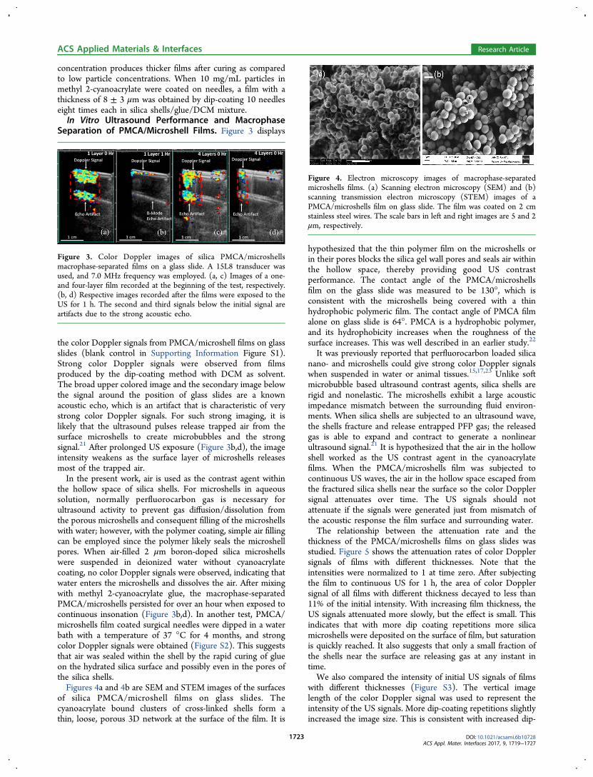
the color Doppler signals from PMCA/microshell films on glassslides (blank control in Supporting Information Figure S1).Strong color Doppler signals were observed from filmsproduced by the dip-coating method with DCM as solvent.The broad upper colored image and the secondary image belowthe signal around the position of glass slides are a knownacoustic echo, which is an artifact that is characteristic of verystrong color Doppler signals. For such strong imaging, it islikely that the ultrasound pulses release trapped air from thesurface microshells to create microbubbles and the strongsignal.21 After prolonged US exposure (Figure 3b,d), the imageintensity weakens as the surface layer of microshells releasesmost of the trapped air.In the present work, air is used as the contrast agent within
the hollow space of silica shells. For microshells in aqueoussolution, normally perfluorocarbon gas is necessary forultrasound activity to prevent gas diffusion/dissolution fromthe porous microshells and consequent filling of the microshellswith water; however, with the polymer coating, simple air fillingcan be employed since the polymer likely seals the microshellpores. When air-filled 2 μm boron-doped silica microshellswere suspended in deionized water without cyanoacrylatecoating, no color Doppler signals were observed, indicating thatwater enters the microshells and dissolves the air. After mixingwith methyl 2-cyanoacrylate glue, the macrophase-separatedPMCA/microshells persisted for over an hour when exposed tocontinuous insonation (Figure 3b,d). In another test, PMCA/microshells film coated surgical needles were dipped in a waterbath with a temperature of 37 °C for 4 months, and strongcolor Doppler signals were obtained (Figure S2). This suggeststhat air was sealed within the shell by the rapid curing of glueon the hydrated silica surface and possibly even in the pores ofthe silica shells.Figures 4a and 4b are SEM and STEM images of the surfaces
of silica PMCA/microshell films on glass slides. Thecyanoacrylate bound clusters of cross-linked shells form athin, loose, porous 3D network at the surface of the film. It is
hypothesized that the thin polymer film on the microshells orin their pores blocks the silica gel wall pores and seals air withinthe hollow space, thereby providing good US contrastperformance. The contact angle of the PMCA/microshellsfilm on the glass slide was measured to be 130°, which isconsistent with the microshells being covered with a thinhydrophobic polymeric film. The contact angle of PMCA filmalone on glass slide is 64°. PMCA is a hydrophobic polymer,and its hydrophobicity increases when the roughness of thesurface increases. This was well described in an earlier study.22
It was previously reported that perfluorocarbon loaded silicanano- and microshells could give strong color Doppler signalswhen suspended in water or animal tissues.15,17,23 Unlike softmicrobubble based ultrasound contrast agents, silica shells arerigid and nonelastic. The microshells exhibit a large acousticimpedance mismatch between the surrounding fluid environ-ments. When silica shells are subjected to an ultrasound wave,the shells fracture and release entrapped PFP gas; the releasedgas is able to expand and contract to generate a nonlinearultrasound signal.21 It is hypothesized that the air in the hollowshell worked as the US contrast agent in the cyanoacrylatefilms. When the PMCA/microshells film was subjected tocontinuous US waves, the air in the hollow space escaped fromthe fractured silica shells near the surface so the color Dopplersignal attenuates over time. The US signals should notattenuate if the signals were generated just from mismatch ofthe acoustic response the film surface and surrounding water.The relationship between the attenuation rate and the
thickness of the PMCA/microshells films on glass slides wasstudied. Figure 5 shows the attenuation rates of color Dopplersignals of films with different thicknesses. Note that theintensities were normalized to 1 at time zero. After subjectingthe film to continuous US for 1 h, the area of color Dopplersignal of all films with different thickness decayed to less than11% of the initial intensity. With increasing film thickness, theUS signals attenuated more slowly, but the effect is small. Thisindicates that with more dip coating repetitions more silicamicroshells were deposited on the surface of film, but saturationis quickly reached. It also suggests that only a small fraction ofthe shells near the surface are releasing gas at any instant intime.We also compared the intensity of initial US signals of films
with different thicknesses (Figure S3). The vertical imagelength of the color Doppler signal was used to represent theintensity of the US signals. More dip-coating repetitions slightlyincreased the image size. This is consistent with increased dip-
Figure 3. Color Doppler images of silica PMCA/microshellsmacrophase-separated films on a glass slide. A 15L8 transducer wasused, and 7.0 MHz frequency was employed. (a, c) Images of a one-and four-layer film recorded at the beginning of the test, respectively.(b, d) Respective images recorded after the films were exposed to theUS for 1 h. The second and third signals below the initial signal areartifacts due to the strong acoustic echo.
Figure 4. Electron microscopy images of macrophase-separatedmicroshells films. (a) Scanning electron microscopy (SEM) and (b)scanning transmission electron microscopy (STEM) images of aPMCA/microshells film on glass slide. The film was coated on 2 cmstainless steel wires. The scale bars in left and right images are 5 and 2μm, respectively.
ACS Applied Materials & Interfaces Research Article
DOI: 10.1021/acsami.6b10728ACS Appl. Mater. Interfaces 2017, 9, 1719−1727
1723
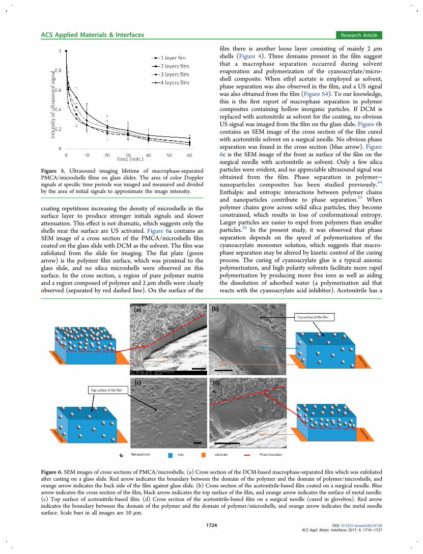
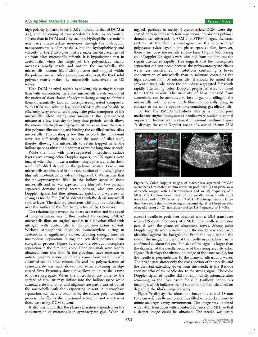
coating repetitions increasing the density of microshells in thesurface layer to produce stronger initials signals and slowerattenuation. This effect is not dramatic, which suggests only theshells near the surface are US activated. Figure 6a contains anSEM image of a cross section of the PMCA/microshells filmcoated on the glass slide with DCM as the solvent. The film wasexfoliated from the slide for imaging. The flat plate (greenarrow) is the polymer film surface, which was proximal to theglass slide, and no silica microshells were observed on thissurface. In the cross section, a region of pure polymer matrixand a region composed of polymer and 2 μm shells were clearlyobserved (separated by red dashed line). On the surface of the
film there is another loose layer consisting of mainly 2 μmshells (Figure 4). Three domains present in the film suggestthat a macrophase separation occurred during solventevaporation and polymerization of the cyanoacrylate/micro-shell composite. When ethyl acetate is employed as solvent,phase separation was also observed in the film, and a US signalwas also obtained from the film (Figure S4). To our knowledge,this is the first report of macrophase separation in polymercomposites containing hollow inorganic particles. If DCM isreplaced with acetonitrile as solvent for the coating, no obviousUS signal was imaged from the film on the glass slide. Figure 6bcontains an SEM image of the cross section of the film curedwith acetonitrile solvent on a surgical needle. No obvious phaseseparation was found in the cross section (blue arrow). Figure6c is the SEM image of the front as surface of the film on thesurgical needle with acetonitrile as solvent. Only a few silicaparticles were evident, and no appreciable ultrasound signal wasobtained from the film. Phase separation in polymer−nanoparticles composites has been studied previously.24
Enthalpic and entropic interactions between polymer chainsand nanoparticles contribute to phase separation.25 Whenpolymer chains grow across solid silica particles, they becomeconstrained, which results in loss of conformational entropy.Larger particles are easier to expel from polymers than smallerparticles.26 In the present study, it was observed that phaseseparation depends on the speed of polymerization of thecyanoacrylate monomer solution, which suggests that macro-phase separation may be altered by kinetic control of the curingprocess. The curing of cyanoacrylate glue is a typical anionicpolymerization, and high polarity solvents facilitate more rapidpolymerization by producing more free ions as well as aidingthe dissolution of adsorbed water (a polymerization aid thatreacts with the cyanoacrylate acid inhibitor). Acetonitrile has a
Figure 5. Ultrasound imaging lifetime of macrophase-separatedPMCA/microshells films on glass slides. The area of color Dopplersignals at specific time periods was imaged and measured and dividedby the area of initial signals to approximate the image intensity.
Figure 6. SEM images of cross sections of PMCA/microshells. (a) Cross section of the DCM-based macrophase-separated film which was exfoliatedafter casting on a glass slide. Red arrow indicates the boundary between the domain of the polymer and the domain of polymer/microshells, andorange arrow indicates the back side of the film against glass slide. (b) Cross section of the acetonitrile-based film coated on a surgical needle. Bluearrow indicates the cross section of the film, black arrow indicates the top surface of the film, and orange arrow indicates the surface of metal needle.(c) Top surface of acetonitrile-based film. (d) Cross section of the acetonitrile-based film on a surgical needle (cured in glovebox). Red arrowindicates the boundary between the domain of the polymer and the domain of polymer/microshells, and orange arrow indicates the metal needlesurface. Scale bars in all images are 10 μm.
ACS Applied Materials & Interfaces Research Article
DOI: 10.1021/acsami.6b10728ACS Appl. Mater. Interfaces 2017, 9, 1719−1727
1724
high polarity (polarity index is 5.8 compared to that of DCM at3.1), and the curing of cyanoacrylate is faster in acetonitrilesolvent than in DCM and ethyl acetate. Hydrophilic acetonitrilemay carry cyanoacrylate monomer through the hydrophilicnanoporous walls of microshells, but the hydrophobicity andviscosity of the DCM/glue mixture make the displacement ofair from silica microshells difficult. It is hypothesized that inacetonitrile, when the length of the polymerized chainsincreases rapidly inside and outside the microshells, themicroshells become filled with polymer and trapped withinthe polymer matrix. After evaporation of solvent, the thick solidpolymer matrix makes the microshells nonactivable to USwaves.With DCM or ethyl acetate as solvent, the curing is slower
than with acetonitrile; therefore, microshells are driven out ofthe matrix of short chains of polymer during curing to obtain athermodynamically favored macrophase-separated composite.With DCM as a solvent, less polar DCM might not be able toefficiently carry monomers through the polar nanopores of themicroshells. Slow curing also maintains the glue−solventmixture at a low viscosity for long time periods, which allowsthe microshells to phase segregate. At the same time, there is athin polymer film coating and binding the air-filled surface silicamicroshells. This coating is too thin to block the ultrasoundwave but sufficiently thick to seal the pores of silica shell,thereby allowing the microshells to retain trapped air in thehollow space as ultrasound contrast agent for long time periods.While the films with phase-separated microshells surface
layers gave strong color Doppler signals, no US signals wereimaged when the film was a uniform single phase, and the shellswere embedded deeper in the polymer matrix. Few 2 μmmicroshells are observed in the cross section of the single phasefilm with acetonitrile as solvent (Figure 6b). We assume thatthe polycyanoacrylate filled in the hollow space of thesemicroshells and air was expelled. The film with two partiallyseparated domains (ethyl acetate solvent) also gave colorDoppler signals, but they attenuated quickly and were not asstrong as for the film (DCM solvent) with the dense microshellsurface layer. The data are consistent with only the microshellsnear the surface of the film being activated by US waves.The relationship between the phase separation and the speed
of polymerization was further probed by coating PMCA/microshells films on surgical needles in a glovebox filled withnitrogen with acetonitrile as the polymerization solvent.Without atmospheric moisture, cyanoacrylate curing inacetonitrile is significantly slower, allowing enough time formacrophase separation during the retarded polymer chainelongation process. Figure 6d shows the obvious macrophaseseparation in the film, and color Doppler signals were readilyobtained from this film. In the glovebox, trace moisture toinitiate polymerization could only come from water initiallyadsorbed on the silica microshells, and the polymerization ofcyanoacrylate was much slower than when air curing the dip-coated films. Extremely slow curing allows the microshells timeto phase segregate. When the microshells are close to thesurface of film, air may diffuse into the hollow space whilecyanoacrylate monomer and oligomer are partly carried out ofthe microshells with the evaporating solvent. A macrophaseseparation was thereby obtained by the slower polymerizationprocess. The film is also ultrasound active, but not as active asthose cast using DCM solvent.It also was found that the phase separation depended on the
concentration of microshells in cyanoacrylate glue. When 10
mg/mL particles in methyl 2-cyanoacrylate/DCM were dip-coated onto needles with four repetitions, no obvious polymerdomain was observed. In SEM and STEM images, the crosssection of the film is analogous to the microshells/polycyanoacrylate layer in the phase-separated film; however,there is no loose microshells surface layer (Figure S5). Strongcolor Doppler US signals were obtained from the film, but thesignals attenuated rapidly. This suggests that the macrophaseseparation did not occur because the polycyanoacrylate chainswere less constrained in solutions containing a lowerconcentration of microshells than in solutions containing thehigh concentration of microshells. It should be noted thatsolvent plays a role, since the non-phase-segregated films withrapidly attenuating color Doppler properties were obtainedfrom DCM solvent. The inactivity of films prepared fromacetonitrile can be attributed to loss of gas and filling of themicroshells with polymer. Such films are optically clear, incontrast to the white opaque films containing gas-filled shells.To test the PMCA/microshells film as a multipurpose
marker for surgical tools, coated needles were hidden in animalorgans and located with a clinical ultrasound machine. Figure7a displays the color Doppler image of a coated 18 mm (3/8
curved) needle in pork liver obtained with a 15L8 transducerwith a US center frequency of 7 MHz. The needle is coplanarparallel with the plane of ultrasound waves. Strong colorDoppler signals were observed, and the needle was very easilyidentified against the background. From the scale bar on theside of the image, the depth of the needle in pork liver can beconfirmed as about 0.5 cm. The size of the signal is larger thanthe diameter of the needle because of the strong acoustic echo.Figure 7b displays the ultrasound image of the same needle, butthe needle is perpendicular to the plane of ultrasound waves.The bright spot shows only the cross section of the needle, andthe dark tail extending down from the needle is the B-modeacoustic echo of the needle due to the strong signal. The colorDoppler signal of needles did not significantly attenuate afterremaining in the liver tissue for 6 h (without continuousimaging), which indicates that tissue or blood has little effect ondegrading the film’s image intensity.Figure 7c displays the ultrasound image of a coated 18 mm
(3/8 curved) needle in a plastic box filled with chicken livers tomimic an organ cavity environment. The image was obtainedwith a 4C1 transducer with a center frequency of 3 MHz so thata deeper image could be obtained. The needle was easily
Figure 7. Color Doppler images of macrophase-separated PMCA/microshells film coated 18 mm needle in pork liver. (a) In-plane viewof needle imaged with 15L8 transducer and an US frequency of 7MHz. (b) Cross-sectional view of the needle imaged with 15L8transducer and an US frequency of 7 MHz. The image sizes are largerthan the needle due to the strong ultrasound signal. (c) In-plane viewof needle using a 4C1 transducer and an US frequency of 3 MHz.
ACS Applied Materials & Interfaces Research Article
DOI: 10.1021/acsami.6b10728ACS Appl. Mater. Interfaces 2017, 9, 1719−1727
1725
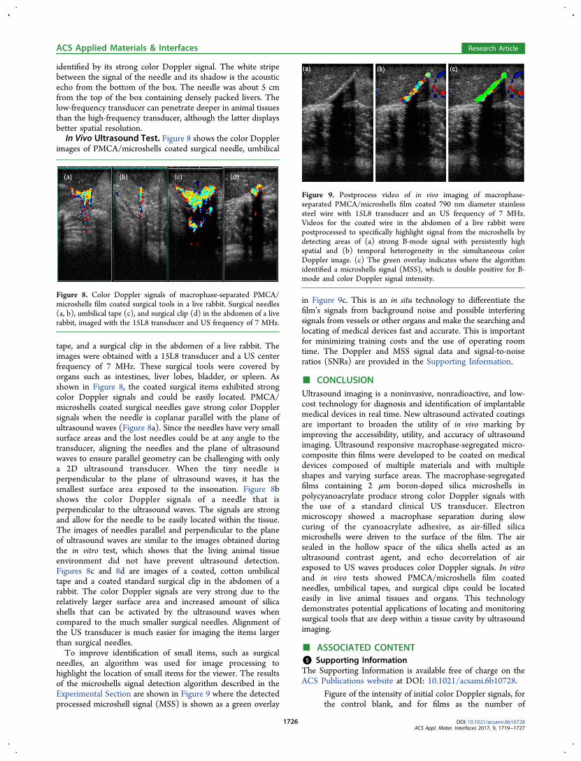
identified by its strong color Doppler signal. The white stripebetween the signal of the needle and its shadow is the acousticecho from the bottom of the box. The needle was about 5 cmfrom the top of the box containing densely packed livers. Thelow-frequency transducer can penetrate deeper in animal tissuesthan the high-frequency transducer, although the latter displaysbetter spatial resolution.In Vivo Ultrasound Test. Figure 8 shows the color Doppler
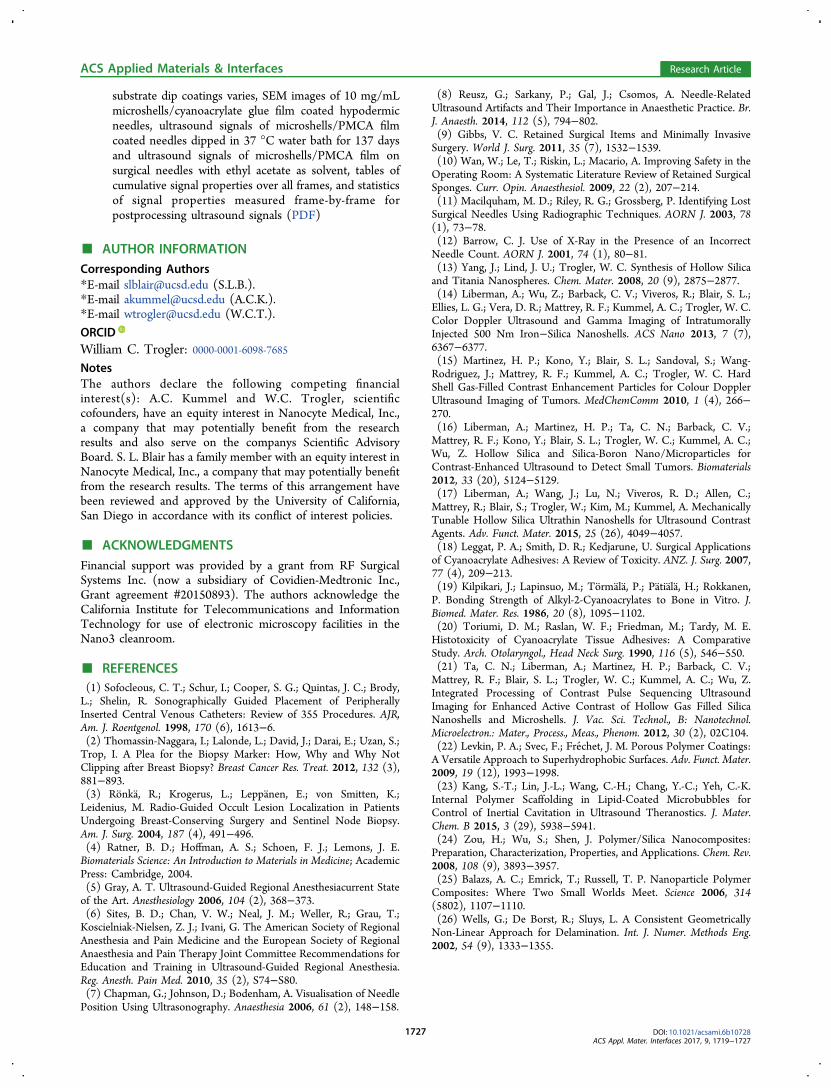
images of PMCA/microshells coated surgical needle, umbilical
tape, and a surgical clip in the abdomen of a live rabbit. Theimages were obtained with a 15L8 transducer and a US centerfrequency of 7 MHz. These surgical tools were covered byorgans such as intestines, liver lobes, bladder, or spleen. Asshown in Figure 8, the coated surgical items exhibited strongcolor Doppler signals and could be easily located. PMCA/microshells coated surgical needles gave strong color Dopplersignals when the needle is coplanar parallel with the plane ofultrasound waves (Figure 8a). Since the needles have very smallsurface areas and the lost needles could be at any angle to thetransducer, aligning the needles and the plane of ultrasoundwaves to ensure parallel geometry can be challenging with onlya 2D ultrasound transducer. When the tiny needle isperpendicular to the plane of ultrasound waves, it has thesmallest surface area exposed to the insonation. Figure 8bshows the color Doppler signals of a needle that isperpendicular to the ultrasound waves. The signals are strongand allow for the needle to be easily located within the tissue.The images of needles parallel and perpendicular to the planeof ultrasound waves are similar to the images obtained duringthe in vitro test, which shows that the living animal tissueenvironment did not have prevent ultrasound detection.Figures 8c and 8d are images of a coated, cotton umbilicaltape and a coated standard surgical clip in the abdomen of arabbit. The color Doppler signals are very strong due to therelatively larger surface area and increased amount of silicashells that can be activated by the ultrasound waves whencompared to the much smaller surgical needles. Alignment ofthe US transducer is much easier for imaging the items largerthan surgical needles.To improve identification of small items, such as surgical
needles, an algorithm was used for image processing tohighlight the location of small items for the viewer. The resultsof the microshells signal detection algorithm described in theExperimental Section are shown in Figure 9 where the detectedprocessed microshell signal (MSS) is shown as a green overlay
in Figure 9c. This is an in situ technology to differentiate thefilm’s signals from background noise and possible interferingsignals from vessels or other organs and make the searching andlocating of medical devices fast and accurate. This is importantfor minimizing training costs and the use of operating roomtime. The Doppler and MSS signal data and signal-to-noiseratios (SNRs) are provided in the Supporting Information.
■ CONCLUSIONUltrasound imaging is a noninvasive, nonradioactive, and low-cost technology for diagnosis and identification of implantablemedical devices in real time. New ultrasound activated coatingsare important to broaden the utility of in vivo marking byimproving the accessibility, utility, and accuracy of ultrasoundimaging. Ultrasound responsive macrophase-segregated micro-composite thin films were developed to be coated on medicaldevices composed of multiple materials and with multipleshapes and varying surface areas. The macrophase-segregatedfilms containing 2 μm boron-doped silica microshells inpolycyanoacrylate produce strong color Doppler signals withthe use of a standard clinical US transducer. Electronmicroscopy showed a macrophase separation during slowcuring of the cyanoacrylate adhesive, as air-filled silicamicroshells were driven to the surface of the film. The airsealed in the hollow space of the silica shells acted as anultrasound contrast agent, and echo decorrelation of airexposed to US waves produces color Doppler signals. In vitroand in vivo tests showed PMCA/microshells film coatedneedles, umbilical tapes, and surgical clips could be locatedeasily in live animal tissues and organs. This technologydemonstrates potential applications of locating and monitoringsurgical tools that are deep within a tissue cavity by ultrasoundimaging.
■ ASSOCIATED CONTENT*S Supporting InformationThe Supporting Information is available free of charge on theACS Publications website at DOI: 10.1021/acsami.6b10728.
Figure of the intensity of initial color Doppler signals, forthe control blank, and for films as the number of
Figure 8. Color Doppler signals of macrophase-separated PMCA/microshells film coated surgical tools in a live rabbit. Surgical needles(a, b), umbilical tape (c), and surgical clip (d) in the abdomen of a liverabbit, imaged with the 15L8 transducer and US frequency of 7 MHz.
Figure 9. Postprocess video of in vivo imaging of macrophase-separated PMCA/microshells film coated 790 nm diameter stainlesssteel wire with 15L8 transducer and an US frequency of 7 MHz.Videos for the coated wire in the abdomen of a live rabbit werepostprocessed to specifically highlight signal from the microshells bydetecting areas of (a) strong B-mode signal with persistently highspatial and (b) temporal heterogeneity in the simultaneous colorDoppler image. (c) The green overlay indicates where the algorithmidentified a microshells signal (MSS), which is double positive for B-mode and color Doppler signal intensity.
ACS Applied Materials & Interfaces Research Article
DOI: 10.1021/acsami.6b10728ACS Appl. Mater. Interfaces 2017, 9, 1719−1727
1726
substrate dip coatings varies, SEM images of 10 mg/mLmicroshells/cyanoacrylate glue film coated hypodermicneedles, ultrasound signals of microshells/PMCA filmcoated needles dipped in 37 °C water bath for 137 daysand ultrasound signals of microshells/PMCA film onsurgical needles with ethyl acetate as solvent, tables ofcumulative signal properties over all frames, and statisticsof signal properties measured frame-by-frame forpostprocessing ultrasound signals (PDF)
■ AUTHOR INFORMATIONCorresponding Authors*E-mail [email protected] (S.L.B.).*E-mail [email protected] (A.C.K.).*E-mail [email protected] (W.C.T.).
ORCIDWilliam C. Trogler: 0000-0001-6098-7685NotesThe authors declare the following competing financialinterest(s): A.C. Kummel and W.C. Trogler, scientificcofounders, have an equity interest in Nanocyte Medical, Inc.,a company that may potentially benefit from the researchresults and also serve on the companys Scientific AdvisoryBoard. S. L. Blair has a family member with an equity interest inNanocyte Medical, Inc., a company that may potentially benefitfrom the research results. The terms of this arrangement havebeen reviewed and approved by the University of California,San Diego in accordance with its conflict of interest policies.
■ ACKNOWLEDGMENTSFinancial support was provided by a grant from RF SurgicalSystems Inc. (now a subsidiary of Covidien-Medtronic Inc.,Grant agreement #20150893). The authors acknowledge theCalifornia Institute for Telecommunications and InformationTechnology for use of electronic microscopy facilities in theNano3 cleanroom.
■ REFERENCES(1) Sofocleous, C. T.; Schur, I.; Cooper, S. G.; Quintas, J. C.; Brody,L.; Shelin, R. Sonographically Guided Placement of PeripherallyInserted Central Venous Catheters: Review of 355 Procedures. AJR,Am. J. Roentgenol. 1998, 170 (6), 1613−6.(2) Thomassin-Naggara, I.; Lalonde, L.; David, J.; Darai, E.; Uzan, S.;Trop, I. A Plea for the Biopsy Marker: How, Why and Why NotClipping after Breast Biopsy? Breast Cancer Res. Treat. 2012, 132 (3),881−893.(3) Ronka, R.; Krogerus, L.; Leppanen, E.; von Smitten, K.;Leidenius, M. Radio-Guided Occult Lesion Localization in PatientsUndergoing Breast-Conserving Surgery and Sentinel Node Biopsy.Am. J. Surg. 2004, 187 (4), 491−496.(4) Ratner, B. D.; Hoffman, A. S.; Schoen, F. J.; Lemons, J. E.Biomaterials Science: An Introduction to Materials in Medicine; AcademicPress: Cambridge, 2004.(5) Gray, A. T. Ultrasound-Guided Regional Anesthesiacurrent Stateof the Art. Anesthesiology 2006, 104 (2), 368−373.(6) Sites, B. D.; Chan, V. W.; Neal, J. M.; Weller, R.; Grau, T.;Koscielniak-Nielsen, Z. J.; Ivani, G. The American Society of RegionalAnesthesia and Pain Medicine and the European Society of RegionalAnaesthesia and Pain Therapy Joint Committee Recommendations forEducation and Training in Ultrasound-Guided Regional Anesthesia.Reg. Anesth. Pain Med. 2010, 35 (2), S74−S80.(7) Chapman, G.; Johnson, D.; Bodenham, A. Visualisation of NeedlePosition Using Ultrasonography. Anaesthesia 2006, 61 (2), 148−158.
(8) Reusz, G.; Sarkany, P.; Gal, J.; Csomos, A. Needle-RelatedUltrasound Artifacts and Their Importance in Anaesthetic Practice. Br.J. Anaesth. 2014, 112 (5), 794−802.(9) Gibbs, V. C. Retained Surgical Items and Minimally InvasiveSurgery. World J. Surg. 2011, 35 (7), 1532−1539.(10) Wan, W.; Le, T.; Riskin, L.; Macario, A. Improving Safety in theOperating Room: A Systematic Literature Review of Retained SurgicalSponges. Curr. Opin. Anaesthesiol. 2009, 22 (2), 207−214.(11) Macilquham, M. D.; Riley, R. G.; Grossberg, P. Identifying LostSurgical Needles Using Radiographic Techniques. AORN J. 2003, 78(1), 73−78.(12) Barrow, C. J. Use of X-Ray in the Presence of an IncorrectNeedle Count. AORN J. 2001, 74 (1), 80−81.(13) Yang, J.; Lind, J. U.; Trogler, W. C. Synthesis of Hollow Silicaand Titania Nanospheres. Chem. Mater. 2008, 20 (9), 2875−2877.(14) Liberman, A.; Wu, Z.; Barback, C. V.; Viveros, R.; Blair, S. L.;Ellies, L. G.; Vera, D. R.; Mattrey, R. F.; Kummel, A. C.; Trogler, W. C.Color Doppler Ultrasound and Gamma Imaging of IntratumorallyInjected 500 Nm Iron−Silica Nanoshells. ACS Nano 2013, 7 (7),6367−6377.(15) Martinez, H. P.; Kono, Y.; Blair, S. L.; Sandoval, S.; Wang-Rodriguez, J.; Mattrey, R. F.; Kummel, A. C.; Trogler, W. C. HardShell Gas-Filled Contrast Enhancement Particles for Colour DopplerUltrasound Imaging of Tumors. MedChemComm 2010, 1 (4), 266−270.(16) Liberman, A.; Martinez, H. P.; Ta, C. N.; Barback, C. V.;Mattrey, R. F.; Kono, Y.; Blair, S. L.; Trogler, W. C.; Kummel, A. C.;Wu, Z. Hollow Silica and Silica-Boron Nano/Microparticles forContrast-Enhanced Ultrasound to Detect Small Tumors. Biomaterials2012, 33 (20), 5124−5129.(17) Liberman, A.; Wang, J.; Lu, N.; Viveros, R. D.; Allen, C.;Mattrey, R.; Blair, S.; Trogler, W.; Kim, M.; Kummel, A. MechanicallyTunable Hollow Silica Ultrathin Nanoshells for Ultrasound ContrastAgents. Adv. Funct. Mater. 2015, 25 (26), 4049−4057.(18) Leggat, P. A.; Smith, D. R.; Kedjarune, U. Surgical Applicationsof Cyanoacrylate Adhesives: A Review of Toxicity. ANZ. J. Surg. 2007,77 (4), 209−213.(19) Kilpikari, J.; Lapinsuo, M.; Tormala, P.; Patiala, H.; Rokkanen,P. Bonding Strength of Alkyl-2-Cyanoacrylates to Bone in Vitro. J.Biomed. Mater. Res. 1986, 20 (8), 1095−1102.(20) Toriumi, D. M.; Raslan, W. F.; Friedman, M.; Tardy, M. E.Histotoxicity of Cyanoacrylate Tissue Adhesives: A ComparativeStudy. Arch. Otolaryngol., Head Neck Surg. 1990, 116 (5), 546−550.(21) Ta, C. N.; Liberman, A.; Martinez, H. P.; Barback, C. V.;Mattrey, R. F.; Blair, S. L.; Trogler, W. C.; Kummel, A. C.; Wu, Z.Integrated Processing of Contrast Pulse Sequencing UltrasoundImaging for Enhanced Active Contrast of Hollow Gas Filled SilicaNanoshells and Microshells. J. Vac. Sci. Technol., B: Nanotechnol.Microelectron.: Mater., Process., Meas., Phenom. 2012, 30 (2), 02C104.(22) Levkin, P. A.; Svec, F.; Frechet, J. M. Porous Polymer Coatings:A Versatile Approach to Superhydrophobic Surfaces. Adv. Funct. Mater.2009, 19 (12), 1993−1998.(23) Kang, S.-T.; Lin, J.-L.; Wang, C.-H.; Chang, Y.-C.; Yeh, C.-K.Internal Polymer Scaffolding in Lipid-Coated Microbubbles forControl of Inertial Cavitation in Ultrasound Theranostics. J. Mater.Chem. B 2015, 3 (29), 5938−5941.(24) Zou, H.; Wu, S.; Shen, J. Polymer/Silica Nanocomposites:Preparation, Characterization, Properties, and Applications. Chem. Rev.2008, 108 (9), 3893−3957.(25) Balazs, A. C.; Emrick, T.; Russell, T. P. Nanoparticle PolymerComposites: Where Two Small Worlds Meet. Science 2006, 314(5802), 1107−1110.(26) Wells, G.; De Borst, R.; Sluys, L. A Consistent GeometricallyNon-Linear Approach for Delamination. Int. J. Numer. Methods Eng.2002, 54 (9), 1333−1355.
ACS Applied Materials & Interfaces Research Article
DOI: 10.1021/acsami.6b10728ACS Appl. Mater. Interfaces 2017, 9, 1719−1727
1727