ultrastructural localization of zinc transporter-3 (znt-3) to synaptic
TRANSCRIPT
Proc. Natl. Acad. Sci. USAVol. 94, pp. 12676–12681, November 1997Neurobiology
Ultrastructural localization of zinc transporter-3 (ZnT-3) tosynaptic vesicle membranes within mossy fiber boutonsin the hippocampus of mouse and monkey
H. JURGEN WENZEL*, TOBY B. COLE†, DONALD E. BORN‡, PHILIP A. SCHWARTZKROIN*§,AND RICHARD D. PALMITER†¶
Departments of *Neurological Surgery, ‡Pathology, §PhysiologyyBiophysics, and †Biochemistry and †Howard Hughes Medical Institute, Box 357370, University ofWashington, Seattle, WA 98195
Contributed by Richard D. Palmiter, September 9, 1997
ABSTRACT Zinc transporter-3 (ZnT-3), a member of agrowing family of mammalian zinc transporters, is expressedin regions of the brain that are rich in histochemically reactivezinc (as revealed by the Timm’s stain), including entorhinalcortex, amygdala, and hippocampus. ZnT-3 protein is mostabundant in the zinc-enriched mossy fibers that project fromthe dentate granule cells to hilar and CA3 pyramidal neurons.We show here by electron microscopy that ZnT-3 decorates themembranes of all clear, small, round synaptic vesicles (SVs)in the mossy fiber boutons of both mouse and monkey.Furthermore, up to 60–80% of these SVs contain Timm’s-stainable zinc. The coincidence of ZnT-3 on the membranes ofSVs that accumulate zinc, and its homology with known zinctransporters, suggest that ZnT-3 is responsible for the trans-port of zinc into SVs, and hence for the ability of these neuronsto release zinc upon excitation.
Most of the zinc in the mammalian brain is associated withmetalloproteins; however, there is also a pool of histochemi-cally reactive zinc that exists in synaptic vesicles (SVs) of asubset of glutamatergic neurons, which has led to classificationof these neurons as zinc-containing or zinc-ergic (1–4). Path-ways utilizing this vesicular form of zinc have been mappedusing histochemical stains such as the neo-Timm’s sulfide-silver method (5), selenium stain (6, 7), and the fluorescentcompound, TSQ (8). One of the best described zinc-ergicsystems is found in the rodent hippocampal formation, wherevesicular zinc can be detected in each component of thetrisynaptic circuit that includes (i) perforant path projectionsfrom the entorhinal cortex to the outer molecular layer of thedentate gyrus, (ii) mossy fiber (MF) projections from granulecells in the dentate gyrus to hilar neurons and pyramidal cellsin the CA3 region (4, 9–11), (iii) projections from CA3pyramidal neurons to neurons in the CA1 region, and (iv)projections from CA1 to subiculum (3, 4). Electron microscopy(EM) has revealed that the Timm’s stain precipitate is presentwithin SVs in the giant axonal boutons of the MFs in the hilusand stratum (s.) lucidum (1, 2, 12). However, only '10–15%of the SVs within a given bouton have been shown to containTimm’s precipitate (2); thus, it has been difficult to ascertainwhether zinc is present in a subset of vesicles or if it is presentin the same vesicles as glutamate.
Accumulation of zinc within SVs presumably depends on theaction of specific transporters, by analogy with the accumulationof other neurotransmitters in SVs (13). A gene, designated zinctransporter-3 (ZnT3), homologous to two established zinc trans-porters (14, 15) was recently cloned (16). ZnT-3 was shown by in
situ hybridization to be expressed at high levels in hippocampusand neocortex. Immunocytochemical studies demonstrated itslocalization to the MFs, where the histochemical Timm’s reactionhas revealed zinc-containing SVs. This profile suggested thatZnT-3 might be the vesicular zinc transporter responsible forsequestration of zinc in MF SVs (16), but also raised questionsregarding the ultrastructural localization of ZnT-3 relative to zincand other neurotransmitters.
MATERIALS AND METHODSTissue Preparation. Eight mice (6 C57BLy6; 2 C57BLy6x129),
ages 8–20 weeks, and five monkeys, (Macaca nemestrina), age 1year were used for light microscopy and EM immunocytochem-ical localization of ZnT-3 and for histochemical localization ofzinc within the MF system of the hippocampus.
Mice were anesthetized with Nembutal (100 mgykg, i.p.), thenperfused with isotonic saline with heparin (100 unitsyml saline),followed by 4% paraformaldehyde in 0.1 M sodium phosphatebuffer (PB), pH 7.4, or by a solution containing 4% paraformal-dehyde, 0.1% glutaraldehyde, and 0.1% picric acid in PB. [Thelow level of glutaraldehyde was necessary to preserve immuno-reactivity (IR) but resulted in suboptimal ultrastructure.] Thebrains were removed and placed in the same fixative for 4 hr at4°C. Monkeys were anesthetized (Nembutal, 50–75 mgykg) andperfused transcardially with 4% paraformaldehyde in PB. Tissueblocks from the temporal lobe, containing the hippocampus, weredissected and cut into 1.5-mm thick slices that were then placedin the same fixative for 24 hr at 4°C.
After postfixation, the brains (mice) or slices (monkey) wererinsed in PB, cryoprotected in 10% sucrose for 1 hr, followed by30% sucrose for 8–12 hr at 4°C, then frozen on dry ice. Transverseserial sections, 30 mm for light microscopy and 80 mm for EM,were cut on a sliding microtome equipped with a freezing stage,and sections were selected for further processing.
EM Detection of ZnT-3. An affinity-purified rabbit antibodyspecific for ZnT-3 (16) was used to determine its ultrastruc-tural localization. Sections were rinsed in PB, followed by 0.1M Tris·HCl buffer (TB), pH 7.4; then endogenous peroxidaseswere inactivated by treatment with 0.5–1% H2O2 in TB for 2hr. Sections were treated with 3% BSA, 3% goat serum, and0.25% dimethyl sulfoxide in TBS (0.05 M TBy0.15 M NaCl, pH7.4) for 1 hr to reduce nonspecific staining. Sections wererinsed in TBS for 30 min and incubated for 20 hr at 4°C inZnT-3 antiserum, diluted 1:100 to 1:300 in TBS containing 1%goat serum, 2% BSA and 0.25% dimethyl sulfoxide. Followingrinses for 2 hr in TBS, sections were incubated in biotinylatedgoat anti-rabbit IgG (diluted 1:500) for 24 hr at 4°C, rinsed 2hr in TBS and then incubated in ABC (Elite ABC Kit; Vector
The publication costs of this article were defrayed in part by page chargepayment. This article must therefore be hereby marked ‘‘advertisement’’ inaccordance with 18 U.S.C. §1734 solely to indicate this fact.
© 1997 by The National Academy of Sciences 0027-8424y97y9412676-6$2.00y0PNAS is available online at http:yywww.pnas.org.
Abbreviations: SV, synaptic vesicle; MF, mossy fiber; MFB, MFbouton; IR, immunoreactivity; ZnT-3, zinc transporter-3; EM, elec-tron microscopy; s., stratum; GABA, g-aminobutyric acid.¶To whom reprint requests should be addressed. e-mail: [email protected].
12676
Laboratories), diluted 1:500 in 1% goat serum, 2% BSA,0.25% dimethyl sulfoxide and TBS for 24 hr at 4°C. Sectionswere rinsed in TB (pH 7.6), then incubated for 15 min in0.025% 3,39-diaminobenzidine in TB. After reaction for 5–10min in fresh 3,39-diaminobenzidine with 0.003% H2O2, sec-tions were rinsed in TB, followed by PB. Specificity of theimmunostaining was evaluated by omitting primary antibody.
Sections were further processed for EM by postfixation in 1%osmium tetroxide in PB for 1 hr at 22°C, followed by alcoholdehydration and flat embedding in Eponate 12 (Ted Pella,Redding, CA) between two aclar sheets for 24 hr at 60°C. Selectedimmunoreactive areas were cut from embedded sections andremounted with Eponate 12 on plastic blocks. Serial ultrathinsections were cut close to the tissue surface, stained with uranylacetate and lead citrate, and examined on a Philips (Eindhoven,The Netherlands) 410 electron microscope.
Postembedding Detection of ZnT-3 and Glutamate. Toinvestigate colocalization of ZnT-3 and glutamate in SVs ofMFs, postembedding immunocytochemistry was performed asdescribed by Wenzel et al. (17). Ultrathin sections fromTimm’s-stained sections or glutaraldehydeyparaformalde-hyde-fixed tissue were etched with redox reagents (periodicacid, sodium metaperiodate) to reduce osmium staining and toexpose antigenic sites. Sections were then incubated with theprimary antibodies—rabbit anti-glutamate (Sigma) diluted1:10,000 or affinity-purified ZnT-3 antibody, diluted 1:50 or1:100—followed by goat-anti-rabbit IgG conjugated to gold(10 nm, Ted Pella), diluted 1:20.
Timm’s Staining. For light microscopy detection in mouseand monkey brains, a Timm’s-staining protocol described byFranck et al. (18) was adapted. Fixed hippocampal slices (1mm) were immersed in 0.4% Na2S for 30 min, then fixed ;16hr in 1% paraformaldehyde and 1.25% glutaraldehyde. Thefixed slices were cryoprotected, 30 mm sections were mountedand dried, and sections were immersed in developer [30 mlgum Arabic (50%), 5 ml citrate buffer (2 M, pH 3.7), 15 mlhydroquinone (5.67%) and 0.25 ml AgNO3 (17%)] for at least60 min. A brief staining period was used in most cases to focuson the densely reactive MF pathway; longer exposure timesrevealed typical laminar Timm’s profile in the dentate molec-ular layer (not shown).
To increase the specific staining of zinc and improve visualiza-tion of the MF system by EM, a variant of the neo-Timm’s stain(2, 5, 19, 20) was modified. After the initial fixation, the tissue wastransferred to a solution containing 3–4% glutaraldehyde, 0.1%Na2S, and 0.136 mM CaCl2 in 0.12 M Millonig’s buffer, pH 7.3, for24 hr at 4°C. The tissue was transferred to cold 0.12 M Millonig’sbuffer with 0.136 mM CaCl2 and sectioned with a vibratome at 80mm. Sections containing the hippocampus were transferred to afresh developing solution [60 ml gum Arabic (50%), 10 ml 2 Mcitrate buffer; 15 ml hydroquinone (5.67%) and 15 ml silver lactate(0.73%)] for 1 hr in the dark, with constant agitation (20), thenwashed for 10 min and postfixed in phosphate-buffered osmiumtetroxide for 1 hr. The tissue was dehydrated through alcohols topropylene oxide, then flat-embedded in Eponate 12.
RESULTSMorphological Characteristics of MF Boutons (MFBs) in
Mouse and Monkey. MF axons of the dentate granule cellsestablish synaptic contacts with neurons in the dentate hilusand with pyramidal cells of the hippocampal CA3 region (seerefs. 21 and 22 for reviews). MF axons ramify within the hilus,and form unmyelinated axon bundles in the CA3 region,forming a major band above the pyramidal cell bodies (s.lucidum) and ramifying within and below the pyramidal celllayer (s. pyramidale and s. oriens, respectively) see Figs. 1A; 2Aand refs. 22 and 23. MF distribution is similar in monkey andrat hippocampus, although there are differences in MF tra-jectories between primates and rodents (24). The characteristicultrastructural appearance of MF synaptic boutons is seen in
both rat and monkey (24). In our study, features of MFs andtheir terminals in the mouse and monkey hippocampus wereconsistent with previous descriptions (see Figs. 1 and 2). TheMFs form two types of boutons; some are relatively smallterminals, whereas others are larger and more irregular inshape, with smaller vesicle-filled extensions arising from alarge varicosity. Both types of boutons have regions filled withdensely packed, clear, spherical SVs (35–45 nm in diameter);a few dense core vesicles (60–140 nm); mitochondria; tubulesof the smooth endoplasmic reticulum; and occasional micro-tubules, neurofilaments, and large clear vesicles (60–200 nm)(23, 25). In both the mouse and the monkey, the large MFBsmake two types of specialized contacts with CA3 pyramidalcells and hilar neurons: (i) symmetrical desmosome-like, non-synaptic junctions (puncta adherentia), characterized by sym-metrical densities in association with the proximal dendriticshafts of CA3 pyramidal cells in s. lucidum (Figs. 2D, 3 B andC); and (ii) asymmetrical synapses with complex branching andinvaginating spines, called ‘‘thorny excrescences’’ by Cajal (26),arising from dendritic shafts and somata of CA3 and hilarneurons (Figs. 1 E and F; 2 E and F, and 3A). For comparisonwith the rat hippocampus, see ref. 25.
Localization of ZnT-3 and Zinc in MFBs from Mice. Insections of mouse hippocampus stained with antiserum to ZnT-3,intense IR was observed within the MF system (Fig. 1B). Inparticular, those regions where MFBs contact the somata anddendrites of hilar neurons and CA3 pyramidal cells stainedintensely with the ZnT-3 antiserum. This pattern of ZnT-3 IR wasidentical to the histochemical localization of zinc by the Timm’sreaction (Fig. 1A) and included suprapyramidal MFBs formingan immunoreactive band in the s. lucidum (arrow in Fig. 1B)reaching from the hilus to CA3a, and intrapyramidal and infra-pyramidal MFBs, the latter primarily within the CA3c subregion(Fig. 1B). S. radiatum and s. oriens of CA1 also stained for ZnT-3,as did the dentate outer molecular layer; the dentate innermolecular layer showed only light staining with the antibody (Fig.1B). Control sections not exposed to the primary ZnT-3 antibodyshowed no IR (Fig. 3C).
At the EM level, ZnT-3 IR was observed in MFBs within thehilus and the CA3 subregion (Figs. 1 D and F). The intense ZnT-3IR corresponded exclusively to MFBs; SVs of interneurons in thesame sections were not stained (Fig. 1D). Electron micrographsof MFBs at higher magnification (Fig. 1F) revealed that ZnT-3IR was localized uniformly to the membranes of spherical, clear(agranular) SVs. ZnT-3 IR associated with round clear SVs wassimilar in both the small boutons on dendritic shafts (Fig. 3B), andlarge boutons associated with complex spines (Figs. 1F and 3A).MFBs sampled from different locations in the hilus and the CA3subregion showed no difference in the pattern or intensity ofZnT-3 IR. In the outer molecular layer of the dentate gyrus, lessintense ZnT-3 IR was observed on SVs in some axon terminals(Fig. 3D).
EM of Timm’s-stained hippocampal sections revealed Timm’ssilver precipitate in regions corresponding to the MFBs (Fig. 1C).At higher magnification, it was apparent that up to 60–80% of theSVs of MFBs contain silver granules (Fig. 1E). In experiments inwhich immunocytochemical staining for ZnT-3 was carried out inTimm’s-stained ultrathin sections (using postembedding tech-niques), the ZnT-3 associated gold particles were found exclu-sively over Timm’s-positive MFBs (Fig. 3G). In addition, ultrathinsections reacted with an antibody against glutamate revealedglutamate IR in MFBs (Fig. 3H).
Localization of ZnT-3 and Zinc in Monkey Hippocampus.Using ZnT-3 immunocytochemistry, the pattern of MF col-lateral extension outside the hilar region was somewhat dif-ferent in primates compared with mice (compare Figs. 1B and2B). In the monkey, MFBs that stain with Timm’s reactionproduct (Fig. 2 A) and ZnT-3 antibody (Fig. 2B) occupy thehilus and entire CA3 region. Outside the MF system, ZnT-3 IR
Neurobiology: Wenzel et al. Proc. Natl. Acad. Sci. USA 94 (1997) 12677
was observed in a prominent band in the dentate innermolecular layer (but not the outer molecular layer); stainingwas also observed in s. radiatum and s. oriens of the CA1region (Fig. 2B). At the EM level, the synaptic and nonsynapticjunctions between MFBs and dendritic shafts, somata, andthorny excrescences of CA3 pyramidal cells were similar tothose seen in mice (Figs. 2 D and F; 3 A and B). AlthoughMFBs were larger in size and exhibited more synaptic contactsper bouton in monkey than in mouse, there was no obviousdifference in SV populations or in the localization of ZnT-3 IR
to the vesicle membrane; the clear spherical SVs revealedZnT-3 IR associated with vesicle membranes, irrespective ofbouton size, location, or species (Figs. 1F and 2F). As in themouse, EM of Timm’s-stained sections of monkey hippocam-pus revealed silver precipitate in many SVs of MFBs, albeit lessfrequently than in mouse (Figs. 2 C and E).
DISCUSSIONThis study reveals that ZnT-3 IR is localized to the membranesof all small, round, clear SVs of MFBs that have been shown
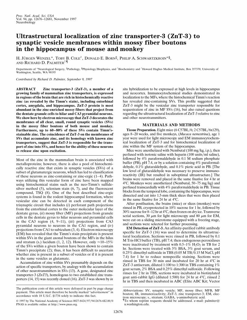
FIG. 1. Localization of histochemically reactive zinc (A, C, E) and IR for ZnT-3 protein (B, D, F) in the mouse hippocampus. (A) Transversesection of the hippocampus demonstrating intense reaction product in the mossy fiber (MF) system (dark band labeled ‘‘mf’’) after Timm’s stainingprocedure; counterstained with cresyl violet. Note the lighter laminar staining in s. radiatum (rad) and oriens (or) of CA1. (B) Transverse sectionof the hippocampus reacted with an affinity-purified antiserum against ZnT-3 protein. IR is intense in the zinc-containing MF projections; arrowsindicate ZnT-3 staining in s. lucidum (luc), pyramidale (pyr) and oriens (or) of CA3. The dentate outer (oml) and inner (iml) molecular layers,and s. radiatum (rad) and oriens (or) of CA1 also appear lightly immunoreactive for ZnT-3. (C) Electron micrograph from the dentate hilus(indicated area in A) showing MF boutons (mfb) with high density of silver granules. (D) Electron micrograph from the dentate hilus (indicatedarea in B) showing intense ZnT-3 IR in MFBs (mfb). Note the probable interneuron terminal (A) without ZnT-3 IR, making a symmetric synapticcontact (arrowhead) onto a dendrite (d). (E, F) Higher magnification of MFBs from CA3 s. lucidum (indicated areas in A and B) to demonstratethe vesicular localization of zinc (E) and ZnT-3 IR on synaptic vesicle membranes (F). Other abbreviations: CA1, CA3 hippocampal regions; DG,dentate gyrus; hi, hilus; M, mitochondrion; S, spine; arrowhead, synaptic contact.
12678 Neurobiology: Wenzel et al. Proc. Natl. Acad. Sci. USA 94 (1997)
previously to contain excitatory amino acids (17, 27). Themajority (up to 60–80% in mouse MFBs) of these SVs alsocontain Timm’s reaction product, indicative of vesicular zinc.The comparison of mouse to monkey revealed no species-related differences in the localization of ZnT-3 to SVs ofMFBs, although there were differences in the general patternof MF distribution. Synapses of nonglutamatergic local circuitneurons, e.g., hilar interneurons (Figs. 1D, 3 E and F), did notshow ZnT-3 IR.
Recent studies demonstrate significant differences betweenrodents and primates with respect to dendritic length (28), thepresence of basal dendrites (29), and basic ultrastructure (30).
Our studies reveal another species difference in the pattern ofMF projections to the pyramidal cells of the CA3 region. Inmice, ZnT-3 and Timm’s-positive MFBs were found in thehilus, s. lucidum, s. pyramidale between the CA3 pyramidalcells, and in s. oriens where MFBs were restricted to the CA3csubfield; in contrast, in the monkey hippocampus the MFBsthat stain with Timm’s reaction product and ZnT-3 antibodyoccupied the hilus and entire CA3 region. The appearance oflarge MFBs in s. lucidum and oriens in the monkey hippocam-pus is consistent with observations by Seress (24), who pointedout that large MFBs form synapses with both apical and basalparts of the dendritic tree in primates, whereas in rodents
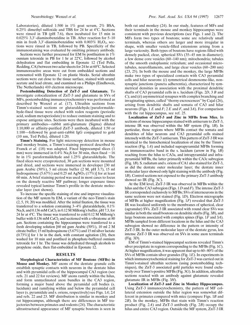
FIG. 2. Localization of histochemically reactive zinc (A, C, E) and IR for ZnT-3 protein (B, D, F) in the monkey hippocampus. (A) Frontalsection of the hippocampus demonstrating intense Timm’s stain for vesicular zinc in the MF projections (mf) within hilus and CA3. Lighter stainingis seen in s. radiatum and oriens and subiculum (Sub). (B) Frontal section of the monkey hippocampus. ZnT-3 IR is present in the zinc-rich MFprojections (mf) in hilus and CA3 region; lighter IR is seen in the dentate iml, in CA1 s. radiatum (rad) and oriens (or) and in subiculum. (C) Electronmicrograph from the dentate hilus (indicated area in A) showing MFBs labeled with silver granules. (D) Electron micrograph from the s. lucidum(indicated area in B) demonstrating ZnT-3 IR in MFBs that surround a dendrite (d). (E) Higher magnification of part of a giant MFB in synapticcontact with spines in CA3 s. lucidum, showing dark silver granules located in the clear round SVs. (F) Higher magnification of a MFB in synapticcontact with a giant spine (arrowheads), also making nonsynaptic contacts onto a dendritic shaft (d, arrows). ZnT-3 IR is localized to the membranesof clear, round SVs within the bouton. Abbreviations as in Fig. 1.
Neurobiology: Wenzel et al. Proc. Natl. Acad. Sci. USA 94 (1997) 12679
synapses with large complex spines are seen only in s. lucidum.Another difference in the pattern of ZnT-3 IR (and Timm’s)was observed in the dentate molecular layer. In mice, synapsesof the perforant path showed ZnT-3 IR in the outer molecularlayer, whereas in the monkey the inner molecular layer stainedmore intensely with the ZnT-3 antibody.
Evidence obtained from kinetic studies of zinc turnover andhistochemical studies of zinc-containing boutons suggest that
zinc is sequestered in SVs, and is released from boutons duringnormal activity by exocytosis of the zinc-filled vesicles (3). Azinc transporter localized to the SV membrane would providean effective means of loading the SVs, and would be consistentwith both light microscopy studies showing ZnT-3 stainingpattern overlapping the MF distribution (16) and with theultrastructural data showing its presence on SV membranes.Various brain regions containing zinc-ergic neurons have been
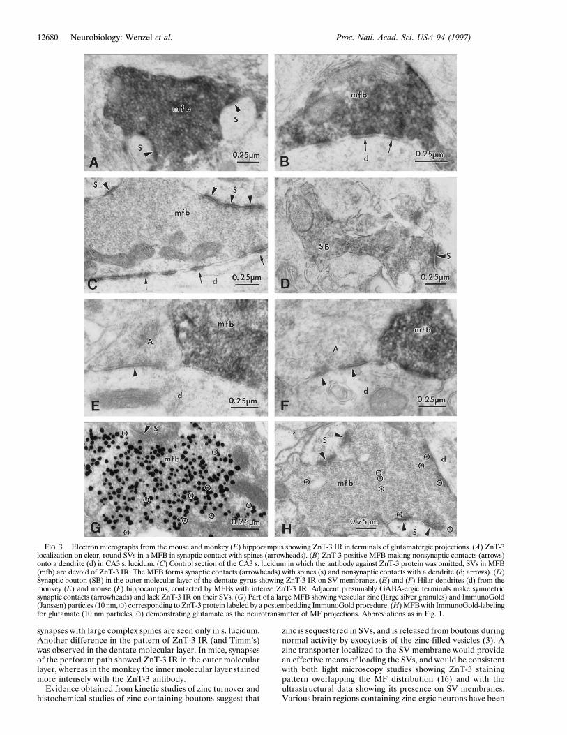
FIG. 3. Electron micrographs from the mouse and monkey (E) hippocampus showing ZnT-3 IR in terminals of glutamatergic projections. (A) ZnT-3localization on clear, round SVs in a MFB in synaptic contact with spines (arrowheads). (B) ZnT-3 positive MFB making nonsynaptic contacts (arrows)onto a dendrite (d) in CA3 s. lucidum. (C) Control section of the CA3 s. lucidum in which the antibody against ZnT-3 protein was omitted; SVs in MFB(mfb) are devoid of ZnT-3 IR. The MFB forms synaptic contacts (arrowheads) with spines (s) and nonsynaptic contacts with a dendrite (d; arrows). (D)Synaptic bouton (SB) in the outer molecular layer of the dentate gyrus showing ZnT-3 IR on SV membranes. (E) and (F) Hilar dendrites (d) from themonkey (E) and mouse (F) hippocampus, contacted by MFBs with intense ZnT-3 IR. Adjacent presumably GABA-ergic terminals make symmetricsynaptic contacts (arrowheads) and lack ZnT-3 IR on their SVs. (G) Part of a large MFB showing vesicular zinc (large silver granules) and ImmunoGold(Janssen) particles (10 nm, E) corresponding to ZnT-3 protein labeled by a postembedding ImmunoGold procedure. (H) MFB with ImmunoGold-labelingfor glutamate (10 nm particles, E) demonstrating glutamate as the neurotransmitter of MF projections. Abbreviations as in Fig. 1.
12680 Neurobiology: Wenzel et al. Proc. Natl. Acad. Sci. USA 94 (1997)
examined using EM techniques after neo-Timm’s or seleniumstaining, and in all cases the reaction product is restricted to thepresynaptic boutons of neurons (3, 19, 31). In addition,Timm’s-stainable zinc is also present in nonneuronal cells inthe brain, e.g., in the choroid plexus. Earlier ultrastructuralinvestigations demonstrated that the zinc in synaptic boutonsis localized in the vicinity of presynaptic vesicles (1, 12) andmore recent studies indicate that the zinc is within individualvesicles (2, 19). However, previous work indicated that only asmall fraction of clear, round vesicles in MFBs were reactivefor zinc (1–3), suggesting that a subclass of vesicles mightcontain zinc. In contrast, our study of Timm’s-stained materialdemonstrates that the majority of SVs in MFBs contain zinc,consistent with the observation that all SVs were ZnT-3positive, and thus presumably capable of facilitating zincuptake. It seems likely that the lack of Timm’s reaction productin individual SVs may represent vagaries of the methodology(e.g., rapidity of fixation), rather than absence of zinc. Wesuggest that all the SVs within MFBs contain both zinc andglutamate. In non-MFBs, e.g., boutons of entorhinal projec-tions in the outer molecular layer of mouse (Fig. 3D), whereboth Timm’s stainable zinc and ZnT-3 IR are less intense, allSVs still appear to react with the antibody. We suggest that thelevel of ZnT-3 expression may vary in different zinc-ergicneurons, but that all of their clear SVs are able to accumulatezinc, perhaps proportionally to the density of ZnT-3; thus, theamount of zinc in SVs would reflect ZnT-3 density. Clearly,zinc is not an obligatory component of SVs containing exci-tatory amino acids, because Timm’s staining is not observed inmany glutamatergic neurons (for review see Frederickson, ref.3). It is not clear what signals direct ZnT-3 to SVs of MFBs.It is also not known whether ZnT-3 can associate with differenttypes of SVs (e.g. clear vs dense core vesicles). Thus, it ispossible that ZnT-3 expression in neurons that produce dif-ferent kinds of vesicles could result in sequestration of zinc inone population of SVs but not another. When ZnT-3 that wastagged with green fluorescent protein was expressed in babyhamster kidney cells it was clearly associated with vesicles;however, ZnT-3 did not facilitate zinc sequestration in babyhamster kidney cells, unlike ZnT-2 expressed in these samecells (15, 16); thus, other components of SVs may be necessaryfor zinc transport by ZnT-3.
Zinc is capable of modulating a variety of voltage andligand-gated ion channels at physiological concentrations (5–500 mM; ref. 32). Zinc inhibition of N-methyl-D-aspartate-typeglutamate receptors has been reported by many authors (33–35), and variable effects of zinc have been observed onnon-N-methyl-D-aspartate glutamate receptors (33, 34, 36, 37).Zinc has also been shown to exert a powerful modulatoryinhibition of g-aminobutyric acid (GABA)A receptors (38).The MF system is particularly interesting considering the rolethat zinc might play in modulating transmitter-mediated ac-tions. First, the MFBs have presynaptic kainate receptors (39),and zinc modulation of kainate-induced currents appears to besensitive to synaptic calcium levels, which may vary in anactivity-dependent manner (40). Second, a-amino-3-hydroxy-5-methyl-4-isoxazolepropionic acid receptors are associatedwith synaptic contacts made by MF onto hilar mossy cells andCA3 pyramidal cells (41–43). Third, N-methyl-D-aspartate andGABA receptor-mediated inputs to CA3 pyramidal cells occurin s. radiatum and oriens, with strong GABAA inhibitionassociated with somatically localized receptors (42, 44, 45).Further, in various forms of hippocampal pathology (e.g.,mesial temporal sclerosis associated with temporal lobe epi-lepsy), there is a sprouting of zinc-rich MFs back on to thegranule cells of origin (18, 20, 46, 47). Their aberrant terminallocalization and their proximity to GABA receptors haveprompted investigators to postulate a role for zinc in thebreakdown of inhibition that is associated with seizure gener-
ation in epileptic hippocampus (48). All of these receptors areprobably within range of zinc diffusion, especially after highlevels of activity that release large amounts of zinc. Given somany potential ‘‘targets,’’ the role of zinc in modulating thereceptor responses to transmitter release during normal syn-aptic transmission remains to be resolved. Inactivation ofZnT-3 by gene-targeting will help define the role of zinc inmodulating synaptic activities in the central nervous system.
The authors thank Norma L. Anderson, Paul Schwartz, and JanetSchukar, for excellent technical assistance. We thank C. J. Fredericksonfor reviewing the manuscript. This work was supported in part by NationalInstitutes of Health Grant NS18895 (P.A.S.) and U.S. Public HealthService National Research Service Award T32 GM07270 (T.B.C.).
1. Haug, F.-M. S. (1967) Histochemie 8, 355–368.2. Perez-Clausell, J. & Danscher, G. (1985) Brain Res. 337, 91–98.3. Frederickson, C. J. (1989) Int. Rev. Neurobiol. 31, 145–238.4. Slomianka, L. (1992) Neuroscience 48, 325–352.5. Danscher, G. (1981) Histochemistry 71, 1–16.6. Danscher, G. (1984) in The Neurobiology of Zinc, eds. Frederickson, C. J.,
Howell, G. A. & Kasarskis, E. J. (Riss, New York), Vol. B, pp. 171–191.7. Slomianka, L., Danscher, G. & Frederickson, C. J. (1990) Neuroscience 38,
843–854.8. Frederickson, C. J., Kasarskis, E. J., Ringo, D. & Frederickson, R. E. (1987)
J. Neurosci. Methods 20, 91–103.9. Haug, F.-M. S., Blackstad, T. W., Simonsen, A. H. & Zimmer, J. (1971)
J. Comp. Neurol. 142, 23–32.10. Crawford, I. L. & Connor, J. D. (1972) J. Neurochem. 19, 1451–1458.11. Zimmer, J. & Haug, F.-M. S. (1978) J. Comp. Neurol. 179, 581–618.12. Ibata, Y. & Otsuka, N. (1969) J. Histochem. Cytochem. 17, 171–175.13. Borowsky, B. & Hoffmann, B. J. (1995) Int. Rev. Neurobiol. 38, 139–199.14. Palmiter, R. D. & Findley, S. D. (1995) EMBO J. 14, 639–649.15. Palmiter, R. D., Cole, T. B. & Findley, S. D. (1996) EMBO J. 15, 1784–1791.16. Palmiter, R. D., Cole, T. B., Quaife, C. J. & Findley, S. D. (1996) Proc. Natl.
Acad. Sci. USA 93, 14934–14939.17. Wenzel, H. J., Buckmaster, P. S., Anderson, N. L., Wenzel, M. E. &
Schwartzkroin, P. A. (1997) Hippocampus 7, in press.18. Franck, J. E., Pokorny, J., Kunkel, D. D. & Schwartzkroin, P. A. (1995)
Epilepsia 36, 543–558.19. Danscher, G. (1996) Histochem. J. 28, 361–373.20. Babb, T. L., Kupfer, W. R., Pretorius, J. K., Crandall, P. H. & Levesque,
M. F. (1991) Neuroscience 42, 351–363.21. Frotscher, M., Soriano, E. & Misgeld, U. (1993) Synapse 16, 148–160.22. Amaral, D. G. & Witter, M. P. (1995) in The Rat Nervous System, ed.
Paxinos, G. (Academic, San Diego), pp. 443–493.23. Claiborne, B. J., Amaral, D. G. & Cowan, W. M. (1986) J. Comp. Neurol.
246, 435–458.24. Seress, L. (1992) in The Dentate Gyrus and its Role in Seizures, eds. Ribak,
C. E., Gall, C. M. & Mody, I. (Elsevier, Amsterdam), pp. 3–28.25. Amaral, D. G. & Dent, J. A. (1981) J. Comp. Neurol. 195, 51–86.26. Ramon y Cajal, S. (1893) Ann. Soc. Esp. Hist. Nat. 22, 53–114.27. Beaulieu, C., Dyck, R. & Cynader, M. (1992) NeuroReport 3, 861–864.28. Claiborne, B. J., Amaral, D. G. & Cowan, W. M. (1990) J. Comp. Neurol.
302, 206–220.29. Seress, L. & Frotscher, M. (1990) J. Comp. Neurol. 293, 253–267.30. Seress, L. & Ribak, C. E. (1992) Brain Res. 569, 353–357.31. Danscher, G., Howell, G., Perez-Clausell, J. & Hertel, N. (1985) Histo-
chemistry 83, 419–422.32. Smart, T. G., Xie, X. & Krishek, B. J. (1994) Prog. Neurobiol. 42, 393–441.33. Peters, S., Koh, J. & Choi, D. W. (1987) Science 236, 589–593.34. Westbrook, G. L. & Mayer, M. L. (1987) Nature (London) 328, 640–643.35. Christine, C. W. & Choi, D. W. (1990) J. Neurosci. 10, 108–116.36. Mayer, M. L., Vyklicky, L., Jr. & Westbrook, G. L. (1989) J. Physiol.
(London) 415, 329–350.37. Hori, N., Galeno, T. & Carpenter, D. O. (1987) Cell. Mol. Neurobiol. 7,
73–90.38. Legendre, P. & Westbrook, G. L. (1991) Mol. Pharmacol. 39, 267– 274.39. Baude, A., Nusser, Z., Molnar, E., McIlhinney, R. A. & Somogyi, P. (1995)
Neuroscience 69, 1031–1055.40. Dreixler, J. C. & Leonard, J. P. (1997) Brain Res. 752, 170–174.41. Schroder, H. (1993) Hippocampus 3, 139–148.42. Petralia, R. S., Wang, Y.-X. & Wenthold, R. J. (1994) J. Neurosci. 14,
6102–6120.43. Catania M. V., Tolle, T. R. & Monyer, H. (1995) J. Neurosci. 15, 7046–7061.44. Siegel, S. J., Brose, N., Janssen, W. G., Gasic, G. P., Jahn, R., Heinemann,
S. F. & Morrison, J. H. (1994) Proc. Natl. Acad. Sci. USA 91, 564–568.45. Johnson, R. R., Jiang, X. & Burkhalter, A. (1996) J. Comp. Neurol. 368,
335–355.46. Sutula, T., Cascino, G., Cavazos, J., Parada, I. & Ramirez, L. (1989) Ann.
Neurol. 26, 321–330.47. Isokawa, M., Levesque, M. F., Babb, T. L. & Engel, J., Jr. (1993) J. Neurosci.
13, 1511–1522.48. Buhl, E. H., Otis, T. S. & Mody, I. (1996) Science 271, 369–373.
Neurobiology: Wenzel et al. Proc. Natl. Acad. Sci. USA 94 (1997) 12681